Pathogenic skin fung i in Australian reptiles Fact sheet
←
→
Page content transcription
If your browser does not render page correctly, please read the page content below
Pathogenic skin fungi in
              Australian reptiles
                  Fact sheet

Introductory statement
Fungi belonging to the genera Nannizziopsis, Paranannizziopsis and Ophidiomyces (formerly members of the
Chrysosporium anamorph of Nannizziopsis vriesii [CANV] complex) are the cause of skin diseases that may
progress to systemic and sometimes fatal disease in a range of reptile species. The disease was formerly
referred to as ‘yellow fungus disease’ due to coloration of the skin lesions. These disease conditions are
relatively newly described, suggesting they are ‘emerging’, although much remains to be learnt about the
aetiological agents, epidemiology, presence, and prevalence of these fungal diseases worldwide. The reasons
for the apparent emergence of these infections in both free-living and captive reptiles are not understood,
however it is likely that global human-assisted movement of reptiles (due to the reptile pet trade) may be a
contributing factor (Paré et al. 2020).

In Australia, pathogenic skin fungi have been reported in a range of captive reptile species and in free-living
Agamids (dragon lizards) and shingleback lizards (Tiliqua rugosa). The focus of this fact sheet is on fungi of the
genera Nannizziopsis, Paranannizziopsis and Ophidiomyces.

Aetiology
The genera Nannizziopsis, and Paranannizziopsis are classified in the family Nannizziopsidaceae of the order
Onygenales1 (Stchigel et al. 2013) and Ophidiomyces is classified in the family Onygenaceae (Onygenales)
(Sigler et al. 2013).

Nine species of the genus Nannizziopsis are associated with skin disease in lizards globally (Sigler et al. 2013;
Paré and Sigler 2016; Peterson et al. 2020). Nannizziopsis barbatae2 has 99% nucleotide similarity to N.
crocodili and is also similar genetically to N. pluriseptata (Peterson et al. 2020).

Genus Paranannizziopsis (four species) are linked to skin disease in Australasian reptiles including tuatara
(Sphenodon punctatus punctatus), aquatic file snakes (Acrochordus sp.) and coastal bearded dragon (Pogona

1
    The fungal agents formerly known as the Chrysosporium anamorph of Nannizziopsis vriesii vriessi (CANV complex) have
been reclassified within the genera Nannizziopsis, Paranannizziopsis and Ophidiomyces (Sigler et al. 2013; Stchigel et al.
2013).
2
    Formerly N. barbata
barbata) (Masters et al. 2016; Paré and Sigler 2016). Ophidiomyces ophidiicola3 (formerly ophiodiicola; the
only species of this genus) is associated with “snake fungal disease” in terrestrial or semiaquatic snake species
(Sigler et al. 2013; Stchigel et al. 2013).

Other onygenalean fungi include Emydomyces testavorans, which has been associated with lesions in turtles
and has no assigned family (Woodburn et al. 2019). Aphanoascella galapagosensis has been isolated from a
captive Galapagos tortoise with carapace keratitis in the USA (Sutton et al. 2013).

Natural hosts
Nannizziopsis spp. have been reported from infections in a wide range of lizard species, including chameleons,
geckos, dragon and iguana lizards, and crocodiles (Paré and Sigler 2016; Peterson et al. 2020). N. guarroi is
associated with skin disease in bearded dragons (Pogona vitticeps) and other lizards in North America (Paré
and Sigler 2016). Nannizziopsis barbatae has been linked to skin and systemic disease in a range of lizards in
Australia (below), including endemic free-living species (Peterson et al. 2020).

Paranannizziopsis spp. have been reported in lizards, snakes and tuatara (Paré and Sigler 2016).

O. ophidiicola has been identified in a wide range of both captive and free-ranging snakes (Sigler et al. 2013;
Stchigel et al. 2013; Lorch et al. 2016; Paré and Sigler 2016).

Emydomyces testavorans is reported to be associated with invasive carapace, plastron and skin lesions in
captive and free-living aquatic and semi-aquatic turtles in North America (Woodburn et al. 2019).

World distribution
Cases of Nannizziopsis spp. and Paranannizziopsis spp. infection in captive reptiles have been reported in
Africa, Asia, Europe, North America, Australia and New Zealand (Sigler et al. 2013; Stchigel et al. 2013; Paré
and Sigler 2016; Peterson et al. 2020). Lesions typical of those associated with O. ophidiicola have been
reported in captive snakes since the 1980s, primarily in North America but also in Europe. Infection has been
identified more recently in wild snakes in North America (Sigler et al. 2013; Stchigel et al. 2013; Lorch et al.
2016; Paré and Sigler 2016; Paré et al. 2020; Snyder et al. 2020).

Occurrences in Australia

Species identified in Australia include N. barbatae, N. crocodili, P. australasiensis and O. ophidiicola (Thomas
et al. 2002; Johnson et al. 2011; Sigler et al. 2013; Paré and Sigler 2016; Peterson et al. 2020).

N. barbatae has been linked to severe disease in four species of free-living lizards in Australia - see below
(Peterson et al. 2020). Other reports of fungal pathogens from these genera in Australia have been in captive
reptiles.

Nannizziopsis: cases of severe, proliferative dermatitis, debility and death associated with N. barbatae were
reported in four species of free-living lizard from at least 8 different locations across Australia, including a
significant outbreak among Eastern water dragons (Intellagama lesueurii) in Brisbane, Qld, beginning in 2013

3
    Formerly O. ophiodiicola

              WHA Fact sheet: Pathogenic skin fungal diseases in Australian reptiles | August 2021 | 2
(Peterson et al. 2020). Affected species included a tommy roundhead (Diporiphora australias) from the
Brisbane areas, an eastern bluetongue skink (Tiliqua scincoides scincoides) from Dubbo NSW and a wild
shingleback lizard from Perth WA (Peterson et al. 2020). Cases came from at least 8 different locations across
Australia. These were the first reports of infection within this genus of fungi in free-living reptiles globally, and
N. barbatae has not been reported outside Australia. A captive Centralian bluetongue lizard (Tiliqua
multifasciata), from Vic was also found to be infected. During this study, many other specimens from
Australian snakes and lizards (collected since 2010 in Australia) were examined, with the cases reported here
the only detections of N. barbatae (Peterson et al. 2020).
A fungus very similar to N. barbatae (but originally thought to be N. pluriseptata) was identified as the cause
of skin disease in eight wild-caught but captively housed shingleback lizards (Tiliqua rugosa) held in Western
Australia in 2019 (WA Wildlife Health Reference Group 2019).
An outbreak (now identified as N. barbatae) was reported in a group of captive coastal bearded dragons (P.
barbata) in NSW in 2008-2009 (Johnson et al. 2011; Paré and Sigler 2016). These were the first known cases
of N. barbatae in Australia (Peterson et al. 2020). A separate case was diagnosed in another captive coastal
bearded dragon (Johnson et al. 2011; Sigler et al. 2013). A isolate very similar to N. barbatae was obtained
from a free-ranging eastern water dragon (Physignathus lesueurii) with skin lesions (Paré and Sigler 2016). N.
barbatae has not yet been reported outside Australia (Paré et al. 2020).
Outbreaks of N. crocodili occurred on two separate occasions in 1994 and 1997 in saltwater crocodiles
(Crocodylus porosus) sourced from the same crocodile farm in northern Qld, with lesions and mortalities
(Thomas et al. 2002; Paré and Sigler 2016). More recent reports have occurred in captive (zoo) juvenile
freshwater crocodiles (C. johnstoni) with skin lesions, at a location geographically far removed from the
reports in farmed saltwater crocodiles (Hill et al. 2019). N. crocodili has not been reported in wild reptiles.
Paranannizziopsis australasiensis was cultured from lesions on two file snakes (Acrochordus arafurae)
housed in captivity (zoo) in Vic (Paré and Jacobson 2007; Sigler et al. 2013; Paré and Sigler 2016).
Ophidiomyces ophidiicola has been diagnosed only in captive snakes in Australia, including a file snake
(Acrochordus arafurae) on display in a crocodile farm in Qld (Sigler et al. 2013) and a broad-headed snake
(Hoplocephalus bungaroides) in a zoo in South Australia, which died with apparent systemic infection
(McLelland et al. 2010).
It has been suggested that mycotic dermatitis attributed to Geotrichum candidum in three captive carpet
pythons (Morelia spilotes variegate) in Qld may have been caused by O. ophidiicola (McKenzie and Green
1976; Paré and Sigler 2016; Sigler 2021). Ophidiomyces ophidiicola has not been reported in wild Australian
reptiles.
Anecdotal evidence suggests that clinical cases consistent with Nannizziopsis and Ophidiomyces fungal
disease may have been occurring in captive lizards and snakes in Australia for several decades (Reiss and
Woods 2018), although Peterson et al. (2020) reports no positive cases from archived samples from 2010
onwards.
WHA Fact sheet: Pathogenic skin fungal diseases in Australian reptiles | August 2021 | 3Epidemiology
The epidemiology of these emerging diseases in reptiles is still poorly understood. Ectothermic taxa such as
reptiles are considered particularly vulnerable to fungal pathogens and these fungal species appear for the
most part to be primary pathogens, rather than opportunists (as fungal pathogens of reptiles have been
traditionally considered) (Mitchell and Walden 2013; Peterson et al. 2020).
Nannizziopsis and Paranannizziopsis: Koch’s postulates have been fulfilled in experimental infection of veiled
chameleons (Chamaeleo calyptratus) with N. guarroi (Paré et al. 2006). In captivity, suboptimal
environmental factors may make reptiles more susceptible to these infections and the pathogens are
contagious between animals in close proximity (Thomas et al. 2002; Paré and Sigler 2016). Cases have been
identified in free-ranging reptiles, but there is little available information on the epidemiology in the wild
(Paré and Sigler 2016; Peterson et al. 2020).
It is not known if N. barbatae was introduced to wild lizards through spillover or if it is an endemic fungus that
has only been detected recently. It is not yet known if it is truly an “emerging disease” among wild lizards, nor
what the factors might be that could be contributing to emergence, however altered host susceptibility or
changing environmental conditions have been suggested as possible contributing factors (Peterson et al.
2020).
Ophidiomyces ophidiicola is now well recognised as a pathogen of snakes, including free-ranging species,
particularly in North America. Experimental infections have fulfilled Koch’s postulates and have demonstrated
action as a primary pathogen (Allender et al. 2015a; Lorch et al. 2015). There is some evidence that some
asymptomatic carrier hosts may exist, but are rare (Paré et al. 2020). It is possible that the fungus can be
vertically transmitted from dam to offspring (around the time of birth) (Stengle et al. 2019) but transmission
routes in general are poorly understood. The fungus is found in copious amounts in the lesions of affected
snakes, and is not thought to be part of the cutaneous mycobiota (Allender et al. 2011; Allender et al. 2015b;
Paré and Sigler 2016). Although the fungus does not grow at temperatures less than 15 C, snakes undergoing
brumation, or held at suboptimal temperature may be susceptible, if the snakes’ immune systems are
depressed (Lorch et al. 2016; Paré and Sigler 2016). Small skin lesions colloquially termed “hibernation” sores
or blisters that have been recognised in snakes for many years also test positive for the fungus, suggesting
infection has been occurring historically, but severity of clinical disease (at least in some incidences) may have
been increasing in recent years. The cause of this increased severity (from small healing sores to a fatal
disease) is unclear (Lorch et al. 2016; Paré and Sigler 2016). Infection may make snakes more susceptible to
predation or other causes of mortality and has been implicated as a cause of wild snake population declines
since 2006 (Allender et al. 2015c; Snyder et al. 2020).
Clinical signs
Lesions are similar for all pathogens, although they appear to vary in severity depending on the host species
and likely other environmental factors (Peterson et al. 2020). Grossly, lesions are initially yellow, and then
thicken to form brown, hyperkeratotic, necrotic plaques. These plaques may crack and seep exudate, or
slough to reveal whitish pink, swollen dermis. Lesions are often found on the head and around the mouth but
can occur anywhere on the body and can involve a whole limb in the case of lizards (Paré and Jacobson 2007;
Lorch et al. 2016; Paré and Sigler 2016; Peterson et al. 2020). Animals may show hyperkeratosis, epidermal
hyperplasia, dermal inflammation, necrosis, ulceration, and emaciation.
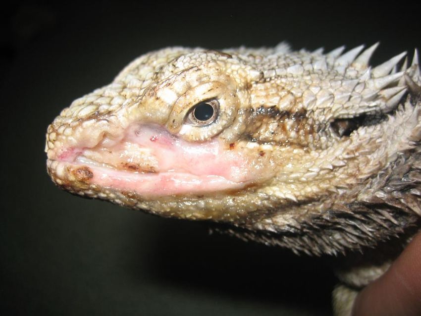
WHA Fact sheet: Pathogenic skin fungal diseases in Australian reptiles | August 2021 | 4Dermatomycoses due to Nannizziopsis spp. are slowly progressive and often fatal skin diseases. Affected
reptiles range in body condition from poor to good. Skin lesions progress over several months from dry and
yellow to hyperkeratotic plaques to exudative and necrotic ulcers (Figure 1) (Johnson et al. 2011; Paré and
Sigler 2016; Peterson et al. 2020). In bearded dragons (Pogona spp.), the mouth and face are commonly
affected but lesions may occur anywhere on the body (Bowman et al. 2007; Abarca et al. 2009; Johnson et al.
2011; Le Donne et al. 2016; Schmidt-Ukaj et al. 2016). Infection is often fatal in bearded dragons, with
infection extending to muscle, bone and internal tissues including liver, heart, kidney, lungs and intestine
(Bowman et al. 2007; Johnson et al. 2011; Masters et al. 2016; Paré and Sigler 2016; Schmidt-Ukaj et al.
2016). In iguanas, infection is often identified in the hind limbs and tail (Han et al. 2010; Kahraman et al.
2015). Hatchling saltwater crocodiles infected with N. crocodili developed multiple leathery plaque-like
lesions on or under the scales, which could be peeled away to reveal white or red tissue. Infection was often
fatal (Thomas et al. 2002).
P. australasiensis in tuatara causes skin lesions similar to those described above. In tuatara, lesions resolved
with treatment in all reported cases. In a coastal bearded dragon, the same agent resulted in fatality, with
similar progression of disease as that described for Nannizziopsis spp. (Masters et al. 2016). In file snakes,
infection results in disseminated punctate or circular, whitish lesions across the epidermis (Paré and Jacobson
2007; Sigler et al. 2013; Paré and Sigler 2016).
O. ophidiicola in snakes mainly affects the head and also the scales of the body, most often on the underside
of the snake. In milder cases, the processes of ecdysis (slough) can resolve the infection, but if deeper tissues
are affected, disease may recur post-slough. Systemic infection with involvement of bone and lung has been
reported, but is rare (Lorch et al. 2016).
Figure 1. Nannizziopsis fungal disease in a captive coastal bearded dragon P. barbata (Courtesy R Johnson).
WHA Fact sheet: Pathogenic skin fungal diseases in Australian reptiles | August 2021 | 5Diagnosis
Definitive diagnosis of skin or systemic disease associated with fungi in the Nannizziopsis, Paranannizziopsis
and Ophidiomyces genera requires both a) identification of the organism through culture and sequencing or
PCR and b) histopathology identifying fungal elements within lesions, in particular the presence of
arthroconidia. Reptile skin may host many fungal elements, but confirmed presence of Nannizziopsis,
Paranannizziopsis or Ophidiomyces is indicative of disease where hyphae and arthroconidia are present in
lesions and the fungus has been isolated or confirmed by PCR. There is increasing evidence that at least some
of these fungi (e.g. N. barbatae) are obligate pathogens (Paré and Sigler 2016; Peterson et al. 2020).
Identification of the organism using matrix-assisted laser desorption/ ionization time-of-flight mass
spectrometry (MALDI-TOF MS), which may be more accurate than sequencing, has been described but may
not yet be available in Australia (Schneider et al. 2017).
Clinical pathology
Although not pathognomonic, cytology of sticky tape preparations or impression smears may reveal presence
of conidia or arthroconidia suggestive of a fungus in the Nannizziopsis, Paranannizziopsis or Ophidiomyces
genera (Le Donne et al. 2016; Paré and Sigler 2016). Conidia are 5-8 μm long by 3-5 μm wide, clavate to ovoid
to cylindrical. Arthroconidia may be arranged in rows of up to 7 conidia, separated or in a chain (Le Donne et
al. 2016; Sigler 2021).
Pathology
Grossly, lesions are as described above. In systemically affected bearded dragons at necropsy, collection of
pale yellow, gelatinous material can be found within the coelomic cavity and pericardial sac and
granulomatous changes have been noted in the liver (Bowman et al. 2007). Histologic lesions include
granulomatous fungal dermatitis, myositis, osteomyelitis, hepatitis, nephritis, coelomitis, myocarditis and
pneumonia (Bowman et al. 2007; Johnson et al. 2011; Paré and Sigler 2016; Schmidt-Ukaj et al. 2016). Hyphae
found within granulomas and in the keratin layer are 2-4 μm wide, septate and exhibit haphazard branching.
Arthroconidia on the skin of reptiles with characteristic lesions is considered pathognomonic for fungi in the
Nannizziopsidaceae and O. ophidiicola. A tape mount could be used to sample the surface of the lesion. (Paré
and Jacobson 2007).
Differential diagnoses
Other dermatomycoses, bacterial dermatitis, stomatitis and osteomyelitis should be excluded from the list of
differential diagnoses.
Laboratory diagnostic specimens
Multiple skin biopsies of dermal lesions, half placed in 10% neutral buffered formalin for histopathology and
half submitted fresh (or less desirably frozen) for PCR and /or culture (Paré and Sigler 2016).
Skin samples from lesions can be submitted for PCR and culture. Swabs should be avoided as the fungus is
difficult to culture from these samples (Paré and Sigler 2016).
WHA Fact sheet: Pathogenic skin fungal diseases in Australian reptiles | August 2021 | 6Sections of multiple internal organs in 10% neutral buffered formalin and fresh/frozen are recommended if
systemic disease is suspected (Sangster 2018).
Laboratory procedures
Histopathological examination should include H&E and either PAS or Grocott-Gomori’s methylene silver stain
for fungal identification (Sangster 2018).
Samples for culture are best treated with enrofloxacin to limit bacterial overgrowth, plated on MycoselTM
agar (Becton, Dickinson and Company, Franklin Lakes, NJ) and incubated at 30°C. White powdery colonies
should be sub-cultured, and speciated by sequencing (Paré and Sigler 2016).
Peterson et al. (2020) recommends an improved method of fungal isolation by modification of the
conventional serial dilution technique.
Treatment
Medical treatment of confirmed cases involves systemic antifungals and topical antifungal or antiseptic
solutions (Paré and Sigler 2016). There is limited information on successful regimens, and treatment may not
be effective or curative (Peterson et al. 2020). Susceptibility testing of N. guarroi has revealed sensitivity to
voriconazole and terbinafine, but less so to itraconazole (Van Waeyenberghe et al. 2010; Paré and Sigler
2016). Pharmacokinetic studies in bearded dragons and green iguanas suggests terbinafine may reach
therapeutic levels in these species after oral administration (McEntire et al. 2020). Serum biochemistry should
be monitored for signs of liver toxicity. Surgical excision or debridement of lesions should be carried out if
possible and in conjunction with medical therapy (Peterson et al. 2020).
Treatment of captive reptiles should also include appropriate cleaning and decontamination of the animal’s
environment, and the maintenance of simple surroundings to facilitate ongoing hygiene and disinfection.
There is currently little information on appropriate disinfections protocols for Nannizziopsis sp. however
laboratory-based studies on O. ophidiicola recommend a minimum of 2 minute environmental exposure to
at least 3% bleach or 70% ethanol or a 10 min exposure to 0.16% Roccal, Lysol products, CLR, NPD, or 409
(Rzadkowska et al. 2016).
Prevention and control
Prevention of pathogenic fungal infection in captive reptiles should focus on reducing the fungal load, with
attention being paid to regular substrate changes and good hygiene in captive situations. In captive reptiles,
infection appears to be more common at low ambient temperatures. Providing optimal husbandry conditions,
including species-appropriate temperature gradients, hydration/humidity and nutrition are important steps in
prevention and control (Paré and Sigler 2016). Affected individuals should be isolated and biosecurity
measures followed as the organism can act as a contagious, primary pathogen (Paré and Sigler 2016).
Prevention and control options for free-living reptiles are limited. Factors that may contribute to increased
susceptibility to infection such as environmental degradation, proximity to humans, and other stressors
should be addressed wherever possible (Peterson et al. 2020). Appropriate disinfection protocols (see above)
should be used for equipment if working with wild reptiles, prior to moving between study sites (Rzadkowska
et al. 2016).
WHA Fact sheet: Pathogenic skin fungal diseases in Australian reptiles | August 2021 | 7Surveillance and management
Wildlife disease surveillance in Australia is coordinated by Wildlife Health Australia. The National Wildlife
Health Information System (eWHIS) captures information from a variety of sources including Australian
government agencies, zoo and wildlife parks, wildlife carers, universities, industry and members of the public.
Coordinators in each of Australia's States and Territories report monthly on significant wildlife cases identified
in their jurisdictions. NOTE: access to information contained within the National Wildlife Health Information
System dataset is by application. See the WHA website for more information:
www.wildlifehealthaustralia.com.au/ProgramsProjects/eWHISWildlifeHealthInformationSystem.aspx#requests.
There are currently no targeted surveillance programs for reptile fungal diseases. There are a number of cases
reported in eWHIS including those reported above, and more recent cases in wild lizards in Australia, and an
earlier suspected case in a wild broad-shelled turtle Chelodina expansa (no culture or PCR).
We encourage those with laboratory confirmed cases of this condition in native Australian or feral animals to
submit this information to the national system for consideration for inclusion in the national database. Please
contact us at admin@wildlifehealthaustralia.com.au.
Research
Molecular differentiation of the taxonomy of this order of fungi has significantly advanced knowledge in this
area, allowing identification of morphological and physiological properties, host trends and sensitivity
patterns for many new species (Paré and Sigler 2016). There are significant knowledge gaps related to the
incidence, host range and epidemiology of disease related to these pathogens, particularly in free-ranging
reptiles (Lorch et al. 2016; Peterson et al. 2020). Further studies are warranted to understand fully the origin
and nature of these organisms in the Australian context, and their significance as primary pathogens in both
captive and wild reptiles.
Human health implications
Molecular characterisation work has revealed cases of Nannizziopsis infection in humans are caused by
species that are distinct from those found in reptiles. The risk of zoonotic transmission of pathogenic skin
fungi from reptiles to humans is considered low since the temperature range for growth of these reptile
associated fungi is generally not compatible with infection in humans (Peterson et al. 2020).
Conclusions
Australia has seen recent confirmed cases of Nannizziopsis infection in wild lizards across a broad geographic
range, as well as sporadic cases and outbreaks of pathogenic skin fungi in captive crocodiles, lizards and
snakes. There is convincing evidence that these fungi are significant primary pathogens of reptiles. Wildlife
carers and veterinarians caring for both captive and free-living reptiles need to be vigilant in preventing the
spread of these pathogens, as they are easily spread by contact. Further work is needed to improve our
understanding of the incidence, host range and epidemiology of these fungal infections, and the risks to
Australian native reptiles.
WHA Fact sheet: Pathogenic skin fungal diseases in Australian reptiles | August 2021 | 8References and other information
Abarca M, Martorell J, Castellá G, Ramis A, Cabañes F (2009) Dermatomycosis in a pet inland bearded dragon
(Pogona vitticeps) caused by a Chrysosporium species related to Nannizziopsis vriesii. Veterinary Dermatology
20, 295-299.
Allender MC, Baker S, Wylie D, Loper D, Dreslik MJ et al. (2015a) Development of snake fungal disease after
experimental challenge with Ophidiomyces ophiodiicola in cottonmouths (Agkistrodon piscivorous). PloS ONE
10, e0140193.
Allender MC, Bunick D, Dzhaman E, Burrus L, Maddox C (2015b) Development and use of a real-time
polymerase chain reaction assay for the detection of Ophidiomyces ophiodiicola in snakes. Journal of
Veterinary Diagnostic Investigation 27, 217-220.
Allender MC, Dreslik M, Wylie S, Phillips C, Wylie DB et al. (2011) Chrysosporium sp. infection in eastern
massasauga rattlesnakes. Emerging Infectious Diseases 17, 2383.
Allender MC, Raudabaugh DB, Gleason FH, Miller AN (2015c) The natural history, ecology, and epidemiology
of Ophidiomyces ophiodiicola and its potential impact on free-ranging snake populations. Fungal Ecology 17,
187-196.
Bowman MR, Paré JA, Sigler L, Naeser JP, Sladky KK et al. (2007) Deep fungal dermatitis in three inland
bearded dragons (Pogona vitticeps) caused by the Chrysosporium anamorph of Nannizziopsis vriesii. Medical
Mycology 45, 371-376.
Han J-I, Lee S-J, Na K-J (2010) Necrotizing dermatomycosis caused by Chrysosporium spp. in three captive
green iguanas (Iguana iguana) in South Korea. Journal of Exotic Pet Medicine 19, 240-244.
Hill AG, Sandy JR, Begg A (2019) Mycotic dermatitis in juvenile freshwater crocodiles (Crocodylus johnstoni)
caused by Nannizziopsis crocodili. Journal of Zoo and Wildlife Medicine 50, 225-230.
Johnson R, Sangster C, Sigler L, Hambleton S, Paré J (2011) Deep fungal dermatitis caused by the
Chrysosporium anamorph of Nannizziopsis vriesii in captive coastal bearded dragons (Pogona barbata).
Australian Veterinary Journal 89, 515-519.
Kahraman BB, Sığırcı BD, Metiner K, Ak S, Koenhemsi L et al. (2015) Isolation of Chrysosporium guarroi in a
Green Iguana (Iguana iguana), in Turkey. Journal of Exotic Pet Medicine 24, 427-429.
Le Donne V, Crossland N, Brandão J, Sokolova Y, Fowlkes N et al. (2016) Nannizziopsis guarroi infection in 2
inland bearded dragons (Pogona vitticeps): clinical, cytologic, histologic, and ultrastructural aspects.
Veterinary Clinical Pathology 45, 368-375.
Lorch JM, Knowles S, Lankton JS, Michell K, Edwards JL et al. (2016) Snake fungal disease: an emerging threat
to wild snakes. Philosophical Transactions of the Royal Society B: Biological Sciences 371, 20150457.
Lorch JM, Lankton J, Werner K, Falendysz EA, McCurley K et al. (2015) Experimental infection of snakes with
Ophidiomyces ophiodiicola causes pathological changes that typify snake fungal disease. MBio 6, e01534-15.
Masters N, Alexander S, Jackson B, Sigler L, Chatterton J et al. (2016) Dermatomycosis caused by
Paranannizziopsis australasiensis in five tuatara (Sphenodon punctatus) and a coastal bearded dragon
(Pogona barbata) in a zoological collection in New Zealand. New Zealand Veterinary Journal 64, 301-307.
McEntire MS, Reinhart JM, Allender MC, Keller KA (2020) Antifungal Susceptibility Patterns of Nannizziopsis
guarroi and the Single-Dose Pharmacokinetics of Orally Administered Terbinafine in the Bearded Dragon
(Pogona vitticeps). In 'American Association of Zoo Veterinarians Conference. Virtual'. (Ed. AAZV) (AAZV).
WHA Fact sheet: Pathogenic skin fungal diseases in Australian reptiles | August 2021 | 9McKenzie R, Green P (1976) Mycotic dermatitis in captive carpet snakes (Morelia spilotes variegata). Journal
of Wildlife Diseases 12, 405-408.
McLelland D, Johnson L, Reuter R (2010) Fatal cutaneous mycosis in a broad-headed snake (Hoplocephalus
bungaroides) caused by the Chrysosporium anamorph of Nannizziopsis vriesii, Proceedings of the Wildlife
Disease Association–Australasian Section. Tasmania, Australia. (WDAA)
Mitchell MA, Walden MR (2013) Chrysosporium anamorph Nannizziopsis vriesii: an emerging fungal pathogen
of captive and wild reptiles. Veterinary Clinics: Exotic Animal Practice 16, 659-668.
Paré J, Coyle K, Sigler L, Maas A, Mitchell R (2006) Pathogenicity of the Chrysosporium anamorph of
Nannizziopsis vriesii for veiled chameleons (Chamaeleo calyptratus). Sabouraudia 44, 25-31.
Paré J, Jacobson E (2007) Mycotic diseases of reptiles. In 'Infectious diseases and pathology of reptiles.' (Ed.
ER Jacobson.) pp. 527-570. (CRC Press: Boca Raton).
Paré J, Sigler L (2016) An overview of reptile fungal pathogens in the genera Nannizziopsis, Paranannizziopsis,
and Ophidiomyces. Journal of Herpetological Medicine and Surgery 26, 46-53.
Paré JA, Wellehan J, Perry SM, Scheelings TF, Keller K et al. (2020) Onygenalean Dermatomycoses (Formerly
Yellow Fungus Disease, Snake Fungal Disease) in Reptiles: Roundtable. Journal of Herpetological Medicine and
Surgery 30, 198-209.
Peterson NR, Rose K, Shaw S, Hyndman TH, Sigler L et al. (2020) Cross-continental emergence of Nannizziopsis
barbatae disease may threaten wild Australian lizards. Scientific Reports 10, 1-12.
Reiss A, Woods R (2018) Clinical cases consistent with Nannizziopsis and Ophidiomyces fungal disease may
have been occurring in captive lizards and snakes in Australia for several decades., Personal Communication.
Rzadkowska M, Allender MC, O'Dell M, Maddox C (2016) Evaluation of common disinfectants effective against
Ophidiomyces ophiodiicola, the causative agent of snake fungal disease. Journal of Wildlife Diseases 52, 759-
762.
Sangster C (2018) Information about pathogenic reptile fungi. Personal Communication.
Schmidt-Ukaj S, Loncaric I, Spergser J, Richter B, Hochleithner M (2016) Dermatomycosis in three central
bearded dragons (Pogona vitticeps) associated with Nannizziopsis chlamydospora. Journal of Veterinary
Diagnostic Investigation 28, 319-322.
Schneider J, Heydel T, Klasen L, Pees M, Schrödl W et al. (2017) Characterization of Nannizziopsis guarroi with
genomic and proteomic analysis in three lizard species. Medical Mycology 56, 610-620.
Sigler L (2021) Taxonomy and pathology of reptile fungi. Personal Communication.
Sigler L, Hambleton S, Paré JA (2013) Molecular characterization of reptile pathogens currently known as
members of the Chrysosporium anamorph of Nannizziopsis vriesii complex and relationship with some
human-associated isolates. Journal of Clinical Microbiology 51, 3338-3357.
Snyder SD, Sutton WB, Walker DM (2020) Prevalence of Ophidiomyces ophiodiicola, the Causative Agent of
Snake Fungal Disease, in the Interior Plateau Ecoregion of Tennessee, USA. Journal of Wildlife Diseases 56,
907-911.
Stchigel A, Sutton D, Cano-Lira J, Cabañes F, Abarca L et al. (2013) Phylogeny of chrysosporia infecting reptiles:
proposal of the new family Nannizziopsiaceae and five new species. Persoonia: Molecular Phylogeny and
Evolution of Fungi 31, 86.
WHA Fact sheet: Pathogenic skin fungal diseases in Australian reptiles | August 2021 | 10Stengle AG, Farrell TM, Freitas KS, Lind CM, Price SJ et al. (2019) Evidence of Vertical Transmission of the
Snake Fungal Pathogen Ophidiomyces ophiodiicola. Journal of Wildlife Diseases 55, 961-964.
Sutton D, Marín Y, Thompson E, Wickes B, Fu J et al. (2013) Isolation and characterization of a new fungal
genus and species, Aphanoascella galapagosensis, from carapace keratitis of a Galapagos tortoise
(Chelonoidis nigra microphyes). Medical Mycology 113-120.
Thomas A, Sigler L, Peucker S, Norton J, Nielan A (2002) Chrysosporium anamorph of Nannizziopsis vriesii
associated with fatal cutaneous mycoses in the salt-water crocodile (Crocodylus porosus). Medical Mycology
40, 143-151.
Van Waeyenberghe L, Baert K, Pasmans F, Van Rooij P, Hellebuyck T et al. (2010) Voriconazole, a safe
alternative for treating infections caused by the Chrysosporium anamorph of Nannizziopsis vriesii in bearded
dragons (Pogona vitticeps). Sabouraudia 48, 880-885.
WA Wildlife Health Reference Group (2019) Skin disease in wild-caught but captively housed shingleback
lizards., Personal Communication.
Woodburn DB, Miller AN, Allender MC, Maddox CW, Terio KA (2019) Emydomyces testavorans, a new genus
and species of onygenalean fungus isolated from shell lesions of freshwater aquatic turtles. Journal of Clinical
Microbiology 57,
Acknowledgements
We are extremely grateful to Robert Johnson, Cheryl Sangster, Lynne Sigler, Nicola Peterson and other
individuals, agencies and organisations who helped to develop and review this fact sheet.
Updated: August 2021
To provide feedback on this fact sheet
We are interested in hearing from anyone with information on this condition in Australia, including laboratory
reports, historical datasets or survey results that could be added to the National Wildlife Health Information
System. If you can help, please contact us at admin@wildlifehealthaustralia.com.au.
Wildlife Health Australia would be very grateful for any feedback on this fact sheet. Please provide detailed
comments or suggestions to admin@wildlifehealthaustralia.com.au. We would also like to hear from you if
you have a particular area of expertise and would like to produce a fact sheet (or sheets) for the network (or
update current sheets). A small amount of funding is available to facilitate this.
Disclaimer
This fact sheet is managed by Wildlife Health Australia for information purposes only. Information contained
in it is drawn from a variety of sources external to Wildlife Health Australia. Although reasonable care was
taken in its preparation, Wildlife Health Australia does not guarantee or warrant the accuracy, reliability,
completeness, or currency of the information or its usefulness in achieving any purpose. It should not be
relied on in place of professional veterinary or medical consultation. To the fullest extent permitted by law,
Wildlife Health Australia will not be liable for any loss, damage, cost or expense incurred in or arising by
reason of any person relying on information in this fact sheet. Persons should accordingly make and rely on
their own assessments and enquiries to verify the accuracy of the information provided.
WHA Fact sheet: Pathogenic skin fungal diseases in Australian reptiles | August 2021 | 11WHA Fact sheet: Pathogenic skin fungal diseases in Australian reptiles | August 2021 | 12
You can also read