Pain After Spine Fusion for Adolescent Idiopathic Scoliosis
←
→
Page content transcription
If your browser does not render page correctly, please read the page content below
Volume 4, Number 2, May 2022

                                           Current Concept Review

Pain After Spine Fusion for
Adolescent Idiopathic Scoliosis
Manaf H. Younis, MD, MPH; Adam L. Haydel, MD; Lauren Saunee, MS; Rutledge C. Clement, MD, MBA

Children’s Hospital New Orleans, LSU Health Sciences Center, New Orleans, LA
Correspondence: Manaf H. Younis, MD, MPH, 217 Penn St., Baltimore, MD 21230.
E-mail: younis.manaf@gmail.com
Received: October 27, 2021; Accepted: March 19, 2022; Published: May 1, 2022
DOI: 10.55275/JPOSNA-2022-0030

 Abstract:
 The prevalence and etiology of chronic back pain following posterior spinal fusion with instrumentation (PSF) for
 adolescent idiopathic scoliosis (AIS) is unknown. We sought to review the prevalence and potential causes of chronic
 back pain following PSF for AIS. Unfortunately, the definition of chronic pain varies and thus the true prevalence
 of chronic pain remains unknown but ranges from 16-64.4% 2 years postoperatively. Many patients did not have
 an obvious etiology of pain identified. Potential causes included mechanical back pain, adjacent segment disease,
 pseudoarthrosis, implant-related failures, infection, and proximal junctional failure. Common risk factors for these
 causes of chronic pain include high preoperative pain levels and the degree and type of curve preoperatively.

 Key Concepts:
 • There is no consensus on the definition of chronic pain after surgery for AIS.
 • The prevalence of chronic pain following PSI for AIS ranges from 16-64.4%.
 • Mechanical back pain, infection, pseudoarthrosis and implant-related factors are some of the most common causes
   of chronic back pain following PSI for AIS.
 • Systematic work up is needed to arrive at a diagnosis.

Introduction
Adolescent idiopathic scoliosis (AIS) is the most common         comparable with general populations.1 Surgical correction
type of scoliosis. Some reports have shown up to 23%             of spinal deformity may decrease pain in those patients
of patients with AIS may have back pain, which is                that have pain in the region of the curves. Using the

Copyright © 2022 JPOSNA®                                    1                                           www.jposna.org
Volume 4, Number 2, May 2022
Scoliosis Research Society (SRS) outcome instrument, years, respectively, the authors found that 16%, 16%, and
recent studies have noted an improvement in pain up to 17% of patients reported moderate-to-severe pain on the
24 months after surgery.2 Yet it is uncertain why some SRS-30 questionnaire.4 From these studies it appears that
patients may develop pain or continue to have chronic some pain can be intermittently present in up to 65% of
pain following surgery.3 While the true prevalence and patients who undergo PSF for AIS.
etiology of chronic back pain following posterior spinal
fusion with instrumentation (PSF) for AIS is unknown, Potential Causes of Chronic Pain
we sought to review the prevalence and potential causes Mechanical Back Pain
of chronic back pain following PSF for AIS. Mechanical back pain is described as nonspecific
pain arising from the spine, intervertebral discs, or
Incidence of Chronic Pain surrounding soft tissues. There are many theories about
The definition of chronic back pain after PSF for AIS has what causes mechanical back pain following spinal
not been established. Bas et al. examined 104 patients surgery. Muscle spasm associated with mechanical back
using the SRS-22 questionnaire and found 30.8% of pain may be caused by inflammation of vertebral column
patients reported mild pain, and 6.7% of patients reported tissues; ischemia may also be responsible for causing
severe pain within the last 6 months, but that there was muscle spasms. Carrilho and Santos suggest that muscle
no significant difference in back pain at 5 years after PSF.4 Bastrom et al. looked at to manipulation of both spinal and peripheral nerves
584 patients with AIS and found that 11% of patients during surgery in the setting of chronic spinal deformity.6
reported pain at least once within 2 years of surgery. Mechanical back pain following PSF has also been
Of these patients who reported pain, 21% reported it attributed to redistribution of load in spinal segments
within 6 months postoperatively and 79% within 6-24 adjacent to the site of fusion which is more likely to
months postoperatively. Within the group that reported be an issue over the long-term than nerve irritation7.
pain from 6-24 months postoperatively, 85% of patients According to Chadbrahman et al., pain following PSF
had no obvious cause of pain.3 A prospective study of can be a result of shoulder and hip imbalance due to
144 patients undergoing PSF for AIS by Chidambaran growth and compensation following surgery.5
et al. found that 37.8% developed “chronic pain” (i.e.,
Mechanical low back pain is a diagnosis of exclusion,
2-3 months postop) and 41.8% developed “persistent
so making this diagnosis requires ruling out other
pain” (i.e., ≥ 1 year postop).5 The authors found that
causes of back pain, especially those associated with
preoperative pain and higher postoperative opioid needs
risk of significant complications. In the postoperative
were significantly predictive of chronic pain—acute
population, plain films are important to rule out other
pain, chronic pain. Childhood anxiety sensitivity index
sources of pain, especially implant-related problems
and surgical duration were also significantly associated
(Figure 1), progressive deformity, or adjacent
with persistent pain. Another study by Helenius et al.
degeneration.
examining 55 patients found that at 2 years after surgery,
8 patients had moderate-to-severe pain.2 Landman et al. Moderate evidence supports the use of NSAIDS
found that at 1 year after surgery, 68.8% of patients and opioids for mechanical back pain in the general
reported mild-to-severe pain within the past 6 months, population.8 Additionally, a systematic review by Chou
and 64.4% of patients reported the same findings 2 years et al. reports evidence that non-pharmacologic therapies,
postoperatively. Sieberg et al. followed 190 patients for such as exercise, physical therapy, spinal manipulation,
at least 2 years postoperatively, and 77 were followed and massage are also effective in relieving mechanical
for at least 5 years postoperatively. After 1, 2, and 5 back pain9.
Copyright © 2022 JPOSNA® 2 www.jposna.orgVolume 4, Number 2, May 2022
specifically addressed adolescents found that 16% of
patients who had normal discs on initial preoperative
imaging developed ASD.13 Most cases of ASD develop
within the first 3-5 years following initial PSF.13
Studies have shown that anywhere from 2.6-27.4% of
adult patients who have been treated with lumbar PSF
undergo additional surgery for ASD that did not respond
to conservative treatment.12 This number is likely
lower in the short term among AIS patients who tend
to be younger and, therefore, have less degeneration;
however, in the long term, it may prove higher as the
fusion constructs are longer and the patients have more
years of life ahead of them to develop degeneration.
Treatment of ASD in AIS is typically based on symptoms
or the severity of the developing deformity; management
involves extending the fusion to incorporate the affected
area to prevent further degeneration and deformity.
Pseudoarthrosis
Although uncommon with modern techniques,
pseudoarthrosis is another potential source of pain
Figure 1. Pedicle screw fracture with low back pain may
indicate pseudarthrosis. following PSF. Pseudoarthrosis has been defined
as “the absence of bony fusion 1 year” post PSF.14
Adjacent Segment Disease (ASD) Pseudoarthrosis typically manifests months to years
ASD is defined by Harrop et al. as “development of after initial PSF as axial or radicular pain or implant
clinically symptomatic junctional degeneration.”10 failure (broken or loosened implants) but can also be
An alternative definition states that ASD involves asymptomatic (Figure 2). The reported incidence of
degeneration of the mobile segments either above or below pseudoarthrosis varies among studies. One systematic
a fused spinal segment.11 Diagnosis involves radiographic review involving 16,938 pediatric patients who
evaluation with x-ray, CT, and MRI. Some parameters underwent spinal surgery for AIS reported a 1.4%
commonly used to diagnose ASD in the adult PSF occurrence of pseudoarthrosis following spinal deformity
population include the development of spondylolisthesis surgery.15 Overall, some risk factors for pseudoarthrosis
>3-4 mm, retrolisthesis >4 mm, a decrease in disk height include insufficient or poor-quality bone grafting,
by more than 3 mm or 10%, complete collapse of the inadequate stability of the fusion construct, and smoking.
disk space, angle change >10 degrees between adjacent Imaging studies, such as x-ray and CT are used in
vertebral bodies on flexion and extension radiographs, diagnosis but are less sensitive than direct surgical
segmental kyphosis >10 degrees, intervertebral angle at exploration. Several studies including one by Quon et al.
flexion 3 mm, and compression fracture.12 diagnosing nonunion following spinal fusion.16
Although the percentage of patients who develop ASD If conservative treatment fails to improve pain in
following PSF varies among studies, one study that symptomatic patients, attempts at a second fusion are
Copyright © 2022 JPOSNA® 3 www.jposna.orgVolume 4, Number 2, May 2022
mal-positioned (Figure 3), based on any breach of the
pedicle. However, in studies where CT scans were
obtained on all patients, the rate of screw malposition
increased to 15.7%. Among the studies reviewed, the
reported rate of revision surgery for mal-positioned
screws was only 0.83%.17
In a study of 120 patients undergoing PSF for AIS,
pedicle screw loosening was more likely at the upper
instrumented vertebra (UIV) or the lowest instrumented
vertebra (LIV) compared to other vertebrae.18 Mal-
positioned screws can cause pain perhaps due to direct
contact with neural elements or inadequate fixation
leading to excess motion.
Infection
Deep surgical site infections (SSIs) are a potential cause
of pain following PSF for AIS. This should be considered
in the patient who has chronic non-specific back pain
that may or may not have redness or drainage but may
have slight elevations of inflammatory parameters such
as the ESR. A retrospective review of 1071 patients
found an overall incidence of 3.6% with an increased
incidence of 8.3% following revision surgery.19 Other
reports have found an incidence between 1.4% and
6.9%.20 Incidence of infection is lower in AIS compared
to neuromuscular, congenital, and syndromic scoliosis.19
These infections are thought to occur through either
direct seeding at the initial operation followed by a latent
period and re-activation or by later hematogenous spread.
Figure 2. Two years after PSF this patient had worsening in A retrospective review of 15 patients that developed a
her chronic back pain. The radiographs demonstrated rod
fracture and CT confirmed pseudarthrosis. late SSI following PSF for AIS found the mean time to
infection was 70 months.21 The most common organisms
associated with late SSIs include Staphylococcus
typically made. The decision to perform additional
epidermidis, Staphylococcus aureus, Propionibacterum
surgery is usually determined by the degree and
acnes, Serratia marcescens, and coagulase negative
persistence of the pain. Rates of successful fusion
staph.19
also depend on addressing the underlying issues that
contributed to the pseudoarthrosis. Specific risk factors for delayed SSI after PSF for AIS
have not been well-established but do include significant
Implant-Related Factors: Failure/ past medical history, increased drainage when a drain
Malposition/Prominence was used, no use of a drain postoperatively, extension
A review of pedicle screw complications in pediatric of the fusion level distally, blood transfusion, increased
scoliosis surgery found that 4.2% of screws were return of cell saver blood intraoperatively, and use of
Copyright © 2022 JPOSNA® 4 www.jposna.orgVolume 4, Number 2, May 2022
Figure 3. In addition to more severe risks, the mal-positioned screw on the left may
be a pain generator. The patient on the right had canal compromise that led to 9
months of severe neuritic pain.
stainless steel alloy implants compared to titanium Proximal Junctional Kyphosis (PJK) and
implants for delated SSIs.21 Proximal Junctional Failure (PJF)
PJK is a sagittal plane deformity that can develop
While no treatment algorithm has been universally
after spinal deformity correction. PJF has recently
accepted, antibiotics with irrigation and debridement is
been defined as symptomatic PJK requiring revision
the most common treatment choice regardless of time
surgery.25 Lee et al. defined PJK as ≥5 degrees kyphosis
from the index procedure. For patients who develop early
greater than normal from T2 to the most proximal
infections (within 90 days of the index procedure), the
level of instrumented fusion in patients with AIS.26
goal should be to retain PSF hardware. Antibiotic therapy
Although it is not uncommon for patients to develop
should be tailored to cultures obtained intraoperatively
radiographic evidence of PJK following PSF for AIS,
and will often require at least 6 weeks of intravenous
the clinical significance of these findings does not
therapy. There is no good evidence to guide when
always correlate with radiographs (Figure 4). Kim et al.
implant removal for persistent infection should be
found no significant differences in pain and self-image
pursued. The fusion mass must be evaluated, typically
SRS subscores between patients who developed PJK
by CT scan, to consider the risk of curve progression
and those who did not following PSF for AIS.27 Since
following implant removal. Implant removal may be
there are multiple ways to define PJK, the prevalence in
complicated by progression of deformity.22 The long-
patients with AIS varies in the literature. Most reports
term follow-up of a cohort of 21 patients showed a
suggest that anywhere from 9-46% of AIS patients who
“settling” effect in the coronal plane of the main thoracic
undergo PSF develop PJK.28
and TL/L curves after instrumentation removal.23
Muschik et al. reviewed 45 patients who underwent Many approaches have been suggested to reduce PJK
PSF for scoliosis and experienced development of late incidence including minimizing paraspinal dissection
infections and after a mean of 3 years after the initial around the UIV, avoiding disruption of the supraspinous
procedure, either underwent implant removal alone and interspinous ligaments, proper end vertebra selection,
or additionally underwent re-instrumentation and “soft landing” at UIV, and restoring sagittal balance.28
fusion. At follow-up, the outcome was clearly better in Multiple risk factors have been linked to PJK including
re-instrumented patients.24 thoracoplasty, preoperative thoracic hyperkyphosis,
Copyright © 2022 JPOSNA® 5 www.jposna.orgVolume 4, Number 2, May 2022
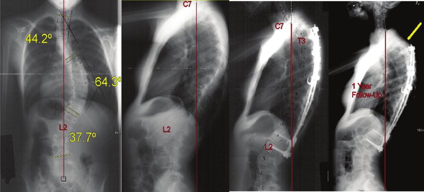
Figure 4. This female with AIS underwent PSF and developed painful PJK.
hybrid instrumentation, combined anterior-posterior Metal hypersensitivity in patients with postoperative pain
spinal fusion, and fusion to the lower lumbar spine and who underwent PSF is another diagnosis of exclusion.
sacrum.28 Overall, metal hypersensitivity should be considered as a
rare cause of pain after PSF in cases where symptoms are
Development of severe symptoms or deformity is often
not explained by other mechanisms such as infection and
used as the benchmark for intervention, and treatment
implant failure. Metal hypersensitivity can cause implant
typically involves extending the fusion construct
loosening and corrosion which both can be identified
proximally to prevent further deformity.
on CT in some cases.29 Biopsy is considered the most
Metal Allergy accurate detector of metal allergy: a predominantly
A local immune reaction to the metal implants used in lymphocytic infiltrate suggests hypersensitivity while a
PSF has been a potential cause of postoperative pain. neutrophilic infiltrate suggests infection.31 The current
Metal hypersensitivity following PSF can present as literature on this condition recommends hardware
pain at the surgical site, swelling, skin reactions, or removal as the only curative intervention.32
radiculopathy making it difficult to differentiate from
other postoperative causes of pain. Metal hypersensitivity
Authors’ Preferred Workup
is thought to involve a local cell-mediated reaction to Algorithm and Management
metal ions that are likely released from the implant Overall, there are no definitive guidelines on timeline
surfaces over time and can lead to aseptic inflammation of the diagnostic workup. Our initial approach is to
and possible loosening of the implants.29 Metal review a detailed history focusing on the course of
hypersensitivity is a delayed Type IV hypersensitivity pain and possibility of psychological causes, including
reaction that gradually affects the surrounding tissues of addiction to pain medications. A thorough review of
the implan.29 Given the gradual nature of the immune symptoms including items such as night sweats, fevers,
reaction, patients with a metal allergy to the implant and chills could indicate possible infection. Physical
typically experience pain-free interval postoperatively exam should include gait assessment, ROM, tenderness,
with symptom onset several months after the initial and neurologic exam. Evaluation of the wound for
surgery.30 any redness, swelling sinus tracts, or dehiscence is
Copyright © 2022 JPOSNA® 6 www.jposna.orgVolume 4, Number 2, May 2022
necessary. Physical examination should also include infection and adjacent segment disease. CT scans are
assessing Waddell signs, which include superficial or used to assess accurate positioning of implants, bone
non-anatomic pain on palpation, pain during painless union, and rule out pseudoarthrosis. Whether to do a CT
evaluation, or overreaction to stimuli which can indicate scan or MRI is guided by presentation and differential
a psychological component. Exam should also evaluate diagnosis: MRI if concerned for infection or degenerative
for sacroiliac or hip pathology that can mimic back pain. disc disease versus CT for mal-positioned implants or
pseudoarthrosis. As a result of image artifact, it can be
Standing AP, lateral, and flexion-extension plain
hard to detect infection on advanced imaging. If infection
radiographs to assess alignment and implant position
is strongly suspected, ultrasound can detect fluid
are needed to rule out obvious issues. We have a collections and guide aspiration and culture.
low threshold for infectious workup with ESR, CRP,
and WBC. If there is a low-grade elevation in these For the patient who is at least a year from PSF and has
parameters, a careful history is taken to include any recent unremitting pain with negative x-rays, blood work, and
illnesses. Often times these tests need to be repeated in a whose CT scan demonstrates solid fusion, we consider
month to demonstrate persistent elevation, which raises exploration, clinical assessment of bone union and possible
the index of suspicion for a nascent infection. implant removal. Shared decision-making is needed to take
this step as some patients can continue to have pain and
Advanced imaging can be ordered to evaluate for pain possible curve gradual curve progression (Figure 5).
generators such as pseudarthrosis, implant failure or
malposition, adjacent segment disease, or infection. An Summary
MRI with and without gadolinium enhancement can After a thorough literature review, the true prevalence
demonstrate a variety of pathologies, such as signs of of chronic pain following surgery remains difficult to
Figure 5. This 14-year-old with AIS and a history of mild back pain and anxiety undergoes PSF for progressive
AIS. She had persistent mid back pain for 5 years despite conservative management. Blood work and CT scan
were negative for infection and implant or pseudarthrosis. At age 20 she undergoes implant removal and has had a
decrease in her pain at most recent follow-up.
Copyright © 2022 JPOSNA® 7 www.jposna.orgVolume 4, Number 2, May 2022
quantify. Each study defines “chronic pain” in the context 8. Will JS, Bury DC, Miller JA. Mechanical low back pain. Am Fam
Physician. 2018;98(7):421-428.
of PSF in their own terms. The true prevalence of chronic 9. Chou R, Deyo R, Friedly J, et al. Nonpharmacologic therapies for low
pain remains unknown but ranged in previous studies back pain: a systematic review for an American College of Physicians
clinical practice guideline. Ann Intern Med. 2017;166(7):493-505.
from 16-64.4% 2 years postoperatively. Common risk 10. Lee CS, Hwang CJ, Lee SW, et al. Risk factors for adjacent segment
factors for these causes of chronic pain include high pre- disease after lumbar fusion. Eur Spine J. 2009;18(11):1637-1643.
11. Hoogendoorn RJ, Helder MN, Wuisman PI, et al. Adjacent segment
operative pain levels and the degree and type of curve degeneration: observations in a goat spinal fusion study. Spine (Phila Pa
preoperatively. Many patients do not have an obvious 1976). 2008;33(12):1337-1343.
12. Charles Malveaux WMS, Sharan AD. Adjacent segment disease after
etiology of pain and causes include mechanical back lumbar spinal fusion: a systematic review of the current literature. Semin
pain, adjacent segment disease, pseudoarthrosis, implant- Spine Surg. 2011;23(4):266-274.
13. Ghandhari H, Ameri E, Nikouei F, et al. Long-term outcome of posterior
related failures, infection, and proximal junctional failure spinal fusion for the correction of adolescent idiopathic scoliosis. Scoliosis
which require a thorough investigative work-up for Spinal Disord. 2018;13:14.
14. Peters M, Willems P, Weijers R, et al. Pseudarthrosis after lumbar spinal
diagnosis. fusion: the role of 18F-fluoride PET/CT. Eur J Nucl Med Mol Imaging.
2015;42(12):1891-1898.
Additional Links 15. How NE, Street JT, Dvorak MF, et al. Pseudarthrosis in adult and pediatric
spinal deformity surgery: a systematic review of the literature and meta-
• POSNAcademy: Anterior Lumbar Vertebral Body analysis of incidence, characteristics, and risk factors. Neurosurg Rev.
2019;42(2):319-336.
Tethering in Adolescent Idiopathic Scoliosis, Courtney
16. Quon A, Dodd R, Iagaru A, et al. Initial investigation of 18F-NaF
E. Baker, MD; Todd A. Milbrandt, MD; D. Dean PET/CT for identification of vertebral sites amenable to surgical
revision after spinal fusion surgery. Eur J Nucl Med Mol Imaging.
Potter, MD; A. Noelle Larson, MD—https://bit.
2012;39(11):1737-1744.
ly/3u78b2Q 17. Hicks JM, Singla A, Shen FH, et al. Complications of pedicle screw
fixation in scoliosis surgery: a systematic review. Spine (Phila Pa 1976).
• W
ebinar: Best Practices for Pediatric, Spine, and Back 2010;35(11):E465-E470.
18. Uehara M, Takahashi J, Ikegami S, et al. Pedicle screw loosening after
Pain, Columbia Orthopedics—https://bit.ly/38wfIQB
posterior spinal fusion for adolescent idiopathic scoliosis in upper and
lower instrumented vertebrae having major perforation. Spine (Phila Pa
• O
rthoKids: Back Pain in Children—https://bit.
1976). 2017;42(24):1895-1900.
ly/3DSyugF 19. Warner SJ, Uppstrom TJ, Miller AO, et al. Epidemiology of deep surgical
site infections after pediatric spinal fusion surgery. Spine (Phila Pa 1976).
Disclaimer 2017;42(3):E163-E168.
20. Coe JD, Arlet V, Donaldson W, et al. Complications in spinal fusion for
No funding was obtained for this study. The authors have adolescent idiopathic scoliosis in the new millennium. A report of the
no conflicts of interest to disclose. Scoliosis Research Society Morbidity and Mortality Committee. Spine
(Phila Pa 1976). 2006;31(3):345-349.
21. Di Silvestre M, Bakaloudis G, Lolli F, et al. Late-developing infection
References following posterior fusion for adolescent idiopathic scoliosis. Eur Spine J.
1. Asher MA, Burton DC. Adolescent idiopathic scoliosis: natural history 2011;20(Suppl 1):S121-S127.
and long term treatment effects. Scoliosis. 2006;1(1):2. 22. Rathjen K, Wood M, McClung A, et al. Clinical and radiographic results
2. Helenius L, Diarbakerli E, Grauers A, et al. Back pain and quality of after implant removal in idiopathic scoliosis. Spine (Phila Pa 1976).
life after surgical treatment for adolescent idiopathic scoliosis at 5-year 2007;32(20):2184-2188.
follow-up: comparison with healthy controls and patients with untreated 23. Potter BK, Kirk KL, Shah SA, et al. Loss of coronal correction following
idiopathic scoliosis. J Bone Joint Surg AM. 2019;101(16):1460-1466. instrumentation removal in adolescent idiopathic scoliosis. Spine (Phila
3. Bastrom TP, Marks MC, Yaszay B, et al. Prevalence of postoperative pain Pa 1976). 2006;31(1):67-72.
in adolescent idiopathic scoliosis and the association with preoperative 24. Muschik M, Luck W, Schlenzka D. Implant removal for late-developing
pain. Spine (Phila Pa 1976). 2013;38(21):1848-1852. infection after instrumented posterior spinal fusion for scoliosis:
4. Sieberg CB, Simons LE, Edelstein MR, et al. Pain prevalence reinstrumentation reduces loss of correction. A retrospective analysis of 45
and trajectories following pediatric spinal fusion surgery. JPain. cases. Eur Spine J. 2004;13(7):645-651.
2013;14(12):1694-1702. 25. Yagi M, Rahm M, Gaines R, et al. Characterization and surgical outcomes
5. Chidambaran V, Ding L, Moore DL, et al. Predicting the pain continuum of proximal junctional failure in surgically treated patients with adult
after adolescent idiopathic scoliosis surgery: a prospective cohort study. spinal deformity. Spine (Phila Pa 1976). 2014;39(10):E607-E614.
Eur J Pain. 2017;21(7):1252-1265. 26. Lee GA, Betz RR, Clements DH, 3rd, et al. Proximal kyphosis after
6. Carrilho PEM, Dos Santos MBM. Focal muscle spasms after thoracic posterior spinal fusion in patients with idiopathic scoliosis. Spine (Phila
spine surgery for schwannoma: the twitching scar. J Mov Disord. Pa 1976). 1999;24(8):795-799.
2020;13(2):168-170. 27. Kim YJ, Bridwell KH, Lenke LG, et al. Proximal junctional kyphosis
7. Rigoard P, Blond S, David R, et al. Pathophysiological characterisation in adolescent idiopathic scoliosis following segmental posterior spinal
of back pain generators in failed back surgery syndrome (part B). instrumentation and fusion: minimum 5-year follow-up. Spine (Phila Pa
Neurochirurgie. 2015;61(Suppl 1):S35-S44. 1976). 2005;30(18):2045-2050.
Copyright © 2022 JPOSNA® 8 www.jposna.orgVolume 4, Number 2, May 2022
28. Lee J, Park YS. Proximal junctional kyphosis: diagnosis, pathogenesis, 31. Davies AP, Willert HG, Campbell PA, et al. An unusual
and treatment. Asian Spine J. 2016;10(3):593-600. lymphocytic perivascular infiltration in tissues around contemporary
29. Shang X, Wang L, Kou D, et al. Metal hypersensitivity in patient with metal-on-metal joint replacements. J Bone Joint Surg Am.
posterior lumbar spine fusion: a case report and its literature review. BMC 2005;87(1):18-27.
Musculoskelet Disord. 2014;15:314. 32. Zairi F, Remacle JM, Allaoui M, et al. Delayed hypersensitivity reaction
30. Guyer RD, Shellock J, MacLennan B, et al. Early failure of metal-on- caused by metal-on-metal total disc replacement. J Neurosurg Spine.
metal artificial disc prostheses associated with lymphocytic reaction: 2013;19(3):389-391.
diagnosis and treatment experience in four cases. Spine (Phila Pa 1976).
2011;36(7):E492-E497.
Copyright © 2022 JPOSNA® 9 www.jposna.orgYou can also read