Original Article Effects of age vestibular and visual systems on the soleus H-reflex
←
→
Page content transcription
If your browser does not render page correctly, please read the page content below
Original Article

Effects of age vestibular and visual systems on the soleus H-reflex

AKSEL CELIK1,2   , FRANCISCO JAVIER ROJAS-RUIZ1,3, MAR CEPERO-GONZALEZ1,4, DAVID M.
KOCEJA1, KOHICHI KITANO1
1Department of Kinesiology, Indiana University, Bloomington, United States of America
2Department of Coaching Education, Dokuz Eylul University, Izmir, Turkey
3Department of Physical Education and Sport ,University of Granada, Granada, Spain
4Department of Musical, Plastic and Corporal Expression, University of Granada, Granada, Spain

                                                              ABSTRACT

The vestibular system, visual and proprioceptive pathways provide information about control of posture, movement and
balance. Loss of postural control directly leads to a greater incidence of falling in the elderly population causing serious
health problems. One important neuromuscular mechanism instrumental in the control of posture and balance is the reflex
system. However, the age-related changes of vestibular and visual systems and their relationship with the reflex system are
not clear. The purpose of this study was to investigate the effects of age, the vestibular and the visual systems on the
modulation pattern of the soleus H reflex. Seventeen neurologically healthy volunteers were categorized by age in two
groups: young (n = 8, mean age = 22.1 ± 5.0 yr.) and elderly (n = 9, mean age = 59.3 ± 12.8 yr.). Maximal soleus H-reflex
(H-max) and motor response (M-max) amplitudes were determined prior to testing at each condition while subjects were
lying supine on a tilt table for standardization. Stimulation intensity was set to evoke a 5-10% M-wave on each trial.
Participants received 5 test H-reflex stimuli in two conditions, static 60º and dynamic 60º on a tilt table. Both tilt conditions
were performed with vision and no vision. A 3-way repeated-measures analysis of variance (ANOVA) 2 (groups: young/old)
x 2 (condition: static/dynamic) x 2(vision: vision/no vision) was used to assess changes in H-reflexes. All data were expressed
relative to the H-reflex amplitude at 0º static on the tilt table. The results showed a significant 3-way interaction (p = .038).
The old group showed greater H-reflex amplitude in the no vision condition at static 60º (vision:0.97; no vision:1.23) whereas
in the young group less modulation was demonstrated in the same condition (vision:1.15; no vision:1.12). These results
suggest in young subjects the vestibular system produced a suppression of the H-reflex with or without visual input; however,
in the old group vision was necessary for this suppression. The interaction between the visual and vestibular systems as we
age needs to be further explored.
Keywords: Balance; Elderly; Electromyography; Motor response; H-reflex suppression.

    Cite this article as:
    Celik, A., Rojas-Ruiz, F.J., Cepero-Gonzalez, M., Koceja, D.M., & Kitano, K. (2021). Effects of age vestibular and visual systems on
        the soleus H-reflex. Journal of Human Sport and Exercise, in press. doi:https://doi.org/10.14198/jhse.2023.181.09

1
 Corresponding author. Department of Coaching Education, Dokuz Eylul University, Izmir 35460, Turkey. https://orcid.org/0000-
0003-0961-7616
   E-mail: aksel.celik@deu.edu.tr
   Submitted for publication April 16, 2021.
   Accepted for publication May 19, 2021.
   Published in press June 15, 2021.
   JOURNAL OF HUMAN SPORT & EXERCISE ISSN 1988-5202.
   © Faculty of Education. University of Alicante.
   doi:10.14198/jhse.2023.181.09
                                                                                        VOLUME -- | ISSUE - | 2021 |               1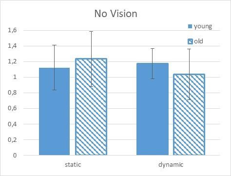
Celik, et al. / Age, vestibular & visual systems                        JOURNAL OF HUMAN SPORT & EXERCISE

INTRODUCTION

Normal aging induces degeneration of numerous structures in the musculoskeletal and nervous system.
These structural changes in turn affect functionality of the different systems. Postural stability is maintained
by the integration of somatosensory, visual and vestibular inputs to the central nervous system, followed by
outputs to the musculoskeletal system. Unfortunately, all components deteriorate with advancing age
(Iwasaki & Yamasoba, 2015). According to Agrawal et al. (2009), the odds of a balance dysfunction increase
with age; nearly 85% of individuals aged 80 years and older have functional evidence of balance dysfunction.

Information from the vestibular system and their central pathways is integrated with the visual and
proprioceptive systems to obtain gaze stabilization and postural stability via the vestibuloocular and
vestibulospinal reflexes, respectively. It is evident that differences in postural control emerge with age
(Vanspauwen, 2018). Loss of postural control directly leads to a greater incidence of falling in the elderly
population causing serious health problems. Numerous studies have suggested an association between age
and falling in the elderly (Schrager, Kelly, Price, Ferrucci, & Shumway-Cook, 2008; Sherrington et al., 2008;
Studenski et al., 2011).

One important neuromuscular mechanism instrumental in the control of posture and balance is the reflex
system. The postural system consists of several sensory systems including the somatosensory, visual and
vestibular. The vestibular system, visual and proprioceptive pathways provide information about the control
of posture, movement, and balance. The vestibular system is a reflex system that contributes to body
equilibrium by adjusting the activity of the postural muscles and also by providing information to supraspinal
centres about head position, motion and spatial orientation (Dizio & Lackner, 1986; Keshner, Allum, & Pfaltz,
1987).

Baloh et al. (2003) (Baloh, Ying, & Jacobson, 2003) showed age- related decreases in vestibular, visual and
somatosensation in normal older people. Teasdale et al. (1991) (Teasdale, Stelmach, & Breunig, 1991) have
demonstrated that alteration in any two of the three sensory inputs (visual, vestibular and somatosensory)
had a significantly greater effect on older subjects than younger subjects. Numerous studies have found that
vestibular function decreases with age (Enrietto, Jacobson, & Baloh, 1999; Paige, 1994; Peterka, Black, &
Schoenhoff, 1990).

Similar, several studies have demonstrated decreased responses of both the spinal stretch reflex and the
Hoffmann (H)-reflex, as well as an inability to properly modulate these spinal pathways in the elderly (Koceja
& Mynark, 2000; Mynark, 2005; Penzer, Duchateau, & Baudry, 2015). Koceja and colleagues demonstrated
that the amplitude of the H-reflex in elderly adults decreased as compared to young adults (Koceja, Markus,
& Trimble, 1995). In a separate study the H-max value was significantly depressed in young subjects but
increased in elderly subjects when standing (Angulo-Kinzler, Mynark, & Koceja, 1998).

Structural changes due to aging in spinal circuits affect their functionality and complicate motor control.
Sensory and motor neurons and interneurons located in the spinal cord are involved and the age-related
structural degeneration resulting in changes in the function of the affected nerves and neural circuits
(Papegaaij, Taube, Baudry, Otten, & Hortobágyi, 2014). The H reflex known to be negatively affected by
advancing age (Kido, Tanaka, & Stein, 2004; Tsuruike, Kitano, Koceja, & Riley, 2012).

Additionally, the greater decrease in the maximum evoked H-reflex (H-max), relative to the decrease in M-
max, suggests that ageing negatively influences the sensory inflow to the motoneurons (Kido et al., 2004).

     2       | 2021 | ISSUE - | VOLUME --                                            © 2021 University of Alicante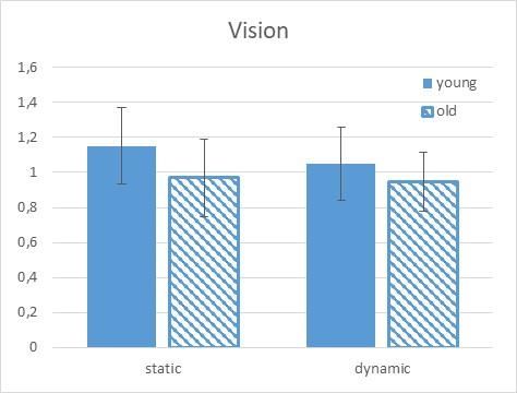
Celik, et al. / Age, vestibular & visual systems                         JOURNAL OF HUMAN SPORT & EXERCISE

This is evident in the differential modulation of H-reflex gain between young and old subjects (Angulo-Kinzler
et al., 1998; Tsuruike et al., 2012). However, the age-related changes of the vestibular and visual systems
and their relationship with the segmental reflex are not clear. Knikou and Rymer (Knikou & Rymer, 2003)
showed that changes in body orientation induced a significant facilitation of the H reflex magnitude in soleus
motoneurons that were essentially independent of angular change in body orientation or of movement
direction. However, Baudry et al. indicated that regardless of age the excitability of the corticomotoneuronal
pathway is not modulated with changes in the sensory conditions during upright standing (S. Baudry, Penzer,
& Duchateau, 2014).

The purpose of this study was to investigate the effects of age-related vestibular and visual systems in static
and dynamic conditioning on the modulation pattern of the soleus H reflex.

MATERIALS AND METHODS

Participants
The experiment was carried out on nine neurologically healthy elderly (mean age = 59.3 ± 12.8 yr.) and eight
healthy young subjects (mean age = 22.1 ± 5.0 yr.), . Subjects completed a general screening questionnaire
and were excluded if they reported any neurological disease, disorder or injury. Participants provided
informed consent prior to participation. All subjects provided informed consent to the procedures as approved
by the University’s Committee for the Protection of Human Subjects.

H reflex and EMG procedures
Surface electrodes were used both for muscle recording and for H-reflex nerve stimulation. For the EMG
recording electrode (Therapeutics Unlimited, Iowa City, IA), Ag/Ag–Cl electrodes with 2 cm intraelectrode
distance were used. The electrodes consisted of an on-site preamplifier, thus minimizing movement artifact.
One of two recording electrodes was positioned over the soleus muscle and the other was placed over the
tibialis anterior muscle of subject’s right leg. Specifically, the electrode on the soleus muscle was adhered
parallel to the muscle fibres at the midpoint between the distal fibres of the gastrocnemius muscles and the
proximal boarder of the Achilles tendon. The electrode for the tibialis anterior was placed lateral to the medial
shaft of the tibia at one third the distance between the knee and the ankle. All electrodes were placed vertically
along the presumed muscle fibre direction. Once in place, the recording and stimulating electrodes were not
removed until the completion of testing, to ensure that exact placement was maintained.

For the H-reflex stimulating electrodes, a 0.8 cm-diameter cathode and a 4 cm diameter anode were used.
To elicit the soleus H-reflex, a cathode electrode was placed in the popliteal fossa and an anode electrode
was placed just superior to the patella of the right leg (Schieppati, 1987). Soleus H-reflexes were evoked
through tibial nerve stimulation with a 1 ms duration pulse.

Maximal soleus H-reflex (H-max) and motor response (M-max) amplitudes were determined prior to testing
at each condition while subjects were lying supine on a tilt table for standardization. The size of the test H -
reflex was measured as the peak-to-peak amplitude. It has been demonstrated earlier that the susceptibility
of the H-reflex to conditioning depends on the size of the control reflex (Crone et al., 1990).

For the H-reflex during the different experimental conditions, the intensity of the H-reflex stimulation was
monitored by keeping constant a 5-10% M-wave preceding each H response (Schieppati, 1987), and
throughout testing, special care was taken to ensure that the size of the small M-wave during each
experimental trial was constant. The EMG signals were DC coupled from the electrodes to a high impedance

                                                                        VOLUME -- | ISSUE - | 2021 |        3
Celik, et al. / Age, vestibular & visual systems JOURNAL OF HUMAN SPORT & EXERCISE
DC amplifier with low bias current requirements. All EMG signals were sampled at 2 kHz, amplified (gain =
1000), and band-pass filtered (20–450Hz).
For H-reflex measurements, the peak-to-peak amplitude of the signal was used as the dependent variable.
To quantify the amount of muscle activity present prior to the H-reflex stimulation, the background EMG
(bEMG) activity in the muscle prior to the H-reflex stimulation for each postural condition was calculated.
After sampling, the bEMG was band passed filtered from 20 to 450 Hz, full-wave rectified and smoothed with
a 25 Hz low-pass filter for the 50 ms prior to H-reflex stimulation.
Experimental protocols on the Tilt table procedures
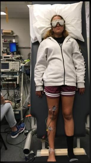
The participants were barefoot their feet side-by-side in a comfortable position and arms hanging relaxed at
their side and they were instructed to keep their head straight on the tilt table. Participants received 5 test H-
reflex stimuli under two randomly administered conditions, static 60º and dynamic 30°-60º on a tilt table. Both
tilt conditions were performed with vision and no vision (Figure 1).
Figure 1. Experimental set-up with participant on the tilt table in a no-vision, static 60° condition.
Statistical analysis
A 3-way repeated-measures analysis of variance (ANOVA) 2 (groups: young/old) x 2 (condition:
static/dynamic) x 2 (vision: vision/no vision/blind) was used to assess changes in H-reflexes applied to test
for interaction and main effects for the dependent variable soleus H-reflex response. Significance was set at
p < .05.
RESULTS
All data were expressed relative to the H-reflex amplitude at static position at 0º lying supine on the tilt table.
The Tests of Within-Subjects Contrasts showed a significant 3-way interaction for the Group x Condition x
Vision (p = .038). This interaction was further analysed. The young group showed an increase in the
amplitude of the H-reflex in the static 60 degree condition whereas the old group inhibited the reflex at this
condition (young: 15% facilitation and old 3% inhibition) when vision was available (first panel, Figure 2).
4 | 2021 | ISSUE - | VOLUME -- © 2021 University of AlicanteCelik, et al. / Age, vestibular & visual systems JOURNAL OF HUMAN SPORT & EXERCISE
When vision was available in the dynamic condition, the young again facilitated the H-reflex (5% facilitation)
whereas the old again inhibited the reflex (5% inhibition; second panel Figure 3).
Figure 2. H-reflex results (standardize to the supine condition) for young and old in the visual experimental
condition. Note that a value of 1.0 represents an H-reflex value equal to the supine condition. The young
subjects produce a facilitation of the H-reflex in both the static and the dynamic conditions whereas the old
produced an inhibition in both the static ad dynamic conditions.
Figure 3. H-reflex results (standardize to the supine condition) for young and elderly groups in the no-vision
experimental condition. Note that a value of 1.0 represents an H-reflex value equal to the static supine
condition. The young subjects produce a facilitation of the H-reflex in both the static and the dynamic
conditions whereas the elderly produce a greater facilitation in the static condition but less facilitation in the
dynamic condition.
VOLUME -- | ISSUE - | 2021 | 5Celik, et al. / Age, vestibular & visual systems JOURNAL OF HUMAN SPORT & EXERCISE
When examining within group differences, vision seemed to play a greater modulatory role for the old and
not the young. When comparing the within group differences between the vision and the no vision conditions
in the static tilt condition, the old group produced 27.3% more facilitation without vision, whereas the young
group produced 3% inhibition. In the dynamic within group comparison, the groups produced similar results.
The young group produced 11.6% more facilitation in the dynamic condition without vision whereas the old
group produced a similar 9.5% greater facilitation in the no-vision condition.
DISCUSSION
The present results suggest differences in the manner in which young and old subjects modulate the soleus
H reflex when the vestibular system is activated. It further demonstrates that the role of vision is more
important in young subjects during dynamic tilt and conversely vision is more important in old subjects during
static tilt. This leads us to conclude that the visual system is important even during periods of stable or static
posture.
It has previously been shown that the ability of elderly subjects to adjust reflex excitability is related to postural
stability (Koceja et al., 1995) and there are several important neural networks that could influence motoneuron
excitability without changing the excitability of the membrane itself. One likely mechanism is presynaptic
inhibition at the Ia terminal. Presynaptic interneurons could receive input from a variety of sensory systems
and/or numerous supraspinal sites responsible for fine movement control; for example, the motor cortex,
cerebellum and basal ganglia. There is evidence that presynaptic interneurons play a role in regulating the
transfer of sensory information to the motoneurons and these interneurons have bene shown to be more
active during periods of increasing postural complexity (Zehr, 2002). However, pertinent to this study, the
role of the vestibular system on reflex modulation is not very well defined and our results add to the complexity
of these interactions. From our results, the young subjects produced an increase in the H-reflex in both the
static and the dynamic conditions, whereas the older subjects demonstrated inhibition in both the static ad
dynamic conditions (see Figure1).
Pinar et al., (2010) (Pinar, Kitano, & Koceja, 2010)originally suggested that presynaptic inhibition is a likely
candidate mediating these changes, but did not specifically examine these interneurons. However, others
have also suggested that presynaptic interneurons play an important role as a segmental gain control
mechanism (Zehr, 2006) and has been suggested in a variety of studies to be an important regulatory spinal
network tasks involving adjustments in postural control (Hultborn, Meunier, Pierrot-Deseilligny, & Shindo,
1987; Meunier & Pierrot‐Deseilligny, 1989; Misiaszek, 2003). Baudry & Duchateau (2012) (Stéphane Baudry
& Duchateau, 2012) proposed that presynaptic interneurons to the soleus Ia afferents increased their activity
when vision was suppressed and when standing on a foam mat, but this adjustment was greater in older
adults.
However, it is important to keep in mind that specific presynaptic interneurons have not been extensively
studies in controlled experiments, and that there are other spinal mechanisms (e.g., reciprocal inhibition,
recurrent inhibition, propriospinal connections) that could influence the excitability of the motoneuron during
various tasks. Given that several experimental have been developed for use in humans that can indirectly
assess presynaptic inhibition, reciprocal inhibition and recurrent inhibition (Pierrot-Deseilligny & Burke, 2005),
future studies will undoubtedly unravel the complexity of motoneuron excitability changes in the intact human,
and identify the exact spinal mechanism(s) responsible for these adjustments.
6 | 2021 | ISSUE - | VOLUME -- © 2021 University of AlicanteCelik, et al. / Age, vestibular & visual systems JOURNAL OF HUMAN SPORT & EXERCISE
The present study was designed to test the interaction effect between aging, vision, and vestibular activation
and there was a significant interaction between these three factors. In fact, in the no vision and dynamic tilt
condition the elderly produce a greater facilitation in the static condition but less facilitation in the dynamic
condition (see Figure 2). This result supports the research of Le Mouel, & Brette (2019) (Mouel & Brette,
2019) in that aging is associated with a progressive shift to a greater reliance on supraspinal pathways
(descending drive) associated with a decreased contribution of Ia afferent input to control leg muscle activity
during standing.
Postural stability depends on the integration of multisensory systems to produce appropriate motor outputs.
Taking into account that in static 60° with vision there is a clear differences between young and old group
(see figure 2) and following Appiah-Kubi (2019) (Appiah-Kubi & Wright, 2019), it can be deduced there is a
reduction of vestibular information with aging, a vestibular and postural training alter sensory organization
after a visual feedback-vestibular activation training protocol, this training should increase the stability
suggesting a possible sensory reweighting through vestibular adaptation. This relation between vestibular
rehabilitation and stability is consistent with the study of Iwasaki and Yamasoba (2015) (Iwasaki & Yamasoba,
2015) that found Vestibular rehabilitation is found to be effective in treating both unilateral and bilateral
vestibular dysfunction.
Furthermore, in agreement with Osoba et al., (2019) (Osoba, Rao, Agrawal, & Lalwani, 2019), results show
that elderly group was particularly dependent on vision to adjust soleus motoneuron excitability, the necessity
of keeping good vision with aging and training vestibular poses a particular challenge for elderly adults and
is linked to decreased falls risk.
CONCLUSIONS
These results suggest in young subjects the vestibular system produced a suppression of the H-reflex with
or without visual input; however, in the old group vision was necessary for this suppression. The necessity of
keep a good vision and the training of vestibular systems shows two main aspects of control posture. The
interaction between the visual and vestibular systems as we age needs to be further explored.
AUTHOR CONTRIBUTIONS
All the authors have contributed substantially to the work reported in conceptualization, methodology,
validation, formal analysis, investigation, procedures, resources, data curation, writing—original draft
preparation, writing—review. All authors have read and agreed to the published version of the manuscript.
SUPPORTING AGENCIES
This work was funded by 2219- International Postdoctoral Research Scholarship Programme of The Scientific
and Technical Research Council Of Turkey (TUBITAK) Scientific Human Resources Development. Award
number:1059B191700345 and by the Spanish Ministry of Science and Innovation (PID2019-110074GB-8
I00/AEI/10.13039/501100011033).
DISCLOSURE STATEMENT
The authors declare that they have no known competing financial interests or personal relationships that
could have appeared to influence the work reported in this paper.
VOLUME -- | ISSUE - | 2021 | 7Celik, et al. / Age, vestibular & visual systems JOURNAL OF HUMAN SPORT & EXERCISE
REFERENCES
Angulo-Kinzler RM, Mynark RG, Koceja DM. (1998) Soleus H-Reflex Gain in Elderly and Young Adults:
Modulation Due To Body Position. Journal of Gerontology 53A(2): M120-MI25.
https://doi.org/10.1093/gerona/53A.2.M120
Agrawal Y, Carey JP, Della Santina CC, Schubert MC, Minor LB (2009) Disorders of Balance and
Vestibular Function in US Adults: Data From the National Health and Nutrition Examination Survey,
2001-2004. Arch Intern Med 169(10):938-44. https://doi.org/10.1001/archinternmed.2009.66
Appiah-Kubi KO, Wright WG (2019) Vestibular Training Promotes Adaptation of Multisensory Integration
in Postural Control. Gait Posture 73:215-220. https://doi.org/10.1016/j.gaitpost.2019.07.197
Baloh RW, Ying SH, Jacobson KM (2003) A longitudinal study of gait and balance dysfunction in normal
older people. Arch Neurol. 60(6):835-9. https://doi.org/10.1001/archneur.60.6.835
Baudry S, Duchateau J (2012) Age-related influence of vision and proprioception on Ia presynaptic
inhibition in soleus muscle during upright stance. Journal of Physiology-London 590(21):5541-5554.
https://doi.org/10.1113/jphysiol.2012.228932
Baudry S, Penzer F, Duchateau J (2014) Input-output characteristics of soleus homonymous Ia afferents
and corticospinal pathways during upright standing differ between young and elderly adults. Acta
Physiol (Oxf) 210(3):667-77. https://doi.org/10.1111/apha.12233
Crone C, Hultborn H, Mazières L, Morin L, Nielsen J, Pierrot-Deseilligny E (1990) Sensitivity of
monosynaptic test reflexes to facilitation and inhibition as a function of the test reflex size: a study in
man and the cat. Exp Brain Res. 81(1):35-45. https://doi.org/10.1007/BF00230098
Dizio PA, Lackner JR (1986) Perceived orientation, motion, and configuration of the body during viewing
of an off-vertical, rotating surface. Perception & Psychophysics 39(1):39-46.
https://doi.org/10.3758/BF03207582
Enrietto JA, Jacobson KM, Baloh RW (1999) Aging effects on auditory and vestibular responses: a
longitudinal study. Am J Otolaryngol 20:371-378. https://doi.org/10.1016/S0196-0709(99)90076-5
Hultborn H, Meunier S, Pierrot‐Deseilligny E, Shindo M (1987) Changes in presynaptic inhibition of Ia
fibres at the onset of voluntary contraction in man. J.Physiol. 389:757-772.
https://doi.org/10.1113/jphysiol.1987.sp016681
Iwasaki S, Yamasoba T (2015) Dizziness and Imbalance in the Elderly: Age-related Decline in the
Vestibular System. Aging Dis. 6(1):38-47. https://doi.org/10.14336/AD.2014.0128
Keshner EA, Allum JHJ, Pfaltz CR (1987) Postural coactivation and adaptation in the sway stabilizing
responses of normals and patients with bilateral vestibular deficit, Exp Brain Res. 69:77-92.
https://doi.org/10.1007/BF00247031
Kido A, Tanaka N, Stein RB (2004) Spinal excitation and inhibition decrease as humans age. Can J
Physiol Pharmacol 82:238-48. https://doi.org/10.1139/y04-017
Knikou M, Rymer WZ (2003) Static and dynamic changes in body orientation modulate spinal reflex
excitability in humans. Experimental Brain Research 152(4):466-475.
https://doi.org/10.1007/s00221-003-1577-3
Koceja DM, Markus CA, Trimble MH (1995) Postural modulation of the soleus H-reflex in young and old
subjects. Electroencephal Clin Neurophysiol. 97:387-393. https://doi.org/10.1016/0924-
980X(95)00163-F
Koceja DM, Mynark RG (2000) Comparison of Heteronymous Monosynaptic Ia Facilitation in Young and
Elderly Subjects in Supine and Standing Positions. International Journal of Neuroscience 104(1),1-
15. https://doi.org/10.3109/00207450009035005
Le Mouel C, Brette R (2019) Anticipatory coadaptation of ankle stiffness and sensorimotor gain for
standing balance. PLoS Comput Biol. 15(11):1-28. https://doi.org/10.1371/journal.pcbi.1007463
8 | 2021 | ISSUE - | VOLUME -- © 2021 University of AlicanteCelik, et al. / Age, vestibular & visual systems JOURNAL OF HUMAN SPORT & EXERCISE
Meunier S, Pierrot‐Deseilligny E (1989) Gating of the afferent volley of the monosynaptic stretch reflex
during movement in man. Journal of Physiology 419:753-763.
https://doi.org/10.1113/jphysiol.1989.sp017896
Misiaszek JE (2003) The h-reflex as a tool ın neurophysıology: ıts lımıtatıons and uses ın understandıng
nervous system function. Muscle Nerve 28:144 -160. https://doi.org/10.1002/mus.10372
Mynark RG (2005) Reliability of the soleus H-reflex from supine to standing in young and elderly. Clinical
Neurophysiology 116(6):1400-1404. https://doi.org/10.1016/j.clinph.2005.02.001
Osoba MY, Rao AK, Agrawal SK, Lalwani AK (2019) Balance and gait in the elderly: A contemporary
review. Laryngoscope Investig Otolaryngol. 4(1):143-153. https://doi.org/10.1002/lio2.252
Paige GD (1992) Senescence of human visual-vestibular interactions, I: vestibulo-ocular reflex and
adaptive plasticity with aging. Exp Brain Res. 2:133-151.
Papegaaij S, Taube W, Baudry S, Otten E, Hortobagyi T (2014) Aging causes a reorganization of cortical
and spinal control of posture. Front. Aging Neurosci. https://doi.org/10.3389/fnagi.2014.00028
Paterka RJ, Black FO, Schoenhoff MB (1990) Age-related changes in human vestibulo- ocular reflexes:
sinusoidal rotation and caloric tests. J Vestib Res.;1:49- 59.
Penzer F, Duchateau J, Baudry S (2015) Effects of short-term training combining strength and balance
exercises on maximal strength and upright standing steadiness in elderly adults. Exp Gerontol.
61:38-46. https://doi.org/10.1016/j.exger.2014.11.013
Pierrot-Deseilligny E, Burke D (2005) The Circuitry of the Human Spinal Cord: Its Role in Motor Control
and Movement Disorders: Cambridge University Press, New York.
https://doi.org/10.1017/CBO9780511545047
Pinar S, Kitano K, Koceja DM (2010) Role of vision and task complexity on soleus H-reflex gain. Journal
of Electromyography and Kinesiology 20(2):354-358. https://doi.org/10.1016/j.jelekin.2009.03.002
Schrager MA, Kelly VE, Price R, Ferrucci L, Shumway-Cook A (2008) The effects of age on medio-lateral
stability during normal and narrow base walking. Gait Posture 28:466-71.
https://doi.org/10.1016/j.gaitpost.2008.02.009
Studenski S, Perera S, Patel K, Rosano C, Faulkner K, Inzitari M et al. (2011) Gait speed and survival in
older adults. JAMA 305:50-8. https://doi.org/10.1001/jama.2010.1923
Sherrington C, Whitney JC, Lord SR, Herbert RD, Cumming RG, Close JC (2008) Effective exercise for
the prevention of falls: a systematic review and meta-analysis. J Am Geriatr Soc. 56(12):2234-4.
https://doi.org/10.1111/j.1532-5415.2008.02014.x
Schieppati M (1987) The Hoffmann reflex: a means of assessing spinal reflex excitability and its
descending control in man. Prog Neurobiol. 28(4):345-76. https://doi.org/10.1016/0301-
0082(87)90007-4
Teasdale N, Stelmach GE, Breunig A (1991) Postural sway characteristics of the elderly under normal
and altered visual and support surface conditions. J Gerontol. 46(6):B238-244.
https://doi.org/10.1093/geronj/46.6.B238
Tsuruike M, Kitano K, Koceja DM, Riley ZA (2012) Differential control of H-reflex amplitude in different
weight-bearing conditions in young and elderly subjects. Clinical Neurophysiology 123(10):2018-
2024. https://doi.org/10.1016/j.clinph.2012.03.006
Vanspauwen R (2018) Dizziness and (Fear of) Falling in The Elderly: A Few Facts. J Int Adv Otol. 14(1):1-
2. https://doi.org/10.5152/iao.2018.0201815
Zehr EP (2002) Considerations for use of the Hoffmann reflex in exercise studies. Eur J Appl Physiol.
86(6):455-68. https://doi.org/10.1007/s00421-002-0577-5
Zehr EP ( 2006) Training-induced adaptive plasticity in human somatosensory reflex pathways.
101(6):1783-94. https://doi.org/10.1152/japplphysiol.00540.2006
VOLUME -- | ISSUE - | 2021 | 9Celik, et al. / Age, vestibular & visual systems JOURNAL OF HUMAN SPORT & EXERCISE
This work is licensed under a Attribution-NonCommercial-NoDerivatives 4.0 International (CC BY-NC-ND 4.0).
10 | 2021 | ISSUE - | VOLUME -- © 2021 University of AlicanteYou can also read