NUCLEOCAPSID VACCINE ELICITS SPIKE-INDEPENDENT SARS-COV-2 PROTECTIVE IMMUNITY
←
→
Page content transcription
If your browser does not render page correctly, please read the page content below
bioRxiv preprint doi: https://doi.org/10.1101/2021.04.26.441518; this version posted April 27, 2021. The copyright holder for this preprint (which
                was not certified by peer review) is the author/funder. All rights reserved. No reuse allowed without permission.

     Nucleocapsid vaccine elicits spike-independent SARS-CoV-2 protective immunity

     William E. Matchetta,b,1, Vineet Joaga,b,1, J. Michael Stolleya,b,1, Frances K. Shepherda,b, Clare F.
     Quarnstroma,b, Clayton K. Mickelsona, Sathi Wijeyesinghea,b, Andrew G. Soerensa,b, Samuel Beckera,b,
     Joshua M. Thiedeb,c, Eyob Weyua,b, Stephen O’ Flanagana,b, Jennifer A. Waltera,b, Michelle N. Vud, Vineet
     D. Menacheryd, Tyler D. Boldb,c, Vaiva Vezysa,b, Marc K. Jenkinsa,b*, Ryan A. Langloisa,b*, and David
     Masopusta,b*
     a
       Department of Microbiology and Immunology, bCenter for Immunology, cDivision of Infectious Disease and
     International Medicine, Department of Medicine, University of Minnesota, Minneapolis, 55455
     d
       Department of Microbiology and Immunology, University of Texas Medical Branch, Galveston, TX, USA
     1
         W.E.M, V.J., and J.M.S. contributed equally

     *Correspondence: masopust@umn.edu, langlois@umn.edu, jenki002@umn.edu

     Author Contributions: W.E.M, V.J., J.M.S., S.W., V.V., M.K.J., R.A.L, and D.M. designed research;
     W.E.M, V.J., J.M.S., F.K.S., C.F.Q, C.K.M, S.W., A.G.S., S.B., E.W., and S.O. performed research;
     J.M.T., J.A.W.,M.V., V.D.M., and T.D.B. contributed reagents/analytic tools; W.E.M, V.J., J.M.S., F.K.S.,
     R.A.L, and D.M. analyzed data; W.E.M, V.J., J.M.S., F.K.S., R.A.L, and D.M. wrote paper.

     Competing Interest Statement: No competing interests

     Classification: Biological Sciences, Immunology, and Inflammation.

     Keywords: SARS-CoV-2, nucleocapsid, vaccine, T cell, protection

     Abstract

     Severe acute respiratory syndrome coronavirus 2 (SARS-CoV-2) is responsible for the COVID-19
     pandemic. Neutralizing antibodies target the receptor binding domain of the spike (S) protein, a focus of
     successful vaccine efforts. Concerns have arisen that S-specific vaccine immunity may fail to neutralize
     emerging variants. We show that vaccination with HAd5 expressing the nucleocapsid (N) protein can
     establish protective immunity, defined by reduced weight loss and viral load, in both Syrian hamsters and
     k18-hACE2 mice. Challenge of vaccinated mice was associated with rapid N-specific T cell recall responses
     in the respiratory mucosa. This study supports the rationale for including additional viral antigens, even if
     they are not a target of neutralizing antibodies, to broaden epitope coverage and immune effector
     mechanisms.

     Introduction
     Vaccine candidates targeting SARS-CoV-2 S protein, which is essential for cell entry, were designed based
     on viral sequences reported in January 2020. Studies of the highly efficacious Pfizer/BioNTech and
     Moderna mRNA vaccines show a strong correlation between neutralizing antibodies (NAb) response and
     protection from disease (1, 2). As the virus continues to spread globally, variants have emerged that evade
     neutralization by vaccine elicited S antibodies (3), which may require continued vaccine adaptation and
     boosting.

     T cells contribute to immunity against respiratory pathogens, including serological variants of influenza
     virus(4). Unlike NAb, T cell immunity is not limited to surface antigens and immunodominant epitopes vary
     considerably between individuals due to recognition in the context of genetically diverse MHC molecules.
     Accordingly, T cell immunity may be less vulnerable to immune selection pressure and viral escape
     mutations. We immunized rodents against SARS-CoV-2-N to address whether protection could be
     conferred independently of spike-specific responses (and thus NAb).

     Results
     Ad5-N vaccine confers protection against SARS-CoV-2 infection
bioRxiv preprint doi: https://doi.org/10.1101/2021.04.26.441518; this version posted April 27, 2021. The copyright holder for this preprint (which
was not certified by peer review) is the author/funder. All rights reserved. No reuse allowed without permission.
Outbred Syrian hamsters are permissive to SARS-CoV-2 infection. We vaccinated hamsters intravenously
(IV) with a human adenovirus serotype 5 (Ad5) vector expressing the N sequence (Ad5-N) derived from
USA-WA1/2021 strain (WA) or a control Ad5 vector lacking a SARS-CoV2 sequence (Ad5-NULL). 7-8
weeks later, animals were challenged intranasally with 6.8x10⁴ pfu WA SARS-CoV-2. Vaccination reduced
weight loss (Fig. 1A, B). Vaccinated hamsters were also challenged with WA or a variant strain B.1.1.7,
which contains two amino acid substitutions in N that do not occur within the N219-227 immunodominant
epitope that we previously defined in C57Bl/6 mice (5). Vaccination elicited a significant 30-fold (WA) and
12-fold (B.1.1.7) reduction in median lung viral titer three days following challenge, though the latter was
not significant. We next immunized transgenic K18-hACE2 (K18) mice that are highly susceptible to SARS-
CoV-2 (6). Ad5-N vaccinated K18 mice experienced significantly reduced weight loss (Fig. 1D, E) and
mortality, with 75% surviving challenge vs. 0% in the Ad5-NULL vaccinated group (Fig. 1F) upon challenge
with 300 pfu WA SARS-CoV-2.
Memory T cells respond to SARS-CoV-2 challenge
Vaccination established SARS-CoV-2-N219-227-specific memory CD8 T cells that persisted in lung, airways,
and spleen from 40 to 86 days, and increased in lung draining mediastinal lymph node (Fig. 2A), which may
result from accumulation of resident memory T cells (7). We examined whether T cells in vaccinated mice
participated in the response four days after WA SARS-CoV-2 challenge. Vaccinated mice showed a ~3.8-
log10 reduction in lung viral load (Fig. 2B). Combined antibody-mediated CD4 and CD8b depletion prior to
challenge partially abrogated protection, although T cell depletion was not absolute in lungs and elevated
N219-227 -specific CD8 T cell responses were still observed in vaccinated mice (Fig. 2B and data not shown).
Intranasal challenge increased granzyme B+ N219-227-specific CD8 T cells in the pulmonary mucosa (Fig.
2C). Total N219-227-specific CD8 T cells substantially increased in both lung parenchyma (IVneg, defined by
the absence of intravascular anti-CD8a staining, as previously described (8)) and draining mediastinal
lymph node, moderately decreased in spleen, and substantially decreased in the lung vasculature (IVpos,
Fig. 2D). These data indicate that vaccine-elicited memory CD8 T cells underwent rapid reactivation and
migration to the site of viral challenge, and that T cells may have contributed to viral control.
Discussion
The rapidity and success of SARS-CoV-2 vaccine development and deployment is unprecedented. Most
strategies, including vaccines licensed for emergency use in the USA, rely solely on the viral spike. Spike
is a logical choice because it contains the receptor binding domain that is the main target of NAb.
Nevertheless, SARS-CoV-2 is likely to become endemic. Viral variants have emerged that escape vaccine-
elicited NAb, and SARS-CoV-2 may continue to evolve with or without selection pressure from increased
global immunity. This study provides evidence in rodents that immunological memory to additional antigens
could provide protection, which may involve memory T cells or non-neutralizing antibodies. It appears likely
that viral evolution will necessitate vaccine evolution and booster immunizations that address emergent
variants. This study supports the rationale for including additional viral antigens into future vaccine
candidates to broaden epitope diversity and protection while limiting opportunities for viral escape.
Materials and Methods
Rodent vaccination, SARS-CoV-2 challenges, assessment of viral titer and T cell responses and other
methods are described in extended methods.
Acknowledgments
We thank the Biosafety Level 3 Program at the University of Minnesota. Supported by the Office of the
Dean of the University of Minnesota Medical School and UMN-Mayo Partnership for Biotechnology and
Medical Genomics. SARS-CoV-2, Isolate USA-WA1/2020 and the B.1.1.7. variant were obtained through
WRCEVA at UTMB. W.E.M., V.J., J.M.S., S.W., and J.M.T. were supported NIH T32 HL007741, the
Canadian Institutes of Health Research Fellowship, NIH T90 DE 022732, F30 DK114942 and NIH T32
AI055433 respectively.bioRxiv preprint doi: https://doi.org/10.1101/2021.04.26.441518; this version posted April 27, 2021. The copyright holder for this preprint (which
was not certified by peer review) is the author/funder. All rights reserved. No reuse allowed without permission.
References
1. A. T. Widge, et al., Durability of Responses after SARS-CoV-2 mRNA-1273 Vaccination. New Engl J
Med 384, 80–82 (2020).
2. F. P. Polack, et al., Safety and Efficacy of the BNT162b2 mRNA Covid-19 Vaccine. New Engl J Med
383, 2603–2615 (2020).
3. W. F. Garcia-Beltran, et al., Multiple SARS-CoV-2 variants escape neutralization by vaccine-induced
humoral immunity. Cell (2021) https:/doi.org/10.1016/j.cell.2021.03.013.
4. A. F. Altenburg, G. F. Rimmelzwaan, R. D. de Vries, Virus-specific T cells as correlate of (cross-)
protective immunity against influenza. Vaccine 33, 500–506 (2015).
5. V. Joag, et al., Cutting Edge: Mouse SARS-CoV-2 Epitope Reveals Infection and Vaccine-Elicited CD8
T Cell Responses. J Immunol, ji2001400 (2021).
6. E. S. Winkler, et al., SARS-CoV-2 infection of human ACE2-transgenic mice causes severe lung
inflammation and impaired function. Nat Immunol 21, 1327–1335 (2020).
7. J. M. Stolley, et al., Retrograde migration supplies resident memory T cells to lung-draining LN after
influenza infection. J Exp Med 217 (2020).
8. K. G. Anderson, et al., Intravascular staining for discrimination of vascular and tissue leukocytes. Nat
Protoc 9, 209–222 (2014).bioRxiv preprint doi: https://doi.org/10.1101/2021.04.26.441518; this version posted April 27, 2021. The copyright holder for this preprint (which
was not certified by peer review) is the author/funder. All rights reserved. No reuse allowed without permission.
Figures
Figure 1. Ad5-N vaccine confers protection against SARS-CoV-2 infection. A) Median and B) peak
weight change after vaccinated hamsters were challenged 7-8 weeks later with WA SARS-CoV-2. C) Lung
viral titers three days post-challenge with WA (left) or B.1.1.7 (right) SARS-CoV-2. D) Median and E) peak
weight change (dashed lines represent humane endpoint), and F) survival of vaccinated K18-hACE2 mice
following intranasal WA SARS-CoV-2 challenge. Bars denote median and interquartile range *, P < 0.05;
**, P < 0.01; ***, P < 0.001. Individual comparisons were analyzed using two-sided Mann–Whitney tests (A-
E) using Bonferroni-Dunn correction for multiple comparisons (A, D) and survival by the log-rank test (F).bioRxiv preprint doi: https://doi.org/10.1101/2021.04.26.441518; this version posted April 27, 2021. The copyright holder for this preprint (which
was not certified by peer review) is the author/funder. All rights reserved. No reuse allowed without permission.
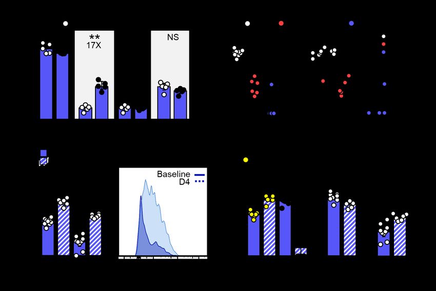
Figure 2. Memory T cells respond to SARS-CoV-2 challenge. A) Number of N219-227-specific CD8 T cells
40 to 86 days after IV Ad5-N vaccination. B) Four days after WA SARS-CoV-2 challenge, viral titers in lungs
of naive and Ad5-N vaccinated K18 mice ± T cell depletion (left) and viral titers plotted against N219-227-
specific CD8 T cells within IVneg lung (right). C) Abundance of granzyme B+ N219-227-specific CD8 T cells in
IVneg lung or mediastinal LN and D) of total N219-227-specific CD8 T cells within indicated compartments
immediately prior (baseline) and four days after challenge. Bars denote median and interquartile range. *,
P < 0.05; **, P < 0.01; ***, P < 0.001 as determined by a two-sided Mann–Whitney test (A-D).You can also read


