NEUROLOGICAL COMPLICATIONS AND INFECTION MECHANISM OF SARS-COV-2
←
→
Page content transcription
If your browser does not render page correctly, please read the page content below

Signal Transduction and Targeted Therapy                                                                                                       www.nature.com/sigtrans

                  REVIEW ARTICLE                          OPEN

                  Neurological complications and infection mechanism of
                  SARS-CoV-2
                                                                                                                              2✉                                 2,3 ✉
                  Dandan Wan1, Tingfu Du2, Weiqi Hong                   1
                                                                            , Li Chen1, Haiying Que1, Shuaiyao Lu                  and Xiaozhong Peng

                      Currently, SARS-CoV-2 has caused a global pandemic and threatened many lives. Although SARS-CoV-2 mainly causes respiratory
                      diseases, growing data indicate that SARS-CoV-2 can also invade the central nervous system (CNS) and peripheral nervous system
                      (PNS) causing multiple neurological diseases, such as encephalitis, encephalopathy, Guillain-Barré syndrome, meningitis, and
                      skeletal muscular symptoms. Despite the increasing incidences of clinical neurological complications of SARS-CoV-2, the precise
                      neuroinvasion mechanisms of SARS-CoV-2 have not been fully established. In this review, we primarily describe the clinical
                      neurological complications associated with SARS-CoV-2 and discuss the potential mechanisms through which SARS-CoV-2 invades
                      the brain based on the current evidence. Finally, we summarize the experimental models were used to study SARS-CoV-2
                      neuroinvasion. These data form the basis for studies on the significance of SARS-CoV-2 infection in the brain.

                      Signal Transduction and Targeted Therapy (2021)6:406                                     ; https://doi.org/10.1038/s41392-021-00818-7
1234567890();,:

                  INTRODUCTION                                                                              receptor,14,17 which may contribute to its stronger transmissibility
                  The emergence of Corona Virus Disease 2019 (COVID-19) in                                  and higher virulence compared with other CoVs.18,19
                  December 2019 was as a result of the outbreak of a novel human                               SARS-CoV-2 has spread fast throughout the world after the first
                  coronaviral pathogen, which was termed severe acute respiratory                           COVID-19 case was detected in Wuhan, infecting a huge number
                  syndrome coronavirus 2 (SARS-CoV-2). This virus has a high                                of people. As of May 2021, according to the World Health
                  similarity with severe acute respiratory syndrome coronavirus                             Organization, there were over 163 million clinically confirmed
                  (SARS-CoV). The virus, SARS-CoV-2, belongs to a beta genus of                             cases with over 3.3 million COVID-19-associated deaths world-
                  coronaviruses and is the seventh member of human corona-                                  wide. COVID-19 was initially defined as a respiratory infection with
                  viruses,1,2 sharing ~80% sequence similarity to SARS-CoV.3,4                              fever, fatigue, abnormal chest X-ray, cough, and shortness of
                  Furthermore, the homology of SARS-CoV-2 is over 90% similar to                            breath.20–22 In addition, a high proportion of COVID-19 patients
                  those of coronaviruses from bats and pangolins, demonstrating its                         exhibit neurological affectations during infection, such as hypo-
                  powerful ability to transmit cross-species.5 The virus shares a                           geusia, dizziness, headaches, myalgia, impaired consciousness,
                  similar spherical structure to other coronaviruses (CoVs) with a                          hyposmia, seizures, and ataxia.23,24 SARS-CoV-2 is extensively
                  diameter of ~100 nm and is a single-stranded positive-sense RNA                           evidenced to cause many neurological diseases,25–28 similar to
                  virus,6,7 composed of four proteins: membrane (M) glycoprotein,                           neurological manifestations previously reported for other respira-
                  nucleocapsid (N) protein, spike (S) glycoprotein, and envelope (E)                        tory viral infections,29–31 however, neurological symptoms of
                  glycoprotein.8 The N protein conjugates with genomic RNA to                               COVID-19 are highly frequent and disabling.32,33 In early
                  form nucleocapsid, while S, M, and E proteins combine to create                           investigations of COVID-19-positive patients in Wuhan, it was
                  an envelope to enclose the nucleocapsid.6 It was revealed that                            demonstrated that 36.4% displayed neurological manifestations,
                  SARS-CoV-2 binds the angiotensin-converting enzyme 2 (ACE2)                               8.9% presented PNS symptoms, the most prevalent of which was
                  receptors under in the presence of the S protein and transmem-                            anosmia (5.1%).34 In particular, in severe COVID-19, partial
                  brane protein serine protease 2 (TMPRSS2) could infect target cells                       neurodegeneration, brain edema, even encephalitis was
                  expressing ACE2 receptors,9–12 including alveolar cells, macro-                           observed.35–37 The neurodegenerative changes of cell death,
                  phages, endothelial cells, kidney cells, intestinal epithelial cells,                     hyperphosphorylation, and dislocation of Tau protein were
                  monocytes, neurons, glial cells, and neuroepithelial cells.13–16                          documented in SARS-CoV-2-infected cells.10 Moreover, some
                  Interestingly, some reports discovered that S protein of SARS-CoV-                        COVID-19 patients were positive for SARS-CoV-2 in the cerebral
                  2 contains specific domains encoding polybasic cleavage sites3                             spinal fluid (CSF) and brain tissue.38–40 These findings imply that
                  and SARS-CoV-2 exhibits a high affinity for human ACE2                                     SARS-CoV-2 infections are not restricted to the respiratory

                  1
                   Laboratory of Aging Research and Cancer Drug Target, State Key Laboratory of Biotherapy, National Clinical Research Center for Geriatricts, West China Hospital, Sichuan
                  University, No. 17, Block 3, Southern Renmin Road, 610041 Chengdu, Sichuan, PR China; 2National Kunming High-level Biosafety Primate Research Center, Institute of Medical
                  Biology, Chinese Academy of Medical Sciences and Peking Union Medical College, Yunnan, China and 3State Key Laboratory of Medical Molecular Biology, Department of
                  Molecular, Biology and Biochemistry, Institute of Basic Medical Sciences, Medical Primate Research Center, Neuroscience Center, Chinese Academy of Medical Sciences, School of
                  Basic Medicine Peking Union Medical College, Beijing, China
                  Correspondence: Shuaiyao Lu (lushuaiyao-km@163.com) or Xiaozhong Peng (pengxiaozhong@pumc.edu.cn)
                  These authors contributed equally: Dandan Wan, Tingfu Du, Weiqi Hong

                  Received: 19 June 2021 Revised: 27 September 2021 Accepted: 2 November 2021

                  © The Author(s) 2021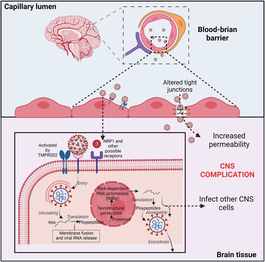
Neurological complications and infection mechanism of SARS-CoV-2
                                                               Wan et al.
2
    systems, but they also can reach the CNS and induce neurological        categories: CNS symptoms, PNS symptoms, and relative skeletal,
    conditions.41                                                           muscular symptoms34,56–58 (Fig. 1 and Table 1).
       However, the neuroinvasive mechanisms of SARS-CoV-2 remain
    unknown. COVID-19 is highly aggressive and is accompanied by
    hypoxia, abnormal clotting, and severe inflammation, so most CNS         CENTRAL NERVOUS SYSTEM DISEASES AND/OR SYMPTOMS
    symptoms are identified as manifestations of peripheral pathol-          Cerebrovascular disease is a group of diseases characterized by
    ogies. Concurrently, ACE2 and TMPRSS2 are vital for SARS-CoV-2          damage to brain tissues due to intracranial blood circulation
    invasion and spread in the body, expressing suppressed levels in        disturbances, which generally occur in the brain’s blood vessels.
    the brain,42 confirming that CNS symptoms are indications of             As we know, virus infections can lead to vascular endothelial as
    peripheral pathologies. However, current research reveals that          well as vascular system damage, resulting in an overactive
    SARS-CoV-2 also can invade cells via Neuropilin-1 (NRP1),43,44          inflammation response, which can lead to ischemic and hemor-
    BASIGIN (BSG),45 Cathepsin L (CTSL), and furin46 which have a           rhagic infarcts and then further develop to thrombosis and
    higher and broader expression in the brain when compared to             vasculitis.59,60
    TMPRSS2 or ACE2.47 This would be one mechanism through which               Although the primary manifestation of COVID-19 is lung
    SARS-CoV-2 propagates in the brain. Recent research has indicated       disease, cerebrovascular disease is also a primary neurological
    that olfactory sensory neurons are a potential route for CNS            complication of COVID-19 that is associated with a high mortality
    invasion.48–51 It is clear that pathological inflammation, as well as    rate.61 A retrospective study suggested that the severe COVID-19
    the cytokine storm initiated by COVID-19 outside the brain,             patients generally had acute cerebrovascular diseases.34 The
    impacted the CNS.52–55 Understanding the invasion mechanisms            pooled prevalence of SARS-CoV-2-associated acute cerebrovascu-
    of SARS-CoV-2 in the nervous system is critical for the rational        lar disease is estimated to be 2.3%.62 An epidemiological and
    treatment of patients. Although the SARS-CoV-2 neuroinvasion            clinical characteristics study revealed that 52% of COVID-19
    mechanism is yet unknown, and considering the highly similar            patients exhibited increased IL-6 levels, with 86% among them
    viral sequence from SARS-CoV, a similar neuroinvasiveness               presenting high CRP levels, indicating a substantive inflammatory
    mechanism may be relevant for SARS-CoV-2.41 Herein, we review           response.63 Concurrently, inflammation is required in developing
    previously documented SARS-CoV-2-associated CNS and PNS                 cerebrovascular diseases, leading to cerebrovascular events.64 A
    complications and further discuss the varieties of potential            Spanish medical center documented that among 1683 hospita-
    neuroinvasiveness mechanisms to help neurologists better under-         lized COVID-19 patients, 1.4% developed cerebrovascular disease,
    stand SARS-CoV-2 influence on the nervous system and facilitate          in which 73.9% of them had ischemic stroke, and 21.7% had
    diagnosis as well as reasonable COVID-19 treatment.                     hemorrhagic stroke with increased levels of ferritin.65 Brain
                                                                            biopsies revealed thrombotic microangiopathy as well as
                                                                            endothelium injury, indicating that due to coagulopathy, seriously
    NEUROLOGICAL SYMPTOMS OF COVID-19                                       infected patients are highly vulnerable stroke.66 Meanwhile,
    SARS-CoV-2 is extensively evidenced to present potential neu-           additional researches have also corroborated this perspective.
    roinvasion. As a result, we reviewed studies on COVID-19, whose         For instance, examining severe patients indicated that these
    neurological manifestations could be assigned into three                people developed coagulation dysfunctions and high D-dimer
                                                                            levels,67 implying that those people may be at an increased risk of

    Fig. 1 SARS-CoV-2-associated neurological symptoms. A variety of neurological manifestations are present in COVID-19 patients, such as
    encephalitis, encephalopathy, ageusia, anosmia, Miller Fisher syndrome, and Guillain-Barré syndrome

                                                                              Signal Transduction and Targeted Therapy (2021)6:406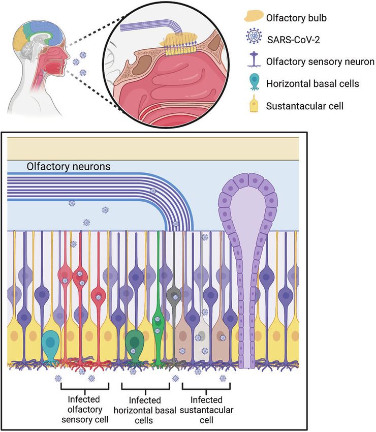
Neurological complications and infection mechanism of SARS-CoV-2
                                                                                  Wan et al.
                                                                                                                                                        3
 Table 1.   Neurological symptoms in COVID-19 patients

Neurological symptom                                       Affected region (reference)                                  Percentage (reference)

Acute cerebrovascular disease                              Cerebral vessels59,60                                        2.8%34
                                                                 39,84
Meningitis/encephalitis                                    CSF                                                          Case report287
                                                                                  288
Acute hemorrhagic necrotizing encephalopathy               Temporal lobe                                                Case report288,289
                                                                    91,290,291
Posterior reversible encephalopathy syndrome               Cortex                                                       Case report91,290,291
                                                                            292
Demyelinating lesion                                       Spinal cord                                                  Case report292
                                                                                               293–295
Seizure                                                    Left temporoparietal lobe                                    0.5%34
Ischemic stroke                                            Cortex34                                                     2.8%34
Dizziness                                                  Whole brain296                                               9.4%297 16.8%34
                                                                             34,298,299
Headache                                                   Whole brain                                                  3.4%300 6.5%297 13.1%34
                                                                             34
Ataxia                                                     Whole brain                                                  0.5%34
                                                                             34
Impaired consciousness                                     Whole brain                                                  7.5%34
                                                                           301
Brain edema                                                Brainstem                                                    Case report301
                                                                                        126
Anosmia                                                    Olfactory neurons                                            5.1%34
                                                                                  106,107,302,303
Ageusia                                                    Tongue nerves                                                5.6%34
Dysopia                                                    Optic nerves34                                               1.4%34
Guillain-Barré syndrome                                    Peripheral nerve demyelination304–310                        Case report310,311
                                                                             312,313
Miller Fisher syndrome                                     Whole brain                                                  Casa report312,313
                                                                                              314,315
Myalgia-muscle pain                                        Neuromuscular junction                                       Case report314,315
                                                                     316
Rhabdomyolysis                                             Muscle                                                       Case report316

venous thrombosis accompanied by hemorrhagic stroke.68 A 75-                      inflammation,          hypercoagulability,      and       pulmonary
year-old COVID-19-related woman who had no notable pre-                           encephalopathy.79
disposing factors but with severe bipartite pneumonia as well as                     Although it is seemly hard for the virus to penetrate the CNS,
acute pulmonary embolism implies that serious infections are an                   CoVs can invade the CNS own their neurotropism;80,81 then may
induced factor of acute venous embolism and associated                            lead to overreacting immune responses and induce fatal
stroke.69,70 During COVID-19 dissemination, patients with cardio-                 encephalitis and meningitis. Encephalitis refers to pathogen-
cerebrovascular risk factors are more susceptible to develop acute                induced inflammatory lesions (neuronal damage and nerve tissue
stroke.71 Six COVID-19 patients had an acute ischemic stroke in                   injury) in brain parenchyma, with typical symptoms, including
large vessels with typical signs of neurological dysfunctions,                    elevated temperature, headache, vomiting, fatigue, and con-
including aphasia, prosopoplegia, sensory loss, dysarthria, and                   sciousness disorders.82 Based on autopsy analyses of 17 cases,
acute confusion. Two patients exhibited multiple infarctions                      eight patients exhibiting cerebral edema and vascular congestion
attributed to coinstantaneous venous as well as arterial thrombo-                 were found SARS-CoV-2 positive in brain tissue.40 Another autopsy
sis. Five cases were positive for lupus anticoagulants, demonstrat-               report also indicated edema in brain tissue and neuronal
ing a coagulation disturbance. Angiograms showed occlusion of                     degenerations in SARS-CoV-2-infected people.83 Meanwhile, a
cerebral artery trunk or branches.72 In the other six cases, four                 Japanese male with developed seizure as well as unconsciousness
patients were diagnosed with ischemic stroke, while the remain-                   after fever and general weakness exhibited a SARS-CoV-2
ing two patients presented with hemorrhagic stroke. They all                      positivity in the CSF and was diagnosed with SARS-CoV-2-related
experienced severe pneumonia and complications in multiple                        meningitis.39 McAbee et al. presented an encephalitis case with
organs, and high transaminase and lactate dehydrogenase (LDH)                     positive SARS-CoV-2 in CSF, accompanied by increased red and
levels, indicating poor clinical outcomes. However, before COVID-                 white cells.38 The team of Beijing Ditan Hospital evidenced SARS-
19 outbreak, only one patient had a possible vascular risk factor                 CoV-2 positivity in the CSF of SARS-CoV-2-infected patients by
associated with stroke.73 Four SARS-CoV-2-infected patients from                  genome sequencing, hence verifying viral encephalitis.84 As
New York demonstrated that two of them developed subarach-                        known, immune cells and viruses can enter the brain via a
noid hemorrhage (SAH) as well as hemorrhagic transformation                       weakened blood–brain barrier (BBB).85 Even though not all
following ischemic stroke, but SARS-CoV-2 remained undetectable                   patients test positive for SARS-CoV-2 in CSF, symptoms of nervous
in the CSF.74 SARS-CoV-2 easily attacks the lung, lowering blood-                 system infections and injuries implies the possibility of COVID-19-
oxygen saturation and then causing hypoxemia, resulting in                        related neurological complications.57
altered consciousness, delirium or confusion, and intracerebral                      Acute necrotizing encephalopathy is a rare severe explosive
hemorrhage or acute/subacute stroke.63,70,75 Moreover, SARS-CoV-                  encephalopathy and that is common as a result of viral infection.
2 impact on ACE2 receptor imbalances the renin–angiotensin                        The pathogenesis of which is generally considered as cytokine
system (RAS), resulting in microcirculation disorders, impairing the              storm-induced breakdown of blood–brain barrier.54 Encephalo-
regulation of cerebral blood flow, and producing an excessive rise                 pathy is recognized as the most common CNS complication of
in blood pressure, hence increasing the risk of encephalorrhagia                  COVID-19, caused by hypoxia or systemic diseases.86 Statistically,
and ischemic stroke.76,77 Hemorrhagic lesions may arise from                      ~50% of hospitalized patients with COVID-19 have encephalo-
coagulation dysfunctions caused by severe systemic infections or                  pathy,87 with common signs including dizziness, cognitive
by invasions of the vascular endothelium by viruses.78 As a result                dysfunction, ataxia, mental disorder, or even impaired conscious-
of cerebral white matter damage and blood vessels, the general                    ness. Old people with hepatic, cardiovascular, and renal comor-
state of CIVD-19 patients may aggravate.66 The stress state caused                bidities, as well as immunosuppressed persons, are more likely to
by injuries to endothelial cells and hypoxia can promote                          develop encephalopathy following COVID-19 infection.34 A 50-

Signal Transduction and Targeted Therapy (2021)6:406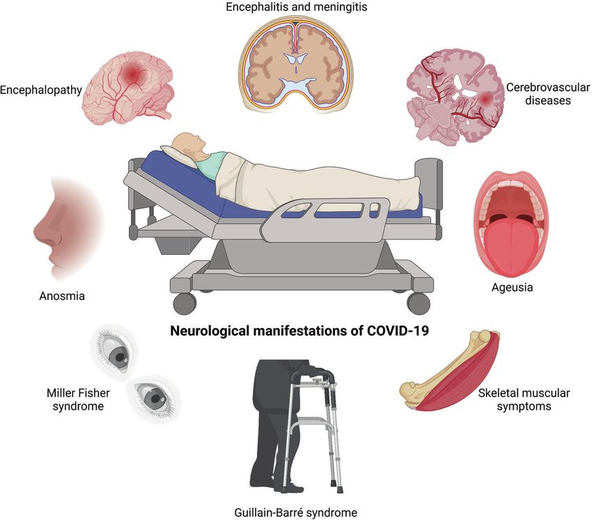
Neurological complications and infection mechanism of SARS-CoV-2
                                                                Wan et al.
4
    year older woman was the first to be diagnosed with acute                 Toscano et al. documented five COVID-19-related cases with limb
    necrotizing hemorrhagic encephalopathy associated with COVID-            or facial paresis as well as paresthesia within some days after the
    19, with primary symptoms of fever, altered mental state, and            appearance of COVID-19 symptoms.108 MRI results revealed caudal
    cough, and neuroimaging indicated symmetrical multifocal                 roots or facial nerve enhancement, indicating that nerves undergo
    changes as well as thalamic invasions.88 Reichard et al. reported        immune response. To date, a rapid increase of GBS cases has been
    that a COVID-19 patient appeared to die of acute disseminated            confirmed in COVID-19 patients.109,110 However, a previous
    encephalomyelitis according to pathological findings.89 Mean-             epidemiological cohort study found no relationship between
    while, some people manifest unexpected seizures owing to high            GBS and COVID-19.111 It is unclear whether the causative agent for
    fever. Combined with analysis of patients previously found to have       GBS is SARS-COV-2 virus itself or other infections secondary to
    epilepsy, apprehension and sleep disorders caused by the COVID-          COVID-19-related patients, therefore, more studies are required to
    19 pandemic may be an additional trigger for epilepsy.90 Four            elucidate on this phenomenon.
    SARS-CoV-2-infected patients aged 60–70 years presented with                SARS-CoV-2 infection can result in dyspnea and decrease the
    seizures and exhibited elevated blood pressure and renal injury,         blood-oxygen saturation by attacking lung.63 Indeed, severe
    systemic inflammation, and hypercoagulopathy and were diag-               COVID-19 patients with acute respiratory distress syndrome
    nosed with posterior reversible encephalopathy syndrome                  (ARDS) can cause serious systemic hypoxemia, which may be
    (PRES).91 Moreover, autopsy analyses of four confirmed COVID-             related to congestion and edema observed in brain tissue.34,112
    19 cases revealed hypoxia-induced cerebral impairments and               Meanwhile, hypoxemia induced by ARDS and lung injury may
    observed small perivascular lesions that appeared in the white           promote SARS-CoV-2 to invade the brain tissue. A study about
    matter.92 However, there is no clear evidence to indicate whether        three COVID-19 patients without breathing difficulties under low
    neuropathologic lesions are caused by primary vascular diseases          oxygen tension has been reported.113 These patients had
    that are a result of damage to white matter or demyelinating             extremely low blood-oxygen levels, which would result in
    diseases through a by-infectious mechanism.                              unconscious or multiple organ failures. However, all of them were
                                                                             awake and had no signs of dyspnea, challenging the under-
                                                                             standing of basic biology nowadays. Notably, patients with
    PERIPHERAL NERVOUS SYSTEM SYMPTOMS                                       hypoxemia may exhibit mild symptoms but may rapidly progress
    The SARS-CoV-2 virus can cause chemosensory disorders, includ-           to multiple organ failure and death.114 Another research indicated
    ing anosmia, ageusia, hyposmia, etc.5,93,94 Over one-third of            that severe systemic hypoxemia would aggravate dementia
    confirmed COVID-19 patients exhibit smell or taste disturbances,62        symptoms in AD patients infected with COVID-19.115,116 To date,
    which can develop prior to, contemporaneously, or after fever and        various hypotheses have been advanced to account for hypox-
    cough.95 Approximately 50% of patients experience anosmia or             emia. One is that SARS-CoV-2 may directly act on oxidation-
    ageusia in the early stages of COVID-19.96,97 It is also reported that   sensitive receptors, alter respiratory center responses to low
    about 11.8% of COVID-19 cases manifested an olfactory disorder           oxygen levels, and modify the way PCO2 to blunt the response of
    before developing other symptoms, implying that anosmia is               the brain to hypoxia.113 Another hypothesis indicates that in a
    crucial for early disease detection.98 Statistics indicate that          corticolimbic network, SARS-CoV-2 can cause neuronal damage
    anosmia and ageusia can be regarded as COVID-19 predictors.              that may alter the secretion of endogenous neuropeptides or
    When combined with fever, discrimination accuracy can increase           neurotransmitters, which are related to perceptual effects.117 More
    up to 75%.99 Although the prevalence of dysosmia and dysgeusia           studies are needed to confirm or disprove these views in the
    decreases in older people,100 female patients have higher smell          future.
    disturbance.101 Another research indicated that about 12% of
    SARS-CoV-2-infected patients develop only anosmia as their initial
    symptom.57 A clinical study analysis with 33 COVID-19 cases              SKELETAL MUSCULAR SYMPTOMS
    reported that ~63.6% of patients developed chemosensitive                Skeletal muscular symptoms are another prevalent PNS complica-
    dysfunction, and 13 patients presented combined anosmia and              tion of COVID-19 patients, presenting tiredness, myositis, myalgia,
    ageusia.102 Moreover, patients may exhibit other related sensory         and skeletal muscle injury. Several cases of skeletal muscular
    dysfunctions, such as tinnitus, sore throat, vertigo, dysphagia, and     symptoms in SARS-CoV-2-positive people have been reported.34
    hearing.103,104 These studies indicate that SARS-CoV-2 virus can         Research of 213 COVID-19 cases indicated that ~85.2% of patients
    cause hyposmia and anosmia, implying that thorough examina-              had significantly elevated serum creatine kinase. Another pub-
    tion of otolaryngologic manifestations may assist in early COVID-        lished study demonstrated elevated lactate dehydrogenase and
    19 diagnosis.                                                            creatine kinase levels, which may be caused by skeletal muscle
       Guillain-Barré syndrome (GBS), a PNS condition, is induced by         injury.118 Mehan et al. described nine COVID-19 cases who had
    overactive immune system, which attacks the PNS by mistake.105           backache, dyskinesia, and paresthesia in lower limbs. Seven
    The initial signs are tingling and weakness in the periphery, but        patients exhibited intramuscular edema through spinal cord MRI,
    they can rapidly distribute leading to whole-body paralysis.             confirming paraspinal myositis.119 In addition, most coronavirus
    Representative neurological manifestations include progressive           infections could result in functional defects as well as myalgias in
    dysergia, flaccid paralysis, and areflexia. The first case of COVID-19-     skeletal muscles with increased CK levels.120 Similar skeletal
    related GBS was a 61-year-old woman from Wuhan, China.106 The            muscular symptoms were found in the studies of SARS-CoV.121
    initial symptom was the weakness of the lower limbs which is in          Virus-induced elevated pro-inflammatory cytokines level might be
    line with the neurological examination: areflexia and symmetric           another reason to aggravate the muscular injury.122 However,
    weakness in both legs and feet. Various examination results              additional research should be conducted to validate whether
    revealed lymphocytopenia, thrombocytopenia, elevated protein             SARS-CoV-2 can cause skeletal muscular sequelae in the
    levels in CSF, and motor and sensory nerve demyelination, all of         long term.
    which were suggestive with GBS. Another GBS patient was a 71-
    year-old male in Italy who had no history of neurological
    disorder.107 Although brain CT scan revealed normal findings,             MECHANISMS THROUGH WHICH SARS-COV-2 INVADES THE
    electroneurography measurements indicated the absence of tibial          NERVOUS SYSTEM
    nerve compound muscle action potential (CMAP) and sural nerve            Generally, the CNS with a very intricate brain barrier system to
    sensory nerve action potential (SAP), indicating peripheral              defend against the virus invasion, including blood-cerebrospinal
    neuropathy caused by demyelination, a typical feature of GBS.            fluid barrier, blood–brain barrier (BBB), and brain-cerebrospinal

                                                                               Signal Transduction and Targeted Therapy (2021)6:406
Neurological complications and infection mechanism of SARS-CoV-2
Wan et al.
5
fluid barrier. Although the CNS is protected by multilayer barriers, neurons and then cause their necrosis in the study of human
where also can be invaded by various viruses involved in the glial ACE2 transgenic mice as well as brain organoids;137,138 however,
cell or neuronal invasions.123 Coronaviruses were reported to the amounts of affected cells were limited due to low TMPRSS2
reach the CNS causing neurovirulence.124 However, the exact and ACE2 levels. The autopsy studies on 32 COVID-19 patients
mechanism of coronaviruses invade the CNS has not been fully indicated thrombotic and thromboembolic signs in olfactory
distinct.125 Mostly, viral infections start from peripheral tissues and mucosa and CNS observed from the section of olfactory mucosa
then spread to the peripheral nerves and finally reaches the and thalamus samples, and SARS-CoV-2 S protein also observed in
central nervous system.126 This process may explain the presence the endothelial cells of small CNS vessels. The levels of virion load
of neurological lesions, like demyelination.127 ACE2 is highly in olfactory mucosa were 124% higher than in the lower
expressed on vascular endothelial cells, and also expressed on respiratory tract,139 demonstrating that nasal epithelium may be
olfactory epitheliums, striatum, cortex, substantia nigra, as well as an entry point for SARS-CoV-2 to reach the brain through the
the brainstem,128 suggesting SARS-CoV-2 can directly infect centripetal route. Notably, no ACE2 mRNA was observed in the
vascular endothelial cells to cross the BBB and then can infect human brain.140,141 To date, ACE2 distribution mostly relies on
cells throughout the CNS. The ACE2 receptor is also expressed on mRNA data analysis, but mRNA incompletely reflects the
glial cells and neurons of various structures, including olfactory distribution of true functional protein. Therefore, numerous
epitheliums, striatum, cortex, substantia nigra, as well as the immunohistochemical characterization researches are urgently
brainstem, implying that SARS-CoV-2 has the potential to infect required.
cells in the CNS. Moreover, in COVID-19 patients, hyposmia is a
frequent complication, indicating the infection of the olfactory Other receptors expressed in the brain
nerve. Therefore, the synaptic connections via olfactory nerves Based on the above description, ACE2 expression in the brain is
would be another mechanism through which SARS-CoV-2 enters very low. However, numerous COVID-19 patients had higher levels
the CNS. SARS-CoV-2-induced inflammation is also considered to of viral modifications in brains than predicted by ACE2 expression
disrupt the BBB allowing virus to enter the CNS.129,130 patterns alone, implying the presence of alternative viral entry
mechanisms besides ACE2. More recently, neuropilin-1 (NRP1) was
identified as a novel mechanism of access to the brain for SARS-
CNS EXPRESSION OF KEY VIRAL INFECTION FACTORS CoV-2, which was expressed in brains and olfactory bulbs (OBs)
Expression of ACE2 and TMPRSS2 (Table 2), at high levels relative to ACE2 and TMPRSS2.43,44,142 The
Currently, it is accepted that SARS-CoV-2 gains entry into cells via autopsies of the olfactory epithelium from COVID-19 patients
ACE29,24 under the participation of TMPRSS249,131 due to the high determined that the infected olfactory epithelial cells with high
expression of both these two proteins in the lung. In addition, expression of NRP1.43 In addition to NRP1, SARS-CoV-2 invades
numerous studies indicate that both ACE2 and TMPRSS2 also cells via BASIGIN (GBS) and Cathepsin L (CTSL), which also
expressed in the brain at suppressed levels (Table 2), so in theory facilitated SARS-CoV-1 to infect cells45,46,143–147 (Table 2). All these
the virus should infect brain cells.14,49,132–134 Lazartigue and proteins have a higher expression in the human brain when
coworkers, according to early immunohistochemical work, found compared to ACE2 or TMPRSS2 and express in the olfactory bulb.
the existence of ACE2 in the neurons of rat brain rather than Accordingly, SARS-CoV-2 can reach the brain possibly through
glia.133 They also established that ACE2 is critical in blood pressure olfactory epithelium (OE), vagus nerve, BBB, or CSF and infect
regulation and in autonomic nerve system diseases. ACE2 is the brain.
expressed both in tractus solitarius and in the areas involved in
blood pressure central regulation, such as paraventricular Transcribial route and neuronal transport dissemination
nucleus.135 Interestingly, this ACE2 is expressed in a professional Numerous evidence indicated that certain CoVs initially invaded
group of CNS structures without the BBB, named circumventricular peripheral nerve terminals and then spread throughout CNS
organs, implying a possible direct path for brain invasion by SARS- through anterograde/retrograde of synapses,41,83,148 including
CoV-2. Recent RNA-Seq studies have found that ACE2 is elevated HEV67,148,149 as well as OC43-CoV.29 In the PNS, the olfactory
in the substantia nigra, ventricles, olfactory bulb, posterior nerve is the main route for SARS-CoV-2 to invade CNS due to high
cingulate cortex, posterior cingulate cortex, choroid plexus, middle levels of TMPRSS2 and ACE2 in olfactory epithelium cells, both of
temporal gyrus, frontal, and motor areas (https://www. which are required for viral binding and accumulation.125,131,150,151
proteinatlas.org/). ACE2 was also found to be expressed in The olfactory nerve is a CNS conduction bundle instead of a real
postmortem frontal cortex vessels with different calibres and nerve, which directly contacts the brain (Fig. 2).152,153 In the nasal
significantly elevated in the brain vasculature of dementia and cavity, the olfactory mucosa is composed of neurons, Bowman’s
hypertension patients.136 SARS-CoV-2 was found to invade glands, basal cells, and epithelial cilia.154–156 The special olfactory
neuroepithelium of the nasal cavity has an apical surface
composed primarily of sustentacular cells.157 Support cells were
Table 2. Receptors or proteins related to SARS-CoV-2 infection in the demonstrated to present high levels of TMPRSS2 and ACE2,49
nervous system indicating that they were predisposed to SARS-CoV-2 infection.48
As observed in the hamster, OE infection would spread to
Receptor or Main expression region Reference horizontal basal cells (HBCs) and then to immature or mature
protein olfactory neurons.51 Meanwhile, the infected HBCs subsequently
ACE2 Pituitary gland, nucleus accumbens, 42,317–321 matured into OSNs, which would reach OB via a synaptic path that
hypothalamus may potentially infect CNS.158 Through this way, a peripheral
49,322,323 infection could access OB and further spread throughout the
TMPRSS2 Pituitary gland, hypothalamus,
cerebellum
brain. Another autopsy study presented that SARS-CoV-2 invaded
43
CNS by traversing the neural mucosal and subsequently
NRP1 Olfactory bulb penetrated the neuroanatomical areas with olfactory tract
45,145–147
BASIGIN Frontal cortex, pituitary gland projections, such as respiratory as well as cardiovascular control
Cathepsin L Pituitary gland, spinal cord 45,143,144
centers in the medulla.139 The authors also observed the
Furin Lung, brain 46 characteristic CoV substructures and SARS-CoV-2 RNA in olfactory
ATR1 Pituitary gland, substantia nigra 324–326 epithelium cells and olfactory mucus cells. The viral load in
olfactory mucosa, medulla oblongata, olfactory tubercle, oral
Signal Transduction and Targeted Therapy (2021)6:406Neurological complications and infection mechanism of SARS-CoV-2
Wan et al.
6
ACE2. Both ACE2 and TMPRRSS2 are expressed in intestinal enteric
neurons and glia, indicating that they are susceptible to SARS-
CoV-2.163 The gut–brain axis is a critical component to cause CNS
disorders.164 A study of 42 COVID-19 patients found that ~66.67%
of them were positive for SARS-CoV-2 RNA in feces.165 In vitro,
SARS-CoV-2 was shown to infect human intestinal epithelium.166
The virus would spread from duodenal cells to the brainstem
neurons via anterograde and retrograde transmission.167 Conse-
quently, it is reasonable that SARS-CoV-2-infected enterocyte
would spread to neuronal and glial cells of enteric nervous
systems and finally invade CNS through the vagus nerve.125,168 In
this route, various data showed that initial SARS-CoV-2 infection in
the lung would cause subsequent viral spreading to the brain,
specifically the areas of the thalamus and brainstem, including the
vagus nerve medullary nuclei.169 Matsuda et al. documented that,
through the vagus nerve, influenza A virus spread from the
respiratory tract to the vagal ganglia.170 Another study demon-
strated ACE2 expression of the vagal complex in rodents.171
However, the data in humans are scarce, and more studies are
required to disseminate SARS-CoV-2 vagus nerve. Similarly,
trigeminal and nasopharyngeal nerves may be another route for
SARS-CoV-2 to reach the brain because both are exposed to the
virus. Aoyagi et al. described a COVID-19 patient who developed
dysphagia and observed dysregulated pharyngolaryngeal sensa-
tions, mesopharyngeal contractile dysfunction, and silent aspira-
tion through the video endoscopy, high-resolution manometry,
and videofluorography implying possible infections of the
Fig. 2 SARS-CoV-2 may invade the brain through the olfactory trigeminal or nasopharyngeal nerves.172 In addition, studies
nerve. SARS-CoV-2 infects the olfactory epithelium via the ACE2 should evaluate the expressions of ACE2, TMPRRSS2, NRP1, and
receptor. The olfactory epithelium surrounds horizontal basal cells other related proteins to confirm whether these mechanisms can
with ACE2 receptor. Human horizontal basal cells express ACE2, cause CNS infections in COVID-19 patients.
suggesting they can be infected by SARS-CoV-2. Horizontal basal
cells can further mature into olfactory neurons. We propose that
infected horizontal basal cells can mature into SARS-CoV-2-infected Hematogenous route
olfactory neurons. These infected olfactory neurons share a synaptic The hematogenous route is a possible route for SARS-CoV-2 to
connection with neurons in the olfactory bulb (OB). This may allow enter the brain as it includes virus circulation into the blood-
for viral spread from the periphery into the CNS. The OB has many stream.123,173,174 In this condition, BBB is a common entry route for
connections throughout the brain. This allows for rapid viral transit virus spread to CNS. Through the hematogenous route, SARS-CoV-
to many areas of the brain 2 invades the CNS through two mechanisms: the infection of
vascular endothelial cells to cross BBB and the induction of
inflammatory responses to disrupt BBB.
mucosa, olfactory bulb, trigeminal ganglion, and cerebellum were
assessed by means of RT-qPCR.139 The human ACE2 transgenic SARS-COV-2 INFECTS VASCULAR ENDOTHELIAL CELLS AND
mice were intranasally administered with SARS-CoV-1, and the CROSSES THE BBB
related antibodies evaluated in OB, parts of cortical areas, as well According to reports, infection and injury of epithelial barrier cells
as the basal ganglia ~60–66 h later.159 Four days after first enable the virus to access the lymphatic system and bloodstream
exposure, the infection was spread through most OB and and further spread to various organs, such as the brain.28 Autopsy
distributed to most areas of the brain as well, including the analyses of lungs of five COVID-19-confirmed patients revealed
hypothalamus, pons, medulla, thalamus, midbrain, amygdala, that viral proteins were observed in lung capillaries and after
basal ganglia, hippocampus, and cortex.159 These data indicate SARS-CoV-2 infection led to endothelial necrosis and capillary
that OB is a route for coronavirus CNS infection. In addition, in the damage.175 SARS-CoV-2 causes damage to lung blood ves-
model of hamster, SARS-CoV-2 was observed to infect OB and the sels.175,176 Although only a small number of COVID-19 patients
brain.48,50,51,160 Moreover, our previous research demonstrated have been shown to be positive for SARS-CoV-2 in blood, it
that SARS-CoV-2 primarily invades the CNS via OB in rhesus suggests that the virus reaches the bloodstream and possibly
monkey and subsequently viruses quickly spread to other areas of infects other organs, like the brain.177 Once the virus has gained
the CNS, including the hippocampus, medulla oblongata, and access to the bloodstream, it can rapidly infect the endothelial
thalamus.161 Although the exact mechanism of early CNS invasion cells in the vasculature due to expressions of ACE2, TMPRSS2, and
remained unclear, when these results are combined, it is plausible NRP142,178 (Fig. 3). Moreover, a structural analysis of the
that SARS-CoV-2 infection can spread to the brain after it reaches postmortem examination of a COVID-19 patient indicated that
OB. In this condition, it is speculated that SARS-CoV-2 infection viral particles presented in neural as well as capillary endothelial
spreads from the olfactory epithelium to the olfactory bulb and cells of the frontal lobe tissue, implying that virus can access the
then to the olfactory nerve, applying endocytosis and exocytosis brain by infecting vascular endothelial cells.16 Autopsy analyses of
for trans-synaptic transfers28,162 (Fig. 2). three COVID-19 patients revealed that SARS-CoV-2 could infect
In addition to the olfactory nerve, SARS-CoV-2 may employ endothelial cells, and endotheliitis were detected in both lungs,
other potential peripheral nerves to reach the brain, including the kidney, heart, small intestines, and liver.179 Additionally, an in vitro
trigeminal, vagus, and nasopharyngeal nerves. Anatomically, the study in human blood vessel organoids demonstrated SARS-CoV-2
vagus nerve is a part of the enteric nervous system and connects invasion and replication, corroborating the mechanism by which
to gastrointestinal tracts with elevated expressions of NRP1 and infected brain endothelial cells allow blood-borne viruses to enter
Signal Transduction and Targeted Therapy (2021)6:406Neurological complications and infection mechanism of SARS-CoV-2
Wan et al.
7
Fig. 3 SARS-CoV-2 possibly directly infects vascular endothelial cells via the ACE2 or NRP1 receptors. Viral particles in the bloodstream can
reach the brain through the blood–brain barrier (BBB) by infecting and replicating inside brain microvascular endothelial cells. Infection of
neurons by SARS-CoV-2 and the increased BBB permeability could be responsible for severe neurological symptoms in COVID-19
the brain.180 Generally speaking, due to the presence of BBB, SARS-COV-2 INITIATED SYSTEMIC INFLAMMATORY
infections do not frequently occur in the brain, even when viruses RESPONSES TO DISRUPT THE BBB
or bacteria are present in the bloodstream. BBB comprises SARS-CoV-2 may also gain entry into the CNS via inflammation,
neurons, pericytes, astrocytes, vascular smooth muscle cells, and which disrupts the blood–brain barrier. Even though it has not
endothelial cells.181 The brain microvascular endothelial cells been conclusively demonstrated that SARS-CoV-2 infection results
(BMVECs) are a major BBB component. The main function of BBB is in inflammation that allows viral entry into CNS, we offer three
to protect the brain by preventing hematogenous entry of probable mechanisms based on existing research.
pathogens and neurotoxic compounds into the brain.182 Conse-
quently, the virus needs to pass blood–brain barrier and infect the
brain through the hematogenous route. Previous research SARS-COV-2 INITIATES CYTOKINE SECRETION BY IMMUNE
confirmed that ACE242 and NRP1178 are expressed in human CELLS
BMVECs, indicating that they may be potential SARS-CoV-2 targets Immune responses resulted from a viral infection can cause
(Fig. 3). To date, the evidence about SARS-CoV-2 infecting BMVECs nervous system damage. It is worth mentioning that SARS-CoV-2
of the BBB are limited; only one autopsy study described the viral- has the ability to infect immune cells, which may subsequently
associated proteins detected in BMVECs of the frontal lobe of a invade CNS. SARS-CoV-2 was demonstrated to activate various
COVID-19 patient.16 This shows that SARS-CoV-2 infects BMVECs of immune cells, including macrophages/monocytes, T cells, neu-
the BBB, even though this infection from the brain or blood is trophils, and natural killer cells (Fig. 4). These activated immune
unclear. Meanwhile, in vitro, JHM OMP1, a kind of coronavirus, was cells, in turn, could kill the virus through cytokine release,185–189
shown to infect isolated BMVECs from humans and rhesus including interleukin (IL), interferon (IFN), tumor necrosis factor
macaques,183 indicating that some coronaviruses can invade (TNF), and chemokine.129 In normal physiological conditions, pro-
BMVECs. Choroid plexus exhibited a highly permeable blood–CSF inflammatory factors and immune cells can form a positive
barrier than BBB and was found to express ACE2 and TMPRSS2,163 feedback cycle to keep the balance of cytokines.185 However,
implying that it may be another potential route through which the SARS-CoV-2 infection can cause excessive immune responses,
virus invades the CNS. According to a study conducted on a triggering a systemic inflammatory response due to cytokine
human choroid plexus model, SARS-CoV-2 not only infected the storms, and then mainly cause damage to blood vessels112,173 (Fig.
choroid plexus cells but also destroyed the blood–CSF barrier, 4). The cytokine storms with remarkable BBB permeability effects
providing another route for the virus to enter the brain.184 All may allow the virus or infected immune cells to reach the brain,
these data show that coronavirus can directly invade as well as promoting related CNS symptoms.129,130 It has been demon-
replicate in vascular endothelial cells and cross the BBB (Fig. 3). strated that infected peripheral lymphocytes and macrophages
Signal Transduction and Targeted Therapy (2021)6:406Neurological complications and infection mechanism of SARS-CoV-2
Wan et al.
8
Fig. 4 SARS-CoV-2 infection can cause excessive peripheral immune responses to result in BBB dysfunction. a The cytokine storms with
remarkable BBB permeability effects may allow the virus or infected immune cells reach the brain. b Possible CNS pathological mechanisms
caused by the severe peripheral hyperinflammation associated with COVID-19. The infected immune cells invade in brain and release
cytokines to activate glial cells to release pro-inflammatory cytokines and VEGF which could cause severe neurological symptoms in COVID-19
act as dissemination vehicles to facilitate the pass across BBB, inflammatory cells are the main source of VEGF in the injured
meninges, and choroid plexus.173,190 SARS-CoV-2 was reported to CNS.194–196 In addition, the renin–angiotensin system was
primarily infect human monocytes, whereas MERS-CoV was found activated following SARS-CoV-2 binding to ACE2, which was
to infect both T cells and monocytes. Meanwhile, SARS-CoV-2 was relevant to the inflammation response, and then through the
determined to infect dendritic cells. However, both monocytes combination of angiotensin II (Ang II) and angiotensin II type 1
and macrophages presented low ACE2 expression, implying that receptor (AT1R) to promote VEGF synthesis. In most brain diseases,
an unknown mechanism might exist in communications between VEGF was found to cause inflammatory responses to disrupt BBB
the host innate immune response and SARA-CoV-2. The exact rather than promoting angiogenesis alone.193,197 Angiogenesis is
mechanism by which SARS-CoV-2 infects immune cells remains invariably associated with inflammation, resulting in increased
unknown. vascular permeability and inflammatory cell recruitment197 (Fig. 4).
ACE2 can respectively catalyze Ang I and Ang II to Ang 1–9 and
Ang 1–7.198 Since SARS-CoV-2 binds to ACE2, an active enzyme
ACTIVATED GLIAL CELLS SECRETE PRO-INFLAMMATORY can boost the signaling of the ACE/Ang II/AT1R axis, resulting in
CYTOKINES excessive Ang II production. Excessive Ang II promoted the growth
Several neurotropic viruses were demonstrated to infect glial cells, of ACE2 in the SARS-CoV-2-infected brain. Consequently, VEGF
causing them to become pro-inflammatory and secrete various further enhanced Ang II, indicating a vicious circle in releasing pro-
cytokines.191 As mentioned above, ACE2 is expressed on glial cells inflammatory cytokines, including IL-8, IL-6, TNF-a, and IL-1b193,199
(Fig. 4). Concurrently, glial cells are a component of BBB. Therefore, (Fig. 4). Among these cytokines, IL-6 is an essential member of the
it may be that SARS-CoV-2 entered in CNS via hematogenous pro-inflammatory cytokine family, inducing various proteins
route, olfactory nerve, or other mechanisms, which also possibly correlated with acute inflammation.200 In addition, the hyper-
infect glial cells and cause a pro-inflammatory state84,192 (Fig. 4). inflammatory syndrome of COVID-19 was reported to mainly
Moreover, Lechien et al. observed numerous inflammatory factors involve IL-6.201 IL-6 was deemed as an indication of respiratory
produced by glial cells after SARS-CoV-2 infection, including failure in hospitalized COVID-19 patients.202 Besides, another
interleukin-6, interleukin-15, tumor necrosis factor α, and so on.98 research indicated that severe COVID-19 symptoms are positively
In turn, these cytokines can disrupt BBB, enabling the virus to correlated with IL-6 levels. As a result, IL-6 may be considered as a
enter the brain and finally induce symptoms of CNS diseases. To predictive marker of COVID-19 severity.84,188
date, there are few reports on glial cells of COVID-19 patients.
Whether SARS-CoV-2 invades glial cells via binding to ACE2 or by
other glial cell receptors remains unclear. Additional research is EXPERIMENTAL MODELS FOR NERVE SYSTEM STUDIES
required to demonstrate SARS-CoV-2 effects on glial cells. Experimental models should be thoughtfully selected due to the
complex interactions between the host and SARS-CoV-2. The
rational use of animal models enables us to obtain reliable
VASCULAR ENDOTHELIAL GROWTH FACTOR INITIATES scientific data and address problems of interest. Although many
INFLAMMATORY RESPONSES animals and humans may exhibit similar physiological, patholo-
Vascular endothelial growth factor (VEGF) with a wide distribution gical, or even therapeutic responses to the same diseases, it is
in CNS mainly regulates angiogenesis, endothelial cell prolifera- essential to keep in mind that variations among species can lead
tion, and vascular permeability.193 It has been reported that to incorrect conclusions.203 Therefore, it is critical to establish a
astrocytes express VEGF and FIt-1 following CNS trauma and that connection between human disease and model.204 To understand
Signal Transduction and Targeted Therapy (2021)6:406Neurological complications and infection mechanism of SARS-CoV-2
Wan et al.
9
the exact mechanism through which SARS-CoV-2 invades the treatment in brain organoids.233 Similarly, Song et al. described
nerve system, animal or cellular models that can mimic the the neuroinvasive capacity of SARS-CoV-2 in brain organoids,
symptoms as well as pathological processes of SARS-CoV-2 nerve especially in NPCs and mature cortical neurons.235 Hypermetabolic
infection patients is urgently needed. Here, we summarize the state in the infected cells and the accumulation of viral particles in
current in vivo and in vitro experimental models applied to study endoplasmic reticulum-like structures were both observed, imply-
SARS-CoV-2 infection of the nervous system. ing virus replication in neural cells. Furthermore, severe SARS-CoV-
2-infected areas accompanied by a hypoxic environment and
massive neuronal death indicated that SARS-CoV-2 infection
CNS CELL LINES causes the death of nearby neural cells.235 Finally, IgG antibodies
Most research methods of SARS-CoV-2 currently available are against SARS-CoV-2 from CSF of patients with COVID-19 were
based on SARS-CoV due to their 78% nucleotide homology.205 demonstrated to block SARS-CoV-2 invasion in human brain
Although the neurotropism of SARS-CoV-2 has been demon- organoids.235 In summary, SARS-CoV-2 is neuroinvasive and may
strated,206 no specialized neural cell line models have been further cause CNS diseases. Concurrently, these studies also reveal
applied to study it. Human-induced pluripotent stem cells that human brain organoids are optimal in vitro model for
(hIPSCs), such as neurons, microglia, and neural progenitor cells studying SARS-CoV-2 infection in CNS.
(NPCs), have been employed as in vitro models to evaluate
nervous infections of SARS-CoV-2.138,207 The HOG cell line is
obtained from human oligodendroglioma and is usually used to ANIMAL MODELS
study neurons, while the cell line C6 derived from glioma is used Due to the interconnected and intricate connections between
to study neural susceptibilities to SARS-CoV-2 infection.208–211 various organs of the human body, the exact mechanism of
However, both of these two cell lines exhibit low viral replication SARS-CoV-2 invasion can only be understood through systemic
levels compared with other susceptible cell lines, such as Caco-2 interaction between virus and host. Although various cell lines
or Vero E6.212 Meanwhile, human H4 brain neuroglioma cells, and organoids as in vitro models are faster systems to study
CHME-5 human fetal microglia cell line, LA-N-5 human neuro- CNS infection of SARS-CoV-2, these studies are only limited to
blastoma cell line, as well as U-87 MG and U-373 MG astrocytic specific cell types and organs. Consequently, the in vivo models
lines have been employed to study CNS virulence abilities of are critical for exploring the complex pathophysiology of SARS-
HCoV-OC43 and HCoV-229E.213–218 In addition, oligodendrocytes, CoV-2. To date, several animal models have been used to
microglia, human primary neurons, and astrocytes have been investigate SARS-CoV-2 brain infection, including mouse,
utilized to investigate these various viruses.219,220 Therefore, nerve hamster, ferret, and non-human primates. SARS-CoV-2 infectiv-
cell lines are possibly appropriate to study SARS-CoV-2, which ity varies due to differences in the capacity of the virus to
requires additional research in the future. combine with various ACE2 species.236 Therefore, in these
in vivo models, it is critical to adopt appropriate approaches to
identify SARS-CoV-2 presence in tissues and explore the
BRAIN ORGANOIDS AS SARS-COV-2 CNS INFECTION MODEL advantages and limitations of these methods. Plaque formation
As in vitro 3D culture systems derived from self-organizing stem indicates virus replication but not cellular localization. PCR
cells, organoids are composed of multiple cell types and can reveals RNA information rather than cellular localization and
imitate the physiological conditions of corresponding human virus replication, and whether subgenomic RNA implies the
organs such as architecture, functioning, and genetic signa- active replication remains unknown.237 Antibodies against viral
ture.221,222 In addition, organoids can overcome the limitations of spike protein or nucleocapsid demonstrate cellular localization
cell culture systems, including the incapacity to study cell–cell but cannot differentiate the whole virus from cleaved proteins
interactions. As a result, organoids are suitable models to study in the brain.238 In situ hybridization provides both tissue
various human physiological or pathological processes, including localization and viral RNA.238
infection, neurotropisms, and possible treatments for SARS-CoV- SARS-CoV-2 exhibits low infectivity to wild-type mice; there-
2.222–224 Although infections by SARS-CoV-2 mainly cause lung fore, to study the infectivity and spread of SARS-CoV-2 in this
injury, other organs like the liver, kidney, cardiovascular system, species, either mice must be engineered to express human
and nervous system are also affected.225–227 Currently, human ACE2, or SARS-CoV-2 virus must be mice-adapted.239,240
lung, liver, kidney, intestine, and blood vessels organoids have Currently, multiple engineered mouse models have been
been deployed to study SARS-CoV-2 infection.138,180,228–230 established to study SARS-CoV-2 invasion.240–244 SARS-CoV-2
Human brain organoids have been applied to study SARS-CoV-2 can infect the olfactory epithelium of mice expressing human
CNS infections. In these brain organoids, viruses were observed to ACE2 (hACE2), and whether infection further progresses to the
mainly infect mature cortical neurons, and neurodegenerative brain is likely dependent on promoter type, which controls
effects were also detected in SARS-CoV-2-infected cells, such as hACE2 expression. Previous researches demonstrated that mild
hyperphosphorylation, cell necrosis, and Tau protein misloca- infection symptoms and few SARS-CoV-2 viral particles were
tion.207 Furthermore, these authors discovered no procreative detected in brains of mice models expressing hACE2 when
virus replication in the first 4 days after infection, corroborating promoters were exogenous or endogenous murine ACE2 or
the postulate that CNS may be a long-term SARS-CoV-2 cytomegalovirus (CMV).137,244–246 In these models, the evidence
reservoir.231 Bullen et al. detected an increased viral titer in neural for the presence of SARS-CoV-2 mostly based on PCR,247,248 but
cells between 6 and 72 h post-brain organoid infections with did not prove through in situ hybridization or immunocyto-
SARS-CoV-2, implying an active viral replication in neural cells chemistry. The K18-hACE2 mice were generated to study SARS-
during initial days.232 Not only were these viral particles found in CoV-1 several years ago, their expressions of hACE2 were
the neuronal soma, but also in the neurites.232 Mesci et al. also controlled by K18 cytokeratin. It has been reported that these
observed SARS-CoV-2 infection in neurons, including mature mice models are frequently fatal, possibly due to infections of
cortical neurons and NPCs, as well as cell death associated with the brain,137,243,244,249–254 but the new mice models con-
the injury of excitatory synapses.233 Moreover, this study structed through CRISPR/Cas9 and knock-in methods do not
evaluated the efficiency of Sofosbuvir in treating SARS-CoV-2 exhibit neuroinvasion.239 Human ACE2 expression replaces
infection, which is a brain-penetrant antiviral drug for RNA virus endogenous ACE2 expression in these new mouse models,
approved by FDA.234 The results demonstrated that viral which are physiologically realistic than K18-hACE2 mice, but the
accumulation and neuronal death were decreased after Sofosbuvir utilized adenoviral vector may occasionally provoke host
Signal Transduction and Targeted Therapy (2021)6:406Neurological complications and infection mechanism of SARS-CoV-2
Wan et al.
10
responses.242 Numerous viruses, including mouse hepatitis thalamus, and hypothalamus.139 In some cases, viruses were also
virus, human coronavirus OC43 (HCoV-OC43), and herpes detected in the cerebral cortex, choroid plexus, and
simplex virus, have previously been reported to readily infect CSF,139,163,238,279–283 but not in CSF in other cases.284,285 However,
olfactory neurons and further spread to secondary and tertiary all these SARS-CoV-2 neuroinvasion data in humans are obtained
olfactory brain centers.29,158,255–258 However, in the new mice on the final outcome, but no time-course data. Overall, it is critical
models and wild-type animals, few SARS-CoV-2 infections were to select an appropriate animal model to better understand SARS-
detected in olfactory circuits, may be due to a deficiency of CoV-2 nerve invasion mechanism.
virus entry proteins in olfactory neurons. Viruses with strong
neuroagression exhibit a common characteristic of high
expression of their entry proteins in infected neu-
rons.29,158,256–258 The studies in mice models using human CONCLUSION AND PERSPECTIVES
ACE2 expression that monitored the arrival time of virus in Although SARS-CoV-2 mainly causes pulmonary disorders, grow-
different brain structures revealed that SARS-CoV-2 infection in ing evidence demonstrate the possible neuroinvasion by SARS-
olfactory bulbs was not more than other brain structures, such CoV-2, as evidenced by isolating SARS-CoV-2 from CSF, SARS-CoV-
as the brainstem, hypothalamus, and thalamic nuclei.249,253,254 2 discovery in the olfactory system, and various neurologic
The limitation of engineered mice models is that hACE2 symptoms in patients with COVID-19. COVID-19 patients have
expression may not occur in the same cell types as expressed been reported to exhibit CNS and PNS complications, including
in humans, and may not have the same expression levels as cerebrovascular diseases, encephalitis and meningitis, encephalo-
well, and hence may not cause SARS-CoV-2 neuroinvasion in pathy, anosmia and ageusia, Guillain-Barré syndrome, hypoxemia,
these mice even with normal ACE2 expression. Thus, experi- and skeletal muscular symptoms. However, it is challenging to
mental data from these mice must be analyzed prudently.259 identify whether CNS symptoms resulted from CNS infection
Hamsters are considered better physiological animal models for rather than peripheral origin due to hypoxia occurrence, blood
exploring SARS-CoV-2 neuroinvasion since their ACE2 expression clots, and cytokine storms in advanced patients. In addition, the
can cause medium-to-high SARS-CoV-2 infectivity, which is similar SARS-CoV-2 neuroinvasion mechanism remains unclear. To date,
to that of humans.51,236,259,260 Seven researches using hamster the possible SARS-CoV-2 neuroinvasion mechanisms include
models determined whether the virus could be present in the invasion by the olfactory nerve, direct infection of vascular
brain following nasal inoculation with SARS-CoV-2.48,50,51,160,260–262 endothelial cells, and invasion through inducing inflammatory
Three of them revealed virus or viral RNA in the brain or olfactory responses that disrupt the BBB. Indeed, other potential peripheral
bulb;260–262 however, four studies observed no brain infec- nerves, such as the trigeminal, vagus, and nasopharyngeal nerves,
tion.48,50,51,160 Using different viral titers might cause these may also be potential routes for SARS-CoV-2 to reach the brain. All
differences between studies during infection. Indeed, different these pathways are associated with ACE2 or NRP1, therefore, the
studies have demonstrated that viral titer varies up to 10000-fold, most effective approach to determine how SARS-CoV-2 invades
from 10 to 1 × 10⁵ plaque-forming units,51,262, and the virus was CNS may be to identify the distributions of NRP1 and ACE2, as well
commonly detected in the brain when higher virus titers were as other possible receptors such as BSG in the human brain and
employed during infection.51 the expression of these proteins in which brain cell types. This
Ferrets have been utilized as animal models to study the article summarized the possible SARS-CoV-2 neuroinvasive
infection mechanism of various viruses, such as SARS-CoV or mechanisms based on the research of SARS-CoV and autopsy
influenza.263–265 In addition, it has been proven that ferrets are studies of SARS-CoV-2 patients. However, SARS-COV-2 is more
sensitive to SARS-CoV-2 infection.236,266,267 In most studies the complex and transmissible than SARS, so studies on SARS are not
inoculation of SARS-CoV-2 was the intranasal route in ferrets.268–270 entirely applicable to SARS-COV-2. At the same time, the almost
Viruses were observed in the brain in some ferrets, and viral insurmountable obstacle of autopsy reports is the relatively long
antigen was detected in the nasal cavity, nasal respiratory, and postmortem interval, especially in a pandemic situation of COVID-
olfactory epithelium.267 In ferret models, viruses were observed in 19. Analysis of these autopsy samples is affected by the well-
the brain only through plaque formation or quantitative PCR, known autolysis of cells and tissues. Therefore, we must also
rather than immunocytochemistry, which caused difficulty in consider other SARS-CoV-2 invasion mechanisms until conclusive
determining the cellular source of viruses. Therefore, more pathological evidence is discovered. Although various in vitro and
evidence are required to demonstrate SRAS-CoV-2 neuroinvasive- in vivo models were employed to study SARS-CoV-2 neuroinva-
ness in ferret models. sion, most animal models do not accurately predict SARS-CoV-2
Since non-human primates have been employed to investigate infection in CNS due to different distributions and density of ACE2,
Middle East respiratory syndrome coronavirus and SARS-CoV,271–273 NRP1, BSG, and TMPRSS2 between these models and the human
these models were considered suitable for studying SARS-CoV-2 as brain. Protein data indicate that low TMPRSS2 and ACE2 levels are
well. Six studies surveyed SARS-CoV-2 infection in the brain expressed in human CNS, while rodents and humanized mice
following nasal or upper respiratory route inoculation. Three of exhibit higher levels than humans.159,286 In addition, the value of
them did not observe SARS-CoV-2 in the brain through quantitative using traditional immunohistochemistry analysis of brains from
PCR.274–276 Another three studies detected viral RNA in several brain people who died due to COVID-19 may be limited. It may be
regions.161,277,278 Recently, using rhesus monkeys, we found that effective to quantify the amounts of cells which express entry
SARS-CoV-2 can also invade the CNS via the olfactory route.161 In proteins. In short, more experimental studies are needed to
this study, SARS-CoV-2 viral protein was observed not only in the unravel the precise neuroinvasion mechanisms of SARS-CoV-2,
olfactory bulb but also in the thalamus, hippocampus entorhinal which may lead to find more efficient treatment strategies for
area, and medulla oblongata. Further research revealed that SARS- these neurological complications.
CoV-2 can invade the CNS but without efficient infection as well as
replication.161 The viruses appeared rapidly in CSF, implying that
neuronal transfers along olfactory nerves may not be the only
ACKNOWLEDGEMENTS
pathway for SARS-CoV-2 to reach the brain and that alternative This work was financially supported by grants from National Key R&D Program of
routes may exist. China (2021YFC0863300, 2020YFA0707602, 2020YFC0846400, 2020YFC0841100),
Most viruses in humans were found in the olfactory epithelium, CAMS Innovation Fund for Medical Sciences (2016-I2M-2-001, 2016-I2M-2-006, and
particularly in sustentacular cells.43,139,260 In some patients, the 2020-I2M-CoV19-012), Yunnan Key R&D Project (202003AC100003). All the figures in
infections occurred in the brain, including the brainstem, this review were created with BioRender.com.
Signal Transduction and Targeted Therapy (2021)6:406You can also read