Neurofibromatosis type 1 manifesting with adolescent idiopathic scoliosis: a case report and literature review
←
→
Page content transcription
If your browser does not render page correctly, please read the page content below
International Journal of Research in Dermatology
Onuoha KM et al. Int J Res Dermatol. 2022 Sep;8(5):493-497
http://www.ijord.com

                                                 DOI: https://dx.doi.org/10.18203/issn.2455-4529.IntJResDermatol20222181
Case Report

          Neurofibromatosis type 1 manifesting with adolescent idiopathic
                   scoliosis: a case report and literature review
                               Kelechukwu M. Onuoha1*, Hakeem D. Badmus2,
                               Oluwaseun M. Oyewumi1, Marvelous O. Afolabi1

  1
      Department of Orthopaedics and Traumatology, Babcock University Teaching Hospital, Ogun State, Nigeria
  2
      Department of Orthopaedics and Traumatology, Hospital for Trauma and Surgery, Lagos, Nigeria

  Received: 25 July 2022
  Revised: 11 August 2022
  Accepted: 16 August 2022

  *Correspondence:
  Dr. Kelechukwu M. Onuoha,
  E-mail: mckelng@yahoo.com

  Copyright: © the author(s), publisher and licensee Medip Academy. This is an open-access article distributed under
  the terms of the Creative Commons Attribution Non-Commercial License, which permits unrestricted non-commercial
  use, distribution, and reproduction in any medium, provided the original work is properly cited.

   ABSTRACT

   Type 1 neurofibromatosis (NF 1) may present with a constellation of symptoms but literature has recorded that the
   commonest manifestations are orthopedic symptoms with spinal presentations taking the lead. Of the spinal
   manifestations, scoliosis is frequently found compared to others which could include spondylolisthesis or defective
   pedicles and dural ectasia on radiographs. We reported is a 15-year-old girl with NF 1 coexisting with severe
   thoracolumbar scoliosis. She complains of dull aching pain in her upper back and hips with progressively worsening
   bending of the back and slight difficulty with breathing. On examination, multiple Café au lait spots on the trunk and
   legs. No neurological deficits noted. There was no family history of neurofibromatosis. There was severe thoracolumbar
   scoliosis on X-ray of the low back. The patient then had long segment fusion and stabilization. Bone graft was used to
   achieve solid arthrodesis. Scoliosis is the commonest manifestation of NF 1 with different severities and degrees of
   Cobb's angle adolescent females are mostly affected and management is dependent of degree of curvature, comorbidities
   like NF 1 and type of scoliosis.

   Keywords: Neurofibromatosis, Café au lait, Spondylolisthesis, Case report

INTRODUCTION                                                         NF. NF 1 is characterized by multiple café-au-lait spots,
                                                                     skinfold (axillary or inguinal) freckling in non–sun
Neurofibromatosis type 1 (NF 1), also known as the Von               exposed areas (lentiginous macules), iris Lisch nodules,
Recklinghausen disease is inherited in an autosomal                  tumors of the nervous system, and other features. 5
dominant pattern. It is the commonest autosomal dominant
disease with a prevalence of about 1:3000. NF 1 gene, the            Orthopedic involvement is the most common clinical
defective gene is located on the long arm of chromosome              presentation of NF 1 patients with the spinal abnormalities
17 whose product is Neurofibromin.1-3 Although a single              more frequently affected. The incidence of spinal
gene mutation, NF 1 can manifest in multiple organ                   deformities in NF 1 patients varies from 2% to 64% with
systems to varying degrees of severity. A diagnostic                 scoliosis being the most recorded.1,4 These deformities are
criterion for NF 1 was first described in 1987. A diagnosis          traditionally classified into non dystrophic and dystrophic
can be made in the presence of two or more parameters of             types based on the radiographic evaluation.6 The non-
the criteria.1,4 There are two types of NF as described by           dystrophic type has the presentations similar to the
literature: type 1 or peripheral NF and type 2 or the central        idiopathic scoliosis, however, the dystrophic type has its

                               International Journal of Research in Dermatology | September-October 2022 | Vol 8 | Issue 5 Page 493
Onuoha KM et al. Int J Res Dermatol. 2022 Sep;8(5):493-497
distinctive features, including vertebral scalloping, rib An assessment of adolescence idiopathic structural
penciling, transverse process spindling, vertebral wedging, scoliosis coexisting with NF 1 was made. Patient and
paravertebral soft-tissue mass, short curve with severe relatives were advised for a spine stabilization surgery to
apical rotation, intervertebral foraminal enlargement. 5 avoid worsening symptoms. Pre-operative bending X-
Other less common distinctive radiographic features, such rays, computed tomography (CT) and magnetic resonance
as dural ectasia, spondylolisthesis, and defective pedicles. imaging (MRI) of the whole spine were done to understand
Familiarity with these deformities is, therefore, the anatomy of the spine and pedicles. Figure 3 shows the
contributed to making an early diagnosis and optimizing bending X-rays of the patient while Figure 4 shows the
treatment.1 lumbosacral and pelvic X-rays indicating riser’s sign (3/5).
Patient was the reviewed by the team of anesthetists and
CASE REPORT was declared fit for surgery.
A 15-year-old high school student who presented with 4 Intra-operatively, under general anesthesia and in prone
years history of upper back pain and hip pain, imbalance position, incision and dissection was made to expose the
in her walking and 3 months history of some difficulty T2 to L4 segments and then instrumentation of T3-L4 was
breathing on exertion. No trauma or falls and she is not a done. Wake up test done and confirmed positive. Rods
known asthmatic. Physical examination revealed multiple were then placed from distal to proximal segments. Intra
dark spots of varying sizes, about twelve on the trunk and operative blood loss was about 500 ml. She had one unit
upper limbs; with some nodules, about six on the upper of blood transfused intraoperatively and another post
body, S-shaped spine, prominence of the inferior angle and operatively. Post-operative intensive care unit (ICU) care
medial border of the right scapula, unevenly balanced hip for 24 hours followed by post-operative check X-rays
region. Adams test was positive for this patient. Figure 1 (Figure 5).
shows the clinical pictures of the patient back with some
features. X-ray revealed an s-shaped spine with Cobbs
angle of 86 degrees in the thoracic spine and 56 degrees in
the lumbar spine (Figure 2).
Figure 3: Bending X-rays of the index patient.
a b
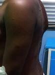
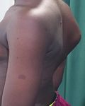
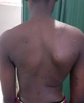
Figure 1: Clinical pictures of the back showing the
lateral and anteroposterior views of the index patient
showing (a) café au lait spot and (b) nodules.
Figure 4: Lumbosacral and pelvic X-rays indicating
riser’s sign (3/5).
Thoracolumbar corset commenced and patient was made
to ambulate about 24 hours post operatively with Zimmer's
Figure 2: X-ray of the whole spine anteroposterior frame and was well tolerated. Alternate day wound
and lateral views. dressing was done for the patient and after about 2 weeks
International Journal of Research in Dermatology | September-October 2022 | Vol 8 | Issue 5 Page 494Onuoha KM et al. Int J Res Dermatol. 2022 Sep;8(5):493-497
wound staples were removed and dressing discontinued. skin lesions were not of primary concern to the patient and
Figure 6 shows the post-operative clinical pictures with parents. NF 1 is a genetic condition that causes tumors to
scars and reduced curvature of the patient’s back. Patient grow along the nerves. The tumors are usually benign but
was discharged from in-patient care about a week after usually cause a range of symptoms. Neurofibromatosis
surgery and then scheduled for follow up on out-patient could manifest with cutaneous and neurological features. 3
basis. As earlier mentioned, scoliosis quite commonly coexists
with NF 1 (as in the index patient as compared to other
possible skeletal anomalies).14
NIH diagnostic criteria for NF 1
Clinical diagnosis based on presence of two of the
following given in Table 1.
Table 1: National Institute of Health: diagnostic
criteria for NF 1.
a S.
Criteria
no.
1 Six or more café-au-lait macules over 5 mm in
diameter in prepubertal individuals and over 15
mm in greatest diameter in post pubertal
individuals
2 Two or more neurofibromas of any type or one
plexiform neurofibroma
3 Freckling in the axillary or inguinal regions
b 4 Two or more Lisch nodules (iris hamartomas)
5 Optic glioma
Figure 5: (a) Post-operative check X-rays; and (b) 6 A distinctive osseous lesion such as sphenoid
Post-operative check X-rays showing the whole extent dysplasia or thinning of long bone cortex, with
of the implants (AP and lateral). or without pseudarthrosis
7 First-degree relative (parent, sibling, or
offspring) with NF-1 by the above criteria
To make a diagnosis of NF 1, two or more of the above-
mentioned criteria must be met.4 In the index patient,
criteria 1 and 2 were present on examination thereby
significantly confirming the diagnosis of NF 1 together
with a Cobb's angle of more than ten degrees in both
thoracic and lumbar regions.
Scoliosis is a term derived from the ancient Greek word
“Skolios” which means curved or crooked. The term was
first established by Galen between 130-201 AD.7 Scoliosis
as we know it now is a medical condition characterized by
the chronic presence of substantial lateral curvature in a
given region of the spine and is usually accompanied by
Figure 6: Post-operative clinical pictures showing rotation of the vertebrae within the curve.
anteroposterior and lateral views of the patient’s back
with surgical scar and reduced curvature. The commonest type is the idiopathic scoliosis that often
occurs in children from the age of eleven, hence the name
DISCUSSION idiopathic structural adolescent scoliosis. Due to the early
growth spurt in girls during puberty, scoliosis is most
Neurofibromatosis presenting with just skin lesions are commonly seen in adolescent girls.7 Adolescent idiopathic
poorly reported in this part of the world as skin lesions scoliosis has a prevalence of 0.47-5.2% with the female to
alone are not life threatening and do not affect quality of male ratio ranging between 1.5:1 and 3:1. These figures
life significantly. In the index patient, the main reason increase with increasing age.7 Scoliosis can be divided
behind presentation was the worsening spine curvature and according to etiology into idiopathic (adolescent or adult),
the surfacing of some breathing difficulties. However, the congenital or neuromuscular.
International Journal of Research in Dermatology | September-October 2022 | Vol 8 | Issue 5 Page 495Onuoha KM et al. Int J Res Dermatol. 2022 Sep;8(5):493-497
The diagnosis of idiopathic scoliosis is made when all have been primed preoperatively and informed or educated
other causes are excluded and it is made up of 80% of all on the process.
cases. Idiopathic scoliosis is further divided to adult and
adolescent idiopathic scoliosis. Of important note is the In the reported case, the wake-up test of Stagnara was
fact that scoliosis could be of adult onset commonly in the performed with the help of the anesthetist. Reversal of
face of degenerative spine diseases. Spinal deformity is the anesthesia was done to a reasonable level of consciousness
commonest orthopaedic manifestation in NF 1. Children where the patient could obey command and was asked to
with NF 1 develop one of two forms of scoliosis; move her legs and hands and the test was positive.
dystrophic or non-dystrophic scoliosis.13 Dystrophic
scoliosis, on the other hand is a form of scoliosis that Post-operative care and ambulation
occurs due to bony changes related to neurofibromas
affecting the spine. Dystrophic scoliosis is identified by Immediately following surgery, the index patient was
picking up specific features on X-rays of the spine. It transferred to the intensive care unit (high dependent unit)
presents with abnormally thin ribs, weakened vertebral for proper supportive care and close monitoring for the
bones and severe spinal curvature including kyphosis and first twenty-four to forty-eight hours. Graded oral intake
rotational deformities and is often associated deformities was commenced the following day beginning with fluid-
and is often associated with dural ectasia.1,13 Non- based diet and then gradually stepped up to full diet.
dystrophic scoliosis, even in children with Ambulating the patient was quite straightforward with
neurofibromatosis is quite similar to the adolescent Zimmer’s frame and a thoracolumbar corset. Limitations
idiopathic scoliosis.1,13 to ambulation would have been poor pain control,
complications arising from surgery (paraplegia,
Cobb's angle respiratory insufficiency), patient’s psychology and many
more which were all taken care of. Post-operative
Dr John Cobb invented the Cobb’s angle in 1948 as a parenteral antibiotics and proper wound care we ensured
standard of measurement for the determination and in the patient to avoid wound breakdown or infections.
tracking of the LATERAL curvature of the spine
(scoliosis).8 Cobb suggested that to measure the angle, the CONCLUSION
upper border of the upper vertebrae; where the curvature
seems to have begun; and the lower border of the lower Scoliosis is the commonest manifestation of NF 1 with
vertebrae, where the curvature ends be taken as reference different severities and degrees of Cobb's angle. An
points in a plain radiograph. A parallel line to these assessment of scoliosis is made when the Cobb's angle
reference points is drawn with another perpendicular line greater than 10 degrees and Cobb’s angles of less than 10
drawn across both lines to cross each other. The angle degrees will only need conservative management and will
between these perpendicular lines is the angle of the usually not need any surgical intervention. Adolescent
curvature.8,9 This method of measurement, though highly females are mostly affected and management is dependent
instrumental, has quite a number of limitations as it is of degree of curvature, comorbidities like NF 1 and type
highly subjective. Intra and inter-observer errors could of scoliosis.
affect the angles significantly.8-10
Funding: No funding sources
Wake up test Conflict of interest: None declared
Ethical approval: Not required
One of the most feared complications of spine surgery is
paraplegia. The scoliosis research society in a case series REFERENCES
that involved over 30,000 patients, the incidence of partial
or complete paraplegia was 0.6%.11 The Stagnara’s wake- 1. Zhao C-M, Zhang W-J, Huang A-B, Chen Q, He Y-
up-test, is a very easy way to detect voluntary motor L, Wei Zhang, Yang H-L. Coexistence of Multiple
function of the lower limb. No know false negative wake rare spinal abnormalities in Type 1
up test as at time of report.12 Wake up test can be used Neurofibromatosis: A case report and Literature
alone or in combination with electrophysiological Review. Int J Clin Exp Med. 2015;8(10):17289-94.
monitoring.11,12 The use of a combined monitoring, 2. Crawford AH, Bagamery N. Osseous manifestations
intraoperative awakening and electrophysiological of neurofibromatosis in childhood. J Pediatr Orthop.
techniques would be optimal; Stagnara’s test alone, 1986;6:72-88.
however, performed with an anesthetic technique allowing 3. Friedman JM. Epidermiology of Nuerofibromatosis
a rapid recovery to a level of consciousness, is a reliable Type 1. Am J Med Genet. 1999;89:1-6.
and practical method to detect as soon as possible 4. Neurofibromatosis Conference Statement. National
neurological problems during major spine surgeries like Institute of Health consensus Development
scoliosis.11,12 Contraindications to the test are mental Conference. Arch Neurol. 1988;45:575-8.
retardation, psychological problems or preexisting 5. Athanasios IT, Asif S, Hilali M. Spinal deformity in
neurological impairment and language.14 Patient must Neurofibromatosis type 1: diagnosis and treatment.
Eur Spine J. 2005;14(5):427-39.
International Journal of Research in Dermatology | September-October 2022 | Vol 8 | Issue 5 Page 496Onuoha KM et al. Int J Res Dermatol. 2022 Sep;8(5):493-497
6. Durrani AA, Crawford AH, Chouhdry SN, Saifuddin 11. Rodola F, D' Avolio S, Cherishing A, Vagnoni S,
A, Morley TR. Modulation of spinal deformities in Forfe E. Wake up test during major spinal surgery
patients with neurofibromatosis type 1. Spine (Phila under Remifentanil balanced anaesthesia. Eur Rev
Pa 1976). 2000;25:69-75. Med Pharm Sci. 2000;4:67-70.
7. Koineczny MR, Senyurt H, Krauspe R, 12. Vauzelle C, Stagnara P, Jouviroux P. Functional
Epidemiology of Adolescent Idiopathic Scoliosis. J monitoring of spinal cord activity during spinal
Chils Orthop. 2013;7(1):3-9. surgery, 1973. Clin Ortgop Rel Res. 1973;93:173-8.
8. Musculoskeletal Consumer Review. Cobb angle and 13. Akbarmia BA, Gabriel KR, Beckman E, Chalk D.
scoliosis. Available at: https://www.coreconcepts. Prevalence of scoliosis in Neurofibromatosis. Spine
com.sg/article/cobb-angle-and-scoliosis/. Accessed (phila Pa 1976). 1992;16:S244-8.
on 11 February 2022. 14. Riccardi VM. Neurofibromatosis: clinical
9. Tan J. Measuring the Cobb angle and scoliosis. heterogeneity. Curr Probl Cancer. 1982;7:1-34.
Available at: http://www.health-articles.co.uk/
measuring-the-cobb-angle-and-scoliosis/. Accessed Cite this article as: Onuoha KM, Badmus HD,
on 11 February 2022. Oyewumi OM, Afolabi MO. Neurofibromatosis type
10. SOSORT guideline committee, Weiss HR, Negrini S, 1 manifesting with adolescent idiopathic scoliosis: a
Rigo M, Kotwicki T, Hawes MC, et al. Indications case report and literature review. Int J Res Dermatol
for conservative management of scoliosis. Scoliosis 2022;8:493-7.
J. 2006;1:5.
International Journal of Research in Dermatology | September-October 2022 | Vol 8 | Issue 5 Page 497You can also read


