Multiple Ruptured Aneurysms Over Basilar Artery Fenestration: Endovascular Management
←
→
Page content transcription
If your browser does not render page correctly, please read the page content below
Open Access Case
                               Report                                                 DOI: 10.7759/cureus.21719

                                                Multiple Ruptured Aneurysms Over Basilar Artery
                                                Fenestration: Endovascular Management
Review began 01/06/2022
                                                Sibasankar Dalai 1 , Madhusudhana Babu Marthati 2 , Aravind Varma Datla 3 , Mohan V. Sumedha Maturu 4 ,
Review ended 01/25/2022                         Ramu Naidu Yellapu 5
Published 01/29/2022

© Copyright 2022                                1. Interventional Neuroradiology, Medicover Hospitals, Visakhapatnam, IND 2. Neurology, Apollo Hospitals,
Dalai et al. This is an open access article     Visakhapatnam, IND 3. Internal Medicine, Medicover Hospitals, Visakhapatnam, IND 4. Neurology, Medicover
distributed under the terms of the Creative     Hospitals, Visakhapatnam, IND 5. Critical Care Medicine, Medicover Hospitals, Visakhapatnam, IND
Commons Attribution License CC-BY 4.0.,
which permits unrestricted use, distribution,
and reproduction in any medium, provided
                                                Corresponding author: Sibasankar Dalai, inrresearch2021@gmail.com
the original author and source are credited.

                                                Abstract
                                                Basilar artery fenestrations (BAF) are rare vascular anomalies. Surgical intervention for aneurysms in this
                                                vascular segment is exceptionally arduous because of the complexity of the neurovascular structures in the
                                                vicinity of the brainstem. Endovascular therapy (ET) is the treatment of choice because of its safety and
                                                efficacy. Here, we report a 62-year-old female presenting with a two-day history of sudden onset severe
                                                headache, vomiting, and altered sensorium. A computed tomography (CT) and subsequent CT angiogram
                                                (CTA) revealed subarachnoid hemorrhage (SAH) and BAF with an aneurysm on each arm of the fenestration.
                                                Digital subtraction angiogram (DSA) with a three-dimensional rotational angiogram (3DRA) was employed
                                                before initiating ET. We used coiling and flow diversion to exclude the aneurysms from circulation. A six-
                                                month follow-up angiography reconfirmed the complete obliteration of the aneurysms. There was no focal
                                                neurological deficit.

                                                Categories: Cardiac/Thoracic/Vascular Surgery, Neurology, Radiology
                                                Keywords: ct angiography, mr angiography, aneurysmal subarachnoid hemorrhage, cerebral digital substraction
                                                angiography, endovascular interventions, flow diversion, aneurysm coiling, aneurysmal rupture, aneurysm, basilar
                                                artery fenestration

                                                Introduction
                                                Basilar artery fenestration (BAF) is a rare intracranial vascular anomaly and is related to embryogenesis. The
                                                reported incidence varies from 0.6% to 2.33% depending on the diagnostic modality [1-3]. Although
                                                challenging, an endovascular approach is the treatment of choice, owing to the complex neurovascular
                                                anatomy in the vicinity of the brainstem [4]. BAF is typically located near the vertebrobasilar artery
                                                junction. Fenestration of the middle and distal segments is a rare occurrence. Currently, the literature
                                                regarding BAF is sparse and is confined mainly to case reports and series [5].

                                                We report a unique case of two small aneurysms, one in each arm of the fenestration. An aneurysmal bleed
                                                occurred, leading to a subarachnoid hemorrhage (SAH). Due to the close anatomical proximity of the
                                                aneurysms, it was difficult to pinpoint the origin of the bleed. Both aneurysms were managed successfully by
                                                coiling the aneurysm in the left arm and flow diversion in the right arm without any complications and an
                                                excellent clinical outcome. At the six-month follow-up, the clinical examination revealed no focal
                                                neurological deficit, and cross-sectional imaging showed complete exclusion of the aneurysms from the
                                                circulation.

                                                Case Presentation
                                                A 62-year-old female patient presented to our hospital with a two-day history of sudden onset severe
                                                headache. She is a known hypertensive on antihypertensive treatment with good compliance. There was no
                                                past medical history of diabetes, smoking, or alcohol consumption. There was no antecedent trauma or fall.
                                                On the second day of her symptoms, she developed vomiting, gradually became drowsy, and was rushed to
                                                our emergency department. Glasgow coma scale (GCS) at presentation was 8/15. Blood pressure was 168/90
                                                mmHg, and heart rate was 59 bpm. Her physical examination revealed neck rigidity and positive Kernig's
                                                sign. A plain CT scan of the brain revealed a subarachnoid hemorrhage (Figure 1).

                                How to cite this article
                                Dalai S, Marthati M, Datla A, et al. (January 29, 2022) Multiple Ruptured Aneurysms Over Basilar Artery Fenestration: Endovascular Management.
                                Cureus 14(1): e21719. DOI 10.7759/cureus.21719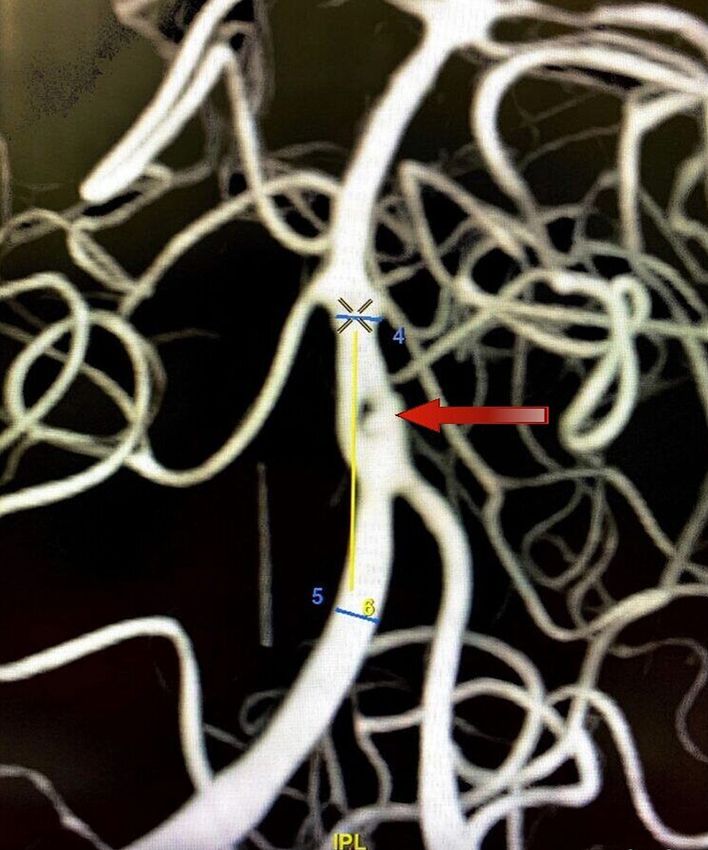
FIGURE 1: Plain CT scan of the brain showing a subarachnoid
                                                    hemorrhage
                                                    CT, Computed tomography.

                                                  A subsequent CT angiogram of the brain revealed a fenestration abnormality of the basilar artery at the
                                                  proximal segment near the vertebrobasilar junction. Each arm of the fenestration revealed a tiny aneurysm,
                                                  measuring 2.5 mm on the right arm and 3.5 mm on the left arm (Figure 2).

2022 Dalai et al. Cureus 14(1): e21719. DOI 10.7759/cureus.21719                                                                                               2 of 10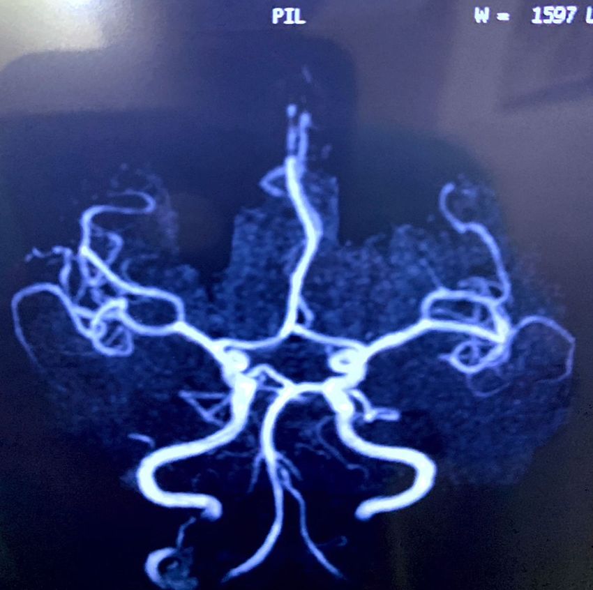
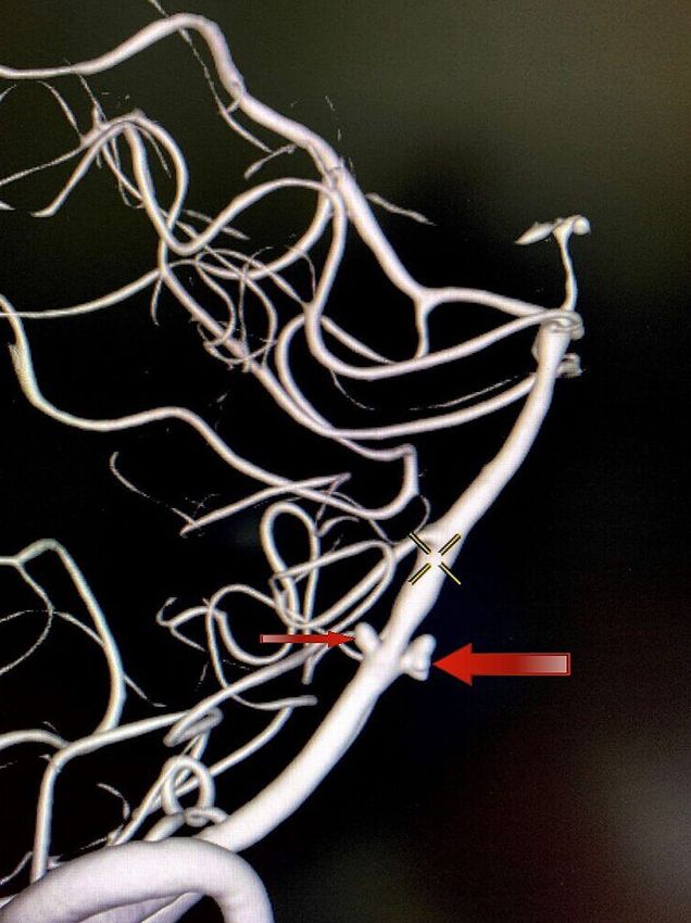
FIGURE 2: DSA lateral view demonstrating an aneurysm directed
                                                    anteriorly (larger arrow) and another aneurysm directed posteriorly
                                                    (smaller arrow)
                                                    DSA, Digital subtraction angiogram.

                                                  A DSA with 3DRA (Figure 3) of the head and neck vessels was done to visualize this complex presentation
                                                  better and to aid in the decision-making process.

2022 Dalai et al. Cureus 14(1): e21719. DOI 10.7759/cureus.21719                                                                                            3 of 10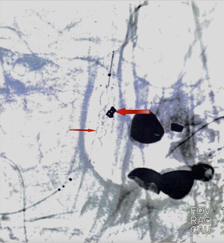
FIGURE 3: DSA-3DRA anterior view showing the anteriorly directed,
                                                    larger narrow-necked left-arm aneurysm
                                                    DSA, Digital subtraction angiogram; 3DRA, three-dimensional rotational angiogram.

                                                  The right aneurysm was directed posteriorly, whereas the left aneurysm was directed anteriorly (Figure 4).
                                                  So, a decision was taken to coil the larger, narrow-necked left aneurysm and achieve flow diversion in the
                                                  smaller, blister-like wide-necked right aneurysm.

2022 Dalai et al. Cureus 14(1): e21719. DOI 10.7759/cureus.21719                                                                                               4 of 10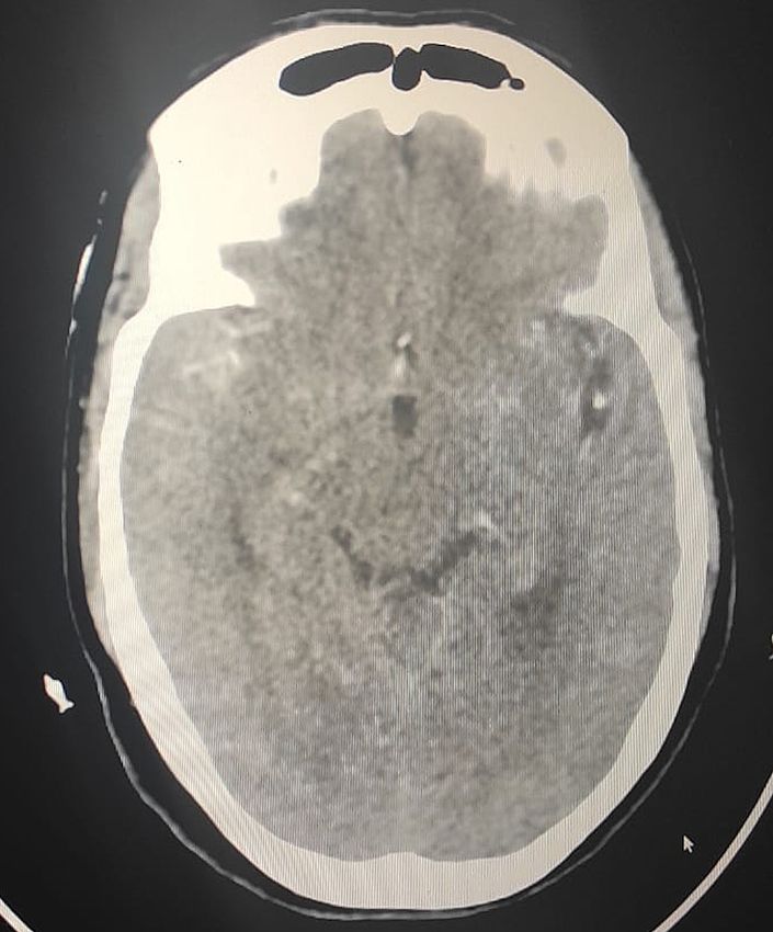
FIGURE 4: DSA-3DRA lateral view demonstrating two aneurysms. The
                                                    left-arm aneurysm (larger arrow) was directed anteriorly. The right
                                                    aneurysm (smaller arrow) was directed posteriorly.
                                                    DSA, Digital subtraction angiogram; 3DRA, three-dimensional rotational angiogram.

                                                  Procedure
                                                  The patient was placed supine on the angiographic table. The patient was intubated, and the procedure was
                                                  performed under general anesthesia. A right transfemoral arterial approach was taken. A 6F short sheath
                                                  was placed into the common femoral artery. The left vertebral artery was accessed with a 5F vertebral artery
                                                  catheter. The vertebral artery catheter was exchanged for a 6F Ballast 088 long sheath (BALT USA, Irvine,
                                                  CA). The Ballast was placed at the distal V2 segment of the vertebral artery. A 6.3F DAC 070 (distal-access
                                                  catheter; Concentric Medical, Mountain View, CA) was taken inside the Ballast and placed in the V4 segment
                                                  of the left vertebral artery. An SL-10 Excelsior microcatheter (Stryker Neurovascular, CA) and Synchro 0.014
                                                  wire (Stryker Neurovascular, CA) were used, and the left aneurysm with a maximum diameter of 3.5 mm was
                                                  canulated. A 3 x 10 coil was placed under continuous road-map guidance. The aneurysm could be entirely
                                                  excluded from the circulation after the coiling (Figure 5).

2022 Dalai et al. Cureus 14(1): e21719. DOI 10.7759/cureus.21719                                                                                                 5 of 10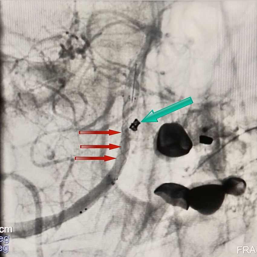
FIGURE 5: DSA anterior view showing a left coiled aneurysmal sac
                                                    (larger arrow) and LVIS stent in the right arm of the fenestration
                                                    DSA, Digital subtraction angiogram; LVIS, low-profile visible intraluminal support.

                                                  The aneurysm of the right fenestrated arm was 2.5 mm in its largest diameter with a wide neck. A Headway
                                                  21 microcatheter (Microvention, Aliso Viejo, CA) with a Synchro 0.014 wire was used, and the aneurysm was
                                                  crossed. The catheter was placed in the mid-basilar artery. A 3.5 mm x 23 mm low-profile visible
                                                  intraluminal support (LVIS) (Microvention, Aliso Viejo, CA) was navigated in the Headway microcatheter.
                                                  The LVIS stent was deployed across the right limb of the BAF. The distal end of the LVIS was placed just
                                                  below the origin of the anterior inferior cerebellar artery, and adequate coverage of the aneurysm was
                                                  achieved (Figure 6).

2022 Dalai et al. Cureus 14(1): e21719. DOI 10.7759/cureus.21719                                                                                              6 of 10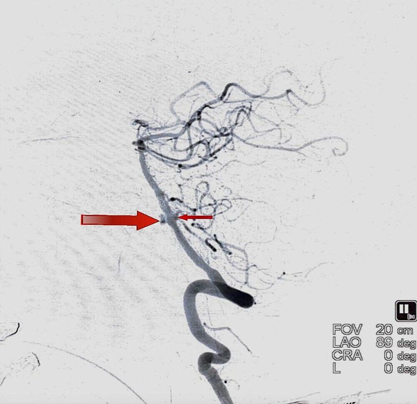
FIGURE 6: DSA anterior view showing the radio-opaque margin of the
                                                    stent used to achieve flow diversion within the right arm of the
                                                    fenestration (red arrows) and the placement of coils within the left
                                                    aneurysmal sac (green arrows)
                                                    DSA, Digital subtraction angiogram.

                                                  The final angiogram demonstrated stagnancy of flow within the right aneurysm. The procedure was
                                                  completed, and the patient was extubated successfully (Figure 7).

2022 Dalai et al. Cureus 14(1): e21719. DOI 10.7759/cureus.21719                                                                                    7 of 10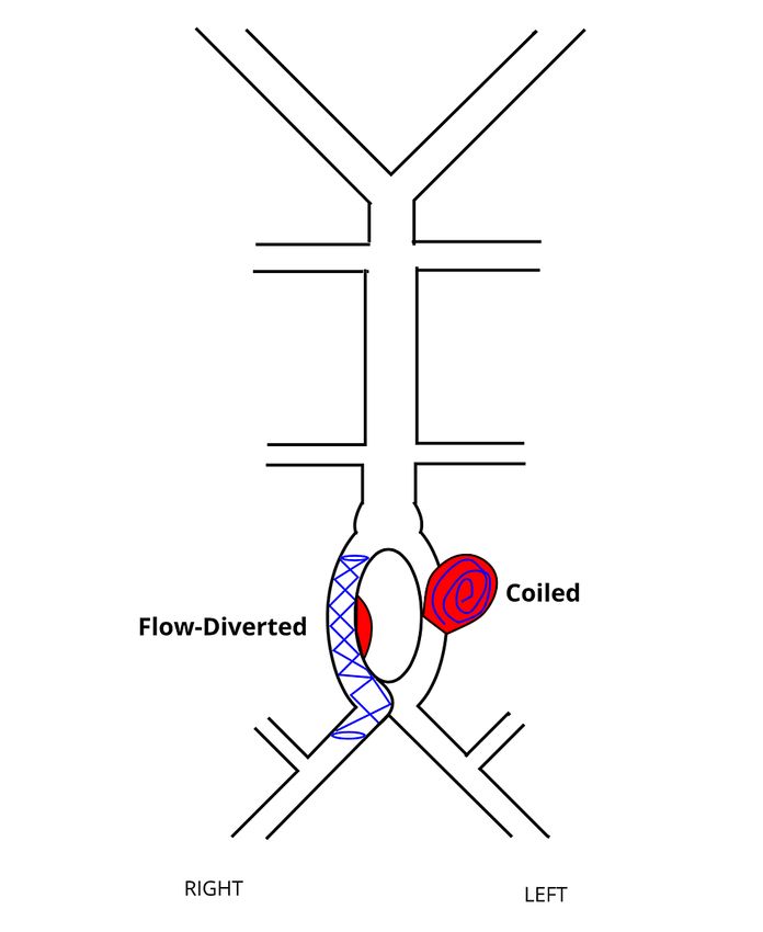
FIGURE 7: A schematic showing flow-diverted right arm of the
                                                    fenestration and coiled left-arm aneurysmal sac

                                                  The patient was shifted to the ICU for further observation. The patient received intravenous hypertonic
                                                  saline, analgesics, and oral nimodipine. A neurological examination 24 hours after the procedure showed no
                                                  focal neurological deficit. There was a significant improvement in her headache and sensorium in the
                                                  following days, and the patient was successfully discharged. A magnetic resonance imaging (MRI) scan and
                                                  magnetic resonance angiogram (MRA) at her six-month follow-up showed complete resolution of the
                                                  SAH and no detectable aneurysmal sacs (Figure 8).

2022 Dalai et al. Cureus 14(1): e21719. DOI 10.7759/cureus.21719                                                                                               8 of 10
FIGURE 8: MRA at six-month follow-up demonstrating complete healing
of the aneurysm
MRA, Magnetic resonance angiogram.
Discussion
Fenestration of the basilar artery develops when the paired fetal longitudinal neural arteries fail to fuse by
the fifth week of gestation. BAF can occur at any segment of the basilar artery with defective midline fusion.
However, it is usually encountered in the proximal segment [6].
The histopathological findings in fenestrations are imperfect medial membrane at both edges of the
fenestration, elastin defect, and thinning of the subendothelial layer at the proximal end, resulting in a
weakened vessel wall. These intrinsic defects coupled with altered hemodynamics (high shear stress and
high-speed turbulence) contribute to aneurysm formation and rupture [5].
Aneurysms are classified based on their size as small (diameter less than 11 mm), large (diameter of 11-25
mm), or giant (diameter greater than 25 mm) [7]. The treatment options for aneurysms include standard
coiling, balloon-assisted therapies, stent-assisted therapies, and flow-diverter systems [8]. Vajpeyee et al. in
2013 used a novel double microcatheter-assisted technique for coiling a BAF aneurysm [9].
Surgery of BAF aneurysms is complicated due to the complex geometry of the fenestration, proximity of the
lower cranial nerves, difficulty in obtaining adequate surgical exposure, and crowding of arteries in this
region [10,11]. In the study by Campos et al., 13 patients had transient lower cranial nerve palsies. One
patient had a permanent neurological deficit, and another patient died following surgical treatment [12]. In
2021, a comprehensive review of literature by Korkmaz et al. reaffirmed the notion that ET is superior to a
microsurgical approach in terms of higher clinical success rate and lower complication rate [8].
In our patient, each arm of the fenestration sported an aneurysm. The larger left aneurysm had a narrow
neck that was ideal for coiling. The smaller right branch aneurysm had a wide neck and was unsuitable for
coiling. Therefore, it was flow-diverted with an LVIS stent. The pliability of the material and relatively
closed cell design (compared to a conventional stent) made flow diversion achievable. There was a
2022 Dalai et al. Cureus 14(1): e21719. DOI 10.7759/cureus.21719 9 of 10significant cost-cut compared to using a traditional flow diverter.
The coiling of an aneurysm promotes thrombosis by diminishing the blood flow going into the aneurysm,
lowering the velocity, prolonging the residence time of blood within the aneurysmal space, and lessening
the aneurysmal wall shear stress. Complete aneurysmal healing might take upward of six months [13]. The
flow diverter covers the neck of the aneurysm, and its mesh reduces the blood flow into the aneurysm. The
stasis of flow within the aneurysm promotes thrombosis. The flow diverter also provides a scaffold for neo-
endothelialization across the aneurysm neck [14-16].
Conclusions
Fenestration of the basilar artery is a rare anatomical anomaly. The endovascular approach is the treatment
of choice because of the inherent complexity of these vascular abnormalities. Proper pre-procedural
planning and selection of ideal endovascular treatment modalities are crucial for therapeutic success and the
best possible clinical outcomes.
Additional Information
Disclosures
Human subjects: Consent was obtained or waived by all participants in this study. Conflicts of interest: In
compliance with the ICMJE uniform disclosure form, all authors declare the following: Payment/services
info: All authors have declared that no financial support was received from any organization for the
submitted work. Financial relationships: All authors have declared that they have no financial
relationships at present or within the previous three years with any organizations that might have an
interest in the submitted work. Other relationships: All authors have declared that there are no other
relationships or activities that could appear to have influenced the submitted work.
References
1. Uchino A, Saito N, Okada Y, et al.: Fenestrations of the intracranial vertebrobasilar system diagnosed by MR
angiography. Neuroradiology. 2012, 54:445-50. 10.1007/s00234-011-0903-x
2. Gao LY, Guo X, Zhou JJ, Zhang Q, Fu J, Chen WJ, Yang YJ: Basilar artery fenestration detected with CT
angiography. Eur Radiol. 2013, 23:2861-7. 10.1007/s00330-013-2890-2
3. Tanaka M, Kikuchi Y, Ouchi T: Neuroradiological analysis of 23 cases of basilar artery fenestration based on
2280 cases of MR angiographies. Interv Neuroradiol. 2006, 12:39-44. 10.1177/15910199060120S103
4. Alqahtani SA, Felbaum DR, Tai A, Liu AH, Armonda RA: Endovascular treatment of large unruptured
fusiform fenestrated vertebrobasilar junction aneurysm. Cureus. 2017, 9:1219. 10.7759/cureus.1219
5. Zhang D, Wang H, Feng Y, Xu N: Fenestration deformity of the basilar artery trunk with an aneurysm: a case
report. Medicine (Baltimore). 2019, 98:16393. 10.1097/MD.0000000000016393
6. Tamrakar K, Chuan Zhi D: Embolization of ruptured aneurysm arising from basilar artery fenestration using
hydrocoils. Cureus. 2015, 7:326. 10.7759/cureus.326
7. Cerebral aneurysms fact sheet. (2018). Accessed: January 13, 2022:
https://www.ninds.nih.gov/Disorders/Patient-Caregiver-Education/Fact-Sheets/Cerebral-Aneurysms-Fact-
Sheet.
8. Korkmaz M, Çınar C, Nas ÖF, Hakyemez B, Oran İ: Endovascular treatment modalities for basilar artery
fenestration aneurysms: experience of two centers and literature review. Turk J Med Sci. 2021, 51:1049-57.
10.3906/sag-2006-352
9. Vajpeyee A, Goyal G, Kant R, Mal N: Double microcatheter-assisted coiling of a basilar artery fenestration
aneurysm. Neurointervention. 2013, 8:125-6. 10.5469/neuroint.2013.8.2.125
10. Kobayashi M, Suzuki M, Sato N, Omama S, Otawara Y, Wada T, Ogawa A: [Fenestrated basilar artery
aneurysm: case report]. No Shinkei Geka. 1999, 27:639-43.
11. Islak C, Kocer N, Kantarci F, Saatci I, Uzma O, Canbaz B: Endovascular management of basilar artery
aneurysms associated with fenestrations. AJNR Am J Neuroradiol. 2002, 23:958-64.
12. Campos J, Fox AJ, Viñuela F, Lylyk P, Ferguson GG, Drake CG, Peerless SJ: Saccular aneurysms in basilar
artery fenestration. AJNR Am J Neuroradiol. 1987, 8:233-6.
13. Brinjikji W, Kallmes DF, Kadirvel R: Mechanisms of healing in coiled intracranial aneurysms: a review of the
literature. AJNR Am J Neuroradiol. 2015, 36:1216-22. 10.3174/ajnr.A4175
14. Pierot L, Wakhloo AK: Endovascular treatment of intracranial aneurysms: current status . Stroke. 2013,
44:2046-54. 10.1161/STROKEAHA.113.000733
15. Meckel S, McAuliffe W, Fiorella D, et al.: Endovascular treatment of complex aneurysms at the
vertebrobasilar junction with flow-diverting stents: initial experience. Neurosurgery. 2013, 73:386-94.
10.1227/01.neu.0000431472.71913.07
16. Dalai S, Datla A, Francis AA, et al.: Endovascular management of mucormycotic aneurysms of the internal
carotid artery in post-COVID-19 patients. Cureus. 2021, 13:20812. 10.7759/cureus.20812
2022 Dalai et al. Cureus 14(1): e21719. DOI 10.7759/cureus.21719 10 of 10You can also read