MiRNA Targets: From Prediction Tools to Experimental Validation - MDPI
←
→
Page content transcription
If your browser does not render page correctly, please read the page content below

Review
miRNA Targets: From Prediction Tools to Experimental Validation
Giulia Riolo            , Silvia Cantara             , Carlotta Marzocchi          and Claudia Ricci *

                                                Department of Medical, Surgical and Neurological Sciences, University of Siena, 53100 Siena, Italy;
                                                riolo@student.unisi.it (G.R.); cantara@unisi.it (S.C.); carlottamarzocchi@libero.it (C.M.)
                                                * Correspondence: claudia.ricci@unisi.it

                                                Abstract: MicroRNAs (miRNAs) are post-transcriptional regulators of gene expression in both ani-
                                                mals and plants. By pairing to microRNA responsive elements (mREs) on target mRNAs, miRNAs
                                                play gene-regulatory roles, producing remarkable changes in several physiological and patholog-
                                                ical processes. Thus, the identification of miRNA-mRNA target interactions is fundamental for
                                                discovering the regulatory network governed by miRNAs. The best way to achieve this goal is
                                                usually by computational prediction followed by experimental validation of these miRNA-mRNA
                                                interactions. This review summarizes the key strategies for miRNA target identification. Several
                                                tools for computational analysis exist, each with different approaches to predict miRNA targets,
                                                and their number is constantly increasing. The major algorithms available for this aim, including
                                                Machine Learning methods, are discussed, to provide practical tips for familiarizing with their
                                                assumptions and understanding how to interpret the results. Then, all the experimental procedures
                                                for verifying the authenticity of the identified miRNA-mRNA target pairs are described, including
                                                High-Throughput technologies, in order to find the best approach for miRNA validation. For each
                                                strategy, strengths and weaknesses are discussed, to enable users to evaluate and select the right
                                                approach for their interests.

                                                Keywords: miRNA target; prediction tools; machine learning; predictive strategies; experimental
                                                validation; validation criteria; high-throughput technologies


Citation: Riolo, G.; Cantara, S.;
Marzocchi, C.; Ricci, C. miRNA
                                                1. Introduction
Targets: From Prediction Tools to
Experimental Validation. Methods
                                                     Among all RNAs, there are non-coding RNAs (ncRNAs). These are functional RNA
Protoc. 2021, 4, 1. https://doi.org/            molecules that do not code for proteins but can modulate protein levels through post-
10.3390/mps4010001                              transcriptional regulation. Non-coding RNA genes consist of highly abundant and func-
                                                tionally important RNAs such as transfer RNA (tRNA) and ribosomal RNA (rRNA), as
Received: 19 November 2020                      well as RNAs such as small interfering RNAs (siRNAs) and microRNAs (miRNAs) [1].
Accepted: 22 December 2020                      Currently, over 35,000 miRNA sequences have been identified in 271 organisms [2].
Published: 24 December 2020                          MiRNAs, approximately 18–26 nucleotides long, are expressed in plants, fungi, ani-
                                                mals, and unicellular organisms and their synthesis is a finely regulated multi-step pro-
Publisher’s Note: MDPI stays neu-               cess [3]. A primary miRNA (pri-miRNA) is transcribed into the nucleus by an RNA
tral with regard to jurisdictional claims       polymerase II. Then, the microprocessor complex, consisting of Drosha and DiGeorge
in published maps and institutional             Syndrome Critical Region 8 (DGCR8), cuts a pri-miRNA to generate a precursor-miRNA
affiliations.                                   (pre-miRNA). The pre-miRNA is exported to the cytoplasm by Exportin5/RanGTP and
                                                processed by the DICER complex to obtain miRNA-miRNA duplexes [3]. This mecha-
                                                nism is called the “canonical pathway” and represents the main modality of miRNAs
Copyright: © 2020 by the authors. Li-
                                                production (Figure 1). Other pathways have recently been discovered and can be “Drosha-
censee MDPI, Basel, Switzerland. This           independent” (i.e., mirtron, t-RNA derived pathways) or “Dicer-independent” (i.e., AGO-,
article is an open access article distributed   tRNaseZ-dependent pathways) [3].
under the terms and conditions of the
Creative Commons Attribution (CC BY)
license (https://creativecommons.org/
licenses/by/4.0/).

Methods Protoc. 2021, 4, 1. https://doi.org/10.3390/mps4010001                                                          https://www.mdpi.com/journal/mps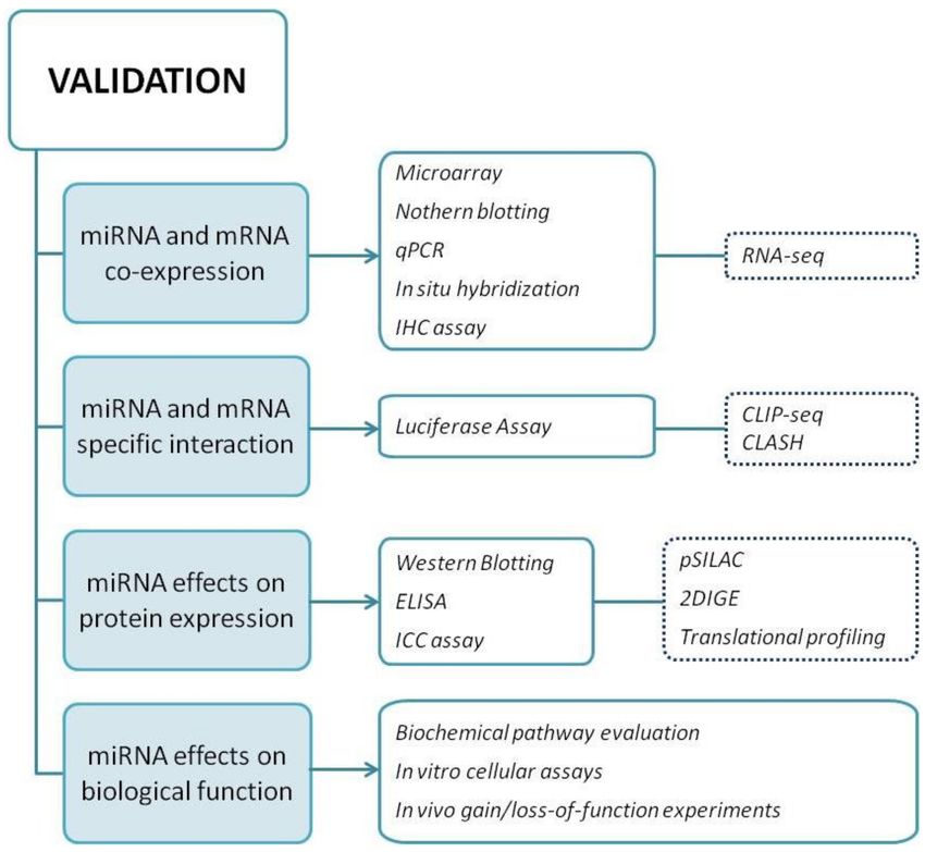
Methods Protoc. 2021, 4, 1                                                                                           2 of 20

                             Figure 1. Canonical pathway of miRNA production.

                                   Whatever the mechanisms, the ultimate goal is the production of a mature, functional
                             miRNA able to regulate protein expression. Bioinformatics analyses have shown that a
                             particular miRNA can regulate expression of up to thousand mRNAs through miRNA-
                             mRNA interaction, and a single mRNA can be controlled by several miRNAs.
                                   Mainly, in mammalian cells, miRNAs bind to the 30 -untranslated region (30 -UTR)
                             of target mRNAs and induce mRNA deadenylation and decapping [4]. Nevertheless,
                             miRNAs can also interact with different regions, such as the 50 -UTR, gene promoter and
                             coding sequence. Pairing with the 30 -UTR was reported to mediate post-transcriptional
                             silencing more effectively than pairing with the 5’-UTR or coding sequence, while miRNA
                             interaction with promoter regions has been shown to induce transcription [4].
                                   However, what are the exact mechanisms of miRNA action? Duplexes of mature
                             miRNA are assembled with an Argonaute (Ago 1–4) protein forming the miRISC complex
                             (miRNA-induced silencing complex). Domain N of Ago fixes miRNA strands in place with
                             the Ago PAZ domain that separates the miRNA duplex. miRISC usually retains the strand
                             with less stable 5’-end base pairing, while the other strand is degraded [4]. The miRNA
                             being partial complementary to a specific mRNA results in translational repression [4]. The
                             process of mRNA silencing also involves other proteins, such as the GW182s [5]. These
                             proteins bridge the link between Ago and downstream effectors, such as the cytoplasmic
                             deadenylase complexes PAN2-PAN3 and CCR4-NOT ensuring mRNA repression. On the
                             other hand, a perfect binding between miRNA-mRNA induces mRNA degradation by
                             enzymes involved in the 50 -to-30 mRNA decay pathway. In this pathway, poly(A) tail is
                             removed from mRNA, which is then decapped and finally degraded starting from the 5’-
                             end. The GW182 proteins also work as shuttle for Ago2, transferring it from the cytoplasm
                             to the nucleus [5].
                                   Once in the nucleus, miRISC complex can induce nuclear mRNA degradation but
                             also participate to mRNA splicing and chromatin state regulation increasing mRNA levels.
                             It is evident that the biological functions of miRNAs are determined by the mRNAs that
                             they control.
                                   Efficient gene regulation is orchestrated by miRNA localization, target mRNA levels,
                             affinity of the miRNA-mRNA interaction, and the presence of multiple factors (e.g., GW182
                             proteins). However, miRNA activity on the mRNA targets is difficult to identify.
                                   Despite the growing knowledge about the mechanisms involved in miRNA-mRNA
                             interaction, the identification of potential targets in the human genome is still a challenge,
                             which has been appropriately described as “a classical needle in a haystack problem” [6].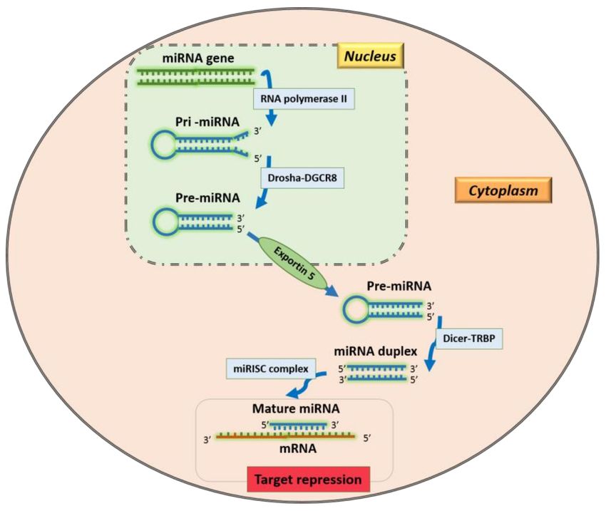
Methods Protoc. 2021, 4, 1                                                                                           3 of 20

                                   Identification and validation of the interactions taking place between miRNAs and
                             their targets is a critical step for defining the regulatory miRNA role in the complex net-
                             works that regulate the biological processes. For any given miRNA, a wide number of
                             potential target sites may be present, and the experimental validation of every potential
                             miRNA target in the laboratory is not feasible, as it is expensive and time-consuming. A
                             computational approach to predict the potential targets of miRNAs can simplify the proce-
                             dure, allowing an initial selection to reduce the number of target sites to be experimentally
                             validated. Several tools for computational analysis exist, each using a different strategy to
                             predict potential miRNA targets, and their number is constantly increasing. Now, users
                             have the opportunity to access a large range of solutions, but they must decide which tool
                             to use. This choice may not be easy; it is at least necessary to be familiar with the basic
                             assumptions and interpretation of the results.
                                   The aim of this review is to provide practical advice for this challenge, in order to
                             make it easier to understand the subject.
                                   In the first part, we depict the main features employed in miRNA target prediction
                             tools. A specific section is dedicated to the Machine Learning (ML) approach. The strengths
                             and weaknesses of each approach are discussed, to enable the users to evaluate and choose
                             the right tool for their interests. In addition, we include a synthesis of the features of the
                             most common computational tools used for miRNA target prediction. Finally, we provide
                             a stepwise scheme, to facilitate the choice of the best approach to use in practice.
                                   Once the overabundance of information from these algorithms has been collected,
                             specific miRNA-mRNA target interactions are selected to proceed investigating with more
                             efficiency. Four criteria need to be fulfilled in order to confirm a miRNA-mRNA interaction
                             as biologically significant. In the second part of this review, we describe the key approaches
                             and the major experimental procedures to meet the criteria for validation. Most importantly,
                             the advantages and drawbacks of “low-yield” techniques and High-Throughput methods
                             are reported, proving details on why miRNA validation is still challenging.

                             2. miRNA Target Prediction
                             2.1. Predictive Methods
                                  Several approaches underlie the development of miRNA target prediction algorithms.
                             These can be divided into two main categories: algorithms derived from characteristics
                             of the mRNA sequence and/or based on the miRNA-mRNA interaction, and statistical
                             inference based on Machine Learning.
                                  In the first case, different features of the miRNA-target complex are taken into account.
                             Pairings of the seed sequence of miRNAs and mRNAs can be analyzed and evaluated.
                             Thermodynamic analysis can be performed by computing the free energy of pair formation
                             and its thermodynamic stability. The evolutionarily conservation of the target sequence
                             across related species can be assessed. Finally, the 30 -UTR structural accessibility for
                             miRNAs can be evaluated, and the number of miRNA target sites can be calculated, since
                             mRNAs may be regulated by the binding of different miRNAs to multiple target sites.
                                  In the case of Machine Learning, the idea is to identify miRNA targets that reference
                             miRNA-mRNA duplexes with proven biological significance, rather than making “de
                             novo” predictions proceeding from sequence features. Machine Learning in general is
                             an application of artificial intelligence that provides systems with the ability to improve
                             automatically through experience; they “learn” from sample datasets and use the acquired
                             information to make predictions on unknown data [7].
                                  The features of these approaches are described in the following sections.

                             2.1.1. Strategies for de novo Predictions
                             1.   Seed pairing
                                  The miRNA sequence is complementary to the sequence of 30 -UTR of potential mRNA
                             targets. The miRNA seed sequence, namely the first 2–7 nucleotides in the miRNA 50
                             region, is essential for binding target mRNAs [8]. Indeed, specific characteristics within
Methods Protoc. 2021, 4, 1                                                                                              4 of 20

                             the seed region, but also within close proximity, have been associated with specific effects
                             on the gene repression induced by miRNAs. Thus, all of these characteristics have been
                             included in target prediction tools [9–11].
                                  The majority of the target prediction algorithms require Watson–Crick pairing between
                             miRNA and mRNA. In other words, guanine (G) must pair with cytosine (C), and adenosine
                             (A) with uracil (U). Different algorithms consider different types of seed matching. The
                             main kinds of seed matches [9,10,12,13] (see Figure 2) are the following:
                             •    8mer site: a perfect Watson-Crick match from nucleotide 2 to nucleotide 8 of the
                                  miRNA seed, with an “A” in mRNA opposite position 1;
                             •    7mer site: Watson-Crick match from nucleotide 2 to nucleotide 8 without the “A”
                                  opposite position 1;
                             •    7mer A1 site: Watson–Crick match from nucleotide 2 to nucleotide 7 with an A
                                  opposite position 1;
                             •    6mer site: position 2–7 match;
                             •    6mer site: position 3–8 match.
                                  The presence of an adenosine opposite position 1 of miRNA is important because this
                             adenosine is specifically recognized within the Argonaute protein [11], independently from
                             the nucleotide in the miRNA sequence [14].

                             Figure 2. Main types of miRNA seed sequences (in order of descending regulatory efficiency).

                             2.   Thermodynamic stability
                                  Whereas sequence complementarity is clearly informative to predict miRNA–mRNA
                             interactions, important indications for evaluating hybridization stability are provided by
                             the thermodynamic properties of the miRNA-mRNA complexes. In the majority of miRNA
                             target prediction tools, the thermodynamic properties of the miRNA-mRNA complex are
                             assessed by evaluating the free energy of the predicted interaction. Free energy is used to
                             evaluate the stability of a biological system. Though the free energy of a miRNA-mRNA
                             complex is lower, more energy is necessary to break the bond. In other words, when the free
                             energy is low, a miRNA-mRNA complex is estimated to be more thermodynamically stable,
                             and the miRNA-mRNA pairing is considered stronger. This is important information,
                             since a stable miRNA-mRNA interaction can be more likely classified as a real interaction.
                             Nevertheless, it is not easy to establish the proper threshold of free energy, since data
                             obtained from known miRNA-mRNA complexes are insufficient [15].
                                  Some algorithms (e.g., MicroTAR [16]) only assess the thermodynamic stability to
                             validate initial predictions based on sequence complementarities; other algorithms (e.g.,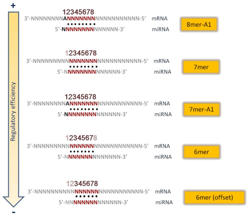
Methods Protoc. 2021, 4, 1                                                                                            5 of 20

                             RNAhybrid [17]) calculate the free energy as the first step for their evaluation. This strategy
                             can be employed to predict non-canonical binding sites and does not need additional
                             information. However, the algorithm itself is based on free energy calculations obtained
                             from computational models of nucleotide association [18].
                                  To improve prediction efficiency, other features, such as sequence conservation, may
                             be considered to effectively predict miRNA targets.
                             3.   Evolutionary conservation
                                  The analysis of sequence conservation among different species is an approach used
                             to try to reduce the number of false positives obtained by prediction algorithms. The
                             principle underpinning this strategy is that miRNA-mRNA interactions with advantageous
                             biological functions are selected and conserved during evolution. It is worth noting
                             that a higher conservation has been reported in the miRNA seed sequence than in other
                             sequences [9].
                                  Several target prediction algorithms identify orthologous 30 -UTR sequences and then
                             evaluate the conservation of the potential miRNA target site in other species, in particular
                             in those that are closely related (e.g., miRanda [19]). This analysis across the species allows
                             reducing the number of false positive predictions [15]. However, this approach can present
                             some limitations, since it only searches for targets of evolutionary conserved miRNAs,
                             but conservation does not necessarily imply functionality. Indeed, previous studies have
                             reported that about 30% of experimentally validated miRNA targets are not actually
                             conserved [20]. The conservation alone, therefore, is not sufficient for obtaining a reliable
                             miRNA target prediction. Thus, it is strongly recommended to combine this approach with
                             non-conservation models when it is necessary to perform de novo predictions.
                             4.   Accessibility of target site
                                   The structural accessibility of the binding sites is another important factor to consider
                             to increase the reliability of predictions. Both miRNA and 30 -UTR of the potential target
                             should be accessible, at least in the regions of the seed sequence. While the accessibility of
                             miRNA seed is ensured, as mature miRNAs are assembled into the RISC [21], the accessi-
                             bility of the 30 -UTR of mRNA needs to be estimated. Some algorithms identify partially
                             accessible binding sites, then rank the targets in accordance with a specific score [22] or
                             the free energy [23]. Other algorithms calculate the free energy necessary to obtain the
                             accessibility of the complementary site and add it to the energy of hybridization. This sum
                             allows defining which binding sites are accessible and ranking the predicted targets [24].
                             5.   Number of target sites in the same 30 -UTR
                                  It is known that more than one miRNA can bind the same target and, therefore, multi-
                             ple miRNAs may cooperate in controlling the expression/repression of target genes [25,26].
                             Therefore, the miRNA-mediated regulation of a specific target gene changes depending
                             on the number of miRNAs binding to that gene [27]. In particular, a relevant element
                             in miRNA-induced silencing seems to be the number and the location of binding sites.
                             Multiple target sites within 10–50 nucleotides of each other usually enhance repression,
                             while sites very close together (< 8 nucleotides) generally operate competitively [14,28].
                             The use of prediction algorithms also considering multi-targeting significantly reduces
                             the number of predictions, increasing the probability of identifying true targets. Thus, it
                             can be very useful for reducing the number of candidates before performing experimental
                             validation studies of miRNA targets [15].
                                  There are plenty of computational tools that use different strategies or a combination
                             of them.
                                  For example, miRanda, one of the first and most frequently used algorithms, is based
                             on an evaluation of complementarity between miRNAs and 3’-UTR regions, thermody-
                             namic stability of the duplex structure, evolutionary conservation of the entire binding site,
                             and its position within 30 -UTR as a final filter [19].
                                  TargetScan primarily predicts potential miRNA targets by looking for the presence of
                             8mer, 7mer, and 6mer sites that match the seed region of every miRNA. It is possible to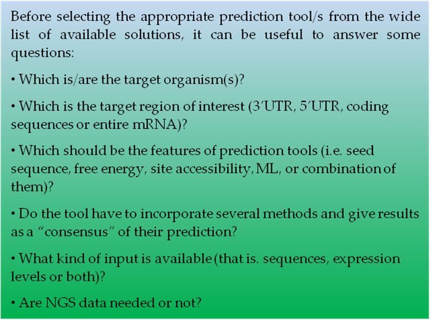
Methods Protoc. 2021, 4, 1                                                                                               6 of 20

                                choose to include only conserved sites. Other features, such as seed-pairing stability, 30
                                compensatory pairing, and target site abundance, may be evaluated. The putative targets
                                are then ranked based on the predicted efficacy of targeting, calculated using cumulative
                                scores considering all the features of the sites. Moreover, TargetScan predicts the secondary
                                structure to evaluate the free energy of predicted complexes [29].
                                       RNAhybrid finds the minimum free energy for short sequences, but also for the whole
                                miRNA-mRNA complex. This tool evaluates the minimum free energy of hybridization
                                for a long and a short RNA, so the short sequence is hybridized to the best fitting part of
                                the long sequence. The user can impose several restrictions, such as the rate of unpaired
                                bases allowed, to reduce the number of predictions [17].
                                       PITA offers a different approach for miRNA target prediction. The main feature
                                assessed by this program is the accessibility of the target site. It evaluates the free energy
                                gained from miRNA–mRNA pair formation and the energy cost of making the target
                                accessible to the miRNA, and computes the difference between these two parameters.
                                It considers also the “flank sites”, the sites around the seed, which are involved in site
                                accessibility. The user can impose restrictions to reduce the number of resulting targets
                                (i.e., minimum seed size and unpaired bases) [30].
                                       All these tools are available online.
                                       Table 1 summarizes the main features and strategies for these and other largely
                                diffused prediction algorithms.

      Table 1. List of miRNA target prediction tools and used strategies. SF: Sequence Features; TS: Thermo-Stability; EC:
      Evolutionary Conservation; SA: Site Accessibility.

   Tool Name             SF     TS         EC         SA                               Web                              Ref.
                         √      √          √
    miRanda                                                             http://www.microrna.org/microrna                [19]
                         √      √          √          √
   TargetScan                                                           http://www.targetscan.org/vert_72               [29]
                                √
   RNAhybrid                                                    https://bibiserv.cebitec.uni-bielefeld.de/rnahybrid     [17]
                                √                     √         https://genie.weizmann.ac.il/pubs/mir07/mir07_
       PITA                                                                                                             [30]
                                                                                   prediction.html
                         √      √
    microTAR                                                           http://tiger.dbs.nus.edu.sg/microtar             [16]
                                √          √
     PicTar                                                                 https://pictar.mdc-berlin.de                [27]
                                √          √          √
    PACMIT                                                                     https://paccmit.epfl.ch                  [31]
                         √      √          √          √        http://www.clipz.unibas.ch/index.php?r=tools/sub/
    MIRZA-G                                                                                                             [32]
                                                                                      mirza_g
                         √      √
     RNA22                                                          https://cm.jefferson.edu/rna22/Interactive          [33]

                                2.1.2. Machine Learning
                                      The term “Machine Learning” (ML) was used for the first time in 1959 by Arthur
                                Samuel, who described ML as the “field of study that gives computers the ability to learn
                                without being explicitly programmed” [34]. ML methods commonly enable computers to
                                analyze collected data in order to build data-driven models, discover statistically significant
                                patterns and relationships, and consequently make predictions on novel data. In practice,
                                ML algorithms are able to “learn” from sets of data and use the acquired knowledge
                                to analyze similar data and make predictions based on the examples that have been
                                provided [7].
                                      In the case of the miRNA target identification, ML approaches do not refer to miRNA-
                                mRNA features, such as sequence or thermodynamic stability, but try to recognize potential
                                miRNA targets by referring to miRNA–mRNA interactions with proven biological signifi-
                                cance. Algorithms are “trained” using experimentally verified miRNA-mRNA interactions
                                as positive examples, and artificially created negative examples. In this way, ML software
                                tries to recognize patterns that discriminate between actual targets and false targets. In
                                the presence of a novel previously unseen dataset, these patterns can be used to correctly
                                predict whether a target is “real” or not.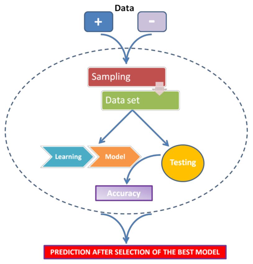
Methods Protoc. 2021, 4, 1                                                                                           7 of 20

                                  Interestingly, ML is not programmed to necessitate stringent seed matches in the
                             30 -UTRs, but it learns by provided examples with biological relevance, so it also allows
                             identifying non-canonical binding sites, including those within the coding regions. On the
                             other hand, ML learns exclusively from the provided examples and consequently it is only
                             able to find results similar to those examples [35].
                                  The general strategy for ML can be summarized as follows (see Figure 3):
                             •    For every miRNA, identify the presumed binding site from validated mRNA targets
                                  (as positive) and non-targets (as negative).
                             •    Extract features from these interactions (regardless of whether they are functional or
                                  nonfunctional).
                             •    Train a classifier to discriminate targets from non-targets.
                             •    Test the classifier.
                             •    Use the classifier to sort unknown miRNA-mRNA interactions as positive (target) or
                                  negative (non-target).

                             Figure 3. Scheme of Machine Learning general strategy.

                                   Some points are of fundamental importance in this process. First, the positive dataset,
                             obtained from experimentally verified databases, largely depends on the features of the
                             database of reference. Several databases of experimentally validated miRNA-mRNA
                             target interactions are available, such as MiRTarBase [36], DIANA-TarBase [37], and
                             miRecords [38]. The use of these validated miRNA-mRNA interactions may introduce
                             some bias, since these data have been produced by different laboratories, using different
                             cell types and approaches [39]. The negative dataset is equally important to obtain a
                             well-trained classifier. If the sequences are too artificial, the Machine-Learning method
                             will probably not be efficiently trained to discriminate between true and false interactions.
                             Conversely, if the negative and the positive datasets are very similar to each other, the ML
                             approach will not be able to distinguish between them. Moreover, an excess of positive
                             or negative dataset can cause underfitting or overfitting models, respectively [35]. Finally,
                             the choice of the validation set is critical for ML: to verify its performance, a model should
                             be assessed on a test dataset that is completely different from the dataset used for the
                             training step (training dataset). A strategy is to exclude a portion of the training set from
                             the training phase and use it as validation set during model optimization [39].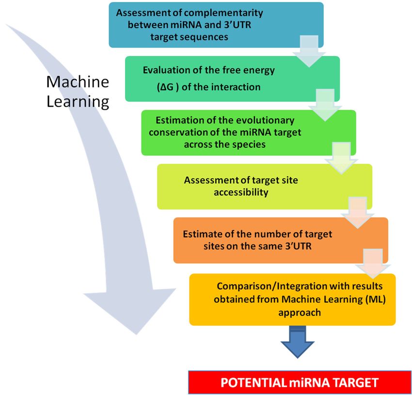
Methods Protoc. 2021, 4, 1                                                                                                           8 of 20

                                        All the ML-based target prediction algorithms are based on a similar set of engineered
                                   features and differ mainly in ML architecture and in the experimental datasets that are used.
                                        For example, MBSTar, a prediction tool based on the random forest algorithm, is
                                   trained on more than 9000 biologically validated miRNA-mRNA interactions, and ap-
                                   proximately 1000 non-interacting pairs, confirmed by data obtained from RISC-associated
                                   immunoprecipitation experiments. A combination of 340 sequence and 31 structural fea-
                                   tures has been applied to an unsupervised learning algorithm to extract 40 putative features
                                   for miRNA-mRNA interactions. Different learning classifiers have been analyzed, and
                                   the random forest algorithm has displayed the highest accuracy rate when used for target
                                   prediction in experimentally validated datasets [40].
                                        Table 2 displays some of the main ML tools used for target prediction, including
                                   details about both negative and positive data sets and ML methods. A brief description of
                                   the features of such methods is reported in Box 1.

         Table 2. List of ML prediction tools with the kinds of used strategies. For an explanation of the algorithms, see Box 1.

        Tool Name                  Algorithm                Positive               Negative                Features             Ref.
                                                                                 Randomly
          MBSTar                Random Forest               MiRbase                                   sequence, structure           [40]
                                                                                 generated
                                                                                 Probability
         NbmiRTar                 Naïve Bayes                TarBase                                       sequence                 [41]
                                                                               Randomization
                                                           let-7, lin-4,
                                    Genetic
        TargetBoost                                         miR-13a,            Random string              sequence                 [42]
                                 Programming
                                                             bantam
                                                           miRecords         Mocking in
        DeepTarget                    RNN                                                                  sequence                 [43]
                                                            miRBase           alignment
                                                                              Randomly
       TargetMiner                    SVM                 miRecords                                          seed                   [44]
                                                                              generated
                                                                             Mocking in               sequence, structure,
        DeepMirTar               Autoencoder              miRecords                                                                 [45]
                                                                              alignment                energy and other
                                  microT-CDS            miRNA regulatory element in both the          sequence, structure,
  DIANA-microT-CDS                                                                                                                  [46]
                                   algorithm                     3’-UTR and CDS                        energy and other
                                                                                                      sequence, structure,
    miRanda-mirSVR           SVR (similar to SVM)          set from transfection experiments                                        [47]
                                                                                                       energy and other

                                   2.1.3. Operative Strategy
                                         The aim of these different approaches is to limit the number of predicted targets
                                   and at the same time reduce the loss of true targets. In other words, it is necessary to
                                   obtain a compromise between sensitivity and specificity. Some tools may be extremely
                                   efficient in identifying true target sites (high sensitivity), but provide a very wide number
                                   of predictions (low specificity) [30,48,49]. On the other hand, other tools present high
                                   specificity, but quite low sensitivity [10,27,50].
                                         All the available tools have their own predictive strengths and limitations, depending
                                   on the strategies they use. A tool exclusively based on seed match, for example, does not
                                   consider whether the miRNA-mRNA complex is thermodynamically stable, or the sequence
                                   of the target site is evolutionarily conserved and accessible for binding. On the other hand,
                                   many non-conserved binding sites in 30 -UTRs have been reported to be functional [51], and
                                   such miRNA-mRNA interactions cannot be captured by using only conservation-based
                                   prediction tools.
                                         To complicate things, target sites for potential miRNA binding can be deeply affected
                                   by other events involving RNA, such as alternative splicing and RNA editing [52,53]. One
                                   solution used by several tools to solve this problem is to select either any 30 -UTR or the
                                   longest one from available databases (i.e., NCBI’s Refseq or Ensembl). In addition, recent
                                   evidence suggests that functional miRNA binding sites are present also within 50 -UTRs and
                                   coding sequences, raising the issue of whether it would be useful to extend the analysis to
Methods Protoc. 2021, 4, 1 9 of 20
the full-length mRNA [54] and of which binding rules and sequence features should be
Methods Protoc. 2021, 4, x FOR PEER REVIEW 9 of 21
used in this case [55].
Box 1. ML Methods: some details.
Box 1. ML Methods: some details.
2.1.3. Operative Strategy
The aim of these different approaches is to limit the number of predicted targets and
A flow-chart summarizing this kind of approach is given in Figure 4.
at the same time reduce the loss of true targets. In other words, it is necessary to obtain a
Considering all these observations, the best results to identify miRNA targets are
compromise between sensitivity and specificity. Some tools may be extremely efficient in
likely to be obtained using a combination of tools that are derived from different prediction
identifying true target sites (high sensitivity), but provide a very wide number of predic-
assumptions. This approach may allow obtaining a good balance between sensitivity and
tions (low specificity) [30,48,49]. On the other hand, other tools present high specificity,
specificity [56]. Also, in this case, it would be necessary to have the foresight to verify that
but quite low sensitivity [10,27,50].
the prediction tools use the same reference databases and tare regularly updated, since
All the available tools have their own predictive strengths and limitations, depend-
miRNA nomenclature is frequently modified and every year new miRNAs are included
ing on the strategies they use. A tool exclusively based on seed match, for example, does
in miRBase [15]. One of the many examples of this strategy is the identification of KRAS
not consider whether the miRNA-mRNA complex is thermodynamically stable, or the
mRNA as a target for miR-193a-3p in non-small cell lung cancer [57]. In this case, miRNA
sequence of the target
target prediction site is evolutionarily
was performed conserved and
using a combination accessible
of three for binding.
different On the
computational
other hand, many non-conserved binding sites in 3′-UTRs have been reported
algorithms based on different approaches (TargetScan, PicTar, and miRanda), and the resultsto be
functional [51], and such miRNA-mRNA interactions cannot be captured by using only
conservation-based prediction tools.
To complicate things, target sites for potential miRNA binding can be deeply af-
fected by other events involving RNA, such as alternative splicing and RNA editing
[52,53]. One solution used by several tools to solve this problem is to select either anyMethods Protoc. 2021, 4, 1 10 of 20
were then confirmed by experimental validation. Thus, the combination of different target
prediction algorithms may significantly help researchers in identifying potential miRNA
targets, as long as users know the fundamental presumptions, strengths, weaknesses, and
limitations of every tool in order to make an informed decision of which ones to use.
Figure 4. Stepwise strategy for miRNA target prediction. (1) Assessment of complementarity between
miRNA and 30 -UTR target sequences. It is advisable to evaluate the score assigned to the prediction,
to have an initial indication of its likelihood. (2) Evaluation of the free energy (∆G) of the interaction.
This may give an indication of thermodynamic stability of the miRNA-mRNA interaction and further
strengthen predictions. (3) Estimate of the evolutionary conservation of the miRNA target across
the species. The more conserved a sequence of DNA, the higher the probability that it corresponds
to functional, transcribed, RNA sequences. (4) Assessment of target site accessibility. This may
provide a ranking of potential target sites. (5) Estimate of the number of target sites on the same
30 -UTR. The use of multitargeting-based algorithms significantly decreases the number of predicted
targets, and raise the chance of identifying true targets. (6) Comparison/Integration with results
obtained from Machine Learning (ML) approach. This allows limiting the number of false positive
and increasing the strength of the prediction.
In addition, data obtained from these computational tools may be coupled and inte-
grated with an ML approach, which can limit the number of false positives and further
strengthen the value of the prediction [58]. Since experimental data sets used by ML come
from a wide range of experiments, as shown in Table 2, also in this case it is strongly
suggested to employ a combination of data sources for ML-based miRNA target prediction
in order to reduce the limitations resulting from specific types of data sets. In fact, an
ML prediction can only be valid if the data on which it is-based are valid [39]. Such an
approach has been successfully used in several cases. An example is the identification
of the potential miR-622 targets in breast cancer, using an integrated miRNA prediction
process, which included miRanda, miRDB [59], RNA22, TargetScan, and the ML algorithm
MiRWalk 3.0 [60]. The intersection of the results obtained from these tools allowed iden-
tifying 77 promising targets and constructing a protein-protein interaction network. The
results were experimentally validated in vitro, revealing the regulatory mechanisms of
miR-622 in breast cancer cells [61].
To avoid getting lost in the sea of available prediction tools, some websites offer
interesting solutions to orientate the choice. For example, tools4miRs [62] provides anMethods Protoc. 2021, 4, 1 11 of 20
Figure 4. Stepwise strategy for miRNA target prediction. 1) Assessment of complementarity be
tween miRNA and 3′-UTR target sequences. It is advisable to evaluate the score assigned to th
prediction, to have an initial indication of its likelihood. 2) Evaluation of the free energy (ΔG) of th
interactiveinteraction.
webpage Thisthat may
allowsgive an indication
selecting of thermodynamic
organisms, stability of the
prediction approaches, miRNA-mRNA
target regions interac
tion and further strengthen predictions. 3) Estimate of the evolutionary conservation of the miRNA
and other features, and gives a list of prediction tools based on the user’s selection.
target across the species. The more conserved a sequence of DNA, the higher the probability that
Another approach was proposed by Kern and colleagues [6], who have set up an
corresponds to functional, transcribed, RNA sequences. 4) Assessment of target site accessibility
interactive webpage,
This freelya ranking
may provide availableof on-line,
potentialwhich helps5)users
target sites. perform
Estimate of the tool selection
number bysites on th
of target
answeringsamea six-step questionnaire based on different criteria.
3′-UTR. The use of multitargeting-based algorithms significantly decreases the number o
Box 2predicted
displaystargets,
some of andthe main
raise thequestions to answer true
chance of identifying for guiding anComparison/Integration
targets. 6) aware choice of wit
the prediction
resultstools.
obtained from Machine Learning (ML) approach. This allows limiting the number of fals
positive and increasing the strength of the prediction.
Box 2. Questions to answer for choosing the prediction tool(s).
Box 2. Questions to answer for choosing the prediction tool(s).
In conclusion, all the tools described may provide important insights about potentia
miRNA targets. The use of a combinatorial approach may increase the true prediction
In conclusion, all the tools described may provide important insights about potential
miRNA targets. The use of a combinatorial approach may increase the true predictions
and limit the number of false positives. However, once bioinformatics analyses have been
performed and the potential miRNA targets have been identified, the biological function of
predicted miRNA-mRNA interactions needs to be verified and confirmed.
3. miRNA Target Validation
3.1. Validation Methods
Whichever strategy has been used to predict miRNA-mRNA interactions, candidate
miRNA targets should be always verified experimentally to obtain full legitimacy.
Depending on information provided, experimental approaches can be classified into
two categories: direct and indirect methods. The first category includes procedures in-
vestigating the existence of an interaction between miRNA and its target, by studying
directly the miRNA-mRNA pair or introducing a specific target site bound by miRNAs,
known as microRNA responsive element (mRE), into a reporter gene, in order to measure
potential miRNA-induced changes at protein levels. In particular, these approaches are
usually limited to mREs that have been obtained from computational predictions.
On the other hand, the second category includes approaches that observe the effects
derived from an altered miRNA expression on mRNA or protein expression, using High-
Throughput technologies (i.e., sequencing and mass spectrometry). However, some of
these methods suffer from ‘knock-on’ effects. This occurs because, when a miRNA engages
its target, it not only induces changes in the protein coded by that gene, but it indirectly
produces consequences also on several different genes, whose expression is regulated byMethods Protoc. 2021, 4, 1 12 of 20
the sequence directly targeted by miRNA [63]. As a consequence, the alteration of some
products obtained by these approaches is not necessarily to be considered to be resultant
from miRNA-mRNA interaction.
As a general approach, the authenticity of the preferred functional miRNA-mRNA
target pair, predicted by computational tools, should be validated in the biological model
of interest by fulfilling four criteria. First, co-expression of miRNA and predicted target
mRNA must be demonstrated. Second, a direct interaction between the miRNA of interest
and a specific region within the target mRNA must be proved. Third, gain- and loss-of-
function experiments must be performed to demonstrate how miRNAs regulate target
protein expression. Fourth, it needs to verify whether the predicted changes in protein
expression are associated with modified biological functions [64,65].
3.1.1. Criterion I: Show Co-Expression of miRNA and Target mRNA In Vivo
Co-expression of a miRNA and its target mRNA is the first crucial point required for
validation. In a recent study, Cava et al. [66] found that only 19.3% of the total mRNAs
is tissue-specific, compared to 39.7% of miRNAs in normal tissues. The co-profiling of
miRNAs and mRNAs can allow a direct assessment of whether mRNAs are in part shaped
by regulatory miRNAs [67] since co-expressed elements share the same transcriptional
program or are regulated by members of the same pathway. Consequently, if they are not
co-expressed or co-expression cannot be verified in cells or tissues, there are no reasons to
move forward with additional experiments.
Microarray profiling and the latest RNA-sequencing (RNA-seq) represent powerful
strategies to carry out large scale studies on the genome. Moreover, Northern blots and
quantitative real-time PCR (RT-qPCR), employing nucleic acids extracted from different
cell cultures, represent the most exploited methods to demonstrate co-expression [68,69].
Northern blotting is useful, although time-consuming, not quantitative, and suffers
from sensitivity issues; RT-qPCR needs low quantities of RNA, taking a short time and low
budget to be performed [70]. However, expression analysis by qPCR has several particular
challenges, mainly due to the short length and the high similarity of certain miRNAs. A
higher number of the available methods analyze mature miRNA levels, although some
techniques for detection and quantification of both pri- and pre-miRNAs exist [71]. In
addition, appropriate probes (SYBR Green or Taqman) or primers for revealing miRNA and
its mRNA target are required. Different approaches employ specific or universal primers
and some of them require the use of more primers to increase specificity. Commonly, use
of SYBR Green dye represents the most cost-effective solution to quantify mature miRNA
levels, although technologies employing probes such as TaqMan are recommended for
providing high reproducibility and additional specificity [65,72,73].
When data are too limited to demonstrate the co-expression of miRNA and its target
mRNA, it may be necessary to analyze many tissues and/or cell lines. In addition, in
situ hybridization and immune-histochemical experiments, using tissues treated with
paraffin and fixed in formalin, may be considered useful methods to face the issue of
co-expression [64,68,69].
3.1.2. Criterion II: Prove Interaction between miRNA and a Specific mRE Target Site
When co-expression is verified, it is needed to proceed investigating the physical
interaction between the miRNA under consideration and the candidate mRE, localized
within target mRNA. The current gold standard procedure to prove a direct miRNA-
mRNA interaction is the reporter gene assay. In this procedure, 30 -UTR region of the gene
of interest, containing the predicted mRE sequence, is cloned immediately downstream of
the luciferase gene, or another reporter gene [74], contained in a plasmid. Then, the plasmid
must be subcloned under the control of a ubiquitous promoter [75]. At this time, plasmid-
containing cells can be co-transfected with miRNA mimics or miRNA inhibitors, in order
to perform gain-/loss-of-function experiments. The rationale for performing luciferase
assay is based on the evidence that though the mRNA is a real target of miRNA underMethods Protoc. 2021, 4, 1 13 of 20
examination, miRNA mimics, as well as miRNA inhibitors, are able to alter endogenous
miRNA concentrations, and probably lead to changes at protein levels [68], as verified by
Criterion III.
3.1.3. Criterion III: Demonstrate miRNA-Mediated Effects on Target Protein Expression
After transfection of either miRNA mimics or inhibitors into the cells, protein expres-
sion needs to be analyzed. Protein changes can be detected by conventional procedures
such as Western blotting, ELISA, and immune-citochemistry experiments [76].
If a given mRNA is a real miRNA target, many changes in levels of proteins should be
detected as a consequence of an interaction occurring between them. An increased miRNA
activity, deriving from transfection of miRNA mimic into cells expressing the target protein,
should decrease target protein expression. On the other hand, a reduced miRNA activity,
due to the use of a miRNA inhibitor for cell transfection, should result in increased target
protein expression [64,65,68].
Various designs exist for miRNA inhibitors. Locked-nucleic acid (LNA) oligonu-
cleotides are synthetic, modified antisense RNAs. When introduced into cells, these
single-stranded molecules perfectly bind to endogenous miRNAs, preventing hybridiza-
tion with its cellular mRNA targets and so decreasing miRNA activity [15,64]. Similarly,
constructs known as ‘sponge’ inhibitors [77] produce RNA sequences containing several
sites specific for miRNA in order to sequester endogenous miRNAs and subsequently
inhibit their regulatory capacity [78].
In this type of methods, appropriate controls need to be employed to determine the
efficiency of transfection. As an example, the transfection of scrambled miRNA sequences
should prove the specificity of a miRNA-mRNA interaction [69], and the use of empty
vector should demonstrate the efficiency of the transfection [79].
3.1.4. Criterion IV: Demonstrate miRNA Effects on Biological Function
Once criterion III is satisfied, it is finally necessary to demonstrate that protein changes
mediated by miRNAs equate to changes in biological function.
On the basis of the target protein, several in vitro and in vivo assays can be performed.
Biochemical assays may be useful to detect miRNA-mediated changes in signaling path-
ways. Moreover, cell capability of proliferation, differentiation, and migration, as well as
receptor binding, can be analyzed in different cellular models [68]. Importantly, miRNA
mimic or inhibitor transfection produces not only direct effects on target protein levels, but
may also indirectly induce phenotypic changes [65].
A wide range of miRNA-dependent biological effects can also be demonstrated carry-
ing out in vivo experiments. Administration of miRNA mimics carried by adeno-associated
viruses (AAV) or lipid-based nanoparticles, in which miRNA mimics are packaged, rep-
resent good alternatives to induce an increased miRNA activity in animal models. miR-
NAs silencing, by contrast, can be obtained by infusion of lipid-based nanoparticles or
cholesterol-based compounds, in which antagomirs are packaged.
3.2. High-Throughput Technologies
3.2.1. Transcriptomic Analysis and Sequencing
Even though reporter gene assay represents the best strategy to mimic predicted
miRNA-mRNA interactions in vivo, one has to keep in mind that results are not completely
free of side effects due to transfection [80–82], and other disadvantages related to this
method can be mentioned:
(1) An artificial increase of miRNA levels over the physiological range may be induced
by miRNA mimics, leading consequently to artificial miRNA-target pairing [65,80,81].
(2) miRNAs may alter the activity of several transcription factors, and in this way indi-
rectly affect miRNA-mRNA interaction [42,83].
(3) These assays are time consuming and labour intensive.Methods Protoc. 2021, 4, 1 14 of 20
(4) Accessibility to mRE sites by miRNA may be very difficult when 30 -UTR sequence
gives rise to a complicated secondary structure [84].
(5) The choice of a cell culture as a suitable model to clearly detect consequences of
miRNA mimic/inhibitor transfection is challenging: it should express appropriate
amounts of endogenous miRNA and target gene.
Recently, in order to overpass the aforementioned limitations, High-Throughput
methods have been applied to validate most of the predicted targets.
Next-Generation Sequencing technologies provide new opportunities in the field of
miRNA research. In particular, as previously mentioned, RNA-seq is of greater sensitivity
and has a larger dynamic range when compared to microarray-based approaches [85]. It
offers the possibility of investigating the genome widely in a more precise manner, with
the advantages of high throughput, high sensitivity, and high speed. However, the analysis
of large-scale data created by this method harbors challenges [86].
Another sequencing-based approach is known as “translational profiling strategy”:
in this case, ribosome-associated mRNAs are studied. Only actively translated mRNAs
are bound to the ribosome and their sequencing may allow identifying the direct effects
of miRNA-induced translational repression [70]. Although this method does not directly
measure protein levels, it provides quantitative data in a much more accurate manner
than some proteomic approaches, providing a powerful strategy to evaluate miRNA
activity [84].
Notwithstanding this, it is not always easy to determine whether the effects of miRNA
activity are mainly detected at mRNA or protein level. To solve these issues and to better
study miRNA-mRNA complex, some biochemical assays based on co-immunoprecipitation
of RISC have been developed.
3.2.2. Biochemical Assays
In this type of approach, after transfecting cells with miRNA mimics or miRNA
inhibitors, highly selective antibodies are used to immunoprecipitate the Ago proteins.
Then, sequencing is employed to identify miRNAs and mRNAs stably associated with RISC,
together with Ago complexes and key proteins that are crucial for RISC function [87–90].
Disadvantages, such as identifying artificial mRNA-miRISC associations or missing
weak interactions between miRNAs and mRNAs, have been recently solved by methods
that use UV light to generate a crosslink between RNA and RNA-binding proteins before
immunoprecipitation. Among these, HITS-CLIP (High-Throughput sequencing of RNA
isolated by crosslinking immunoprecipitation) uses UV irradiations at 254 nm to generate
a RNA-miRNA-Ago complex before immunoprecipitation with Ago-specific antibodies.
RNA not incorporated in the complex is degraded, while RNA of interest is purified and
then subjected to deep sequencing [64]. Thanks to UV light, HITS-CLIP produces much
more reproducible miRNA targets rather than immunoprecipitation alone [91]. Importantly,
it represents a good technology to validate the interaction of a given miRNA with its mRE
target in vivo [65].
PAR-CLIP (photoactivatable-ribonucleoside-enhanced CLIP) is an alternative pro-
cedure to HITS-CLIP. It uses 365 nm UV light and 4-thiouridine (or analogs), which is
randomly incorporated into RNA during transcription, causing thymidine to cytidine tran-
sitions (T to C). Deep sequencing can then reveal where transitions occur. T to C transitions
are significantly higher in crosslinked RNA [92], mapping more accurately miRNA-mRNA
interactions when compared to HITS-CLIP [70,93].
Additional refinements of the method have led to iCLIP (individual nucleotide resolu-
tion CLIP), which uses deletions to drastically increase the resolution of pairing between
miRNA sequence and target sites, up to individual nucleotide resolution [94]. Further de-
velopments have brought to the crosslinking, ligation, and sequencing of hybrids (CLASH)
method, where a ligation step has been added to CLIP procedure. Once miRNA and
its target sites within the miRISC complex are ligated, the hybrids are sequenced. How-Methods Protoc. 2021, 4, 1 15 of 20
ever, CLASH efficiency is quite low, so further improvements are necessary to define
miRNA-target site with a higher resolution [95].
There are some drawbacks inherent to the immunoprecipitation-based approaches.
First of all, they are time-consuming procedures. Then, these strategies identify the in-
teractions between miRNAs and their targets, but do not have a sufficient resolution to
perfectly define the mREs. Improved strategies need to be developed in order to experi-
mentally identify miRNA-specific models of interaction with targets and improve miRNA
target prediction.
3.2.3. Proteomic Analysis
High-Throughput proteomic methods are available as an alternative to conventional
mass spectrometric analysis. In the same way as microarrays search for differences in gene
expression, pulsed stable isotope labeling with amino acids in cell culture (pSILAC) allows
detecting differences at the protein level [70]. This methodology measures changes in
protein production in cells transfected with miRNAs, which have been grown in medium-
containing amino acids labelled with light or heavy isotopes. MALDI (matrix-assisted
laser desorption/ionization) technology is able to detect the ratio between the light/heavy
isotope signals of proteins purified from gels, which seem to be altered due to higher
miRNA expression [75,96]. This quantitative method provides a wide view of how miRNAs
regulate gene expression at different stages of cell cycle. However, it does not discriminate
direct and indirect miRNA targets and does not provide indications about the real site of
interaction between miRNA and mRNA [96].
A different but appropriate proteomic approach for validation of miRNA targets
is represented by the two-dimensional differentiation in-gel electrophoresis (2D-DIGE),
which separate proteins according to isoelectric point and size. If different fluorophores are
employed for labeling proteins of different samples, then fluorescent spots obtained from
electrophoresis can be analyzed by MALDI. The aim of this strategy is to compare cells
with increased or reduced miRNA activity to control cells (for example, cells transfected
with a non-targeting control oligonucleotide or treated with scrambled miRNA sequences).
This approach has successfully identified a direct target of miR-21 in cancer [79,97].
A flowchart summarizing the key approaches for miRNA validation, including all the
aforementioned experimental techniques, is shown in Figure 5.
3.3. Strengths and Limitations
Compared with the prediction tools, the availability of methods to experimentally
verify miRNA targets remains even more challenging [75].
High-Throughput methods may serve as a bridge between computational prediction
and experimental validation. They provide higher resolution in detecting true miRNA-
mRNA interactions and determining miRNA-induced effects. However, the deep sequenc-
ing not only requires time and expensive equipment, but also leads to identify several
miRNAs whose biological role is still not known [70,96] and generate a large amount of
data that is not easy to interpret. Anyway, High-Throughput data needs to be confirmed
by using conventional techniques to determine specific miRNA-mRNA interactions [85].
Predicted interactions are commonly validated by Microarrays, qRT-PCR, Western
blots, Luciferase assays, and some HT-methods including CLIP-seq and CLASH [6].
Despite strengths and weaknesses characterizing each strategy, the difference between
transcriptomic and proteomic outcomes is crucial. Transcriptomic procedures search for
mRNAs of interest and cannot detect repressed targets; on the other hand, proteomic
procedures fail to differentiate cleavage from repression events, but certainly do not miss
any regulatory effect [6].
Moreover, it is important to remember that gene expression and biochemical ap-
proaches investigate two different aspects of miRNA function. The first strategy focuses on
analysis of regulatory outcome (at mRNA or protein level), whereas biochemical assays
aim to identify miRNA targets, with less consideration toward regulatory outcome [78].Methods Protoc. 2021, 4, 1 16 of 20
Figure 5. Summary of miRNA validation approach. In the flowchart, all experimental procedures
that need to be performed in order to meet the four criteria required for miRNA validation are
indicated. Dotted rectangles indicate methods belonging to High-Throughput technologies.
4. Conclusions
In recent years, the study of miRNAs has raised growing interest and miRNA regu-
latory function has been extensively investigated. Research of miRNA target genes has
proved to be more complicated than predictable, so it is necessary to continue working to
discover the complex rules governing the interaction between miRNAs and their targets.
This has supported the development of several computational tools for miRNA target pre-
diction, based on different strategies. Among these, the Machine Learning approaches learn
from sample datasets and use the acquired knowledge to make predictions about unknown
data. An integration of prediction algorithms based on different rules may significantly
improve the prediction accuracy and reduce the number of potential targets. All candidate
miRNA targets must be experimentally validated to verify their role. A functional miRNA-
mRNA pair must fulfill four criteria. First, both miRNA and the mRNA target gene must
be expressed. Second, the miRNA-mRNA interaction must be verified. Third, the effect
of miRNA on target protein expression must be demonstrated by using miRNA mimics
and miRNA inhibitors. Fourth, miRNA regulation of target gene expression should be
related to modified biological functions. Several different methods can be used to perform
the validation. High-Throughput methods may be an important resource for this kind or
research. Also in this case, computational approaches, which work extremely well in the
analysis of genomic, transcriptomic, and proteomic data, could provide precious tools for
handling all these data.
The availability of confidently validated miRNA targets is of fundamental impor-
tance for the development of effective strategies for miRNA target identification. All the
prediction tools are based on, or learn from, verified miRNA-mRNA interactions; thus,
they can be improved only by obtaining more validated experimental data. On the other
hand, effective and performant predictive tools may provide reliable predictions and allowMethods Protoc. 2021, 4, 1 17 of 20
limiting the number of targets to validate. In the future, it will be more and more necessary
to integrate the results obtained from prediction tools and experimental validation, to build
a virtuous circle that will aid in improving and deepening our knowledge of miRNA role,
action, and network, in physiological and pathological conditions.
Author Contributions: Writing: G.R., S.C. and C.R.; figures, editing and revision: G.R., S.C., C.M. and
C.R.; supervision: C.R. All authors have read and agreed to the published version of the manuscript.
Funding: This research received no external funding.
Conflicts of Interest: The authors declare no conflict of interest.
References
1. Zhang, Y. Non-coding RNA. In Encyclopedia of Systems Biology; Dubitzky, W., Wolkenhauer, O., Cho, K.H., Yokota, H., Eds.;
Springer: New York, NY, USA, 2013.
2. Kozomara, A.; Birgaoanu, M.; Griffiths-Jones, S. miRBase: from microRNA sequences to function. Nucleic Acids Res. 2019, 47,
D155–D162. [CrossRef] [PubMed]
3. O’Brien, J.; Hayder, H.; Zayed, Y.; Peng, C. Overview of microRNA biogenesis, mechanisms of actions, and circulation. Front.
Endocrinol. 2018, 9, 402. [CrossRef]
4. Stroynowska-Czerwinska, A.; Fiszer, A.; Krzyzosiak, W.J. The panorama of miRNA-mediated mechanisms in mammalian cells.
Cell. Mol. Life Sci. 2014, 71, 2253–2270. [CrossRef] [PubMed]
5. Jonas, S.; Izaurralde, E. Towards a molecular understanding of microRNA-mediated gene silencing. Nat. Rev. Genet. 2015, 16,
421–433. [CrossRef]
6. Kern, F.; Backes, C.; Hirsch, P.; Fehlmann, T.; Hart, M.; Meese, E.; Keller, A. What’s the target: Understanding two decades of in
silico microRNA-target prediction. Brief. Bioinform. 2019, 21, 1999–2010. [CrossRef]
7. Bishop, C.M. Pattern Recognition and Machine Learning, 1st ed.; Springer: Cambridge, UK, 2006.
8. Bartel, D.P. MicroRNAs: Target recognition and regulatory functions. Cell 2009, 136, 215–233. [CrossRef]
9. Lewis, B.P.; Shih, I.H.; Jones-Rhoades, M.W.; Bartel, D.P.; Burge, C.B. Prediction of mammalian microRNA targets. Cell 2003, 115,
787–798. [CrossRef]
10. Lewis, B.P.; Burge, C.B.; Bartel, D.P. Conserved seed pairing, often flanked by adenosines, indicates that thousands of human
genes are microRNA targets. Cell 2005, 120, 15–20. [CrossRef]
11. Schirle, N.T.; Sheu-Gruttadauria, J.; MacRae, I.J. Structural basis for microRNA targeting. Science 2014, 346, 608–613. [CrossRef]
12. Brennecke, J.; Stark, A.; Russell, R.B.; Cohen, S.M. Principles of microRNA-target recognition. PLoS Biol. 2005, 3, e85. [CrossRef]
13. Friedman, R.C.; Farh, K.K.-H.; Burge, C.B.; Bartel, D.P. Most mammalian mRNAs are conserved targets of microRNAs. Genome
Res. 2009, 19, 92–105. [CrossRef] [PubMed]
14. Nielsen, C.B.; Shomron, N.; Sandberg, R.; Hornstein, E.; Kitzman, J.; Burge, C.B. Determinants of targeting by endogenous and
exogenous microRNAs and siRNAs. RNA 2007, 13, 1894–1910. [CrossRef] [PubMed]
15. Akhtar, M.M.; Micolucci, L.; Islam, M.S.; Olivieri, F.; Procopio, A.D. A practical guide to miRNA target prediction. Methods Mol.
Biol. 2019, 1970, 1–13. [CrossRef] [PubMed]
16. Thadani, R.; Tammi, M.T. MicroTar: Predicting microRNA targets from RNA duplexes. BMC Bioinform. 2006, 7, S20. [CrossRef]
[PubMed]
17. Krüger, J.; Rehmsmeier, M. RNAhybrid: microRNA target prediction easy, fast and flexible. Nucleic Acids Res. 2006, 34,
W451–W454. [CrossRef]
18. Yue, D.; Liu, H.; Huang, Y. Survey of computational algorithms for microRNA target prediction. Curr. Genom. 2009, 10, 478–492.
[CrossRef] [PubMed]
19. John, B.; Enright, A.J.; Aravin, A.; Tuschl, T.; Sander, C.; Marks, D.S. Human microRNA targets. PLoS Biol. 2005, 3, e264, Erratum
in 2005, 3, e264. [CrossRef]
20. Sethupathy, P.; Corda, B.; Hatzigeorgiou, A.G. TarBase: A comprehensive database of experimentally supported animal microRNA
targets. RNA 2006, 12, 192–197. [CrossRef]
21. Kobayashi, H.; Tomari, Y. RISC assembly: Coordination between small RNAs and Argonaute proteins. Biochim. Biophys. Acta
2016, 1859, 71–81. [CrossRef]
22. Robins, H.; Li, Y.; Padgett, R.W. Incorporating structure to predict microRNA targets. Proc. Natl. Acad. Sci. USA 2005, 102,
4006–4009. [CrossRef]
23. Long, D.; Lee, R.; Williams, P.; Chan, C.Y.; Ambros, V.; Ding, Y. Potent effect of target structure on microRNA function. Nat. Struct.
Mol. Biol. 2007, 14, 287. [CrossRef] [PubMed]
24. Marìn, R.M.; Vanìcek, J. Efficient use of accessibility in microRNA target prediction. Nucleic Acids Res. 2010, 39, 19–29. [CrossRef]
[PubMed]
25. Enright, A.J.; John, B.; Gaul, U.; Tuschl, T.; Sander, C.; Marks, D.S. MicroRNA targets in Drosophila. Genome Biol. 2003, 5, R1.
[CrossRef] [PubMed]You can also read