Measurement of the Femoral Anteversion Angle in Medium and Large Dog Breeds Using Computed Tomography
←
→
Page content transcription
If your browser does not render page correctly, please read the page content below
ORIGINAL RESEARCH
                                                                                                                                                published: 05 March 2021
                                                                                                                                          doi: 10.3389/fvets.2021.540406

                                             Measurement of the Femoral
                                             Anteversion Angle in Medium and
                                             Large Dog Breeds Using Computed
                                             Tomography
                                             Ahmad Al Aiyan 1*, Ken Richardson 2 , George Manchi 3 , Mário Ginja 4 and Leo Brunnberg 3
                                             1
                                               Department of Veterinary Medicine, College of Food and Agriculture, United Arab Emirates University, Al Ain, United Arab
                                             Emirates, 2 College of Veterinary Medicine, School of Veterinary and Life Sciences, Murdoch University, Perth, WA, Australia,
                                             3
                                               Department of Veterinary Medicine, Small Animal Clinic, Freie University Berlin, Berlin, Germany, 4 Department of Veterinary
                                             Science, Centre for the Research and Technology of Agro-Environmental and Biological Sciences (CITAB), University of
                                             Trás-os-Montes and Alto Douro, Vila Real, Portugal

                                             To promote the development of an optimally functional total hip prosthesis for medium
                                             and large dog breeds, accurate measurements of the normal anatomy of the proximal
                                             femur and acetabular retroversion are essential. The aim of the current study was
                                             to obtain precise normal values of the femoral anteversion angle using computed
                                             tomography on cadavers of mature dogs with normal hip joints of both medium and
                                             large breeds. Based on the length of their femora 58 dogs were allocated either to
                           Edited by:        group I: ≤195 mm or group II: >195 mm. In the study the femoral anteversion angle
                  Fintan John McEvoy,
                                             (FAA) was measured on each femur using multi-slice spiral computed tomography
  University of Copenhagen, Denmark
                                             (CT). The data were processed as multi-planar and three-dimensional reconstructions
                        Reviewed by:
                          James Miles,       using Advantage Workstation software. The CT measurements showed that the mean
  University of Copenhagen, Denmark          ± standard deviation (SD) FAA of group I was 31.34 ± 5.47◦ and in group II it was
                Sheila Canevese Rahal,
     São Paulo State University, Brazil      31.02 ± 4.95◦ . There were no significant mean difference associations between the
                  *Correspondence:           length of the femur and the femoral neck angle in either group (P > 0.05). The data
                        Ahmad Al Aiyan       suggest that a prosthesis FAA of 31 degrees would be suitable for a wide range of
                 a.alaiyan@uaeu.ac.ae
                                             dog sizes.
                    Specialty section:       Keywords: computed tomography, total hip replacement, canine, femoral anteversion angle, femoral morphology
         This article was submitted to
                    Veterinary Imaging,
                a section of the journal     INTRODUCTION
        Frontiers in Veterinary Science

          Received: 04 March 2020            The hind limb is frequently affected by several orthopedic diseases, such as hip dysplasia especially
        Accepted: 09 February 2021           in medium and large dog breeds (1–6). The femoral anteversion angle (FAA) is a significant and
         Published: 05 March 2021            frequently used measure for understanding the orientation of the proximal end of the femur
                              Citation:      (7–9). It plays an important role in the assessment of the health of the hip joint due to its
 Al Aiyan A, Richardson K, Manchi G,         involvement in the development of coxarthrosis in dogs (1, 10). The FAA is defined as the angle
     Ginja M and Brunnberg L (2021)          formed by the intersection of the axis of the femoral neck and the transcondylar axis of the
         Measurement of the Femoral
                                             femur, which is the axis parallel to the medial and lateral posterior edges of the condyles in the
    Anteversion Angle in Medium and
 Large Dog Breeds Using Computed
                                             condylar plane (4). It indicates the degree of torsion of the femoral neck and head cranially and
                         Tomography.         represents external rotation of the femoral neck and head relative to the distal femur (11–13). It
            Front. Vet. Sci. 8:540406.       is important biomechanically in the transfer of forces from the femur to the acetabulum (14). In
     doi: 10.3389/fvets.2021.540406          a larger than normal FAA, the lever arm between the center of the femoral head and the greater

Frontiers in Veterinary Science | www.frontiersin.org                                1                                             March 2021 | Volume 8 | Article 540406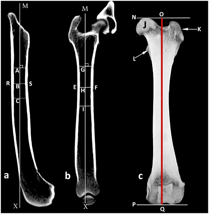
Al Aiyan et al. Femoral Anteversion Angle in Dogs
trochanter is shortened (14). Thereby, the pressure, that acts no clinical history of pelvic limb lameness. Dogs with orthopedic
on the femoral head in the acetabulum, is higher. Anatomists abnormalities or signs of hip joint disease were excluded from
and surgeons have long been interested in the FAA since it is the study.
considered an important factor for hip joint stability (1, 2, 4, 8, The dogs used in this study were assigned into two groups
9, 15). according to the length of their femora measured in CT (32).
Surgical treatment of serious hip joint problems often requires The CT scanning was conducted at the Small Animal Clinic,
total hip arthroplasty. Both the femoral neck angle and the Düppell, Free University of Berlin. The CT scanning of the
femoral anteversion angle, that describe the relationship between femora was done at a setting of 0.3 mm slice thickness, multi-slice
the femoral head, neck and the femur shaft, must be taken into spiral “Lightspeeds” QXi (General Electric Healthcare, GE), 120
account in the development of hip endoprostheses in order to kV, 130 mAs. The dogs were positioned in dorsal recumbency
reduce the risk of hip luxation following the implantation of on the CT scanner table. The pelvic limbs were pulled back
the prosthesis (16). Using a total hip replacement prothesis with and tied at the tarsal level with adhesive straps (Tesa AG
an inappropriate FAA value may result in premature wear and Humburg) to ensure that the femora were parallel to each other
loosening between the prothesis stem and the internal surface of and parallel to the CT scanner table. Advantage Workstation
the femoral shaft due to the increased pressure which finally cause software (Advantage Workstation 4.2, GE Healthcare) was used
failure of the prosthesis. to analyse the images. The data record was processed as multi-
Many different methods have been used to determine the planar and three-dimensional reconstructions using Advantage
FAA, including standard radiography (1, 12, 17), biplanar Workstation software.
planar radiography (4, 7, 18–21), computed tomography (CT) The sequence of measurements were done in the following
(9, 12, 22, 23), magnetic resonance imaging (24), three- order as some measurements were reliant on values of earlier
dimensional modeling (25) and three-dimensional (3D) laser measurements: determination of the axis of the femoral shaft,
scanner techniques (8, 26). length of the femur, center of the femoral head, axis of the femoral
Using single standard radiographic imagery to measure the neck, condylar axis, femoral anteversion angle. All measurements
FAA does not truly reflect spatial relationships between pertinent were performed by an experienced veterinarian and repeated
landmarks, due to a lack of depth information (27). CT imaging after 24 h. The mean of the two measurements of the FAA was
is considered to be a reliable and an accurate method for used to ensure data accuracy.
measuring the FAA because it allows accurate 3D volumetric
femoral reconstructions of the femur and avoids artifacts due Medullary Axis and Length of the Femur
to incorrect positioning thus improving the precision of FAA To ensure consistency in femoral measurements, an exact sagittal
measurements (20, 28–30) with average errors of 0.45◦ (30). plane view was obtained by aligning the caudal aspects of both
The main aim of this work was to use CT to obtain precise femoral condyles, and to avoid cranial or caudal inclination of
data of the femoral anteversion angle in cadavers of medium and the femur, the femoral axis was identified as the line connecting
large dog breeds in support of the development of an optimally the three central points shown in Figure 1a and was placed
functioning total hip replacement prothesis. In addition we vertically (Figure 1a). From here the femur was rotated exactly
provide a detailed description of the methodology using CT to 90◦ cranially to be able to obtain an accurate frontal plane view of
measure the femoral anteversion angle. the femur without external or internal rotation (Figure 1b).
In the sagittal and frontal planes, the center of the intracortical
width was created at the narrowest point of the femoral shaft.
MATERIALS AND METHODS Using similar methodology, additional central points were placed
2 cm proximal and 2 cm distal. The axis of the femoral shaft
The cadavers used in this study have been reported earlier in was identified as the line connecting the three central points
a previous article where the femoral neck inclination angle was (Figures 1a,b). Using a three dimensional model in a frontal
studied (31). view, the length of the femur was determined to be the line
Femora from 58 cadavers of orthopedically healthy adult dogs parallel to the femoral axis that connects the orthogonal lines
of medium and large breed size were studied using computed at the most proximal point of the femoral head and at the most
tomography. The dogs used in this study were obtained from the distal end of the femoral condyles (Figure 1c).
Small Animal Clinic of the Free University of Berlin. The dogs
had either died or were euthanased for reasons unrelated to this Center of the Femoral Head
study. For each individual dog the research ethics code of the Using a 3D transverse plane, the center of the femoral head was
institution was met and accompanied by written consent from identified by using annotation software to generate concentric
the dog’s owner. circles of best fit and superimpose these onto the femoral head
Post-mortem examination was conducted on each dog to (Figure 2a).
establish the absence of orthopedic abnormalities and disease.
The Ortolani and Barlow tests were conducted immediately post- Axis of the Femoral Neck and the Condylar
mortem. Radiography and CT examination of the hip joint was Axis
conducted post-mortem immediately after the death to establish In the transverse femoral neck planes the lesser trochanter
the absence of hip joint dysplasia. The dogs used in this study had appears at the transition from the medial to the caudal contour
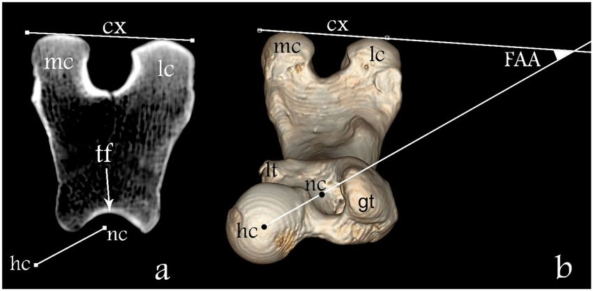
Frontiers in Veterinary Science | www.frontiersin.org 2 March 2021 | Volume 8 | Article 540406Al Aiyan et al. Femoral Anteversion Angle in Dogs FIGURE 1 | Determination of the medullary axis of the femoral shaft. (a) Sagittal plane view of the femur where line RS represents the intracortical width at the narrowest point of the femoral shaft and B is its central point; A and C are central points 2 cm proximal and 2 cm distal to (B) respectively; MX is the medullary sagittal axis. (b) Frontal plane view of the femur where EF represents the intracortical width at the narrowest point of the femoral shaft and H is its central point; G and I are central points 2 cm proximal and 2 cm distal to H, respectively; MX is the medullary frontal axis. (c) Frontal view of the femur where N and P are the proximal and distal orthogonal lines to medullary axis, respectively; J femoral head; K great trochanter; L lesser trochanter; OQ length of the femur. of the femur and disappears in more distal sections. A computer- The FAA was measured between the femoral neck axis and the generated circle was placed in the section with the maximum condylar axis (Figure 3b). extent of the lesser trochanter and the center of the circle was determined and presents the base of the femoral neck Statistical Analysis (Figure 2b). The axis of the femoral neck was defined as the The statistical analysis was based in the comparison of FAA line passing from the center of the femoral head to the base in the two groups with different femoral length. The intra- of the femoral neck in the transverse view of the femur and class correlation coefficient (ICC) and Kendall’s tau was used in remained visible on the monitor (Figures 2a, 3a). More distally, order to evaluate the intra-observer independent measurement the sectional view with the maximum caudal curvature of the repeatability. The Pearson correlation was used to study condyles was defined to represent the condylar axis (Figure 3a). the association between the length of the femur and the Frontiers in Veterinary Science | www.frontiersin.org 3 March 2021 | Volume 8 | Article 540406
Al Aiyan et al.                                                                                                                            Femoral Anteversion Angle in Dogs

  FIGURE 2 | Determination of the axis of the femoral neck. (a) Transverse plane of the proximal femur where “gt” is the great trochanter and “hc” is the center of the
  femoral head. (b) Transverse plane of the proximal femur on the level of the lesser trochanter where “lt” is the lesser trochanter and “nc” is the center of the base of the
  femur neck.

  FIGURE 3 | Determination of the FAA. (a) Transverse plane overlap view of the distal femur and (b) three-dimensional dorsoventral view of the femur. Here “hc”
  represents the femoral head center, “nc” the femoral neck center, “tf” the femoral trochlea, “mc” medial condyle, “lc” lateral condyle, “gt” greater trochanter, “lt” lesser
  trochanter; line between “nc” and “hc” femoral neck axis and line “cx” condylar axis. The FAA angle is defined between lines “nc-hc” and cx.

femoral anteversion angle. Pearson and Kendall’s tau results                               RESULTS
of −1 or 1 indicate perfect negative or positive association
between variables. A P-value smaller than 0.05 was considered                              In this study a total of 116 femora were measured from
significant. The statistical analysis was performed using the                              58 medium to large breed dogs. Twenty-three dogs were
Statistical Packages for Social Science software (SPSS Inc. Version                        excluded from the study due to orthopedic abnormalities or
26, Chicago II, USA). Values were reported as mean +/–                                     signs of hip joint disease which had been detected. The most
standard deviation.                                                                        common breed measured was the German Shepherd followed

Frontiers in Veterinary Science | www.frontiersin.org                                  4                                               March 2021 | Volume 8 | Article 540406
Al Aiyan et al.                                                                                                                    Femoral Anteversion Angle in Dogs

  FIGURE 4 | Boxplot with medians and data ranges representing the Femoral Anteversion Angle (FAA) in relation to the Length of the Femur for Group I and Group II.

by Staffordshire Terriers, Boxers, Rottweilers, Bullmastiffs,                         in this study shows no significant difference between group I and
and Weimaraners.                                                                      group II. Likewise, there was no correlation between the length
   Dogs were divided into two groups according to the length                          of the femur and the FAA (Figure 4). This is consistent with the
of their femora. Group I (n = 25) included all the dogs with a                        results of Palierne et al. (32).
femoral length ≤195 mm. Group II (n = 33) included all the dogs                           In adult medium and large dog breeds with normal hip
with a femoral length >195 mm.                                                        joint morphology, the FAA has been measured using several
   The age of dogs in Group I ranged from 2 to 16 years old, mean                     different imaging methods as well as anatomical preparation and
7.6 ± 4.15 years and in Group II ranged from 1.5 to 16 years old,                     reported in the literature to be within the range of 7.6 to 34.2◦
mean 8.4 ± 3.95 years. The body mass of dogs in Group I ranged                        (3, 4, 7–9, 12, 23, 24, 33–35). The results reported vary greatly
from 17 to 45 kg, mean 27.8 ± 7.53 kg, and in Group II ranged                         in these studies (Table 1). However, there are many relatively
from 22 to 60 kg, mean 42.3 ± 8.37 kg.                                                common congenital and developmental conditions where the
   All the measurements performed in the two independent                              FAA deviates significantly from the normal such as canine hip
sessions had adequate repeatability as the Kendall’s tau test                         dysplasia associated with a larger than normal FAA, that tends to
showed strong correlation (tau = 0.956, P = 0.000). The femur                         rotate the femoral head out of the acetabulum (1, 2). The different
length in Group I was 175.29 ± 12.29 mm and in Group II 213.44                        measurement methodologies as well as the body size, age profile,
± 15.77 mm.                                                                           gender, and breeds of dog populations, may explain the different
   The mean values of FAA obtained in this study were 30.99 ±                         results (15, 27).
4.02◦ for Group I and 31.58 ± 5.09◦ in Group II. No correlation                           Accurate measurement of the FAA using classical radiography
was found between the length of the femur and the FAA (P =                            relies on precise positioning of the femur to obtain a true
0.136) (Figure 4).                                                                    axial projection of the femur from distal to proximal, which
                                                                                      is technically challenging due to the difficulties encountered in
DISCUSSION                                                                            patient positioning (12). Often multiple attempts are necessary;
                                                                                      consequently such radiographic studies can often be time-
Because the dogs used in this study varied in their nutritional                       consuming (12).
status and history, body mass was not included as a                                       Due to the complex three-dimensional configuration of the
morphological parameter (32). The medium to large breed                               femur, CT imaging is considered to be the most reliable and
dogs used in the present study were assigned to two groups based                      accurate method to measure the FAA (9, 12, 20, 26, 28, 29).
solely on the total length of their femur (32). The FAA measured                      This allows accurate 3D volumetric femoral reconstructions of

Frontiers in Veterinary Science | www.frontiersin.org                             5                                            March 2021 | Volume 8 | Article 540406
Al Aiyan et al. Femoral Anteversion Angle in Dogs
TABLE 1 | Mean (SD) femoral anteversion angle reported in dogs by other studies, measured by standard radiograph (RAD), computed tomography (CT), magnetic
resonance imaging (MRI) and anatomical preparation (AP).
Authors N Method FAA (SD)
Adams et al. (8) five mongrel dogs 3D scanner and 3D animation software 23.4◦ ± 3.5
Bardet et al. (19) 15 mixed, medium to large Fluoroscopic method 31.31◦
Bardet et al. (19) 15 mixed, medium to large RAD biplanar 30.8◦
Bloebaum et al. (34) 21 greyhound RAD biplanar 27◦ ± 6.3
Dudley et al. (12) nine medium to large RAD, Fluoroscopic method 16◦ ± 6.4
Dudley et al. (12) nine medium to large CT 19.6◦ ± 7.9
Dudley et al. (12) nine medium to large AP 18.9◦ ± 5.4
Ginja et al. (23) 23 estrela Mountain Dogs, 7–8 week RAD biplanar 29.9◦ ± 4.8
Ginja et al. (23) 23 estrela Mountain Dogs, 7–8 week CT 30.4◦ ± 4.2
Griffon et al. (7) 160 labrador Retrievers RAD biplanar 29.67◦ ± 6.44
Hauptman et al. (3) 75 medium to large RAD biplanar 15.2◦
Kaiser et al. (24) 40 small, medium to large MRI 7.6◦ ± 5.5
Kara et al. (9) 75 mixed breeds CT 26.86◦ ± 11.46
Löer (36) large breeds CT 33.8◦
Löer (36) small breeds CT 33.2◦
Madsen and Svalastoga (37) 41 medium to large RAD biplanar 30◦ -43◦
Mahringer, (38) 105 medium to large AP 33◦ ± 8.66
Montavon et al. (4) 30 mongrel dogs, medium to large RAD biplanar 31.3◦ ± 6.2
Martins et al. (21) 126 young normal joints RAD biplanar 31.4◦ ± 4.8
106 young abnormal joints 32.6◦ ± 4.9
158 adult normal joints 26.4◦ ± 4.5
232 adult abnormal joints 27.7◦ ± 5.0
Nunamaker et al. (1) 34 various breeds adults RAD, Fluoroscopic method 26.97◦ ± 6.52
Palierne et al. (35) 82 medium to large RAD biplanar 30◦ ± 6.32
Palierne et al. (32) 206 small, medium to large RAD biplanar 29.40◦ ± 6.35
Savio et al. (26) 16 medium to large 3D scanner and design software 45◦ ± 4.5
Schawalder et al. (11) 50 medium to large RAD biplanar 30.1◦
Sumner et al. (33) 15 medium to large RAD biplanar 34.2◦ ± 5.7
the femur and obviates artifacts related to animal position and in dogs with a femoral length of between 196 and 240 mm (group
thereby increases the precision of the FAA measurement (12, 20, II) is 31.58 ± 5.09◦ . The mean FAA reported in the present study
28, 29) and can be used for clinical or research purposes without are in close agreement with those of Schawalder and Sterchi (11),
the need of additional radiographic exposures (12, 23). Bardet et al. (19), Montavon et al. (4), Sumner et al. (33, 38)), Löer
The patient preparation and the time required for (36), Palierne et al. (35), Ginja et al. (23), Palierne et al. (32), and
radiographic and CT examinations are similar (23). Even (7) (Table 1).
using the same imaging technique could result in different Our findings are inconsistent with (1, 3, 12, 24, 26, 34,
values, due to the different methodologies used to estimate 37) (Table 1). The use of different measurement techniques
the center of the base of the femoral neck (27). Minor can explain the different results of the FAA values. In the
variations in radiographic positioning and selection of current study we found that accurate identification of the
landmarks affect the correctness and variability of radiographic sagittal and frontal planes as demonstrated in this study are
measurements (13). necessary to delineate the intramedullary axis of the femur.
In the present study, the precise FAA was obtained using The transverse plane is the appropriate plane to identify the
a CT scan data set of 116 femora of 58 mature dogs, all free center of the femoral head, the femoral neck axis and the
of hip dysplasia. Multi-slice spiral computed tomography and condylar axis to be able to measure the FAA. In addition
Advantage Workstation software were used for the analysis. A the size, age, gender, and breed of the dog population also
set of five landmarks; the center of the femoral head, center contribute to variations in the FAA (4, 11, 15, 19, 24, 25, 27,
of the base of the femoral neck, lesser trochanter, medial and 36).
lateral aspect of the femoral condyles were found to be readily Martins et al. (21) described a significant reduction in FAA in
identifiable and suitable for our CT measurements. adult animals compared to younger dogs. In contrast, the mean
In this study the mean value of the FAA in dogs with a femoral FAA of 7.6◦ in the Kaiser et al. (24) study, during which magnetic
length of between 145 and 195 mm (group I) is 30.99 ± 4.02◦ and resonance imaging (MRI) was used, is considerably lower than
Frontiers in Veterinary Science | www.frontiersin.org 6 March 2021 | Volume 8 | Article 540406Al Aiyan et al. Femoral Anteversion Angle in Dogs
the mean FAA seen in other studies, this could be due to fact that DATA AVAILABILITY STATEMENT
the femoral head center lies cranially to the plane in which we can
define the center of the femoral neck (23, 24). The datasets generated for this study are available on request to
Some authors confirm a link between an increased anteversion the corresponding author.
angle and the incidence of degenerative hip diseases such
as hip joint dysplasia (1, 2, 4) whilst some others do not ETHICS STATEMENT
(21). This could confirm the high FAA measured by Savio
et al. (26) (45◦ ) and by Madsen and Svalastoga (37) (30– Ethical review and approval was not required for this animal
43◦ ). study as cadavers were obtained from euthanized animals. All
The FAA can support the development of a durable and research activities were done in accordance with The Central
optimally functional hip prosthesis. The use of correctly designed Ethics Committee of Freie Universität Berlin. Written informed
hip prostheses plays an active role in lowering the risk of consent was obtained from the animal’s owner for their use in
postsurgical complications associated with hip arthroplasty in this study.
medium and large dog breeds. According to this study, using
the methodology described, the measurement of the FAA can AUTHOR CONTRIBUTIONS
be made with good repeatability by a single observer based
on using femoral length as a proxy for dog size, a prosthesis All authors have contributed to the conception, design,
FAA of 31 degrees would be suitable for a wide range of acquisition of data, analysis and interpretation of data, drafting
dog sizes. or revising, and final approval of the manuscript.
REFERENCES techniques in Labrador Retrievers with and without cranial cruciate ligament
disease. Vet Surg. (2014) 43:534–41. doi: 10.1111/j.1532-950X.2014.12096.x
1. Nunamaker DM, Biery DN, Newton CD. Femoral neck anteversion 14. Prieur WD. Coxarthrosis in the dog part I: normal and abnormal
in the dog: Its radiographic measurement. Vet Radiol. (1973) 14:45–8. biomechanics of the hip joint. Vet Surg. (1980) 9:145–9.
doi: 10.1111/j.1740-8261.1973.tb00647.x doi: 10.1111/j.1532-950X.1980.tb01671.x
2. Dueland DJ. Femoral torsion and its possible relationship to canine hip 15. Gulan G, Matovinovi,ć D, Nemec B, Rubini,ć D, Ravlić-Gulan J. Femoral
dysplasia. Vet Surg. (1980) 9:48–52. neck anteversion: values, development, measurement, common problems.
3. Hauptman J, Cardinet G, Morgan JP, Guffy MM, Wallace LJ. Angles of Coll Antropol. (2000) 24:521–7.
inclination and anteversion in hip dysplasia in the dog. Am J Vet Res. 16. Guerrero TG, Montavon PM. Zurich cementless total hip
(1985) 46:2033–6. replacement: retrospective evaluation of 2nd generation implants in
4. Montavon PM, Hohn RB, Olmstead ML, Rudy RL. Inclination and 60 dogs. Vet Surg. (2009) 38:70–80. doi: 10.1111/j.1532-950X.2008.
anteversion angles of the femoral head and neck in the dog evaluation 00466.x
of a standard method of measurement. Vet Surg. (1985) 14:277–82. 17. Towle HA, Griffon DJ, Thomas MW, Siegel AM, Dunning D,
doi: 10.1111/j.1532-950X.1985.tb00883.x Johnson A. Pre- and postoperative radiographic and computed
5. Ginja MMD, Silvestre AM, Gonzalo-Orden JM, Ferreira AJA. Diagnosis, tomographic evaluation of dogs with medial patellar luxation.
genetic control and preventive management of canine hip dysplasia: a review. Vet Surg. (2005) 34:265–72. doi: 10.1111/j.1532-950x.2005.
Vet J. (2010) 184:269–76. doi: 10.1016/j.tvjl.2009.04.009 00040.x
6. Hayes GM, Ramirez J, Langley Hobbs SJ. Use of the cumulative summation 18. Ogata K, Goldsand EM. A simple biplanar method of measuring femoral
technique to quantitatively assess a surgical learning curve: canine total hip anteversion and neck-shaft angle. J Bone Joint Surg Am. (1979) 61:846–51.
replacement. Vet Surg. (2011) 40:1–5. doi: 10.1111/j.1532-950X.2010.00752.x doi: 10.2106/00004623-197961060-00007
7. Griffon DJ, Cunningham D, Gordon-Evans WJ, Tanaka R, Bruecker KA, 19. Bardet JF, Rudy RL, Hohn RB. Measurement of femoral torsion
Boudrieau RJ. Evaluation of a scoring system based on conformation factors in dogs using a biplanar method. Vet Surg. (1983) 12:1–6.
to predict cranial cruciate ligament disease in Labrador Retrievers. Vet Surg. doi: 10.1111/j.1532-950X.1983.tb00693.x
(2016) 46:206–12. doi: 10.1111/vsu.12593 20. Kuo TY, Skedros JG, Bloebaum RD. Measurement of femoral
8. Adams RW, Gilleland B, Monibi F, Franklin SP. The effect of valgus and varus anteversion by biplane radiography and computed tomography imaging:
femoral osteotomies on measures of anteversion in the dog. Vet Comp Orthop comparison with an anatomic reference. Invest Radiol. (2003) 38:221–9.
Traumatol. (2017) 30:184–90. doi: 10.3415/VCOT-16-09-0138 doi: 10.1097/01.RLI.0000059542.90854.EF
9. Kara ME, Sevil-Kilimci F, Dilek ÖG, Onar V. Proximal and distal alignment of 21. Martins J, Ferreira AJ, Ginja MM. Morphometric assessment of the hip joint
normal canine femurs: a morphometric analysis. Ann Anat. (2018) 217:125–8. in the Estrela Mountain Dog breed. Vet Comp Orthop Traumatol. (2012)
doi: 10.1016/j.aanat.2018.02.006 25:202–10. doi: 10.3415/VCOT-11-07-0101
10. Schawalder P, Spreng D, Dietschi E, Dolf G, Gaillard C. Die 22. Hernandez RJ, Tachdjian MO, Poznanski AK, Dias LS. CT
Hüftgelenksdysplasie im Umfeld von sekundären Einflüssen und ektopischen determination of femoral torsion. Am J Roentgenol. (1981) 137:97–101.
Ursachen. Kleintierpraxis. (1996) 41:625–38. doi: 10.2214/ajr.137.1.97
11. Schawalder P, Sterchi HP. Der Centrum-Collum-Diaphysenwinkel (CC’D) 23. Ginja M, Gonzalo-Orden JM, Jesus SS, Silvestre AM, Llorens-Pena
und der Antetorsionswinkel (AT) beim Hund. Kleintierpraxis. (1981) 26:151– MP, Ferreira A. Measurement of the femoral neck anteversion angle
62. in the dog using computed tomography. Vet J. (2007) 174:378–83.
12. Dudley RM, Kowaleski MP, Drost WT, Dyce J. Radiographic and computed doi: 10.1016/j.tvjl.2006.08.002
tomographic determination of femoral varus and torsion in the dog. Vet 24. Kaiser S, Cornely D, Colder W, Garner MT, Wolf K-J, Waibl H, et al.
Radiol Ultra. (2006) 47:546–52. doi: 10.1111/j.1740-8261.2006.00184.x The correlation of canine patellar luxation and the anteversion angle as
13. Mostafa AA, Griffon DJ, Thomas MW, Constable PD. Radiographic measured using magnetic resonance images. Vet Radiol Ultra. (2001) 42:113–
evaluation of femoral torsion and correlation with computed tomographic 8. doi: 10.1111/j.1740-8261.2001.tb00913.x
Frontiers in Veterinary Science | www.frontiersin.org 7 March 2021 | Volume 8 | Article 540406Al Aiyan et al. Femoral Anteversion Angle in Dogs
25. Hartel MJ, Petersik A, Schmidt A, Kendoff D, Nüchtern J, Rueger JM, et al. 34. Bloebaum RD, Ota DT, Skedros JG, Mantas JP. Comparison of human
Determination of femoral neck angle and torsion angle utilizing a novel three- and canine external femoral morphologies in the context of total hip
dimensional modeling and analytical technology based on CT datasets. PLoS replacement. J Biomed Mater Res A. (1993) 27:1149–59. doi: 10.1002/jbm.
ONE. (2016) 11:e0149480. doi: 10.1371/journal.pone.0149480 820270905
26. Savio G, Baroni T, Concheri G, Baroni E, Meneghello R, Longo 35. Palierne S, Asimus E, Mathon D, Meynaud-Collard P, Autefage
F, et al. Computation of femoral canine morphometric parameters A. Geometric analysis of the proximal femur in a diverse sample
in three-dimensional geometrical models. Vet Surg. (2016) 45:987–95. of dogs. Res Vet Sci. (2006) 80:243–52. doi: 10.1016/j.rvsc.2005.
doi: 10.1111/vsu.12550 07.010
27. Chimhundu C, Sivarasu S, Steiner S, Smit J, Douglas TS. Femoral neck 36. Löer B. Computertomographische Torsionsmessung an Femur und Tibia
anteversion measurement using linear slot scanning radiography. Med Eng des Hundes: Methode und klinische Anwendung bei der Luxation patellae
Phys. (2016) 38:187–91. doi: 10.1016/j.medengphy.2015.11.017 congenita. Munchen: Ludwig Maximilians Universitat (1999).
28. Weiner DS, Cook AJ, Hoyt WA, Oravec CE. Computed tomography in the 37. Madsen JS, Svalastoga E. Inclination and anteversion of collum femoris
measurement of femoral anteversion. Orthopedics. (1978) 1:299–306. in hip dysplasia and coxarthritis. Acta Vet Scand. (1994) 35:115–9.
29. Anda S, Terjesen T, Kvistad KA, Svenningsen S. Acetabular angles and femoral doi: 10.1186/BF03548337
anteversion in dysplastic hips in adults: CT investigation. J Comput Assist 38. Mahringer C. Längen- und Winkelverhältnisse am proximalen Femurende des
Tomogr. (1991) 15:115–20. doi: 10.1097/00004728-199101000-00018 Hundes und deren biomechanische Aspekte. (1991).
30. Kim JS, Park TS, Park SB, Kim IY, Kim SI. Measurement of femoral neck
anteversion in 3D. Part 2: 3D modelling method. Med Biol Eng Comput. (2000) Conflict of Interest: The authors declare that the research was conducted in the
38:610–6. doi: 10.1007/BF02344865 absence of any commercial or financial relationships that could be construed as a
31. Al Aiyan A, Richardson K, Manchi G, Plendl J, Brunnberg L. Measurement potential conflict of interest.
of the femoral neck angle in medium and large dog breeds using computed
tomography. Acta Vet Hung. (2019) 67:22–33. doi: 10.1556/004.2019.003 Copyright © 2021 Al Aiyan, Richardson, Manchi, Ginja and Brunnberg. This is an
32. Palierne S, Mathon D, Asimus E, Concordet D, Meynaud-Collard P, Autefage open-access article distributed under the terms of the Creative Commons Attribution
A. Segmentation of the canine population in different femoral morphological License (CC BY). The use, distribution or reproduction in other forums is permitted,
groups. Res Vet Sci. (2008) 85:407–17. doi: 10.1016/j.rvsc.2008.02.010 provided the original author(s) and the copyright owner(s) are credited and that the
33. Sumner DR, Devlin TC, Winkelman D, Turner TM. The geometry original publication in this journal is cited, in accordance with accepted academic
of the adult canine proximal femur. J Orthop Res. (1990) 8:671–7. practice. No use, distribution or reproduction is permitted which does not comply
doi: 10.1002/jor.1100080508 with these terms.
Frontiers in Veterinary Science | www.frontiersin.org 8 March 2021 | Volume 8 | Article 540406You can also read