Increased Pulmonary Embolism in Patients with COVID-19: A Case Series and Literature Review
←
→
Page content transcription
If your browser does not render page correctly, please read the page content below
Increased Pulmonary Embolism in Patients with
COVID-19: A Case Series and Literature Review
Sonia Hesam-Shariati
University of New South Wales
Poya Fatehi
Kurdistan University of Medical Science: Kurdistan University of Medical Sciences
Morteza Abouzaripour
Kurdistan University of Medical Science: Kurdistan University of Medical Sciences
Fardin Fathi
Kurdistan University of Medical Science: Kurdistan University of Medical Sciences
Negin Hesam-Shariati
University of New South Wales
mohamad bakhtiar hesam shariati ( b.hesamshariati@gmail.com )
Kurdistan University of Medical Science: Kurdistan University of Medical Sciences
https://orcid.org/0000-0002-2000-5197
Case report
Keywords: COVID-19, Venous Thromboembolism, Pulmonary Embolism, Computed Tomography
Angiography
Posted Date: March 22nd, 2021
DOI: https://doi.org/10.21203/rs.3.rs-314021/v1
License: This work is licensed under a Creative Commons Attribution 4.0 International License.
Read Full License
Page 1/12Abstract
Recently, there is evidence that the coronavirus disease 2019 (COVID-19) increases the risk of venous
thromboembolism by creating a prothrombotic state. COVID-19 and pulmonary embolism (PE) are both
associated with tachypnoea, hypoxemia, dyspnoea, and increased D-dimer. Diagnosis of pulmonary
embolism in a patient with COVID-19 compared to a patient without it using the conventional clinical and
biochemical evidence is challenging and somehow impossible. In this study, we report 4 male cases
affected by COVID-19, admitted to hospitals in Sanandaj, Iran. The patients were all older adults (ranged
between 56 and 95 years of age). Fever, chills, muscle aches, and cough were evident in all of them. Red
blood cell levels were low, while pulmonary embolism was clearly seen on spiral computed tomographic
(CT) angiography of the pulmonary circulation of all patients. These cases demonstrated that COVID-19
may lead to pulmonary embolism by causing blood coagulation problems. As COVID-19 continues to
cause considerable mortality, more information is emerging which reveals its complicated pathogenicity.
In the meantime, venous thromboembolism remains an uncommon finding in patients with COVID-19. It
is essential that health care providers perform the necessary diagnostic evaluations and provide
appropriate treatment for patients.
Introduction
Coronavirus disease 2019 (COVID-19), caused by the new coronavirus, usually appears with mild
symptoms; however, in 14% of cases, it can lead to a serious illness that requires hospitalization [1].
Severe hypoxemia is the main characteristic of the severity of this disease [2]. To this date, COVID-19 has
infected about 95 million people and killed over 2 million of them worldwide [3]. Studies have shown that
COVID-19 infection increases the risk of venous thromboembolism (VTE) in patients with an increased
disseminated intravascular coagulation, inflammation, hypoxemia, and immobility [4, 5]. The incidence
rate of VTE in COVID-19 is still unknown. However, emerging data show an increased incidence of venous
thromboembolism in COVID-19, especially in more severe cases [6].
So far, there have been several reports of coagulation in patients with COVID-19[7–9]. It has been
suggested that vascular endotheliitis due to an activated immune response or an infection of the
vascular endothelium with acute coronavirus syndrome 2 (SARS-CoV-2) or COVID-19 may lead to blood
clotting. Nevertheless, the pathophysiology of coagulation associated with coronavirus is not yet well
understood. The incidence of pulmonary embolism (PE) in COVID-19 patients has been reported in many
countries, mainly in Europe and the United States [10]. Unfortunately, due to the lack of large prospective
studies, there is little information on the epidemiology and pathophysiological mechanisms of COVID-19-
associated PE. Timely understanding of these mechanisms is extremely important for the proper
diagnosis and management of the deadly complications of this disease. In addition, proper dosage and
duration of prophylactic anticoagulation are the main concerns for controlling this disease [11, 12]. In this
article, we have reported four cases of coronavirus patients with pulmonary embolism admitted to
hospitals in Sanandaj, Iran.
Page 2/12Case 1
A 60-year-old man was presented to the medical unit with symptoms of respiratory problems, severe
headache, cough, dizziness, and frequent vomiting. Initial physical and clinical examinations of the
patient were normal and there was no underlying disease. The patient had no history of alcohol or
tobacco use and was not taking any specific medications at the time. His blood pressure was 120/80
mmHg with a regular pulse rate of 112 beats/min, a respiratory rate of 22 cycles/min, and a temperature
of 36°C. While the patient had no symptoms of arrhythmia, he had mild hypoxemia with an oxygen level
of 85–92%. Important laboratory findings of the patient are listed in Table 1. PCR on the nasopharyngeal
swab sample was performed on the day of hospitalization, which confirmed the diagnosis of COVID-19.
The patient was discharged from the hospital after 2 days because his symptoms were relatively mild
and there were not any serious symptoms. He was admitted to the hospital five days later with respiratory
problems, and initial examinations revealed that his oxygen saturation was now 82% on air. The patient
underwent high-resolution computed tomography (CT) scans of the lungs and CT pulmonary
angiography. CT scans of the lungs (Fig. 1) showed several diffuse areas of opacity in both right and left
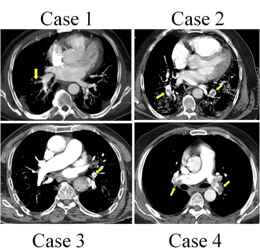
lungs, which could indicate viral pneumonia. In addition, on CT angiography of the lungs (Fig. 2), several
filling defects were visible in the branch of the pulmonary artery leading to the lower lobe of the right lung,
which may indicate acute pulmonary embolism. The patient was started on medications such as
Naproxen, Hydroxychloroquine, Famotidine, Zinc, Neurobion, and some anticoagulant including injected
heparin and acetylsalicylic acid tablets, and high-flow oxygen. Fortunately, he did not require mechanical
ventilation or intensive care unit (ICU) management and was released from the hospital 15 days after
partial recovery.
Page 3/12Table 1
The results of laboratory findings
Test Name Unit Reference Range Case 1 Case 2 Case 3
1 BUN mg/dl 7-16.8 24 Hi 28 Hi 20
2 Ca mg/dl 8.6–10.3 8.5 Low - -
3 p mg/dl 2.7–4.5 4.9 Hi - -
4 Na(ser) mEq/L 138–145 139 139 140
5 K(ser) mEq/L 3.6–5.9 4.3 4.3 3.7
6 MCH pg 27.5–33.2 28.2 28.2 32.5
7 MCHC g/dL 30.0–38.0 34.6 31.6 32.6
8 Plt ×1000/µL 140–440 252 167 72
9 Cr mg/dl male:0.8–1.3 mg/dl 1 1.1 1.4 Hi
10 SGOT(AST) IU/L Male:Test Name Unit Reference Range Case 1 Case 2 Case 3
26 PT 11–13 second 13.6 - 15
27 INR 1.2 - 1.6
28 PT Control % 12 - 11
29 Amylase IU/L < 100 U/L 98 - -
30 HBSAg MIU/ml < 1 non reactive > = 1 Negative Negative Negative
CLIA reactive
31 HCV-Ab MIU/ml < 1 non reactive > = 1 Negative Negative Negative
CLIA reactive
32 HIV-Ab MIU/ml < 1 non reactive > = 1 Negative Negative Negative
CLIA reactive
Case 2
A 56-year-old man was hospitalized due to the persistence of high fever started 5 days before. In physical
and clinical examinations of the patient, symptoms such as fever, chills, muscle pain, weakness, cough,
tachycardia, and acute respiratory syndrome were reported, while there was no report of underlying
disease in the patient's file. Investigations showed no history of alcohol and tobacco intake or any
particular medication use at the time. A polymerase chain reaction (PCR)-based test for SARS-CoV-2 was
done, and he was diagnosed with COVID-19. Initial examinations of the patient in the hospital revealed a
blood pressure of 130/90 mmHg with a regular pulse rate of 109 beats/min, a respiratory rate of 28
cycles/min, and a temperature of 39°C. All laboratory findings of the patient are presented in Table 1. The
patient's electrocardiography (ECG) was normal. There were no changes in the patient's hemodynamics
or respiratory status (oxygen saturation: 84% on room air), and in later stages, due to persistent
respiratory problems, CT pulmonary angiography of the patient was ordered by a pulmonologist for
further examination. As in CT angiography (Figs. 1 and 2): (a) the diameters of the main pulmonary
arteries were normal; (b) defective filling in the lobar, segmental, and sub-segmental branches of the
upper, middle, and lower lobes was evident in both lungs, which may indicate thrombosis (embolism); (c)
multiple confluent patchy Ground-glass opacification (G.G.O) and consolidation was found in both lungs.
Due to the existence of a pandemic around the world and the positive result of COVID-19 test, the COVID-
19 diagnosis was confirmed, while no deep vein thrombosis or other thrombosis were detected.
Continuous heparin injection was performed according to the doctor's instructions to treat pulmonary
embolism. Supportive care, antibiotics, and other treatments were used to treat the patient, and he
eventually was discharged after 18 days of hospitalization.
Case 3
Page 5/12A 95-year-old male patient with a history of severe dyspnea for 2 days was referred to a medical center.
Examination of the patient's file showed that he had experienced several episodes of diarrhea a few days
prior to the onset of shortness of breath, which resolved on its own. According to the patient's history, his
wife had contracted coronavirus two weeks before. The study also found that the patient had symptoms
of fever, cough, and chest pain. His previous medical history showed low blood pressure and
hyperlipidemia, but no history of malignancy. Examination of the family history revealed no signs of
coagulation disorders or thromboembolism. The patient never smoked or consumed alcohol. His vital
signs included blood pressure of 100/65 mmHg, pulse rate of 88 beats/min, respiratory rate of 60/min,
oximetry 76% on room air, and a temperature of 38.5°C. There was no lower extremity edema or calf
tenderness. The most common blood and electrolyte tests are listed in Table 1. Testing for viral diseases,
including influenza was negative. The initial results of the coronavirus test were negative, but in the
second test, which was taken from the back of the throat, the coronavirus was detected. According to the
patient's respiratory rate and old age, a CT scan of the lung was performed on the order of the treating
physician, and CT images showed a specific sign of the virus (Fig. 1) .Due to the deterioration of the
patient and having respiratory problems, he was transferred to the hospital’s ICU, approximately 4 days
after hospitalization. CT pulmonary angiography was also performed by the order of a pulmonologist. As
shown in the CT scan images of the patient, there is a defect of contrast material in the middle lobe
branch of the left pulmonary artery, which can indicate chronic thrombosis in this artery (Fig. 2). The
patient was started on Naproxen, Hydroxychloroquine, Famotidine, Zinc, Neurobion, and anticoagulant
treatments. Unfortunately, he died due to respiratory failure and intubation after 15 days of
hospitalization in the ICU.
Case 4
A 72-year-old man was hospitalized in Besat Hospital in Sanandaj, Iran after 11 days of cough, fever,
weakness, palpitation, and respiratory problems, but no chest pain. The COVID-19 nucleic acid diagnostic
test was positive before hospitalization. After the final diagnosis of COVID-19, the patient was transferred
to the quarantine ward of the hospital. At the time of admission, his clinical and physical examination did
not show any irregularity in heartbeat, which could be a sign of atrial fibrillation. Other vital symptoms
were blood pressure of 62.95 mmHg, temperature of 39°C, respiration rate of 23 breaths/min, and oxygen
saturation rate of 87%. After admission, the laboratory test of COVID-19 was re-confirmed in the hospital.
Unfortunately, we could not have accessed to the results of the routine blood tests and the patient’s
serum biochemical analyses. Due to respiratory problems, CT angiography of the pulmonary arteries was
ordered by his doctor. A defect of filling in the right and left main pulmonary arteries as well as the lobar
and segmental branches of both sides was quite obvious (Figs. 1 and 2), which suggested pulmonary
embolism. Examination of the lung parenchyma tissue also revealed multifocal turbidity in both lungs,
which indicated infection. According to these findings, antiviral, antibacterial, anticoagulant, symptomatic
and supportive treatments were started for the patient. Fortunately, with proper and timely treatment, the
patient was discharged from the hospital 12 days after admission and the rest of the treatment was
continued at home.
Page 6/12Discussion
Coronaviruses are a large family of enveloped ribonucleic acid (RNA) viruses found in animals such as
pigs, camels, bats, and cats. Entering these viruses into the human body can cause mild to moderate
upper respiratory illnesses [13]. Several of these coronaviruses are known worldwide, including acute
respiratory syndrome (SARS), Middle East respiratory syndrome (MERS), and SARS-CoV-2, which often
lead to severe and deadly diseases in humans [13].
Patients with COVID-19 often have respiratory symptoms, which make it hard to distinguish from
pulmonary embolism (PE) in severe cases. Studies have shown that coronaviruses increase the risk of
arterial and venous thromboembolism by causing inflammatory reactions, immobility, hypoxia, and
disseminated intravascular coagulation (DIC) in patients [14]. The co-occurrence and clinical
symptomatic overlaps between pulmonary embolism and COVID-19 has made the diagnosis and
treatment of PE more difficult. The presence of COVID-19 pneumonia can be easily detected with RT-PCR
and CT scan [15, 16]. However, it is much more difficult to confirm PE. This is because factors such as
lactate dehydrogenase, ferritin, C-reactive protein, and interleukin levels that influence pro-inflammatory
and hypercoagulability processes increase in patients with coronavirus [17, 18]. In addition, recent studies
have shown that levels of D-dimer and fibrinogen and fibrin degradation products increase in patients
with COVID-19 [19]. It has also been shown that even in the absence of pulmonary embolism in patients
with COVID-19, the level of D-dimer increases [18]. An increase in D-dimer (> 1 mg/dL) may indicate
mortality in these patients but is not a reliable indicator in the diagnosis of venous thromboembolic [5,
20]. As a result, CT angiography can be helpful in diagnosing VTE in patients with coronavirus [21].
The coagulation mechanism in COVID-19 is unknown. Some theories introduce cytokines as possible
factors in the coagulation process in this disease, while others believe that hepatic dysfunction may be
involved [22]. Regardless of the coagulation mechanism in patients with coronavirus, it is known that the
incidence of thrombosis increases in these patients and so that this coagulation often extends to
intravascular coagulation and this expansion results in venous and arterial thrombosis. In addition, it has
been shown that 71.4% of patients who died of COVID-19 met the criteria for diffuse intravascular
coagulation [23]. Many patients with COVID-19 face sepsis and septic shock [24]. In the septic process,
DIC is a major cause of organ dysfunction, so undergoing anticoagulant therapy in this situation can be
very challenging [25].
There are currently no specific criteria for the use of anticoagulants in COVID-19 patients. Therefore, more
clinical trials are needed to determine whether all patients with coronavirus need to be treated with
anticoagulants. In general at this point, using PE prophylaxis based on clinical manifestations and D-
dimer level, even in mild cases of COVID-19 seems to be important and necessary [10].
Due to the lack of sufficient information, using or not using anticoagulants to improve the overall
symptoms of the disease and its complications in patients with COVID-19 is still highly controversial [26].
Two recent published studies by Klok et al. and Middeldorp et al. advised against prophylactically
initiating treatment-dose anticoagulation in all patients with COVID-19 and in opposite recommended
Page 7/12using a lower threshold for proper diagnostic tests in assessing thrombotic complications including deep
vein thrombosis and PE [27, 28].
Thus, two aims are recommended to be considered in the treatment of patients with COVID-19: the first
goal is to protect the organs and timely diagnosis of events caused by the disease and the second goal is
to strengthen the immune system to prevent the formation of cytokine storms and blood clotting. Further
research into clinical trials is needed to clarify whether prophylactic treatment with anticoagulants leads
to clinically beneficial outcomes in patients with COVID-19 infection.
Conclusion
Coronavirus continues to cause significant consequences, while more data are emerging that could help
investigate the effects of this disease. The little emerging information suggests the role of this
coronavirus in increasing systemic coagulation activation, which generally leads to thromboembolic
complications such as pulmonary embolism. Here, we presented four rare and notable cases of
pulmonary embolism. Therefore, early and timely treatment and diagnosis of this disease and its
complications can be a useful prognosis. Furthermore, studies have shown that this virus can weaken
and destroy the immune system by causing pathogenic infections and the associated complications in
the body. Thus, strengthening the immune system is one of most important ways of fighting this disease.
Further research is needed on the role of prophylactic anticoagulants in COVID-19 patients and the
pathogenesis of hypercoagulability in this disease.
Declarations
Ethical Approval and Consent to participate: This research has been confirmed by the Research Center of
Kurdistan University of Medical Sciences and Ethics Committee with the file number
IR.MUK.REC.1399.253.
Consent for publication: Written informed consent was obtained from a legally authorized
representative(s) for anonymized patient information to be published in this article which was approved
by the Research Center of Kurdistan University of Medical Sciences.
Availability of data and materials: The patients’ data are presented in Table 1 and Figures 1 and 2 of the
manuscript.
Competing interests: All authors declare that there is no conflict of interest that prejudices the impartiality
of this scientific work.
Funding: No source of funding
Authors' contributions: MBHS supervised the study and drafted the manuscript; PF collected the clinical
data; MA and FF analyzed the data and images; SHS and NHS edited and critically reviewed the
Page 8/12manuscript.
Acknowledgements: The authors thank all the teaching and medical staff of Kurdistan University of
Medical Sciences for their effort in eradicating the virus around the clock.
References
1. Greenan-Barrett, J. and A. Perera, COVID-19 and Pulmonary Emboli: A Case Series and Literature
Review. Clinical practice and cases in emergency medicine, 2020. 4(3): p. 299.
2. Chen, N., et al., Epidemiological and clinical characteristics of 99 cases of 2019 novel coronavirus
pneumonia in Wuhan, China: a descriptive study. The Lancet, 2020. 395(10223): p. 507-513.
3. Rahimzadeh, M. and P. Pooprasert, Pulmonary Embolism in a Post Covid-19 Patient-A Case Report.
The Physician, 2020. 6(1).
4. Guan, W.-j., et al., Clinical characteristics of coronavirus disease 2019 in China. New England journal
of medicine, 2020. 382(18): p. 1708-1720.
5. Zhou, F., et al., Clinical course and risk factors for mortality of adult inpatients with COVID-19 in
Wuhan, China: a retrospective cohort study. The lancet, 2020.
6. Wang, T., et al., Attention should be paid to venous thromboembolism prophylaxis in the
management of COVID-19. The Lancet Haematology, 2020. 7(5): p. e362-e363.
7. Fei, Y., et al., Coagulation dysfunction: a hallmark in COVID-19. Archives of pathology & laboratory
medicine, 2020. 144(10): p. 1223-1229.
8. Giannis, D., I.A. Ziogas, and P. Gianni, Coagulation disorders in coronavirus infected patients: COVID-
19, SARS-CoV-1, MERS-CoV and lessons from the past. Journal of Clinical Virology, 2020: p. 104362.
9. Zou, Y., et al., Analysis of coagulation parameters in patients with COVID-19 in Shanghai, China.
Bioscience trends, 2020.
10. Akiyama, Y., et al., A case of non‐severe COVID‐19 complicated by pulmonary embolism. Respirology
case reports, 2020. 8(7): p. e00622.
11. Helms, J., et al., High risk of thrombosis in patients with severe SARS-CoV-2 infection: a multicenter
prospective cohort study. Intensive care medicine, 2020: p. 1-10.
12. Sakr, Y., et al., Pulmonary embolism in patients with coronavirus disease-2019 (COVID-19)
pneumonia: a narrative review. Annals of intensive care, 2020. 10(1): p. 1-13.
13. Bhatt, H. and S. Singh, Venous thromboembolism and COVID-19: a case report and review of the
literature. Journal of Medical Case Reports, 2020. 14(1): p. 1-4.
14. Klok, F.A., et al., Confirmation of the high cumulative incidence of thrombotic complications in
critically ill ICU patients with COVID-19: an updated analysis. Thrombosis research, 2020.
15. Chung, M., et al., CT imaging features of 2019 novel coronavirus (2019-nCoV). Radiology, 2020.
295(1): p. 202-207.
Page 9/1216. Lei, J., et al., CT imaging of the 2019 novel coronavirus (2019-nCoV) pneumonia. Radiology, 2020.
295(1): p. 18-18.
17. Han, H., et al., Prominent changes in blood coagulation of patients with SARS-CoV-2 infection.
Clinical Chemistry and Laboratory Medicine (CCLM), 2020. 1(ahead-of-print).
18. Chen, J., et al., Findings of acute pulmonary embolism in COVID-19 patients. Available at SSRN
3548771, 2020.
19. Tang, N., et al., Anticoagulant treatment is associated with decreased mortality in severe coronavirus
disease 2019 patients with coagulopathy. Journal of thrombosis and haemostasis, 2020. 18(5): p.
1094-1099.
20. Wang, D., et al., Clinical characteristics of 138 hospitalized patients with 2019 novel coronavirus–
infected pneumonia in Wuhan, China. Jama, 2020. 323(11): p. 1061-1069.
21. Hekmat, M., et al., COVID-19 Complicated by Massive Acute Pulmonary Embolism: A Case Report.
Iranian Red Crescent Medical Journal, 2020. 22(6).
22. Bikdeli, B., et al., COVID-19 and Thrombotic or Thromboembolic Disease: Implications for Prevention,
Antithrombotic Therapy, and Follow-Up: JACC State-of-the-Art Review. Journal of the American
College of Cardiology, 2020. 75(23): p. 2950-2973.
23. Tang, N., et al., Abnormal coagulation parameters are associated with poor prognosis in patients with
novel coronavirus pneumonia. Journal of thrombosis and haemostasis, 2020. 18(4): p. 844-847.
24. Singer, M., et al., The third international consensus definitions for sepsis and septic shock (Sepsis-3).
Jama, 2016. 315(8): p. 801-810.
25. Iba, T., et al., Diagnosis and management of sepsis‐induced coagulopathy and disseminated
intravascular coagulation. Journal of Thrombosis and Haemostasis, 2019. 17(11): p. 1989-1994.
26. Atallah, B., S.I. Mallah, and W. AlMahmeed, Anticoagulation in COVID-19. European Heart Journal—
Cardiovascular Pharmacotherapy, 2020.
27. Middeldorp, S., et al., Incidence of venous thromboembolism in hospitalized patients with COVID‐19.
Journal of Thrombosis and Haemostasis, 2020.
28. Klok, F., et al., Incidence of thrombotic complications in critically ill ICU patients with COVID-19.
Thrombosis research, 2020.
Figures
Page 10/12Figure 1
Axial non-contrast-enhanced image of computed tomography (CT) scan of the chest, showing the COVID-
19 infection.
Page 11/12Figure 2
CT angiography images of pulmonary embolism, showing pulmonary artery embolism.
Page 12/12You can also read


