Immunodiagnosis and Immunotherapeutics Based on Human Papillomavirus for HPV-Induced Cancers
←
→
Page content transcription
If your browser does not render page correctly, please read the page content below
REVIEW
                                                                                                                                           published: 08 January 2021
                                                                                                                                     doi: 10.3389/fimmu.2020.586796

                                            Immunodiagnosis and
                                            Immunotherapeutics Based
                                            on Human Papillomavirus
                                            for HPV-Induced Cancers
                                            Zhen Dong 1,2,3*†, Renjian Hu 1,4†, Yan Du 5, Li Tan 1,2, Lin Li 1,2,6, Juan Du 1,7,
                                            Longchang Bai 1,2, Yingkang Ma 1,2 and Hongjuan Cui 1,2,3*
                                            1 State Key Laboratory of Silkworm Genome Biology, Institute of Sericulture and Systems Biology, College of Sericulture &
                                            Textile & Biomass Science, Southwest University, Chongqing, China, 2 Cancer Center, Reproductive Medicine Center,
                            Edited by:      Medical Research Institute, Southwest University, Chongqing, China, 3 NHC Key Laboratory of Birth Defects and
                          Franz Rödel,      Reproductive Health (Chongqing Key Laboratory of Birth Defects and Reproductive Health, Chongqing Population and
University Hospital Frankfurt, Germany      Family Planning Science and Technology Research Institute), Chongqing, China, 4 School of Pharmacy and Bioengineering,
                                            Chongqing University of Technology, Chongqing, China, 5 Department of Ultrasound, Chongqing University Central Hospital
                        Reviewed by:
                                            (Chongqing Emergency Medical Center), Chongqing, China, 6 Department of Immunology, School of Basic Medicine,
                         Daniel Martin,
                                            Southwest Medical University, Luzhou, China, 7 Department of Dermatology, The Third Hospital of Hebei Medical University,
University Hospital Frankfurt, Germany
                                            Shijiazhuang, China
                Ekaterina Dadachova,
 University of Saskatchewan, Canada
                   *Correspondence:         Infection with human papillomavirus (HPV) is one of the main causes of malignant neoplasms,
                           Zhen Dong        especially cervical, anogenital, and oropharyngeal cancers. Although we have developed
                zdong007@swu.edu.cn
                         Hongjuan Cui
                                            preventive vaccines that can protect from HPV infection, there are still many new cases of
                     hcui@swu.edu.cn        HPV-related cancers worldwide. Early diagnosis and therapy are therefore important for the
     †
      These authors have contributed        treatment of these diseases. As HPVs are the major contributors to these cancers, it is
                 equally to this work
                                            reasonable to develop reagents, kits, or devices to detect and eliminate HPVs for early
                     Specialty section:
                                            diagnosis and therapeutics. Immunological methods are precise strategies that are promising
           This article was submitted to    for the accurate detection and blockade of HPVs. During the last decades, the mechanism of
                        Cancer Immunity
                                            how HPVs induce neoplasms has been extensively elucidated, and several oncogenic HPV
                    and Immunotherapy,
                 a section of the journal   early proteins, including E5, E6, and E7, have been shown to be positively related to the
                Frontiers in Immunology     oncogenesis and malignancy of HPV-induced cancers. These oncoproteins are promising
              Received: 24 July 2020        biomarkers for diagnosis and as targets for the therapeutics of HPV-related cancers.
         Accepted: 24 November 2020
          Published: 08 January 2021
                                            Importantly, many specific monoclonal antibodies (mAbs), or newly designed antibody
                             Citation:
                                            mimics, as well as new immunological kits, devices, and reagents have been developed for
     Dong Z, Hu R, Du Y, Tan L, Li L,       both the immunodiagnosis and immunotherapeutics of HPV-induced cancers. In the current
          Du J, Bai L, Ma Y and Cui H
                                            review, we summarize the research progress in the immunodiagnosis and
         (2021) Immunodiagnosis and
       Immunotherapeutics Based on          immunotherapeutics based on HPV for HPV-induced cancers. In particular, we depict the
                 Human Papillomavirus       most promising serological methods for the detection of HPV infection and several
            for HPV-Induced Cancers.
          Front. Immunol. 11:586796.
                                            therapeutical immunotherapeutics based on HPV, using immunological tools, including
    doi: 10.3389/fimmu.2020.586796          native mAbs, radio-labelled mAbs, affitoxins (affibody-linked toxins), intracellular single-chain

Frontiers in Immunology | www.frontiersin.org                                      1                                      January 2021 | Volume 11 | Article 586796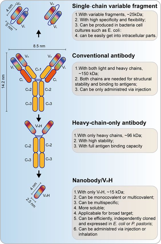
Dong et al.                                                                                                      Immunological Methods for HPV-Induced Cancers

                                          antibodies (scFvs), nanobodies, therapeutical vaccines, and T-cell-based therapies. Our
                                          review aims to provide new clues for researchers to develop novel strategies and methods
                                          for the diagnosis and treatment of HPV-induced tumors.
                                          Keywords: cervical cancer, human papillomavirus, monoclonal antibody, immunodiagnosis, immunotherapeutics

INTRODUCTION                                                                       sequence analysis, HPVs have been divided into five genera—a, b, g,
                                                                                   Nu, and Mu—each with different life cycle characteristics and related
Every year, more than 4.5% (8.6% in women and 0.8% in men) of all                  diseases (8–10). Epidemiological studies show that HPV types 16, 18,
cancers worldwide (630,000 new cancer cases), such as cervical                     31, 33, 35, 39, 45, 51, 52, 56, 58, and 59 are carcinogenic, and HPV68
cancer, vulvar cancer, vaginal cancer, penile cancer and anal cancer               are probably carcinogenic (6). Among them, HPV16 is the most
1), oropharyngeal cancers (OPC; including tumors derived from the                  prevalent worldwide and the major cause of HPV-associated
base of the tongue and tonsils), and even esophageal                               cancers (11).
adenocarcinoma (EAC) (2, 3), are attributed to the human                               The HPV genome has approximately 7800 to 7900 base pairs
papillomaviruses (HPVs) (Figure 1). Recently, HPV has also been                    (bp) and a molecular weight of 5 × 10⁶ Daltons. It consists of 72
found to be present and active in lung cancer 4) and may also                      shell particles comprising three-dimensionally symmetrical
contribute to skin cancers (5). HPVs are a kind of small,                          icosahedrons with a diameter of about 55 nm. It has a
unenveloped, and highly host-specific double-stranded circular                      lipoprotein-free membrane, a core, and a protein capsid. The
DNA viruses (6). They are not only microorganisms that are                         HPV genome has four parts (Figure 2): an early transcribed
sexually transmitted via genital contact but also a kind of viruses                region encoding six early proteins, including E1, E2, E4, E5, E6,
that can be passed on by skin and mouth (7). HPVs belong to                        and E7, a late transcribed region encoding two late proteins,
subgroup A of the papillomavirus family. To date, the genome of 189                including L1 and L2, a non-transcribed region containing the cis-
HPV types have been completely sequenced. According to DNA                         elements necessary for replication and transcription, and a small

 FIGURE 1 | Human papillomavirus (HPV)-induced cancers in human beings. HPV commonly induced oropharyngeal cancer and esophageal adenocarcinoma (also
 referred to as head and neck cancer) in the orpharynx and esophagus and cervical cancer, vaginal cancer, vulvar cancer, anal cancer, and penile cancer in the
 reproductive system.

Frontiers in Immunology | www.frontiersin.org                                  2                                      January 2021 | Volume 11 | Article 586796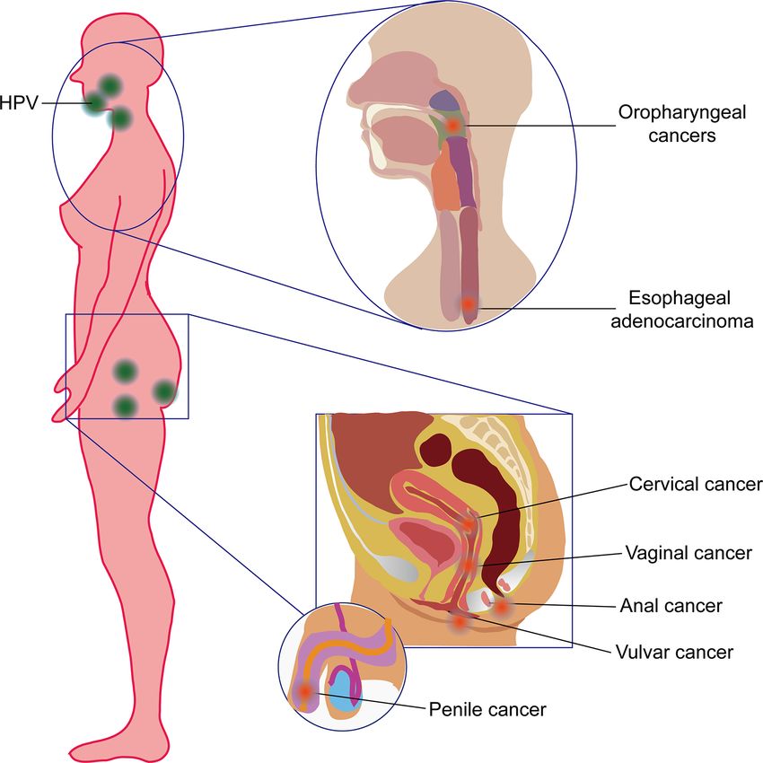
Dong et al.                                                                                                                Immunological Methods for HPV-Induced Cancers

                                                                                            HPV is cost‐effective in almost all countries (16). Currently, the
                                                                                            newest HPV vaccine is Gardasil-9 (Merck & Co), which can
                                                                                            protect against nine different types of HPV: type 6, 11, 16, 18, 31,
                                                                                            33, 45, 52, and 58 (17). However, progress toward prevention is
                                                                                            often frustrating because of the low access to vaccine and the
                                                                                            limitations of use for HPV-positive cancer screening, especially
                                                                                            in less developed countries (18, 19).
                                                                                                At present, the clinical detection method of HPV is mainly
                                                                                            based on the polymerase chain reaction (PCR) method. This
                                                                                            method can only be used to detect HPV DNA and HPV types
                                                                                            and cannot be used to accurately predict HPV-positive cancers (20,
                                                                                            21). For instance, the serum DNA level of HPV has been shown to
                                                                                            have a significant prognostic impact on advanced anal carcinoma
                                                                                            before first-line chemotherapy, and HPV-circulating tumor DNA
                                                                                            (ctDNA) becomes negative after chemotherapy completion (22).
                                                                                            However, the HPV oncoproteins, especially E6 and E7, are more
                                                                                            important during the HPV-induced carcinogenesis (23, 24). The
                                                                                            integration and hypermethylation mechanisms of the HPV viral
                                                                                            genome make the expression and lifecycle of the E5, E6, and E7
                                                                                            oncoproteins different from each other (25). These oncoproteins
                                                                                            are therefore probably biomarkers for diagnosis and prognosis and
                                                                                            may even be drug targets for therapeutics. Besides, their
                                                                                            downstream factors can also be used for both diagnosis and
                                                                                            therapeutics. For instance, the p16 INK4A protein, the
 FIGURE 2 | Schematic view of gene structure of human papillomavirus (HPV)                  immunohistochemical overexpression of which may be a useful
 genome. The HPV genome has an early transcribed region encoding six early                  screening test for HPV-induced cancers, is one of the important
 proteins, including E1, E2, E4, E5, E6, and E7, a late transcribed region encoding         downstream factors of E7. In normal cells, p16 can be negatively
 two late proteins, including L1 and L2, a non-transcribed region containing the
                                                                                            transcriptionally regulated by active pRb. However, in HPV-
 cis-elements necessary for replication and transcription and a small highly variable
 non-coding region located between E5 and L2. LCR, long control region.
                                                                                            positive cells, the pRb is inactivated by E7, thus resulting in the
                                                                                            significant overexpression of the p16-encoded protein in HPV-
                                                                                            positive cancer cells (26). Although p16 is a putative biomarker for
highly variable non-coding region located between E5 and L2                                 HPV-transformed cervical neoplasia, it has some drawbacks, such
(12, 13). The functions and characteristics of HPV-encoded                                  as insufficient standardization and interpretation of the different
proteins are shown in Table 1.                                                              immunoreactive stain, that make this method controversial (27).
    Cervical cancer, the fourth most common type of cancer in                               The possibility of HPV proteins as promising diagnostic and
women worldwide, is one of the most preventable HPV-induced                                 therapeutic targets is thus under reconsideration. Because of high
cancers. Unfortunately, cervical cancer remains a major public                              affinity, high specificity, and biocompatibility, immunological
health problem affecting middle-aged women (14). Aside from                                 methods based on antigens and antibodies are very useful to
cervical carcinomas, a substantial proportion of neoplasms of the                           develop both diagnostic and therapeutic strategies for HPV-
vulva, vagina, penis, anus, and oropharynx are also highly                                  related neoplasms (28).
correlated with HPV, mainly HPV16 (6, 15). A comprehensive                                      Over the decades, many kinds of HPV protein antibodies,
strategy based on vaccination against HPV and screening of                                  including both polyclonal and monoclonal antibodies (pAbs and

TABLE 1 | HPV proteins and their function.

Gene location in HPV genome                 Protein                                                           Function

Early transcribed region, also known as       E1      E1 and E2 are involved in the completion of viral DNA replication and the life cycle, and play a key role in the virus’s
the E region that consists of 4,500 bp                initiation of replication
                                              E2      The full-length E2 protein functions as a transcriptional activator, which binds to the DNA of the upstream regulatory
                                                      region to increase transcription in the early region, and the small E2 protein inhibits transcription in the early region
                                              E4      E4 is associated with viral replication mutations and is expressed late in the infection when the virus is assembled
                                              E5      E5 interacts with cell surface receptors such as EGF and PDGF and may stimulate the proliferation of infected cells
                                              E6      The combination of E6 and P53 causes the degradation of P53 protein and plays an important role in virus
                                                      replication, host cell immortality and transformation
                                              E7      E7 binds to the Rb protein in the host cell and causes dissociation of the E2F-Rb complex that stimulates host cell
                                                      transcription and plays a key role in viral replication, host cell immortality, and transcription
Late transcription region, also known         L1      Major capsid protein, constituting the capsid of the virus, and involving in the proliferation of the virus
as L region that consists of 2500 bp          L2      Minor capsid proteins, making up the capsid of the virus and involving in the proliferation of the virus

Frontiers in Immunology | www.frontiersin.org                                           3                                        January 2021 | Volume 11 | Article 586796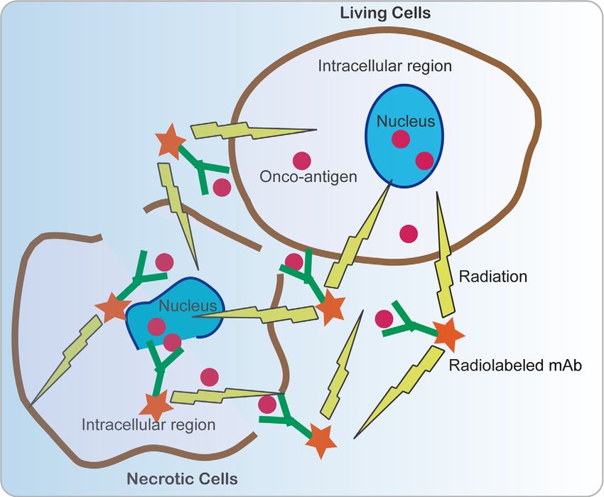
Dong et al.                                                                                                    Immunological Methods for HPV-Induced Cancers

mAbs) against various types of HPV proteins, have been                            independently of one another (32). It is therefore hard to select
developed for both diagnosis and therapy of HPV-positive                          an appropriate sample types for the diagnosis of HPV-positive
carcinomas (21, 29–31). Even other kinds of biomimetic                            cancers. According to present studies, there are mainly three
antibodies, such as affibody, nanobody, intracellular single-                      kinds of sample types, including tumor tissues/cells, exocrine
chain antibodies (scFvs), as well as T-cell-based therapies, have                 samples and sera, to perform the detection of HPV antigen or
also been developed for the diagnosis or therapeutics of HPV-                     antibodies (Figure 3).
associated cancers. In this review, we aim to summarize the
frontiers in immunological methods that are developed based on
                                                                                  Detection of HPV Protein Antigens in
the HPV proteins for both diagnosis and therapy.
                                                                                  Tumor Tissues and Cells
                                                                                  Tumor tissues are mainly used for validation of the detection
                                                                                  methods based on antibodies or antigens, which can be used for
IMMUNOLOGICAL METHODS BASED ON                                                    prognosis, tumor typing, and medication instruction after
HPV PROTEINS FOR DIAGNOSIS                                                        surgery. For instance, two mouse mAbs induced by a
                                                                                  recombinant HPV16 E7 oncoprotein were used to establish a
There are many kinds of sample types including cervicovaginal,                    chemiluminescent immunoassay based on a labeled streptavidin-
oral and serum samples for HPV detection in cervical cancers.                     biotin (LSAB)- enzyme-linked immunosorbent assay (ELISA)
However, a study shows that immunoglobulin-A and -G (IgG                          method and a luminol detection system that was suitable for the
and IgA) responses to HPV16 in different kinds of sample types                    detection of HPV16 E7 oncoprotein in HPV16-positive cervical
of women with cervical intraepithelial neoplasia (CIN) function                   carcinoma tissues. In this method, an IgG2a-type anti-HPV16 E7

 FIGURE 3 | Schematic view of different samples for immunodiagnosis of human papillomavirus (HPV)-induced cancers. Detection of HPV antigen or antibodies was
 applied for immunodiagnosis in three kinds of sample types, including tumor tissues/cells, exocrine samples, and sera from patients.

Frontiers in Immunology | www.frontiersin.org                                 4                                      January 2021 | Volume 11 | Article 586796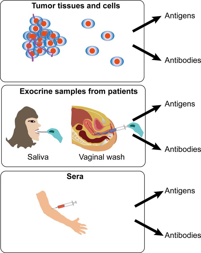
Dong et al.                                                                                              Immunological Methods for HPV-Induced Cancers

mAb was used as the detection antibody, and an IgM-type anti-                  look for other more accurate detection methods for the prediction
HPV16 E7 mAb was used as the capture antibody (21). A Rabbit                   of tumors after HPV infection.
polyclonal antibody to the HPV58 E7 protein was also developed                     Recently, biomimetic antibodies have also been designed for the
for detection of cervical cancer; it can specifically recognize the             detection of HPV antigens in tumors. For example, affibody
endogenous and the recombinant HPV58 E7 proteins (33).                         molecules have a promising potential for E7 detection in vivo. As
    In addition to oncogenic early proteins, many other kinds of               an antibody mimetic, affibodies contain three helical bundles with
anti-HPV antibodies, such as HPV L1 and L2, were also developed                58 amino acids in structure (40). Compared with conventional
for diagnosis, evaluation of vaccines, or basal biomedical research.           antibodies, affibodies are low in cost, highly affinitive, specifically
Two mAbs, specifically recognized as HPV18 L1 and VLPs, were                    and lowly immunogenic, small in size (around 6 kDa), and rapid in
used to develop an ELISA kit based on HRP-conjugated antibodies,               bio-distribution and elimination in vivo (41), making them an
and this method displayed good linearity, repeatability, and                   attractive candidate in biotechnological, pharmaceutical, and
sensitivity for detecting HPV18 L1 pentamer and HPV18 VLP                      clinical developments, such as in vivo imaging, diagnosing,
(31). Antibodies against HPV16 L1 can actively interact with the               targeting, and treatment of tumors. Four HPV16 E7-binding
HPV18 L1, HPV31 L1, HPV45 L1, and HPV6 L1 (34). In addition                    affibody molecules (ZHPV16 E7127, ZHPV16E7301, ZHPV16E7384, and
to L1, L2 was also trialed for use as an antigen for diagnosis, although       ZHPV16E7745) were screened from a phage-displayed peptide library
L2 epitopes are mainly within the canyons of pentavalent capsomers             and used for molecular imaging in tumor-bearing mice. Biosensor
(35). However, antibody competition reveals that minor HPV L2                  binding analysis showed that the four affibody molecules were
capsid protein residues 17–36 also locate at the surface (35). A panel         bound to HPV16 E7 with very high affinity and specificity. Affibody
of 30 mAbs, which target the N-terminus of L2 amino acids 11–200,              ZHPV16E7384 was conjugated with Dylight755 and showed great
was developed; selected mAbs were processed with enzymes, and                  potential to be used for in vivo tumor imaging and diagnosis of
anti-L2 Fabs were also generated (30).                                         HPV-induced cancers (42). Further study by using whole animal
    The antibodies developed underpin the basis for tumor typing,              imaging analysis indicated that a ZHPV18E7 affibody-targeted tumor
diagnosis, prognosis, and further development of the therapeutic               tissue specifically appeared about 10 minutes after injection, and the
tools. However, it is assumed that detection of oncogenic antigens in          affibody reached the highest level of tumor tissues 45 minutes after
tumor samples may be an auxiliary method for pathological                      injection. At total of 24 hours after injection, the affibody still
identification and diagnosis. Besides, there are many limitations               maintained a certain level in tumor tissues compared with other
for early-stage detection or prediction of an HPV-positive tumor               organs (43).
since the samples are acquired after the tumor size is large enough.               Collectively, these lines of evidence show that tumor tissues
They can be obtained only for a more accurate pathological analysis.           and cells are mainly used for validation of the antibodies
Besides, it is difficult to acquire the valid tumor samples without             generated. Detection of HPV protein antigens in tumor tissues
an operation.                                                                  and cells is not only important for tumor staging and prognosis
    Interestingly, the exfoliated cervical cytological examination             prediction but can also be used as a promising tool for tumor
may be used for detection in the early stage of HPV-positive                   imaging and orientation. However, the causality between HPV
tumors, such as cervical cancers of the Ia stage. It can be used as            protein expression and tumorigenesis, staging and prognosis
an adjuvant method for cervical cytological tests. For instance, a             should be further interpreted. Another problem is that it is
mouse mAb panel against a C-terminal conserved sequence                        hard to detect HPV antigens for early diagnosis because the
polypeptides of human papillomavirus (HPV) L1, a major capsid                  methods of obtaining samples are based on the current
protein, by using the IHC method used for the detection of HPV in              pathological diagnostic method used in the clinic.
exfoliated cervical cells. The detection rate was comparable to that
obtained using the commercial HPV genotyping kit currently used                Detection of Anti-HPV Protein Antibodies
in clinical practice (36). Another study also showed that HPV L1               in Exocrine Samples of Patients
capsid detection in cervical exfoliated cells is useful for screening          In addition to detecting the antigens, an easier and more effective
cervical lesions in high-risk HPV-positive women, and it may be a              way has been developed to detect the content of anti-HPV protein
promising triage for high-risk women with HPV-positive atypical                antibodies. There are some different exocrine samples that have
squamous cell of undetermined significance (ASCUS) (37). A study                been tried for the detection of anti-HPV protein antibodies, such as
also used immunohistochemistry for the detection of HPV E7                     saliva for OPC and vaginal wash for cervical cancer. For instance,
protein in pre−malignant and malignant lesions of the uterine                  anti-HPV E7 antibodies were detected in the saliva of patients with
cervix, and the results showed that this method has high specificity            oropharyngeal squamous cell carcinoma and were shown to be
and feasibility for detecting precancerous cells in cervical exfoliated        associated with HPV status. Besides, the median longitudinal
epithelial cells. Antibody staining of cervix biopsies can indicate the        salivary E7 antibody levels decreased significantly in patients after
pathological grade of cervical cancer and precancerous lesions (38).           chemotherapy treatment (44). For cervical cancer, vaginal wash can
However, the sensitivity and specificity of HPV E6/E7 protein                   be used to detect HPV protein antibodies. An ELISA using
testing are less useful than the telomerase reverse transcriptase              recombinant HPV 16 E7 oncoprotein showed that levels of HPV-
(hTERT) and Ki67 expression levels in distinguishing between                   16 E7-specific IgG in vaginal wash were significantly higher, while
cancerous and normal specimens (39), as this method cannot                     the levels of the HPV-16 E7-specific IgA were lower in women
accurately detect the intraepithelial neoplasia. This leads us to              undergoing radical hysterectomy for cervical cancer (HCC) or loop

Frontiers in Immunology | www.frontiersin.org                              5                                  January 2021 | Volume 11 | Article 586796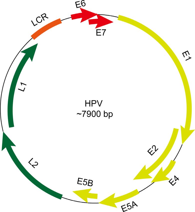
Dong et al.                                                                                            Immunological Methods for HPV-Induced Cancers

conization due to cervical dysplasia (LOOP), as compared with that           cancer was greater than that of all other groups, including patients
of patients undergoing hysterectomies for other reasons (45). This           with HPV16-associated CIN and invasive cervical cancer patients
may be due to the selective downregulation of local HPV-specific              without HPVs (46). High HPV E7 oncoprotein levels are necessary
IgA responses in women with cervical cancer (45). This method                for cervical cancers and are apparently essential as tumor markers
may be used for prediction of HPV-induced cancer in the early stage          (47). HPV16 E7 and/or HPV18 E7 antibodies in the blood are
in a non-invasive manner. However, since this detection method is            significantly related to cervical cancer risk (48). HPV 16 E7
relatively inconvenient to obtain valid samples to make sure the             antibody positivity detected by using a peptide ELISA and
high detection efficacy, it is important to set up specific criteria for       radioimmunoprecipitation assay (RIPA) may be associated with
obtaining valid samples.                                                     the stage of cervical cancer (49). Previous evidence also showed that
                                                                             an antibody response to HPV16 E6 is more frequent than to E7,
Detection of Anti-HPV Protein Antibodies                                     especially in cervical carcinoma at the early stages (50). However,
in the Sera of Patients                                                      there is also a point of view arguing that E7 functions as a valid
Anti-HPV antibodies are generated by the human body when                     candidate biomarker for all the stages of the malignant progression
infected with HPV and circulate in the blood, thus underpinning              of cervical cancer (51). Antibodies to HPV16 capsids and
the basis of serological detection of HPV antibodies in the sera.            oncoproteins E6 and E7 or types of HPV DNA in the blood
However, characterization of serological anti-HPV antibody                   samples do not appear to be useful as indicators of cervical cancer
levels is challenging due to several limited factors, including              prognosis (52). However, antibodies that respond to several linear
complexity of oncoprotein bioactivities, pre-existing anti-HPV               and conformational HPV epitopes are independently associated
titers, cross-reactivity, polyclonal responses, and low affinity of           with cervical cancer, and the combined analysis of several HPV
non-specific antibodies. Firstly, the oncoprotein bioactivities are           antibody responses can result in better predictive values for HPV-
complex and varied. An oncoprotein can interact with many                    associated cancer (53).
oncogenic pathways in HPV-induced cancers, but there are                          In addition to exploring the causality between serological
different levels of pre-oncogenic backgrounds in each                        positivity of HPV proteins and cervical cancers, researchers also
individual. That means that the presentence of an anti-HPV                   tried to unravel the association between serological positivity of
antibody in sera cannot accurately predict the risk of                       HPV proteins and other HPV-induced cancers, such as OPC. It is
carcinogenesis of HPV-induced cancer. Secondly, in the sera of               found that HPV16 E1, E2, E4, E5, E6, E7, and L1-specific IgG levels
some people who have previously been infected with HPVs but                  in the sera are tightly associated with increased risk for HPV OPC.
where the viruses are cleared, the serum level of the anti-HPV               Among patients with OPC, HPV16 Abs are associated with tumor
antibody still exists and affects the result of the prediction of            HPV status, particularly among HPV positive patients with no or
tumorigenesis by using anti-HPV antibody level. Thirdly, HPV                 little smoking history (54), as smoking may induce impaired
E6 and E7 have some similar sequences, and their antibodies in               antibody response in HPV16/18-infected young women below 30
sera may be cross-reactive with each other and may be                        years of age (55). Besides, some reports also showed that smoking
polyclonally responsive. Finally, the existence of non-specific               and HPV infection may lead to a kind of different OPC compared
antibodies in the sera can also be an obstacle for the accurate              with HPV-induced OPC with little or no smoking (56, 57). Positive
detection of anti-HPV antibodies in the sera of patients for the             HPV16 antibodies in the sera are strongly associated with HPV16-
diagnosis of HPV-induced carcinomas.                                         induced oropharyngeal cancer (58). Seropositivity of HPV16 E6
    Many efforts have been made, including exploration of the                with not other HPV antigens was higher among people with than
relationship between serological positivity of specific anti-HPV              without oral HPV16 infection (59). Men with circulating L1
antibodies and carcinogenesis, the design of highly specific                  antibodies in the sera to HPV-6, -11, -16, or -18 are not less likely
antigens for the successful detection of anti-HPV antibodies in              to acquire type-specific oral HPV than men without antibodies (60).
the sera, and the development of a detection system with high                Transcervical sonography and seropositivity for HPV 16 E6
efficacy for the quantification of HPV antibodies in the sera.                 antibodies are sensitive to the detection of OPC (61).
From current studies, we can see there are some achievements                      Aside from the HPV E6 and E7 oncoproteins, some other
that have been obtained.                                                     proteins may also be suitable for biomarkers of HPV-related
                                                                             cancers. Serological E1, NE2, and E6 antibody positivity was
Exploration of the Relationship Between                                      strongly associated with improved prognosis of patients with
Serological Positivity of Anti-HPV Protein                                   HPV16-positive tumors (62). HPV E2 proteins are usually
Antibodies and Tumorigenesis                                                 expressed during the lytic stage of HPV infection in cervical
The association of carcinogenesis with serological positivity of             cancers. Serological E2 antibody positivity was also shown to be
specific anti-HPV antibodies should be explored before it is used             strongly associated with tumor HPV status and prognosis (63).
for diagnosis. Immunological responses in different people vary,             Sometimes, only one biomarker is unable to precisely predict the
and it is difficult to set up a clear causality between serum HPV             tumorignesis. However, when combined with another biomarker or
protein antibodies and tumorigenesis. However, there are many                other biomarkers it can be more accurate. For instance, a study
studies that have tried to find some clues.                                   showed that concurrent of antibodies against E2 and p16INK4A, the
   In an ELISA assay using four HPV-16 E6–E7 peptides, the                   overexpression of which is also a biomarker of HPV-positive
seroreactivity of patients with HPV16-associated invasive cervical           cervical neoplasia, was significantly associated with HPV

Frontiers in Immunology | www.frontiersin.org                            6                                  January 2021 | Volume 11 | Article 586796
Dong et al. Immunological Methods for HPV-Induced Cancers
infection and precancerous cervical lesions (64). However, some other is that patients with higher responses of anti-HPV antibodies
HPV proteins are quite type specific. For instance, E4 cannot be may be due to better patient immune systems, resulting in a quicker
detected in HPV-18 DNA-positive CIN3 lesions but can be detected elimination of viruses in the body and a decreased possibility of
in 76% HPV16 and 55.6% HPV58 CIN3 (65). carcinogenesis. For instance, serological positivity for IgA and IgG
Besides, the detection of anti-HPV antibodies before and after in the patients with cervical neoplastic lesions showed higher titers
treatment can also be used as a prediction for the recurrence of than those in the normal group (75). This complexity of anti-HPV
HPV-induced tumors. The serum E7 antibody, once positive, antibodies is therefore an obstacle for the development of a
could be detected for a long time after surgical removal of the serological method to detect anti-HPV antibodies for an effective
cancers (66). A study showed that E6 and/or E7-positive/p16- diagnosis of HPV-associated neoplasms. This may be the reason
positive cases have better disease-specific and recurrence-free why 20–40% of patients with HPV-16 DNA-positive cervical
survival rates compared with E6-/E7-/p16- cases by using a GST carcinoma lack serum antibodies against E6 or E7 or both since
capture ELISA system in HPV-positive head and neck cancer this kind of absence of anti-HPV 16 E6 and E7 antibodies in patients
(HNC) (67). with HPV-induced cervical cancer is not due to the sequence
Nevertheless, E6 and E7 antibody levels undergo decay after variations (76). Besides, this may also be one of the possible
cervical cancer treatment (68). Recurrence of squamous high-grade reasons why controversial evidence is shown by using antibody
intraepithelial neoplasia (VIN3), which happens in approximately levels in prediction of the recurrences of HPV-associated tumors.
30% of women after treatment, was less frequent among those with
natural HPV16 antibodies in the sera (69). Antibodies against E6 and Design of Highly Specific Antigens for
E7 (HPV16/18/31/33/35), E1 and E2 (HPV16/18) were assessed in the Successful Detection of Anti-HPV
the sera of patients with neck lymph node metastasis of squamous cell Protein Antibodies in the Sera
carcinoma (SCC) from unknown primary tumor (NSCCUP) and in The successful detection of anti-HPV antibodies in the sera is also
follow-up sera from five patients. The results showed that HPV based on the use of highly specific antigens. HPV-16-like particles
antibody levels decreased after curative treatment. Recurrence was (VLPs), which are similar to native virions in structure but have no
associated with increasing levels in an individual case. However, viral genome, were firstly used for reaction with the IgA and IgG
HPV-seropositive patients had a better overall and progression-free responses in the sera. For example, VLPs were tested by ELISA for
survival (70). These results indicate that the anti-HPV antigen detecting antibodies in the sera of HPV 16 in women with cervical
antibodies in the sera of patients with HPV-induced tumors may cancer and CIN, and the results showed that this assay using HPV-
be more complex than previous expected. 16 VLP may be useful as a diagnostic tool to supplement cervical
A similar phenomenon is seen in HPV-positive oropharyngeal cytological tests (77). However, since the expression modes of HPV
cancer (HPV-OPC). An increased level of pretreatment log-unit E6 proteins are highly different during the life cycle of HPV infection,
titer was significantly associated with increased risk of disease integration and replication, different antigens also play different
recurrence of HPV-OPC (71). After treatment, average serum E6 roles during HPV-induced carcinogenesis. The most important
and E7 antibody levels decreased significantly over time in the sera thing is therefore to figure out which protein is the specific
of patients with HPV-OPC (71). Patients with HPV-OPC whose biomarker for HPV-associated tumors. What is more, as the
disease recurs have a lower clearance of E6 and E7 antibodies than cross-reactivity and polyclonal response of anti-HPV antibody
patients who do not have recurrence. The ratio of E7 antibodies at are also important for accurate detection, it is important to
disease recurrence compared with baseline is potentially a clinically develop highly specific antigens for the detection of serum anti-
significant measurement of disease status in HPV-OPC (72). HPV antibodies. Many more specific HPV antigens are used for
Another study showed that HPV16 E6 antibody levels decrease developing new methods for the diagnosis of serum anti-HPV
after treatment in patients with HPV-OPC, but most cases remain antibodies. Antibodies to HPV-16 E6 and/or E7 represent a more
seropositive for up to 2 years. HPV16 E6 antibody levels at diagnosis specific biomarker than anti-HPV-16 VLP of an HPV-related head
did not appear to be a strong predictor of recurrence (73). This and neck cancer (HNC) (78). L1/L2 VLPs and in vitro-translated E6
evidence makes the prognostic value of E6/E7 antibodies levels and E7 proteins of HPV-16 were also used and showed high
before and after treatment of HPV-driven cancer controversial (74). serological positivity in the sera of a large amount of patients with
We suspect that the levels of the anti-HPV antigen antibodies in the cervical carcinoma generated (79), indicating that these HPV-16-
sera of patients with HPV-induced tumors may be dynamic and associated proteins might be specific markers that are useful in an
may be due to different immune responses of different people. A adjunctive diagnostic assay and a seroepidemiologic study of HPV-
more comprehensive study should therefore be performed for the related cervical neoplasia. In particular, HPV-16 E7 protein seems
better understanding of the causality between HPV antibody to be valuable in the monitoring of antibody for the proper
serological positivity and the diagnosis and prognosis of HPV- management of cervical cancers (79).
induced cancers. In addition to VLPs that similar with natural proteins, chimeric
High serological positivity of anti-HPV antibodies may be one particles containing HPV-16 L1 protein fused with E6 and E7
cause of HPV infection, but this leads to two different results for seroreactive epitopes show a much better effect for detection of IgG
carcinogenesis. One is that patients with higher serological antibodies in the sera of patients HPV16-positive CIN1 than those
positivity of anti-HPV antibodies may be caused by too much obtained with VLPs containing only the HPV-16 L1 protein (80). A
virus load in the body, resulting in high-risk of carcinogenesis. The recombinant HPV16 E7 and the N-terminal and C-terminal
Frontiers in Immunology | www.frontiersin.org 7 January 2021 | Volume 11 | Article 586796Dong et al. Immunological Methods for HPV-Induced Cancers
fragments of gp96 (NT-gp96 and CT-gp96) protein were used in the (ECL) was developed by using multiplex technology from Meso Scale
Western blot and ELISA methods to test serum antibody, and the Diagnostics (MSD, Rockville, MD) with maltose-binding protein
results showed significantly higher levels of these markers in cervical (MBP)-tagged E6 and E7 oncoproteins. This is a high-throughput
cancer patients in squamous cell carcinoma only but not in method, but it requires lower sample quantity input with greater
adenocarcinoma and control groups (81). dynamic range to detect type-specific anti-HPV E6 and E7
Intriguingly, some biomimetic peptides are also used in oncoproteins (87).
serological detection of anti-HPV antibodies. Recently, a Collectively, the present detecting methods are mainly based on
nonapeptide (16L1) derived from the HPV-16 major capsid chemiluminescence and ECL that can magnify the signals of
protein was used in an ELISA system to detect potential cross- immunological reactions. The success of novel detection systems also
reaction of serum IgG and cervical IgA antibodies HPV-associated depends on the clear understanding of the biomarkers in HPV-induced
low-grade squamous intraepithelial lesions (LSIL) and cervical cancers. The current biggest obstacle of immunodiagnosis of HPV-
cancer patients. The results showed that the 16L1 peptide is a induced cancers is therefore the appropriate specific biomarkers.
high-risk epitope that induces cross-reactive antibodies in patients
with high-risk, but not low-risk, HPV-induced LSIL, indicating that
this method is suitable for distinguish high- and low-risk infected THERAPEUTICS
women in the stage of low grade (82).
In summary, the selection of valid natural HPV antigens and HPV oncoproteins, such as E5, E6 and E7, are not only the
design of biomimetic peptides are important for better detection etiological factors for carcinogenesis for HPV-associated cancers,
of HPV protein antibodies. It is therefore urgent to explore how but some of them are constantly expressed during tumor
the antibodies circulating in the blood are induced by the HPV development and exhibit malignant characteristics via interacting
proteins, and the epitope profiles should be widely studied for a with multiple signaling pathways and regulatory modes that
better understanding of the immunological responses upon HPV participate in the modulations of intraepithelial neoplasia (IN),
infection and what the differences in immunological responses tumorigenesis, cell cycle progression, cell survival, and metastasis
during carcinogenesis are compared with those of the non- (88–90) (Figure 4).
tumorigenic HPV infection. For instance, HPV16 E6/E7 could promote the invasive potential
of cancer cells and epithelial-mesenchymal transition (EMT) via
Development of Detection System With High turning on the cadherin switch, downregulating NHERF1, and/or
Efficacy for the Quantification of Anti-HPV activating the STAT3 signaling pathway (91–93). HPV E6 protein
Protein Antibodies in the Sera expression enriches the differentiation (CD) 55-positive population,
The magnifying and accurate detection system used also accounted which are possible cancer stem-like cells that contribute to
for the application of HPV antibodies detection. Currently, many tumourigenesis and radioresistance in cervical cancer cells (94).
detection methods for the diagnosis and prediction of HPV- HPV16 E6/E7 induces O-linked GlcNAcylation (O-GlcNAc) and
associated tumors are tried in basic research. For instance, a O-GlcNAc transferase (OGT), thereby elevating c-MYC and
luciferase-based detection method for determining serum transcriptional co-regulator host cell factor 1 (HCF-1) via increased
antibodies is possible for the diagnosis of HPV-associated head protein stability, which results in tumor transformation and
and neck squamous cell carcinoma (83). A new LSAB capture tumorigenesis (95, 96). E6 induces proteasome-dependent p53
ELISA method based on the recombinant HPV16/18 E7 degradation via recruiting the intracellular ubiquitin ligase E6AP
oncoproteins was used to investigate anti-HPV E7 antibody (97). Recently, HPV16/18 E5 is also expressed in the early stage of
prevalence in the sera of patients with cervical cancer. It is shown carcinogenesis (98) and is shown to promote cervical cancer cell
that this assay could potentially be used as an adjunctive tool to proliferation, migration, and invasion in vitro, and it accelerates
monitor the type of response to treatment, possibly detect antibody tumor growth in animal models (99).
induction in cervical cancer patients after vaccination, and to These oncoproteins are potential drug targets for the therapeutics
function as a potential method to evaluate its efficacy (84). A of HPV-associated cancer. Among them, immunotherapy-based on
custom HPV protein microarray based on the ELISA method, HPV is very important. From current studies, we can see there are
which is also called programmable protein arrays (NAPPA), was many kinds of immunotherapeutical methods or immunotherapy-
also established, displaying 98 proteins as C-terminal GST fusion like methods based on HPV studied in basal research, including anti-
proteins, representing eight antigens of two low-risk HPV types, HPV protein mAbs, radioimmunotherapy, affitoxins, single-chain
such as HPV6, and 11 and 10 high-risk oncogenic HPV types, such antibodies (scFvs), nanobodies, therapeutic vaccines, and T-cell-
as HPV16, 18, 31, 33, 35, 39, 45, 51, 52, and 58. Then, NAPPA was based therapies.
used to detect antigens in the sera of patients with cervical cancer
and oropharyngeal cancer. The results showed this method had a Native Anti-HPV Protein mAbs for the
great potential for rapid identification of serologic responses to 12 Therapeutics of HPV-Induced Cancers
HPV types and could be used as a valuable high-throughput tool for Anti-HPV E6 and HPV E7 mAbs via intraperitoneal or intratumoral
measuring the breadth, specificity, and heterogeneity of the injections in mice models significantly inhibited cervical tumor
serologic response to HPV in cervical disease (85, 86). growth (100). Another study also showed that mAbs specifically
However, the ELISA assay allows only one antigen to be evaluated targeting HPV6 E7 49-57 peptides lead to a significant tumor-
at a time per well. An assay based on electrochemiluminescence suppressing effect in animal models (101). Other mAbs that target
Frontiers in Immunology | www.frontiersin.org 8 January 2021 | Volume 11 | Article 586796Dong et al. Immunological Methods for HPV-Induced Cancers
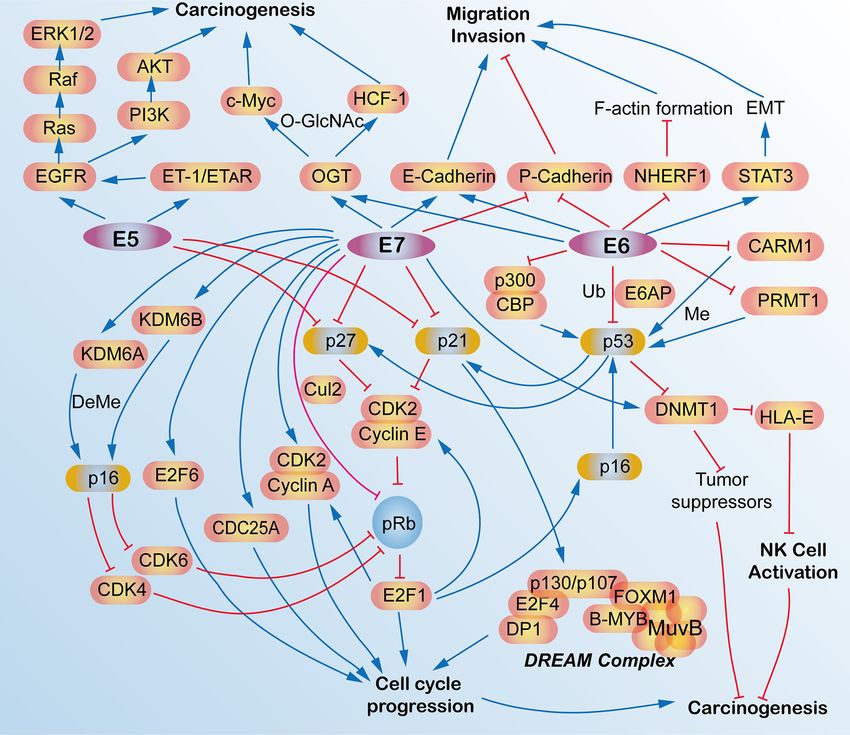
FIGURE 4 | Schematic view of the molecular network and functions of human papillomavirus (HPV) E5, E6, and E7 oncoproteins in HPV-induced cancers. E5, E6, and E7
oncoproteins are responsible for the carcinogenesis and tumorigenesis of HPV-induced cancers via activating multiple signaling pathways, such as cell cycle signaling, EGFR
pathway, migration, and invasion pathways, p53 pathway and some epigenetic pathway. AKT, AKT serine/threonine kinases; B-MYB, MYB proto-oncogene like 2, MYBL2;
CARM1, coactivator-associated arginine methyltransferase 1; CBP, CREB binding protein; CDC25A, cell division cycle 25A; CDK2, cyclin-dependent kinase 2; CDK4,
cyclin-dependent kinase 4; CDK6, cyclin-dependent kinase 6; C-Myc, avian myelocytomatosis viral oncogene homolog; Cul2, cullin 2; DeMe, demethylation; DNMT1, DNA
(cytosine-5-)-methyltransferase 1; DP1, transcription factor DP-1, TFDP1; E2F1, E2F transcription factor 1; DREAM, dimerization partner, RB-like, E2F and multi-vulval class
B; E2F4, E2F transcription factor 4; E2F6, E2F transcription factor 6; E6AP, E6-associated protein, ubiquitin protein ligase E3A, UBE3A; E-Cadherin, epithelial cadherin;
EGFR, epidermal growth factor receptor; EMT, epithelial mesenchymal transition; ERK1/2, mitogen-activated protein kinase 1/2; ET-1, endothelin 1; ETAR, endothelin A
receptor; F-actin, filamentous actin; FOXM1, forkhead box M1; HCF-1, host cell factor 1; HLA-E, HLA class I histocompatibility antigen, alpha chain E; KDM6A, lysine
demethylase 6A; KDM6B, lysine demethylase 6B; LIN54, protein lin-54 homolog; Me, methylation; MuvB, MuvB complex of five proteins including LIN9, LIN37, LIN52,
RBBP4 and LIN54; NHERF-1, Na+/H+ exchanger regulatory factor; NK, natural killer; O-GlcNAc, O-Linked b-N-acetylglucosamine; OGT, protein O-GlcNAc transferase;
p16, p16INK4A, p14ARF, cyclin dependent kinase inhibitor 2A, CDKN2A; p21, p21WAF1/CIP1, cyclin-dependent kinase inhibitor 1A, CDKN1A; p27, p27KIP1, cyclin dependent
kinase inhibitor 1B, CDKN1B; p53, tumor protein p53, TP53; p300, E1A binding protein p300; P-Cadherin, placental cadherin; PI3K, phosphoinositide 3-kinase; pRb,
phosphorylated retinoblastoma transcriptional corepressor 1; PRMT1, protein arginine N-methyltransferase 1; Raf, Raf proto-oncogene, serine/threonine kinase; Ras, RAS
proto-oncogene, GTPase; STAT3, signal transducers and activators of transcription 3; Ub, ubiquitination.
HPV E6/E7 also have an anti-tumor effect to some extent according tumor-associated antigens (40, 106). Conventionally, traditional
to several reports (102–105). Theoretically, the use of anti-HPV RIT aims for cell-surface-associated tumor markers; however,
mAbs has limited potential in cancer therapy because its size is too big targeting viral antigens within the tumors is fundamentally
to enter into the intracellular regions where the HPV oncoproteins different from that because the oncotargets are of viral origin
are located. However, these samples showed that native anti-HPV while not “self” human antigens, which minimizes cross-
mAbs also have some anti-tumor effects, and this may be because reactivity with host tissues. Nevertheless, the difficult condition
cellular turnover occurs as cervical cancer solid tumors grow, making is that the viral oncoproteins normally reside in intracellular
it possible for anti-HPV E6 and E7 to be accessible to these two compartments, which is thought to be beyond the reach of
oncogenes via necrosis (100). In contrast to this, some other immunogloblulins. For instance, the E6 and E7 oncoproteins
researchers have pointed out that the deposition of C3 complement are usually located in the intranuclear location. Intriguingly, this
and lymphocytes infiltration, which potentially exert significant approach really works due to many non-viable and necrotic cells
antitumor effects, is induced by mAbs treatment rather than with permeable membranes in tumors that allow mAbs access to
necrosis (105). interact with the intracellular antigens. In addition, due to the
renewal of cells in a rapidly growing tumor, the cellular
Radioimmunotherapy for the Therapeutics membrane has provided some crevasses for some intracellular
of HPV-Induced Cancers antigens E6 and E7 to be released from cancer cells. Radiolabeled
Radioimmunotherapy (RIT) is a kind of therapeutic method that E6 or E7 specific mAbs bind to extracellular E6 and E7 and
systemically administers radiolabeled mAbs to bind to specific deliver cytotoxic radiation to this area. Surviving tumor cells,
Frontiers in Immunology | www.frontiersin.org 9 January 2021 | Volume 11 | Article 586796Dong et al. Immunological Methods for HPV-Induced Cancers
including weak or no E6 or E7 expression, were killed by epidermal growth factor receptor (EGFR), and insulin-like
radiation via the “cross-fire” effect produced by radiation in growth factor type 1 (IGF1R) (109, 110). These affibodies showed
360° spheres (Figure 5) (107). a promising future for early-stage cancer diagnosis and treatment in
For example, E6 and E7 oncoproteins in experimental cervical the clinic. Interestingly, affibodies were also applied in HPV-
cancer can be targeted by using radiolabeled mAbs with a beta- positive tumors via targeting HPV oncoproteins. In a mouse
emitter 188-Rhenium (188Re) attached to HPV E6, making them cervical cancer model, Z HPV18E7 and Z HPV16E7 affibodies
selective mediators of tumor destruction even in experimental connected with Pseudomonas exotoxin, also known as affitoxins,
cervical tumors expressing low levels of E6 (102, 103). In were able to deliver Pseudomonas exotoxin, a clinically used anti-
addition, the radiolabeled anti-HPV E6 mAb alone and in cancer agent to tumor tissues effectively, showing great potential for
combination with MG-132, a proteasome inhibitor, was used HPV-induced cancer treatment (43, 111). These results exemplify
to treat cervical tumor-bearing mice and showed a significant the potential use of affitoxins for HPV-induced cancers.
suppression of tumor growth (103, 104). Radioimmunotherapy
based on 188Re directed toward HPV E7 oncoprotein also Intracellular Single-Chain Antibodies
inhibits experimental tumors growth (105). Besides, beta (scFvs) for the Therapeutics of
emitters lutetium-177 ( 177 Re) are equally effective for HPV-Induced Cancers
radioimmunotherapy based on an anti-HPV E6 mAb in HPV- Single‐chain variable fragment (scFv) antibodies are the smallest
positive cervical cancer (108). These results mean that immunoglobulins, but they have a high antigen‐binding affinity
radiotherapies based on both E6 and E7 are probably potential (Figure 6). Compared with mAbs, scFvs can access the intracellular
therapeutic methods for HPV-positive cancers. region and bind to intracellular oncoantigens. It can be well
produced by using prokaryotic expression in Escherichia coli.
Affitoxins for the Therapeutics of Applications of scFv against fibroblast growth factor receptor
HPV-Induced Cancers (FGFR) and fibroblast growth factor 1 (FGF-1) have shown a
In addition to applications in tumor imaging and diagnosis, great anti-tumor effect both in vitro and in vivo (112–114).
affibodies can also be used as therapeutic tools for tumor Recently, scFvs have also been applied in HPV-induced
treatment. Clinical and pre-clinical studies showed that several neoplasms by targeting oncoproteins, including HPV E5, E6, and
affibodies have been developed to target some oncogenic proteins, E7. The HPV16 E5 protein is a small hydrophobic protein, the
such as human epidermal growth factor receptor 2 (HER2), expression of which generally decreases as the infection progresses
FIGURE 5 | Schematic view of anti-cancer mechanism of radiolabeled mAbs. Since many non-viable and necrotic cells with permeable membranes are present in
tumors, allowing mAbs to access to interact with the intracellular antigens. In addition, intracellular antigens E6 and E7 can be released from cancer cells via necrosis
and cell turnover. E6 or E7 specific mAbs bind to extracellular E6 and E7 and deliver cytotoxic radiation to this area. Surviving tumor cells including weak or no E6 or
E7 expression were killed by radiation by the “cross-fire” effect produced by radiation in 360° spheres.
Frontiers in Immunology | www.frontiersin.org 10 January 2021 | Volume 11 | Article 586796Dong et al. Immunological Methods for HPV-Induced Cancers to malignancy. ScFvs were used to recognize HPV16 E5 in W12 cells inhibition of tumor growth of these cells. These two scFvs do not by fluorescent microscopy and its colocalization with one of its host correspond to the pRb binding site, but they can be non- substrates (115). Functional scFvs against the E6 oncoprotein can competitively inhibited by pRb (120). These results showed a also be produced in E. coli, and it impairs growth of HPV16-positive great potential for scFvs in the treatment of HPV-positive tumors. tumor cells in mouse models (116, 117). Anti-HPV16 E7 scFvs expressed in HPV DNA-containing cell lines showed a significant Nanobodies for the Therapeutics of decrease in cell proliferation via altering the level of HPV16 E7 HPV-Induced Cancers oncoprotein (118). Besides, anti-HPV16 E7 scFvs also exerted an in Different from scFvs, heavy chain antibodies (HCAbs) are vivo antitumor effect (119). Two scFvs binding E7, designed and another kind of small antibodies (approximately 15 kD) that generated by using distinct but overlapping epitopes, were are naturally devoid of light chains, the antigen-binding selectively expressed in the nucleus and the endoplasmic fragment of which consists of only one single domain referred reticulum (ER) of cervical cancer cells, leading to the selective to as nanobody (Nb) or the heavy chain antibody variable region FIGURE 6 | Schematic view of single‐chain variable fragment (scFv) antibody and nanobody. VH, heavy chain variable domain; VL, light chain variable domain; CH, heavy chain constant domain; CL, light chain constant domain; VHH, single-domain antibody. Frontiers in Immunology | www.frontiersin.org 11 January 2021 | Volume 11 | Article 586796
Dong et al.                                                                                            Immunological Methods for HPV-Induced Cancers

(VHH, Figure 6) (121). This kind of antibody, which is also                      In addition to bacterial vectors, viral vector-mediated vaccines
referred to as camelid single-domain antibody (sdAb), is inspired            against HPV oncoantigens are also tested for cancer therapy.
by the HCAbs that can be found in the Camelidae family (122).                Decades ago, a live recombinant vaccinia virus vaccine encoding
Nb is more soluble and stable than conventional antibodies, and              HPV16 and 18 E6 and E7 oncoproteins, also known as TA-HPV,
a high yield can be found in bacteria (E. coli) or yeast (Pichia             induced a specific cytotoxic T-cell response in three patients and
pastoris). Recently, Nbs also function important therapeutic                 eliminated tumors of two patients at 15 and 21 months after
approaches for cancer treatment (123). Nbs that target HPV                   vaccination (134). Furthermore, TA-HPV was shown to reduce
L1 have been successfully achieved (124). Importantly, Nbs are               HPV-associated lesion size and to stimulate HPV 16 E6 and E7-
also applied for targeting of HPV E7 in cervical cancers. A phage-           specific T-cell immunity in patients with HPV-16+ vulval
display approach was used to select the high-affinity HPV16 E7-               intraepithelial neoplasia (VIN) grade III and a single patient with
specific Nbs and expressed by using prokaryotic expression.                   HPV-16+ vaginal intraepithelial neoplasia (VAIN) grade (135). In
Then, a high-affinity Nb was expressed in cervical cancer, and                addition to vaccinia virus, a replication-deficient adenovirus-
it induced a significant decrease of tumor growth (125). These                encoding fusion vaccine consisting of calreticulin (CRT) and
studies underpin the basis of the applications of Nbs in HPV-                HPV16 E7 can also eliminate E7-expressing tumors in mice
induced tumors.                                                              (136). Other kinds of chimeric vaccines mediated by viruses, such
                                                                             as adenovirus vaccine encoding hepatitis B virus surface antigen
Therapeutic Vaccination for the                                              (HBsAg) and HPV16 E7 proteins as well as alphavirus vaccine
Therapeutics of HPV-Induced Cancers                                          encoding recombinant Semliki Forest virus (rSFV) particles and
Unlike preventive vaccines, therapeutic vaccines fight against an             HPV16 E6/E7, can increase anti-HPV E7 antibody and cytotoxic T-
existing disease rather than immunizing for protection against               cell responses in mice (137–139). Importantly, a clinical study
future disease by utilizing a patient’s own immune system. These             showed that a modified vaccinia virus Ankara (MVA)-based
therapeutic vaccines deliver antigens to antigen-presenting cells            vaccine, which has a suspension of MVATG8042 particles with
(APCs), where these antigens are processed and digested into                 attenuated recombinant MVA that carry sequences encoding
short peptides by proteasome, which can stimulate antigen                    modified HPV-16 E6/E7 and human IL-2, also referred to as
presentation via major histocompatibility complex class I and                TG4001, can clear both HPV16 DNA and mRNA, with no
II (MHC I/II), leading to CD8+ cytotoxic T-cell or CD4+ helper               recurrence of high-grade lesions one year after treatment in
T-cell responses. This method has also been trialed for cancer               patients with CIN 2/3 (140). In addition to HPV E6 and E7, HPV
therapy (126). Recently, this method has also been introduced for            E2 was also used in an MVA-based vaccine and the results
the therapy of HPV-induced tumors (127). There are several                   demonstrated a strong therapeutic effect via stimulating the
kinds of therapeutic vaccines, including live vector (bacterial or           immune system in a phase III clinical study for the treatment of
viral vector), protein or peptide, nucleic acid, and whole cell-             HPV-induced anogenital intraepithelial lesions (141).
based vaccines (128).
                                                                             Peptide/Protein-Based Therapeutic Vaccines
Live Vector-Based Therapeutic Vaccines                                       Another important kind of vaccine is peptide/protein-based
Although live vector-based vaccines may pose a safety risk, they can         vaccine. Those antigens derived from HPV can be processed by
induce strong cellular and humoral immune responses. The vectors             dendritic cells and presented on MHC class I/II molecules, which
can be either bacteria, such as Listeria monocytogenes, Lactobacillus        further can stimulate CD8+ or CD4+ T cell responses (142). They
lactis, Lactobacillus plantarum, and Lactobacillus casei, or viruses,        are safe, stable, and easy to produce, but they have poor
such as adenoviruses, adeno-associated viruses, alphaviruses, and            immunogenicity. A clinical study showed that HPV16-synthetic
vaccinia viruses. They can infect macrophages and stimulate                  long-peptide (SLP) vaccination combined with standard
immune responses that kill tumor cells. Listeria-based E7                    carboplatin and paclitaxel (CarboTaxol) chemotherapy can
vaccines were reported as a promising method for the treatment               significantly improve immunity via fostering robust T-cell
of HPV E6/E7-expressing autochthonous solid tumors in mice                   responses (143). Other several HPV16 peptide-based vaccines,
(129). Besides, Listeria can secrete a kind of toxin, listeriolysin O        such as TA-CIN (a subunit vaccine composed of HPV16 E6E7L2
(LLO), which can make the bacteria escape from phagosomal lysis              fusion protein) (144), PepCan (a therapeutic HPV vaccine
and decrease the CD4+FoxP3- and CD8+ regulatory T cells,                     containing four synthetic peptides covering HPV-16 E6 and
resulting in HPV vaccine-mediated tumor regression (130). A                  Candin) (145) and GTL001 (a therapeutic HPV protein vaccine
phase I study on the intravenous Listeria monocytogenes-based                targeting both HPV16 and HPV18) (146) showed remarkable
fusion vaccine consisting of HPV16 E7 antigen and LLO in cervical            potentialities for HPV-positive malignancies. Recently, a well-
cancer showed a reduction of total tumor size that was observed in           documented HPV-E7 SLP) therapeutic vaccine was performed to
four of 15 patients, but this method also induced some adverse               conjugate to ultra-small polymeric nanoparticles (NP), thus
effects, such as pyrexia, vomiting, chills, headache and anemia,             enhancing their antitumor efficacy in different HPV-positive
nausea and tachycardia, and musculoskeletal pain (131, 132).                 tumor-bearing animal models. This synergetic effect is due to a
Further study showed that oral administration of the Listeria                larger pool of E7-specific CD8+ T cells with increased anti-tumor
casei-based HPV16 E7 vaccines exerted a remarkable E7-specific                efficacy induced by conjugated E7 SLPs than unconjugated ones. A
cell-mediated immune responses in patients with HPV16+ CIN III               robust infiltration of CD8+ T cells was also observed at the tumor
lesions (133).                                                               site treated with conjugated E7 SLPs; however, concomitant

Frontiers in Immunology | www.frontiersin.org                           12                                 January 2021 | Volume 11 | Article 586796
You can also read