Hypertrophic Scar Formation and Wound Healing Modulation Fatty Acids as Modulators of Severe Scars
←
→
Page content transcription
If your browser does not render page correctly, please read the page content below
Modern Plastic Surgery, 2023, 13, 41-51
                                                                                            https://www.scirp.org/journal/mps
                                                                                                         ISSN Online: 2164-5280
                                                                                                           ISSN Print: 2164-5213

Hypertrophic Scar Formation and Wound
Healing Modulation Fatty Acids as Modulators
of Severe Scars

Bárbara Díaz, Valerie Nuñez

Department of General Surgery, Burn Unit Care Dr. Ricardo Gutiérrez Children’s Hospital, Buenos Aires, Argentina

How to cite this paper: Díaz, B. and Nuñez,   Abstract
V. (2023) Hypertrophic Scar Formation and
Wound Healing Modulation Fatty Acids as       Scar tissue usually generates severe discomfort in the short and long
Modulators of Severe Scars. Modern Plastic    term. Common symptoms include anesthetics sequelae, pruritus, joint mal-
Surgery, 13, 41-51.
                                              function, new wounds on the scar surface, and pain. There are several treat-
https://doi.org/10.4236/mps.2023.131005
                                              ments for scars, like compression, topical or intralesional steroid infiltration,
Received: November 1, 2022                    5-fluorouracil, dermabrasion, and surgeries with new scar tissue. For adult
Accepted: January 16, 2023                    patients, it is easier to choose the treatment. However, compression is com-
Published: January 19, 2023
                                              monly applied in children to prevent treatments that have adverse effects.
Copyright © 2023 by author(s) and             This study reports the outcomes of 15 patients submitted to abdominoplasty,
Scientific Research Publishing Inc.           traumatic wounds and post-burn scar treatments, which showed significant
This work is licensed under the Creative
                                              changes after the continuous use of an ointment composed of petrolatum,
Commons Attribution International
License (CC BY 4.0).                          cod liver oil, BHT, Chamomilla recutita (chamomile) oil, Helianthus annuus
http://creativecommons.org/licenses/by/4.0/   (sunflower) oil, and Prunus amygdalus dulcis (sweet almond) oil. As compo-
                Open Access                   nents of the stratum corneum, unsaturated fatty acids influence the cutaneous
                                              structural and immune status and permeability. They also interfere with the
                                              maturation and differentiation of the stratum corneum and inhibit the pro-
                                              duction of proinflammatory eicosanoids, reactive species (ROS and RNS),
                                              and cytokines, thereby influencing the inflammatory response and possibly
                                              wound healing. This article aims to share our experience with the regular use
                                              of an ointment in adult and pediatric patients for three months. The increase
                                              in proinflammatory cytokine production at wound sites, resulting in a non-
                                              invasive, therapeutical, and effective cutaneous wound healing and scarring
                                              modulation, may provide a physiopathological explanation for the fast im-
                                              provement of scars.

                                              Keywords
                                              Scarring, Burn Scar, Inflammatory Modulation, Cytokine, Sequelae, Fatty

DOI: 10.4236/mps.2023.131005 Jan. 19, 2023                       41                                      Modern Plastic Surgery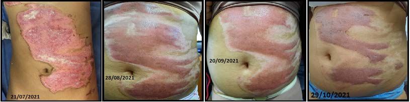
B. Díaz, V. Nuñez

                                 Acids, Eicosanoids, Non-Adverse Effects, Aesthetics, Hypertrophic

                               1. Introduction
                               Burn surgeons observe daily that, immediately after the acute treatment, burn
                               patients experience an undetermined evolution stage. That stage is challenging
                               to manage, as it requires long rehabilitation times, particularly in flexion-extension
                               areas, and surgical revisions.
                                  In developing countries, scar treatment is usually challenging due to long dis-
                               tances to specialized centers, treatment costs, difficult access to medical tech-
                               nology, and insufficient recognition of these pathologies by the public health
                               systems.
                                  In Latin America and our practice, phototypes IV to VI of the Fitzpatrick scale
                               are prevalent as brown skin tends to scar pigmentation and has a greater risk of
                               forming hypertrophic or keloid scars [1] [2].
                                  In cosmetic surgery, hypertrophic scars and keloids pose an aesthetic and
                               functional problem. One study found that 60% of the patients were unsatisfied
                               with the cosmetic results of their scars after dermatologic and cosmetic surgery
                               procedures, and up to 90% wanted to improve their appearance [3].
                                 Curefini was initially, and it is currently used as a topical treatment for epi-
                               dermolysis bullosa and chronic wound healing. No adverse effects and good
                               wound-healing outcomes have been reported. Our team has a long experience in
                               surgical treatments, and we started using the product to treat small, ulcerated le-
                               sions that remain in the final stage of burn healing. We observed that healed
                               areas improved faster, showing more elasticity, better texture, less itching, and
                               reduced post-inflammatory vascularity and pigmentation.
                                 Therefore, planning scars is just as important as planning flaps. An efficient
                               topical medication, combined with usual therapies, may aid the modulation of
                               healing stages.

                               2. Objectives
                               This article aims to demonstrate that the use of Curefini®, associated with tradi-
                               tional scar modulation therapies, is more efficient in producing better scar cos-
                               metic and functional outcomes than only moisturizing creams associated with
                               those treatments.
                                 The evaluated treatment complied with the specifications for the clinical use
                               of the drugs. In this regard, all clinical research should safeguard the dignity of
                               participating subjects, ensuring their rights, particularly their autonomy and
                               physical, mental, and oral integrity. [4]

                               3. Material and Methods
                               This study included 15 patients under acute stage resolution, treated or not with

DOI: 10.4236/mps.2023.131005                         42                                       Modern Plastic Surgery
B. Díaz, V. Nuñez

                               grafts, presenting hypertrophic scars with erythema or pigmented scars up to six
                               months from the start of healing.
                                  Patients with scars older than two years and stable were excluded because they
                               presented no measurable differences suitable for this study, keloid scars that could
                               not be partially resected or submitted to z-plasty, or keloids requiring immuno-
                               modulatory treatment.
                                  Both adult patients and pediatric patient surrogates granted their consent to
                               participate in this study, per the ethical-legal regulations.
                                  The topical medication used was Curefini®, an ointment composed of sun-
                               flower oil, cod liver oil, beeswax, sweet almond oil, petroleum jelly (Vaseline®),
                               and butylated hydroxytoluene (BHT). [5]
                                  The protocol was applied for three months. Curefini® was applied to hyper-
                               trophic scars resulting from burns or cosmetic surgeries. Photographic records
                               were made at 30, 60, and 90 days of treatment to evaluate scar evolution in terms
                               of scar pigmentation, itching reduction, and elasticity improvement. The Van-
                               couver Scar Scale [6] [7] [8] (Table 1) and photographic records of the parame-
                               ters specified in the protocol were used as a reference for scar evolution.

                                          Table 1. Vancouver scar scale.

                                                                   Scar characteristic
                                                     Vascularity                         Score
                                                      Normal                              0
                                                         Pink                             1
                                                          Red                             2
                                                         Purple                           3
                                                    Pigmentation
                                                      Normal                              0
                                                 Hypopigmentation                         1
                                                 Hyperpigmentation                        2
                                                      Pliability
                                                      Normal                              0
                                                         Supple                           1
                                                      Yielding                            2
                                                         Firm                             3
                                                         Ropes                            4
                                                    Contracture                           5
                                                    Height (mm)
                                                          Flat                            0
                                                          5                              3
                                                     Total score                          13

DOI: 10.4236/mps.2023.131005                        43                                           Modern Plastic Surgery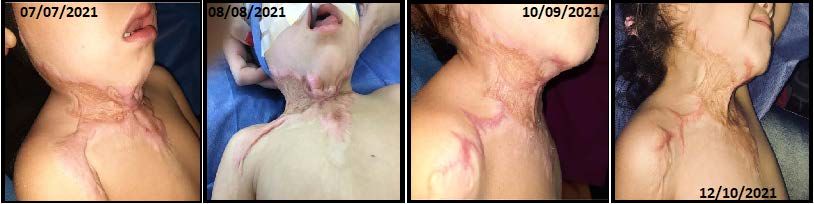
B. Díaz, V. Nuñez

                                  Despite being a subjective clinical assessment tool designed to describe scars
                               in general—not hypertrophic scars—we chose the Vancouver Scar Scale in this
                               study because it is simple, easy to apply in low-technology settings, and possibly
                               the most recognized burn scar assessment method. The VSS remains widely ap-
                               plicable to evaluate therapy and as a measure of outcome in burn studies. [9]

                               3.1. Pathophysiology of Healing and Role of Polyunsaturated
                                    Fatty Acids
                               Pathological scars are caused by an excessive response to the activity of TGF-β1.
                               Connective tissue growth factors are overexpressed 100- to 150-fold in hyper-
                               trophic and keloid scars, respectively, in response to this cytokine compared
                               with normal fibroblasts. [10] [11]
                                 Our treatment was based on studies that showed that the concentration of
                               polyunsaturated fatty acids (PUFA, EPA, DHA) influences the synthesis and ac-
                               tivity of proinflammatory cytokines and inhibits the expression of the gene in-
                               duced by TGF-β1, inhibiting pro-fibrogenesis. [12] [13]
                                 Those fatty acids may partially inhibit some inflammation processes, such as
                               leukocyte chemotaxis, adhesion expression molecules and leukocyte interactions
                               with the endothelium, the production of eicosanoids, such as prostaglandins and
                               leukotrienes, from arachidonic n-6 PUFA, and T-cell inflammatory cytokine pro-
                               duction and reactivity. Eicosapentaenoic acid (EPA) stimulates the biological ac-
                               tivity of arachidonic acid (AA) derivatives and the synthesis of active mediators
                               of inflammation resolution, such as resolvins and protectins. [14]
                                 Those mediators compete with cyclooxygenases and lipoxygenases and reduce
                               the expression of COX-2 and 5-lipoxygenase, with a beneficial anti-inflammatory
                               effect of n-3 PUFAs. [15] [16]

                               3.2. The Role of Vitamin D in Inflammation
                               Supplied by sunflower, sweet almond oil, and cod liver oil, vitamin D signifi-
                               cantly reduces IL-6 and TNF-α levels. Using cholecalciferol receptors, it directly
                               binds to leukocyte DNA, activating the MKP-1 gene and thereby interfering in
                               the inflammatory cascade. [17] [18]

                               3.3. Virgin Beeswax
                               It contains long-chain polysaccharides, long-chain free fatty acids, palmitic acid,
                               and exogenous compounds. It is associated with other components as a skin
                               protector, as it is highly hydrophobic. Studies carried out in 2016 report that it
                               has antibacterial activity against S. aureus and antifungal activity against C. albi-
                               cans. [5]

                               3.4. Study Description
                               Our experience with Curefini® in the combined treatment for burn sequelae be-
                               gan in 2019.

DOI: 10.4236/mps.2023.131005                         44                                      Modern Plastic Surgery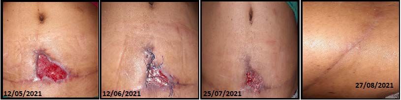
B. Díaz, V. Nuñez
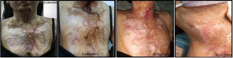
Our “patient 0” was an adolescent female admitted on 08/20/2019 due to an
extensive burn by direct fire. She remained hospitalized for a long time due to
Pseudomonas A infection acquired in the hospital. She was submitted to grafting
and discharged on 10/25/2019 (Figure 1). On 03/12/2020, she started elastic
bandage compression and Curefini® treatment (Figure 2).
The patient discontinued compressive treatment three months after the start
and revisited the hospital on 04/19/2021, showing an elastic, hypopigmented
scar with some hypertrophic nuclei under regression (Figure 3).
The patient was maintained under the ointment treatment only.
We then decided to apply an evaluation protocol of the topical treatment
without interrupting the usual therapies.
4. Results
Healing is an active process that lasts 12 - 18 months.
Figure 1. Thorax and neck burn.
Figure 2. Neck graft.
Figure 3. Thorax and neck burn 18 months later.
DOI: 10.4236/mps.2023.131005 45 Modern Plastic Surgery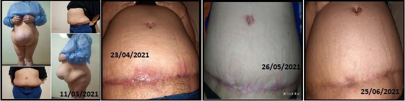
B. Díaz, V. Nuñez

                                             Although between the first 15 to 30 days the patients without compressive
                                          treatment showed an increase in purplish-red scar pigmentation, after 90 days
                                          the scars showed less erythema due to a decrease in the vascular scar component.
                                             A notable decrease was observed in the Vancouver Scar Scale score in the
                                          treated patients during that period. (Table 2)
                                             Although some scars initially showed severe pliability scores, the firmness,
                                          pigmentation, itching, and superficial lesions commonly observed in hyper-
                                          trophic scars were reduced.
                                             Patients who started topical treatment with Curefini® and required surgical
                                          treatment (surgical skin flaps, z-plasty, new grafts) showed better post-surgical
                                          healing of the new scars.
                                             One of the adult patients presented wound dehiscence due to infection with
                                          Staphylococcus A (MRSA) and was submitted to surgical debridement and sec-
                                          ondary intention wound healing, showing a rapid regression of the initial sur-
                                          gical wound.
                                             It was not possible to evaluate one of the abdominoplasties at 60 days, but at
                                          90 days post-surgery, the wound already had the characteristics of a mature scar.
                                             Only one patient showed no changes between 60 to 90 days, but at 120 days,
                                          the wound remained stable.
                                             No patient presented any irritation, pruritus, or allergy signs to the ointment
                                          used.

                                          Photographic Record
                                          Wound evolution between 30 and 90 days is shown below. Due to the vast
                                          number of records, only the most significant are shown. (Figures 4-18)

Table 2. Vancouver scar scale results.

   Patient    Age (yrs.)   Start treat.     30 D       60 D         90 D           Scar           Compression      Graft
     AL             46          6             2          0           0        Abdominoplasty           No           No
      SS            43          9            10          4           2        Abdominoplasty           No           No
     FA             49          8             8          4           0        Abdominoplasty           No           No
     ID             2           8             6          4           3       Ant. Thorax burn          Yes          No
     MM             10          8             8          7           7        Abdomen burn             Yes          No
     CM             2           12            8          6           6          Neck burn              No           Yes
     AL             18          12            8          6           4          Neck burn              No           Yes
      FJ            6           13           11          8           4          Hand burn              Yes          Yes
     CS             12          7             4          4           4          Hand burn              No           No
     SV             5           9             7          5           3         Forearm burn            Yes          Yes
     FA             4           12            8          6           6          Ankle burn             No           Yes
     LB             16          9             8          8           ?           Foot burn             No           Yes
     MO             20          5             1          0           0          Ciliary burn           No           No
     SM             45          5             3          2           0      Lower eyelid tumor         No           Yes
     PS             82          5             5          2           0       Lower limb wound          No           No

DOI: 10.4236/mps.2023.131005                                   46                                     Modern Plastic Surgery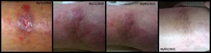
B. Díaz, V. Nuñez

                    Figure 4. Abdominoplasty dehiscence by MRSA infection. Scar surface improvement.

                    Figure 5. Abdominolplasty. Diabetic and hypothyroid patient.

                    Figure 6. Abdominoplasty hypothyroid patient.

                    Figure 7. Thorax burn. Compressive treatment missed.

                    Figure 8. Neck and shoulder burn ropes. Z plasty and grafting.

                    Figure 9. Abdominal 2nd degree burn without grafting. Scar surface improvement.

DOI: 10.4236/mps.2023.131005                                47                                        Modern Plastic Surgery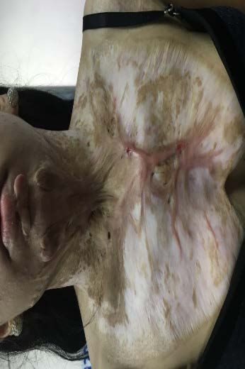
B. Díaz, V. Nuñez

  Figure 10. Fire 3er degree neck and thorax burn. Scar surface improvement. Jugular vein prominence.

  Figure 11. Fire burn hand sequelae. Grafting without compressive treatment.

  Figure 12. Fire acute 2nd degree burn wrist.

  Figure 13. Arm sequelae. Grafting and compressive treatment.

  Figure 14. Acute 2nd degree foot burn. Grafting.

  Figure 15. Acute 3rd degree foot burn.

DOI: 10.4236/mps.2023.131005                                  48                                        Modern Plastic Surgery

B. Díaz, V. Nuñez

       Figure 16. Traumatic wound and friction burn. Scar surface improvement.

       Figure 17. Lower lid Basal Cell Carcinoma. Epidermolysis on the full thickness graft.

       Figure 18. Traumatic wound in Chronic Venous Insufficiency.

                                       5. Conclusions
                                       Using a topical treatment with minimal risk of adverse effects as a complemen-
                                       tary therapy to inhibit chronic inflammation significantly reduces the number
                                       and complexity of scar revision surgeries, provides better comfort for patients
                                       requiring compressive dressings, and produces more elastic and less visible scars
                                       in a shorter time.
                                          Based on our experience, surgery planning may be now different, as the use of
                                       Curefini® before surgery allowed for obtaining more elastic scars and planning
                                       flaps with the same grafted skin as the new scars are easier to conceal.
                                          In cosmetic surgery, the use of Curefini® immediately post-op shortens heal-
                                       ing time and provides satisfying scar results.

                                       Conflicts of Interest
                                       The authors declare no conflicts of interest regarding the publication of this pa-
                                       per.

                                       References
                                       [1]   Berardesca, E. and Maibach, H. (1996) Racial Differences in Skin Pathology. Journal
                                             of the American Academy of Dermatology, 34, 667-672.
                                             https://doi.org/10.1016/S0190-9622(96)80070-3

DOI: 10.4236/mps.2023.131005                                  49                                        Modern Plastic Surgery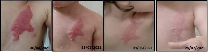
B. Díaz, V. Nuñez

                                [2]   Ribera, N.M. (2016) Características y dermatosis propias de la piel oscura. Medicina
                                      Cutánea Ibero-Latino-Americana, 44, 11-23.
                                [3]   Travis, T., Ghassemi, P., et al. (2015) A Multimodal Assessment of Melanin and
                                      Melanocyte Activity in Abnormally Pigmented Hypertrophic Scar. Journal of Burn
                                      Care & Research, 36, 77-86. https://doi.org/10.1097/BCR.0000000000000154
                                [4]   Fratini, F., Cilia, G., Turchi, B. and Felicioli, A. (2016) Beeswax: A Minireview of Its
                                      Activity and Its Application in Medicine. Asian Pacific Journal of Tropical Medi-
                                      cine, 9, 839-843. https://doi.org/10.1016/j.apjtm.2016.07.003
                                [5]   Krakowski, A.C., Shumaker, P.R., Feldstein, S.I. and Nguyen, T.A. (2016) A Review
                                      of Scar Assessment Scales. Seminars in Cutaneous Medicine and Surgery, 34, 28-36.
                                      https://doi.org/10.12788/j.sder.2015.0125
                                [6]   Chae, J.K., Kim, J.H., Kim, E.J. and Park, K. (2016) Values of a Patient and Observer
                                      Scar Assessment Scale to Evaluate the Facial Skin Graft Scar. Annals of Dermatolo-
                                      gy, 28, 615-623. https://doi.org/10.5021/ad.2016.28.5.615
                                [7]   Baryza, M.J. and Baryza, G.A. (1995) The Vancouver Scar Scale: An Administration
                                      Tool and Its Interrater Reliability. Journal of Burn Care and Rehabilitation, 16,
                                      535-538. https://doi.org/10.1097/00004630-199509000-00013
                                [8]   Colwell, A.S., Phan, T.T., Kong, W., Longaker, M.T. and Lorenz, P.H. (2005) Hyper-
                                      trophic Scar Fibroblasts Have Increased Connective Tissue Growth Factor Expres-
                                      sion after Transforming Growth Factor-β Stimulation. Plastic and Reconstructive
                                      Surgery, 116, 1387-1390. https://doi.org/10.1097/01.prs.0000182343.99694.28
                                [9]   McDaniel, J.C., Belury, M., Ahijevych, K. and Blakely, W. (2008) Omega-3 Fatty
                                      Acids Effect on Wound Healing. Wound Repair & Regeneration, 16, 337-345.
                                      https://doi.org/10.1111/j.1524-475X.2008.00388.x
                               [10]   Pesce, M.S.C. and D’Agostini, S.M.D. (2014) Ácidos Grasos Omega 3: Respuesta
                                      Inmune y su Efecto Sobre Algunas Enfermedades. Enfermería, 3, 33-37.
                               [11]   Calder, P.C. (2013) Omega-3 Polyunsaturated Fatty Acids and Inflammatory Processes:
                                      Nutrition or Pharmacology? British Journal of Clinical Pharmacology, 75, 645-662.
                                      https://doi.org/10.1111/j.1365-2125.2012.04374.x
                               [12]   Hu, S., Bae, M., Park, Y. and Lee, J. (2020) n-3 PUFAs Inhibit TGFβ1-Induced
                                      Profibrogenic Gene Expression by Ameliorating the Repression of PPARγ in He-
                                      patic Stellate Cells. The Journal of Nutritional Biochemistry, 85, Article ID: 108452.
                                      https://doi.org/10.1016/j.jnutbio.2020.108452
                               [13]   Ràdmark, O., Werz, O., Steinhilber, D. and Samuelsson, B. (2015) 5-Lipoxygenase, a
                                      Key Enzyme for Leukotriene Biosynthesis in Health and Disease. Biochimica et Bi-
                                      ophysica Acta (BBA)-Molecular and Cell Biology of Lipids, 1851, 331-339.
                                      https://doi.org/10.1016/j.bbalip.2014.08.012
                               [14]   Calton, E.K., Keane, K.N., Newsholme, P. and Soares, M.J. (2015) The Impact of
                                      Vitamin D Levels on Inflammatory Status: A Systematic Review of Immune Cell
                                      Studies. PLOS ONE, 10, e0141770. https://doi.org/10.1371/journal.pone.0141770
                               [15]   Secretariat, M.A. (2010) Clinical Utility of Vitamin D Testing: An Evidence-Based
                                      Analysis. Ontario Health Technology Assessment Series, 10, 1-93.
                               [16]   Fitch, N., Becker, A.B. and HayGlass, K.T. (2016) Vitamin D [1,25(OH)2D3] Diffe-
                                      rentially Regulates Human Innate Cytokine Responses to Bacterial versus Viral Pat-
                                      tern Recognition Receptor Stimuli. The Journal of Immunology, 196, 2965-2972.
                                      https://doi.org/10.4049/jimmunol.1500460
                               [17]   Olson, K.C., Larkin, P.M.K., et al. (2018) Vitamin D Pathway Activation Selectively
                                      Deactivates Signal Transducer and Activator of Transcription (STAT) Proteins and

DOI: 10.4236/mps.2023.131005                            50                                           Modern Plastic Surgery
B. Díaz, V. Nuñez
Inflammatory Cytokine Production in Natural Killer Leukemic Large Granular Lym-
phocytes. Cytokine, 111, 551-562. https://doi.org/10.1016/j.cyto.2018.09.016
[18] Cornara, L., Biagi, M., Xiao, J. and Burlando, B. (2017) Therapeutic Properties of
Bioactive Compounds from Different Honeybee Products. Frontiers in Pharmacol-
ogy, 8, Article 412. https://doi.org/10.3389/fphar.2017.00412
DOI: 10.4236/mps.2023.131005 51 Modern Plastic SurgeryYou can also read