Hepatic and gastrointestinal manifestations in Mexican patients with juvenile systemic lupus erythematosus
←
→
Page content transcription
If your browser does not render page correctly, please read the page content below
Hepatic and gastrointestinal manifestations in
Mexican patients with juvenile systemic lupus
erythematosus
Elizabeth Hernández Chavez ( elizabethernandezchavez@hotmail.com )
IMSS: Instituto Mexicano del Seguro Social https://orcid.org/0000-0002-6908-7146
José María Alarid Coronel
Instituto Mexicano del Seguro Social
Myriam Mendez Nuñez
IMSS: Instituto Mexicano del Seguro Social
Research Article
Keywords: hepatitis, Mexican children, gastrointestinal system, paediatric-onset SLE (pSLE)
Posted Date: March 28th, 2022
DOI: https://doi.org/10.21203/rs.3.rs-1455340/v1
License: This work is licensed under a Creative Commons Attribution 4.0 International License.
Read Full License
Page 1/12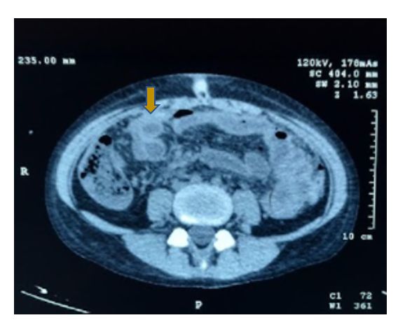
Abstract
Background: Systemic lupus erythematosus (SLE) is a multisystem disorder which can affect the
gastrointestinal system (GI), symptoms can manifest in 50% of patients with systemic lupus
erythematosus (SLE), these have barely been reviewed due to difficulty in identifying different causes [1].
The causes of abdominal pain in SLE patients are diverse, including lupus-related and non–lupus-related
etiologies, as well as infections and medication side effects [2]. Children more often have lupus-related
causes such as lupus mesenteric vasculitis, ascites peritonitis, and pancreatitis [2,3]. Liver
manifestations, generally mild elevations in liver transaminases, have been reported in approximately 25-
57.9% in paediatric-onset SLE (pSLE).

Methods: Retrospective study, with review of 63 medical records of patients diagnosed with paediatric-
onset SLE (pSLE) according to ACR 1997 criteria treated at the UMAE Hospital Pediatría CMNO, during the
period from 2018 to 2020.

Results: 63 patients were included, 49 (77.8%) girls and 14 (22.2%) boys, age range 5-17 years, mean 14
years (DS±2.9). Gastrointestinal involvement was recorded in 50.8% (n= 32) children. Lupus hepatitis, the
most frequent digestive disorder (12.7%). Elevation of liver enzymes was detected in 31.7% with a higher
elevation in patients with digestive disorders, in the case of AST (p= 0.001) and ALT (p= 0.024)
Abdominal pain was the most frequent symptom, present in 38.1% (n= 24) patients, especially related to
multiple medication 46% (n= 29), pancreatitis 7.9% (n=5), lupus enteritis 3.2% (n=2), intestinal pseud
occlusion 1.6% (n=1) and ascites 1.6% (n= 1). A significant association was found (p= 0.03), among
those patients with gastrointestinal affection and management with 4 or more drugs. Azathioprine was
the drug most associated with abdominal pain (p= 0.07). Pancreatitis was associated with higher levels
of disease activity by MEX SLEDAI score (p= 0.016). Of the manifestations not related to SLE, gastritis
was the most frequent in 7.9% which was associated with a longer disease evolution time (p= 0.01).

The decrease in C4 was the only immunological parameter significantly associated in patients with lupus
and digestive disorders (P = 0.033).

Conclusions: Gastrointestinal manifestations are common in paediatric-onset SLE (pSLE), but most of
them are mild and due to adverse reactions to medications. Moderate elevation of liver enzymes occurred
in 31.7% with significantly higher mean values of ALT and AST in patients with digestive disorders.

Background:
Systemic lupus erythematosus (SLE) is a multisystem disorder which can affect the gastrointestinal
system (GI), symptoms can manifest in 50% of patients with systemic lupus erythematosus (SLE), these
have barely been reviewed due to difficulty in identifying different causes [1]. The causes of abdominal
pain in SLE patients are diverse, including lupus-related and non–lupus-related etiologies, as well as
infections and medication side effects [2], children more often have lupus-related causes such as lupus
mesenteric vasculitis, ascites, peritonitis, and acute pancreatitis (AP) [2,3].
                                                 Page 2/12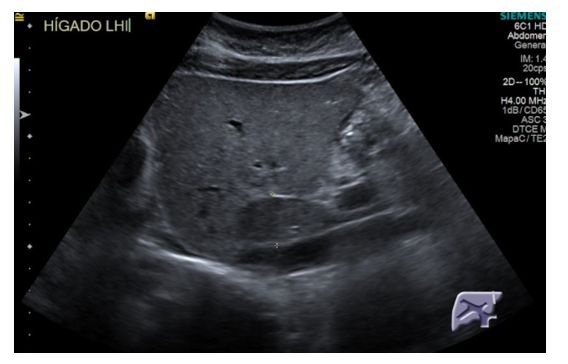
Exist a limited number of studies, mostly case reports, to report GI involvement in pSLE, were identified
only 3 large pediatric cohorts regarding this topic. Tahernia et al, published a retrospective study of 138
pediatric patients with SLE in Iran, as the main finding, liver involvement was found in 48.55% of children
with abnormal liver enzyme levels. Abdominal pain was reported in 37%, mainly in the epigastrium (25%)
[4]. Sonmez et al. in Ankara, Turkey, conducted a retrospective review of 69 medical records of children
with SLE with gastrointestinal involvement, finding gastrointestinal manifestations in 27.5% of patients,
the most frequent being autoimmune hepatitis (42.1%) and lupus enteritis (5.2%). [5]. In France, Richer et
al. published a multicenter, retrospective study of 201 patients with SLE, 39 with gastrointestinal
involvement were identified, with an overall prevalence of 19%. Gastrointestinal symptoms were present
or occurred 1 month after diagnosis in 32% of patients. Abdominal pain was the most frequent symptom
in 34 (87%) patients. They were mainly related to ascites (n= 14) and pancreatitis (n= 12); more rarely
with events induced by treatment (n= 1) or infection (n= 1) and never with events unrelated to SLE [6].

Methods:
In this observational study were collected from medical records 63 pediatric cases of pSLE attending in
Pediatric Rheumatology Department at UMAE Pediatric Hospital CMNO in Guadalajara, México. The
outcomes, frequency, and type of hepatic and gastrointestinal manifestations involvement were assessed
as the main purpose of this study, but other functional gastrointestinal disorders were analyzed:
constipation, dyspepsia and irritable bowel syndrome. In addition to the nutritional status of the patients.
Information was collected from imaging studies performed: abdominal tomography and
ultrasound. pSLE cases were diagnosed based on the presence of 4 of 11 American College of
Rheumatology (ACR) criteria. ACR SLE classification criteria were revised and validated by Systemic
Lupus International Collaborating Clinics (SLICC) group in 2012. According to SLICC, a person is
classified as having SLE in presence of biopsy-proven lupus nephritis with ANA or anti-ds-DNA antibodies
or 4 of diagnostic criteria, including at least one clinical and one immunologic criterion The clinical
parameters analyzed were presence/absence of any gastrointestinal manifestations: related or not
related to lupus and other functional digestive disorders (dyspepsia, constipation, irritable bowel);
nutritional status of the patient, number of drugs consumed, imaging studies performed. Disease activity
measured by Mexican SLE disease activity index score (Mex-SLEDAI).

Laboratory parameters included: SLE-specific antinuclear antibodies by ELISA, AST and ALT levels with a
cut-off point >40 u/L, and GGT with a cut-off point >30 u/L. Hypoalbuminemia was considered a value
(
variables the Chi2 test. All analyzes will be performed using the IBM SPSS® v25.1. A p value less than
0.05 was considered statistically significant.

Results:
A total of 75 files were reviewed, of which 63 patient files met the inclusion criteria, 49 were girls (77.8%)
and 14 boys (22.2%), with a male-female ratio of 1: 3.5. The ages ranged from 5 to 17 years, with a mean
of 14 years and a standard deviation of 2.9 years. The majority adolescents in 77.8%.

Nutritional alterations were found in most of the patients (57.1%), overweight and obesity were the most
frequent in one (41.3%); followed by growth retardation and malnutrition. Table 1. We found a significant
association between patients with altered nutritional status and use of 4 or more drugs (p= 0.03), as well
as higher levels of proteinuria (p= 0.049). Regarding the clinical presentation, gastrointestinal signs and
symptoms were present in 50.8% (n= 32). The most frequent digestive symptom was abdominal pain in
38%. Hepatomegaly was the most frequent gastrointestinal sign unrelated to SLE, found in 4.8%, jaundice
and ascites were less frequent, in 3.2 and 1.6%, respectively. Table 2.

 A significant association was found (p= 0.03), among those patients with gastrointestinal signs and
symptoms and management with 4 or more drugs, no statistical significance was found when correlated
with time of disease evolution, antinuclear antibody positivity, complement levels or activity of the illness.
Abdominal pain and digestive disorders were significantly associated with patients diagnosed with
depression and higher levels of proteinuria with p= 0.033 and p= 0.01, respectively.

Functional digestive disorders appeared in 12.7%, constipation was the most frequent, followed by
dyspepsia and irritable bowel syndrome Table 4.

Elevated aminotransferases were the most frequent biochemical alteration in liver function tests, in 31.7%
of patients, taking into account ALT and AST with a cut-off point >40 u/L and GGT with a cut-off point
>30 u/L. Hypoalbuminemia (
The most frequently found antibodies were antinuclear antibodies (ANA) by indirect immunofluorescence
homogeneous pattern in 93.7%, antiDNAdc (58%), antiRNP (42.9%), anti SM (42.9%). Associations were
made between antibodies and complement levels with digestive disorders, C4 was the only
immunological parameter significantly associated in patients with lupus and digestive disorders (P =
0.033).

Regarding imaging studies, abdominal ultrasound was performed in 8 patients, with altered results in 5 of
them, hepatic steatosis was present in 2 patients, followed by free abdominal fluid, alteration in hepatic
echogenicity suggestive of chronic liver disease and thickening of the liver gut wall.

Abdominal tomography was performed on 2 patients, reporting thickening of the large intestine wall in
one and changes in the caliber of hepatic vessels suggestive of portal hypertension in another. Likewise,
2 patients underwent endoscopy, altered only in one patient, reporting erosive gastritis. Colonoscopy was
performed in 2 patients, without alterations and finally liver biopsy in only 1 patient, with a report of
hepatic steatosis.

Discussion:
This retrospective study emphasises that abdominal manifestations are common in pSLE. The GI
involvement in pSLE is variable. Sönmez et al., in a series of cases carried out in Turkey, described
gastrointestinal involvement in 27.5%. Bader-Meuner et al., in 17% of children with SLE, and in this study
it was found in 50.8% of children. [5, 7]

The most common symptom in the largest pediatric series is abdominal pain. The causes of abdominal
pain in SLE patients are more frequent lupus-related etiologies, among which are: lupica enteritis (EL),
protein-losing enteropathy (PLE), intestinal pseudo-obstruction (IPO), acute pancreatitis (AP). Its
prevalence varies in variable ranges between 15.8–100% of cases. Richer et al. [6] reported that 87% of
the causes of abdominal pain were due to acute pancreatitis, Sönmez et al. [5] and Tu et al. [8] to lupus
enteritis in 5.2% and 31.6% respectively and in Hispanic children it was found in 38.1%, the most frequent
cause was associated with the use of multiple medications. Gastrointestinal symptoms in Hispanic
children with SLE were significantly associated with multiple medication (p = 0.033), without finding a
correlation with disease activity levels, time of evolution or antinuclear antibody positivity. [6, 9]

Sönmez et al. reported autoimmune hepatitis as the most frequent disorder associated with SLE, in
42.1%. [5] In contrast, in our series no patient was found. Lupus hepatitis (LH) was the most frequent
(12.7%), its evolution was subclinical with a course favorable to the use of steroids, none with
progression to end-stage liver disease, and although the real prevalence of hepatitis autoimmune (AIH)
with SLE in our patients is unknown, it was not necessary liver biopsy for diagnosis, since the evolution
was satisfactory, unlike those reported by other authors in which there is overlap of both entities and
therefore the need for histological study. [4, 5]

                                                    Page 5/12
Globally, the estimated prevalence of LE ranges between 0.2% and 14% among all SLE patients [16] and
accounts for 29–65% cases of acute abdominal pain [10], in our review it was found in only 3.2% of our
patients. Intestinal wall edema and ascites are important for diagnosis, and imaging studies are
necessary for a definitive diagnosis. The two patients with lupus enteritis were the only ones who
underwent imaging studies and responded satisfactorily to treatment with steroids and alkylating agents.
Patients with lupus and severe abdominal pain require a thorough investigation as elevated disease
activity is related to the presence of intra-abdominal vasculitis. [11]
Reported prevalence of AP in pSLE ranges between 2.6% and 6% (12–16). In the cohort described by
Bader-Meunier et al., AP was the most common cause of abdominal pain [5]. In our study, it occurred in
7.9% of patients and it was confirmed that it is significantly associated with lupus activity, in our case by
the MEX-SLEDAI score (p = 0.016). [7, 11]. Figure 1. Likewise, we corroborate that the association between
the administration of steroids or azathioprine and the development of pancreatitis cannot be proven
significantly.
Mild elevation of transaminases occurs in up to 57.9% of patients, most of the time asymptomatically, in
our case it was the most frequent biochemical alteration in 31.7% of patients and a greater elevation was
demonstrated in mean values of transaminases in patients with digestive disorders, however, to attribute
it to SLE, it is due to hepatotoxicity due to drugs, autoimmune hepatitis, viral hepatitis and fatty liver. [4,
11]
In our study, 58.7% of patients presented some type of nutritional impairment, in contrast to other studies
conducted in adults, where it is described in less than 10% of patients. Possibly this difference is due to
the fact that the disease is more serious and has greater renal involvement in children, since we
significantly correlated those patients with altered nutritional status and higher levels of proteinuria (p =
0.049), on the other hand, they were not significantly associated with the time of evolution or the activity
of the disease. [7]
Although gastrointestinal manifestations are frequent, functional digestive disorders have hardly been
studied in children with SLE, in our study they occurred in 12.7% of patients, constipation being the most
frequent, it is likely that medications alter gastrointestinal homeostasis, such as glucocorticoids and non-
steroidal anti-inflammatory drugs, and these are potential risk factors for its development.
In our study, associations were made between antibodies and complement levels with digestive disorders,
only the decrease in C4 was the only immunological parameter significantly associated in patients with
lupus and digestive disorders (p = 0.033).
Abdominal ultrasound shows abnormalities in up to 79% of patients with gastrointestinal manifestations,
increased echogenicity of the liver parenchyma is the most common alteration in more than 50% of
patients. [5]. On the contrary, hepatic steatosis was the most frequent alteration in our patients, and
ultrasound alterations in general were described less frequently than those described in other series
(62.5%). Figure 2.
Page 6/12Abdominal tomography has been described as abnormal in 5.5% of patients, the most common finding
being thickening of the intestinal wall. In our study, only 2 patients underwent abdominal tomography,
due to the severity of their symptoms, reporting thickening of the wall of the large intestine. Figure 3, in
one and changes in the caliber of hepatic vessels suggestive of portal hypertension in another.
Abdominal tomography should be mandatory in children with lupus who have unexplained abdominal
pain to rule out findings suggestive of ischemic bowel disease.[6]
Liver biopsies performed on patients with SLE mainly report autoimmune hepatitis (42%), hepatocellular
damage (10%), toxic hepatitis (5%) and cholestatic hepatitis (5%). [4] In our series, only 1 patient
underwent liver biopsy, with a report of hepatic steatosis. Hepatic steatosis is rarely described in children
with SLE, secondary to lupus per se or corticosteroid therapy. Histopathological approach is necessary in
children with SLE and elevated transaminase levels unexplained by other causes, due to the high
frequency of autoimmune hepatitis and associated histopathological alterations [5].
Conclusion:
Gastrointestinal manifestations in Hispanic children are mostly mild and secondary to the use of multiple
medications, and hepatic non related of SLE,
Most SLE GI manifestations our children responded well to glucocorticoids and immunosuppressants
In view of the limited available data, further studies in children are needed to better explore the
prevalence, prognosis and treatment recommendations for GI involvement in pSLE.
Limitations:
Among the limitations of our study were incomplete data in the clinical records, so it was not possible to
include the total number of SLE patients treated at our reference center. In addition, it was not possible to
perform imaging studies on all patients who presented some gastrointestinal symptom, because most of
the symptoms were mild, including liver disease. Therefore, the performance of a histological study was
not justified.
Abbreviations:
Systemic lupus erythematosus (SLE)
Paediatric-onset SLE (pSLE)
Gastrointestinal system (GI)
Paediatric-onset SLE (pSLE).
American College of Rheumatology (ACR)
Page 7/12Systemic Lupus International Collaborating Clinics (SLICC)
Protein-losing enteropathy (PLE)
Intestinal pseudo-obstruction (IPO)
Enteritis lupica (EL)
Acute pancreatitis (AP).
Autoimmune hepatitis (AIH)
Lupus hepatitis (LH)
Mexican SLE disease activity index score (Mex-SLEDAI)
Antinuclear antibodies (ANA)
Declarations:
Availability of supporting data: the statistical analysis and the databases are available and updated if
required by the publisher.
Competing interests: the authors declare that they have no conflicts of interest.
Funding: the project received no funding of any kind, neither government nor sponsorship from the
pharmaceutical industry.
Authors' contributions: The main author is responsible for the design of the study, revision and editing of
the database for its final statistical analysis. Third year resident of pediatrics: review of the literature,
collection of sociodemographic information, elaboration of databases of the patients included in the
study.
Co-author review of the literature and research protocol, revision and final edition of the manuscript.
Acknowledgments: We appreciate the cooperation of clinical file staff for the facilities for reviewing
clinical records; as well as the head Chief of the service Dr. Pacheco, and pediatric gastroenterologists
attached to the hospital's gastroenterology service.
Authors' information:
Elizabeth Hernández Chávez, pediatric gastroenterologist associate researcher at UMAE Hospital de
Pediatria CMNO at Instituto Mexicano del Seguro Social IMSS. Guadalajara Mexico • Associate Professor
of the specialization course in pediatric gastroenterology subspecialty of IMSS • Fellowship in pediatric
Page 8/12liver diseases in Hospital Universitario La Paz, Madrid, Spain, care, research in liver disease and liver
transplantation.
Myriam Mendez Nuñez, MD. Pediatric Rheumatologist associate researcher at UMAE Hospital de
Pediatria CMNO at Instituto Mexicano del Seguro Social IMSS. Guadalajara Mexico • Associate Professor
of the specialization course in Pediatric Rheumatology of IMSS • Fellowship in the pediatric
rheumatology unit Isstituto Gannina Gaslini Genoa Italy, care, research and ultrasound activities in JIA.
José María Alarid Coronel, MD. Pediatrician graduated in UMAE Hospital de Pediatria CMNO at Instituto
Mexicano del Seguro Social IMSS. 2020. Current certification by the Mexican Council of Pediatrician.
References:
1. Gastrointestinal system involvement in systemic lupus erythematosusZ Li1, D Xu1, Z Wang1, Y
Wang2, S Zhang1, M Li1, X Zeng1. lupus 2017 Oct;26(11):11271138. doi:
10.1177/0961203317707825.
2. MEXMEX O. Richer, T. Ulinski, I. Lemelle, et al., Abdominal manifestations in childhoodonset systemic
lupus erythematosus, Ann. Rheum. Dis. 66 (2007) 174–178.
3. Y.L. Tu, K.W. Yeh, L.C. Chen, et al., Differences in disease features between childhoodonset and
adultonset systemic lupus erythematosus patients presenting with acute abdominal pain, Semin.
Arthritis Rheum. 40 (2011) 447–454.
4. Tahernia L, Alimadadi H, Tahghighi F, Amini Z, Ziaee V. Frequency and Type of Hepatic and
Gastrointestinal Involvement in Juvenile Systemic Lupus Erythematosus.Autoimmune Dis.
2017;80(9)7273.
5. Sönmez H, Karhan A, Batu E, Bilginer Y, Gümüş E, Demir H, et al.Gastrointestinal system
manifestations in juvenile systemic lupus erythematosus. Clin Rheumatol. 2017;36(7):15211526.
6. Richer O, Ulinski T, Lemelle I, Ranchin B, Loirat C, Piette J, et al. Abdominal manifestations in
childhoodonset systemic lupus erythematosus. Ann Rheum Dis. ;66(2):174178.
7. BaderMeunier B, Armengaud J, Haddad E, Salomon R, Deschênes G, KonéPautI, et al.Initial
presentation of childhoodonset systemic lupus erythematosus: A French multicenter study. J Pediatr.
2005;146(5):648653.
8. Tu Y, Yeh K, Chen L, Yao T, Ou L, Lee W, et al. Differences in Disease Features Between
ChildhoodOnset and AdultOnset Systemic Lupus Erythematosus Patients Presenting with Acute
Abdominal Pain. Semin Arthritis Rheum. 2011;40(5):447454.
9. ALVES SC, FASANO S, ISENBERG DA: Autoimmune gastrointestinal complications in patients with
systemic lupus erythematosus: case series and literature review. Lupus 2016; 25: 150919.
10. JU JH, MIN JK, JUNG CK et al.: Lupus mesenteric vasculitis can cause acute abdominal pain in
patients with SLE. Nat Rev Rheumatol 2009; 5: 27381.
Page 9/1211. Brewer B, Kamen D. Gastrointestinal and Hepatic Disease in Systemic Lupus Erythematosus. Rheum
Dis Clin North Am. 2018;44(1):165175.
12. RICHER O, ULINSKI T, LEMELLE I et al.:Abdominal manifestations in childhoodonset systemic lupus
erythematosus. Ann Rheum Dis 2007; 66: 1748.
13. HUGGINS JL, HOLLAND MJ, BRUNNER HI:Organ involvement other than lupus nephritis in
childhoodonset systemic lupus erythematosus.Lupus 2016; 25: 85763.
14. MARQUES VLS, GORMEZANO NWS, BONFÁ E et al.: Pancreatitis subtypes survey in 852
childhoodonset systemic lupus erythematosus patients. J Pediatr Gastroenterol Nutr 2016; 62:
32834.
15. CAMPOS LMA, OMORI CH, LOTITO APN,JESUS AA, PORTA G, SILVA CAA: Acute pancreatitis in
juvenile systemic lupus erythematosus: A manifestation of macrophage activation syndrome? Lupus
2010; 19: 16548.
16. GORMEZANO NWS, OTSUZI CI, BARROS DL et al.: Macrophage activation syndrome: a severe and
frequent manifestation of acute pancreatitis in 362 childhoodonset compared to 1830 adultonset
systemic lupus erythematosus patients. Semin Arthritis Rheum 2016; 45: 70610.
17. Trapani S, Rubino C, Simonini G, Indolfi G. Gastrointestinal and hepatic involvement in paediatric
systemic lupus erythematosus. Clin Exp Rheumatol. 2021 JulAug;39(4):899906. Epub 2021 Mar 2.
PMID: 33666164.
Tables:
Tables 1-6 are not available with this version
Figures
Page 10/12Figure 1
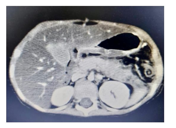
Computerized axial tomography in a 15-year-old boy with Balthazar C pancreatitis; to the onset of
systemic lupus.
Page 11/12Figure 2
Abdominal ultrasound: showing increased hepatic echotexture due to moderate-grade hepatic steatosis
with hepatomegaly in a 15-year-old girl with lupus.
Figure 3
Abdominal contrast-enhanced computed tomography: demonstrating multiple segments of focal bowel
wall thickening, diffuse bowel wall enhancement with peripheral rim enhancement (target sign).
Page 12/12You can also read