Hecate-CGb conjugate and gonadotropin suppression shows two distinct mechanisms of action in the treatment of adrenocortical tumors in transgenic ...
←
→
Page content transcription
If your browser does not render page correctly, please read the page content below
Endocrine-Related Cancer (2009) 16 549–564

Hecate-CGb conjugate and gonadotropin
suppression shows two distinct
mechanisms of action in the treatment of
adrenocortical tumors in transgenic mice
expressing Simian Virus 40 T antigen under
inhibin-a promoter
Susanna Vuorenoja1,2, Bidut Prava Mohanty1, Johanna Arola3,
Ilpo Huhtaniemi1,4, Jorma Toppari1,2 and Nafis A Rahman1
Departments of 1Physiology and 2Pediatrics, University of Turku, Kiinamyllynkatu 10, FIN-20520 Turku, Finland
3
 Department of Pathology, University of Helsinki and HUSLAB, Helsinki, Finland
4
 Institute of Reproductive and Developmental Biology, Imperial College, London, UK
(Correspondence should be addressed to N A Rahman; Email: nafis.rahman@utu.fi)

Abstract
Lytic peptide Hecate (23-amino acid (AA)) fused with a 15-AA fragment of human chorionic
gonadotropin-b (CG-b), Hecate-CGb conjugate (H-CGb-c) selectively binds to and destroys tumor
cells expressing LH/chorionic gonadotropin receptor (Lhcgr). Transgenic mice (6.5 month old)
expressing SV40 T-antigen under the inhibin-a promoter (inha/Tag) presenting with Lhcgr
expressing adrenal tumors were treated either with H-CGb-c, GnRH antagonist (GnRH-a),
estradiol (E2; only females) or their combinations for 1 month. We expected that GnRH-a or E2 in
combination with H-CGb-c could improve the treatment efficacy especially in females by
decreasing circulating LH and eliminating the potential competition of serum LH with the H-CGb-c.
GnRH-a and H-CGb-c treatments were successful in males (adrenal weights 14G2.8 mg and
60G26 vs 237G59 mg in controls; P!0.05). Histopathologically, GnRH-a apparently destroyed
the adrenal parenchyma leaving only the fibrotic capsule with few necrotic foci. In females, H-CGb-c
was totally ineffective, whereas GnRH-a (19G5 mg) or E2 (77G50 mg) significantly reduced
the adrenal weights compared with controls (330G70 mg). Adrenal morphometry, cell
proliferation markers, post-treatment suppression of serum progesterone, and quantitative
RT-PCR of GATA-4, Lhcgr, and GATA-6 further supported the positive outcome. H-CGb-c
selectively killed the Lhcgr expressing tumor cells, whereas GnRH-a blocked tumor progression
through gonadotropin suppression, emphasizing the gonadotropin dependency of these
adrenocortical tumors. If extrapolated to humans, H-CGb-c could be considered for the treatment
of gonadotropin-dependent adrenal tumors in males, whereas in females gonadotropin
suppression, but not H-CGb-c, would work better.
Endocrine-Related Cancer (2009) 16 549–564

Introduction                                                              & Brennan 1999a,b), and high probability around
Adrenocortical tumors are rare and aggressive as they                     menopause (Mijnhout et al. 2004). The incidence of
often are diagnosed late and have poor survival rate                      these malignancies increases during chronic gonado-
(Schulick & Brennan 1999a). These tumors are 1.5-                         tropin overload, e.g. in pregnancy (Sheeler 1994) or
fold more common in females than males with peak                          after menopause (Mijnhout et al. 2004), resulting in
incidences in the first and fifth decades of life (Schulick               ACTH-independent macronodular adrenocortical
Endocrine-Related Cancer (2009) 16 549–564                                                        Downloaded from Bioscientifica.com at 09/15/2021 10:40:17PM
                                                                                                                     DOI: 10.1677/ERC-08-0232
                                                                                                                                                via free access
1351–0088/09/016–549 q 2009 Society for Endocrinology Printed in Great Britain          Online version via http://www.endocrinology-journals.org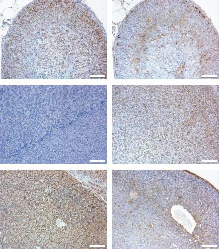
S Vuorenoja et al.: Treatment strategies for adrenocortical tumors

hyperplasia and Cushingoid symptoms maintained by             and testicular cancer cells (Bodek et al. 2005b).
ectopic adrenal Lhcgr expression (Lacroix et al. 1999,        H-CGb-c has been shown to induce dose-dependent
Bourdeau et al. 2001, Miyamura et al. 2002, Feelders          rapid and cell-specific membrane permeabilization of
et al. 2003, Goodarzi et al. 2003). Also LH-dependent         Lhcgr expressing cells in vitro, resulting in necrotic cell
adrenal adenomas producing aldosterone (Saner-Amigh           death without activation of apoptosis (Bodek et al. 2005b).
et al. 2006) or androgens (Werk et al. 1973, Givens et al.    More generally, our model is able to provide the proof
1975, Larson et al. 1976, Smith et al. 1978, Takahashi        of principle for the efficacy of receptor-mediated
et al. 1978, de Lange et al. 1980, Leinonen et al. 1991)      targeting of toxic molecules in the abolition of tumors.
and adrenocortical carcinomas (Wy et al. 2002, Barbosa            TG mice of both sexes expressing the inhibin-a
et al. 2004) with abundant Lhcgr expression have been         promoter/Simian Virus (SV40) T-antigen (inha/Tag)
described. Moreover, the normal human adrenal cortex          were originally found to produce gonadal tumors with
has been shown to express low levels of Lhcgr (Pabon          100% penetrance by the age of 6 months, but when
et al. 1996). The only effective cure for adrenocortical      gonadectomized prepubertally they produced adrenal
tumors so far is their total surgical removal (Kuruba &       tumors by the same age (Kananen et al. 1996a,
Gallagher 2008). Adjuvant therapies such as radio-            Rilianawati et al. 1998, Rahman et al. 2004). The TG
therapy and mitotane after surgery have been tested           adrenal tumors and a tumor-derived cell line (Ca1) were
(Fassnacht et al. 2006, Terzolo & Berruti 2008), but there    found to express high levels of Lhcgr (Rilianawati et al.
is still the need for better curative methods of treatment.   1998, Rahman et al. 2004), and their growth was found
   Hecate is a 23-amino acid (AA) amphipathic,                to be gonadotropin dependent (Kananen et al. 1997).
positively charged lytic peptide, a synthetic analogue        Adrenal tumors failed to appear if the post-gonadect-
of melittin, which is the principal toxic component of        omy increase in gonadotropin secretion was blocked
natural honeybee venom (Arrowood et al. 1991a,b,              either by administration of GnRH antagonist (GnRH-a)
Henk et al. 1995, Baghian et al. 1996). Its mechanism
                                                              or by cross-breeding the TG mice into the hypogonado-
of action depends on the formation of pores or channels
                                                              tropic hpg genetic background (Cattanach et al. 1977,
in cell membranes by wedge-shaped insertion of
                                                              Kananen et al. 1997). Hence, the post-gonadectomy
monomers of the lytic peptide. This causes aggregation
                                                              elevation of LH levels apparently induced ectopic Lhcgr
of the membrane proteins and expulsion of phospho-
                                                              expression in the adrenal cortex, which together with
lipids (Leuschner & Hansel 2004, Bodek et al. 2005a),
                                                              co-expression of the potent oncogene Tag triggered the
leading to osmotic cytolysis and cell death through
                                                              tumorigenesis (Rahman et al. 2001, 2004, Mikola et al.
necrosis. The binding of lytic peptides is mainly
                                                              2003). The adrenocortical tumorigenesis occurs in a
subjected to negatively charged membranes typical of
                                                              slow hyperplasia-adenoma-carcinoma sequence
the outer leaflet of tumor cells due to altered
distribution of their phosphatidylserines (Utsugi et al.      following prepubertal gonadectomy of the TG mice;
1991). In order to specifically target the action of          hyperplasia is seen at 4 months and discernible tumors
Hecate, its 23 AA sequence was fused with a 15-AA             at 6 months (Rahman et al. 2004). The chronically
segment (81–95) of the b-subunit of human chorionic           elevated LH level (Kananen et al. 1996a) and ectopic/
gonadotropin (CG-b), which possesses high affinity for        up-regulated Lhcgr, with low (5–10%) metastasis
Lhcgr (Leuschner et al. 2001). Hecate-CGb conjugate           frequency (Rahman et al. 2004, Vuorenoja et al.
(H-CGb-c) selectively kills tumor cells expressing            2008), makes these TG mice a relevant experimental
Lhcgr but spares the healthy receptor-positive and            model for human adrenal tumors.
-negative cells (Leuschner et al. 2001, Bodek et al.              This study represents continuation to our previous
2005b). The cytotoxic activity of the H-CGb-c induces         work on the use of H-CGb-c in treating the Lhcgr
plasma membrane disruption within minutes (Chen               expressing adrenal tumors of the inha/Tag TG mice,
et al. 2003, Bodek et al. 2005b, Hansel et al. 2007b),        where the treatment effectively killed tumor cells in
it is not antigenic, and it has not been shown to have        males but was ineffective in females (Vuorenoja et al.
any clear side effects (Bodek et al. 2005b, Hansel            2008). We hypothesized that treatment of the mice
et al. 2007b, Vuorenoja et al. 2008). H-CGb-c has             with estradiol (E2) or GnRH-a, alone or in combination
been found to selectively kill Lhcgr expressing               with H-CGb-c, would decrease the circulating serum
prostate (Hansel et al. 2001, Leuschner et al. 2001,          LH and consequently eliminate the potential compe-
2003, Bodek et al. 2005a), mammary gland (Bodek               tition between H-CGb-c and LH. This could increase
et al. 2003, Leuschner et al. 2003, Zaleska et al.            the efficacy of the H-CGb-c treatment in particular in
2004, Leuschner & Hansel 2005, Hansel et al. 2007a,b),        the female TG mice. We also compared the effects of
ovarian (Gawronska et al. 2002, Bodek et al. 2005b),          GnRH-a treatment with H-CGb-c in TG males.
                                                                                    Downloaded from Bioscientifica.com at 09/15/2021 10:40:17PM
                                                                                                                                  via free access
550                                                                                             www.endocrinology-journals.org
Endocrine-Related Cancer (2009) 16 549–564

Materials and methods                                       were injected once per week for three consecutive
                                                            weeks, according to earlier protocols for in vivo
Experimental animals
                                                            treatment for nude mouse xenografts (Leuschner et al.
Adrenal tumors were induced by prepubertal gonadect-        2001), TG mice with gonadal tumors (Bodek et al.
omy of inha/Tag TG mice as described earlier (Kananen       2005b), and for adrenal tumors (Vuorenoja et al. 2008).
et al. 1996a). Discernible adrenocortical tumors with       In addition to H-CGb-c treatment, a group of TG males
100% penetrance appear in these mice by the age of          and females and WT controls were injected subcu-
6 months (Kananen et al. 1996a, Rilianawati et al. 1998,    taneously with GnRH-a (10 mg/kg) every 84 h accor-
2000, Kero et al. 2000, Rahman et al. 2001, 2004).          ding to a previously established protocol (Kananen
Gonadectomy was performed under Avertin anesthesia          et al. 1997). Another group of TG females, and controls,
(Hogan et al. 1994) prepubertally between 21 and            were treated with E2 silastic implants. After 4 weeks of
24 days of life, and burprenorphine (Schering-Plough,       treatment, blood was collected by cardiac puncture
Brussels, Belgium) was administrated as post-operative      under Avertin anesthesia and the mice were killed by
analgesia. Six to eight mice per treatment group were       cervical dislocation. Total body weights, adrenal tumor,
used for the experiments. Wild-type (WT) control            and selected organ weights were recorded. Tissues were
littermate mice (C57Bl/6N) were used as controls            either snap-frozen in liquid nitrogen or fixed in 4%
(nZ6). For routine genotyping, PCR analysis was carried     paraformaldehyde and embedded in paraffin. Paraffin
out as previously described (Kananen et al. 1995) using     sections of 5 mm thickness were stained for histological
DNA extracts from ear biopsies. After weaning at the        analysis after hematoxylin–eosin (HE) staining, or used
age of 21 days, the mice were housed two to four per        for immunohistochemical analysis. For each tissue and
cage, females and males separately, in a room of            treatment group, at least four independent specimens
controlled light (12 h light:12 h darkness) and tempera-    were examined.
ture (21G1 8C). The mice were fed with mouse chow
SDS RM-3 (Whitham, Essex, UK) and tap water                 Hormone measurements
ad libitum, kept in a specific pathogen-free surrounding
and routinely screened for common mouse pathogens.          Serum levels of LH (Haavisto et al. 1993) and FSH
Ethics Committees for animal experimentation of the         (van Casteren et al. 2000) were measured by
Turku University and the State Provincial Office of         immunofluorometric assays (Delfia; Wallac, Turku,
Southern Finland approved all the animal experiments.       Finland) as described previously. Serum progesterone
                                                            was measured using the Delfia Progesterone Kit
                                                            (Wallac). The approximate assay sensitivity for LH
Preparation of drugs
                                                            was 0.1 mg/l, for FSH 0.03 mg/l, and for progesterone
Hecate and H-CGb-c were synthesized and purified in the     0.5 nmol/l. The intra- and interassay coefficients of
Peptide and Protein Laboratory, Department of Virology,     variations for these assays were below 10%.
Hartman Institute, University of Helsinki as described
earlier (Bodek et al. 2003). Silastic implant tubes, 8 mm   Immunohistochemistry
in length (inner diameter 1.58 mm and outer diameter
                                                            The paraffin sections (5 mm) of tumors and wild-type
2.41 mm) were filled with 8 mg of E2 powder (Sigma
                                                            control adrenals were deparaffinized and rehydrated.
Chemical Co.) and sealed at both ends with silastic
                                                            Three percent H2O2 in water was used to block
adhesive (Elastosil RTV-1 Silicone Rubber, Wacker-
                                                            endogenous peroxidases, and the sections were boiled
Chemie GmbH, Munich, Germany) as described earlier
                                                            by microwave treatment for 10 –15 min in 10 mM
(Pakarainen et al. 2005). GnRH-a (cetrorelix acetate,
                                                            citric acid (pH 6.0) for antigen retrieval. Two washes
Cetrotide) was purchased from Merck Serono.
                                                            were carried out after each step with 0.05 M Tris and
                                                            150 mM NaCl with 0.1% Tween (TBS-T). Serial
H-CGb-c, GnRH-a, and E2 treatments
                                                            sections were subjected to immunohistochemistry
Male and female mice (inha/Tag TG; nZ6–8 per                (IHC). To detect GATA-6, slides were incubated
group) were treated at the age of 6.5 months with either    with the primary goat polyclonal anti-GATA-6
H-CGb-c or Hecate (12 mg/kg b.w.) by i.p. injections        antibody (dilution 1:250; Santa Cruz Biotechnology,
as described earlier (Vuorenoja et al. 2008). As no         Santa Cruz, CA, USA) at 4 8C overnight. After
significant weight changes of the mice could be             incubation and washings with TBS-T buffer, the slides
observed between Hecate and H-CGb-c by adding               were incubated with the anti-goat (dilution 1:400;
hCG to Hecate, both Hecate and H-CGb-c were given           Santa Cruz Biotechnology) secondary antibody for
in the same dose (Leuschner et al. 2001). The mice          30 min. The avidin–biotin immunoperoxidase system
                                                                               Downloaded from Bioscientifica.com at 09/15/2021 10:40:17PM
                                                                                                                             via free access
www.endocrinology-journals.org                                                                                             551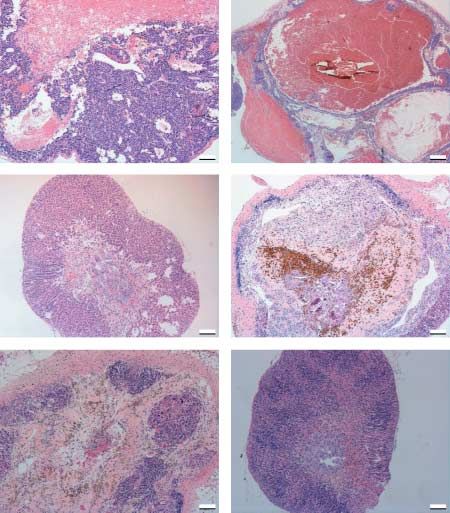
S Vuorenoja et al.: Treatment strategies for adrenocortical tumors

was used to visualize bound antibody (Vectastain Elite          the specific types of tissue of interest (i.e., tumor,
ABC Kit, Vector Laboratories, Burlingame, CA, USA)              healthy, fibrotic/necrotic, cyst-type formation) and
with 3,3 0 -diaminobenzidine (Sigma) as a substrate. For        thereafter by dividing the points of each tissue type
Ki-67, a rabbit monoclonal anti-Ki-67 SP6-clone                 of interest by the total number of points.
(dilution 1:2000; NeoMarkers, Fremont, CA, USA)
with EnVisionCSystem-HRP labeled Polymer (Dako-                 Quantitative RT-PCR
Cytomation, Inc., Carpinteria, CA, USA) was used. In
                                                                We extracted total RNA from snap-frozen whole adrenal
this case, the microwave treatment was done in 10 mM
                                                                tumors after different treatments (nZ5 for each treatment
Tris–EDTA buffer (pH 9.0), and the primary antibody
                                                                group) for the quantitative (q)RT-PCR analysis, as
was diluted in 3% BSA. Novolink Polymer Detection
                                                                described earlier (Kero et al. 2000, Rahman et al.
System Kit (Novocastra, Benton Lane, UK) was used
                                                                2004). Total RNA was extracted with RNeasy Mini Kit
for the detection of GATA-4 with a rabbit polyclonal
                                                                (Qiagen) according to the manufacturer’s instructions
anti-GATA-4 IgG (dilution 1:800; Santa Cruz
                                                                and treated with amplification grade DNaseI
Biotechnology) and PowerVisionCPoly-HRP IHC
                                                                (Invitrogen). For cDNA synthesis and subsequent
Kit for goat (ImmunoVision Technologies, Hague,
                                                                qRT-PCR, the SYBR Green DyNAmo HS qRT-PCR
The Netherlands) for the detection of p53 using a goat
                                                                kit (Finnzymes, Espoo, Finland) was used with 1:50
polyclonal antibody anti-p53 IgG (dilution 1:100;
                                                                diluted aliquots. qRT-PCR analysis was performed using
Santa Cruz Biotechnology). The protocols followed
                                                                the DNA Engine Thermal Cycler (BioRad) with
the manufacturer’s instructions except for endogenous
                                                                continuous fluorescent detection. The program was by
peroxidase blocking that was done after the first
                                                                the manufacturer’s recommendations as follows: 15 min
antibody incubation. The antibodies were diluted to
                                                                in 95 8C to activate the hot start DNA polymerase and to
PBS with 0.1% Triton X-100 (anti-GATA-4) or TBS
                                                                denaturate the template, 10 s in 94 8C to denaturate, 30 s
(anti-p53). As a control for the antibodies, adjacent
                                                                in 56–61 8C, depending on the primers for annealing, 30 s
sections were incubated with either 1% normal goat
                                                                in 72 8C for extension, and 1 s in 78 8C for data
serum in PBS or rabbit IgG instead of primary antibody
                                                                acquisition. The above protocol was repeated for 44
to differentiate unspecific from specific staining (data
                                                                times after which there was 10 min incubation at 72 8C
not shown).
                                                                for final extension. Melting curve was between 72 and
                                                                90 8C, in 0.5 8C intervals for 1 s. The protocol ended by
Morphometric analyses                                           5 min in 72 8C to re-anneal. Primer pairs and annealing
                                                                temperatures are shown in Table 1, and the samples and
Serial sections (nZ6 per group) of HE-stained slides
                                                                standards were run in triplicates. The housekeeping gene
from each treatment group were analyzed morphome-
                                                                L19 was analyzed to normalize the results between
trically by a point counting technique as described
                                                                samples. Amplification products were separated on 1%
earlier (Haapasalo et al. 1990, Howard & Reed 1998)
                                                                agarose gel and stained with ethidium bromide.
in order to quantify the histological differences
between the study groups. A grid of orthogonal lines
                                                                Statistical analysis
was placed on top of the tissue section (magnified
under light microscope !5). The volume fraction                 Statistical analyses were carried out by ANOVA with
estimation (in %) was done by counting the number of            the post-hoc Bonferroni test, using SAS Enterprise
crossing points of the whole section and points that hit        Quide 3.0 program (SAS Institute, Inc., Cary, NC, USA).

Table 1 Oligonucleotides used in quantitative RT-PCR analysis

Gene           Primers       Sequence                                   Product size (bp)        Temperature (annealing) (8C)

L19                F         5 0 -GGACAGAGTCTTGATGATCTC-3 0                   195                                 56
                   R         5 0 -CTGAAGGTCAAAGGGAATGTG-3 0
GATA-4             F         5 0 -TCTCACTATGGGCACAGCAG-3 0                    246                                 61
                   R         5 0 -CGAGCAGGAATTTGAAGAGG-3 0
GATA-6             F         5 0 -GAGCTGGTGCTACCAAGAGG-3 0                    193                                 61
                   R         5 0 -TGCAAAAGCCCATCTCTTCT-3 0
LHR                F         5 0 -CAATGGGACGACGCTAATCT-3 0                    204                                 56
                   R         5 0 -CTGGAGGGCAGAGTTTTCAG-3 0

F, forward; R, reverse.

                                                                                       Downloaded from Bioscientifica.com at 09/15/2021 10:40:17PM
                                                                                                                                     via free access
552                                                                                                www.endocrinology-journals.org
Endocrine-Related Cancer (2009) 16 549–564
Logarithmic transformations were carried out for the H-CGb-c. E2 alone reduced the adrenal weight
analysis of groups with unequal variations. P values significantly compared with controls (77G50 vs
!0.05 were regarded as statistically significant. All 330G70 mg, P!0.05), probably due to its negative
values are presented as meanGS.E.M. feedback on gonadotropin secretion. The adrenal weights
and tumor burden decreased significantly (P!0.01) in
the groups receiving E2, and the additional effect of
Results H-CGb-c was not significant (Fig. 1C). As a sign of
effects of the E2 treatment, the uterine weights in the
H-CGb-c and GnRH-a treatments were effective in
E2 treatment groups increased (160G16 vs 63G30 mg,
males, while in females only GnRH-a but not
E2 versus control; or 173G29 vs 63G30 mg, E2 and
H-CGb-c was successful
H-CGb-c versus control; P!0.01).
Treatments with GnRH-a or H-CGb-c alone were
highly effective with significant antineoplastic effect in
male TG mouse adrenal glands (Fig. 1A). Combination Endocrine consequences of the treatments
of H-CGb-c with GnRH-a did not show further The adrenal tumors and Ca1 cells derived from them
improvement of treatment efficacy in males compared secrete progesterone (Kananen et al. 1996a, Vuorenoja
with H-CGb-c or GnRH-a treatment alone (Fig. 1A). et al. 2008). In TG males, treatment with either
Following a 1-month treatment of males with H-CGb-c, H-CGb-c, GnRH-a or their combination significantly
the adrenal tumor weights were reduced from decreased serum progesterone in comparison with
237G59 mg in controls to 60G26 mg in treated ones control- or Hecate-treated mice (P!0.01), reaching
(P!0.01), but after GnRH-a alone or together with the levels of gonadectomized WT mice (Fig. 2a). The
H-CGb-c the weights were further reduced down to same phenomenon could be seen in females: both
14G2.8 and 13G0.9 mg respectively (P!0.001; GnRH-a and E2 treatments, either alone or combined to
Fig. 1A). Owing to the age-related variability of H-CGb-c, significantly reduced the progesterone levels
tumor progression rate between inha/Tag TG mice, a in comparison with control- or H-CGb-c-treated
common phenomenon for SV40/Tag effects (Hanahan groups (P!0.01), where GnRH-a and E2 blocked
1989, Kananen et al. 1995, 1996b, Rahman et al. progesterone production apparently by blocking
1998), we also analyzed the tumor burden (tumor gonadotropin secretion. The progesterone levels corre-
weight/body weight). It also decreased significantly lated with the decrease of tumor weight and thus were
after combination treatment of H-CGb-c and GnRH-a helpful in monitoring the treatment outcome. In
or GnRH-a alone in males, in comparison with control comparison, elevated LH level in the WT group was
or Hecate treatments (P!0.001; Fig. 1A). In TG due to the lack of negative feedback from the gonads.
females, the poor response to H-CGb-c (Vuorenoja GnRH-a treatment effectively blocked gonadotropin
et al. 2008) was confirmed (330G65 vs 249G64 mg, secretion especially in males, and the levels of LH in
control versus H-CGb-c treatment). GnRH-a either all the GnRH-a-treated groups were nearly unmeasur-
alone or combined to H-CGb-c drastically reduced the able (P!0.05; Fig. 2a). Serum FSH showed similar
tumor weight in females by 95% (249G64 vs pattern to LH (Fig. 2a and b).
10G0.4 mg or 12G1 mg; H-CGb-c versus GnRH-a
or H-CGb-c and GnRH-a; P!0.001; Fig. 1B).
As Hecate alone previously had no effects in females H-CGb-c selectively destroyed the Lhcgr
(Vuorenoja et al. 2008), we did not include an expressing adrenal tumor cells
additional Hecate only treatment group for them. In TG males, qRT-PCR analysis revealed a concomi-
Also the treatment effect for tumor burden in TG tant significant decrease in Lhcgr and GATA-4
females was significant in the GnRH-a treatment expression in the H-CGb-c and GnRH-a groups in
groups in comparison with controls or H-CGb-c comparison with control- and/or Hecate-treated
(P!0.001; Fig. 1B). The treatment did not affect tumors (P!0.01). Lhcgr mRNA was undetectable in
body weights in any of the groups (data not shown). the groups treated with H-CGb-c (Fig. 3), indicating
selective destruction of Lhcgr and GATA-4 positive
adrenocortical tumor cells by this lytic peptide (Fig. 3).
H-CGb-c did not enhance the antitumor effect of
In mice treated with GnRH-a alone, low levels of
E2 in females
Lhcgr and GATA-4 message could be detected. In WT
Groups of TG female mice (nZ6–8 per group) were control male adrenals, Lhcgr and GATA-4 mRNA
treated with E2 alone or with a combination of E2 and expression were hardly detectable.
Downloaded from Bioscientifica.com at 09/15/2021 10:40:17PM
via free access
www.endocrinology-journals.org 553S Vuorenoja et al.: Treatment strategies for adrenocortical tumors
Figure 1 Adrenal weights and tumor burden (total adrenal weight/body weight) after the treatments. (A) Total adrenal weights of
male GnRH antagonist treated inha/Tag TG and wild-type (WT) mice after 1 month treatment (nZ6–8 per group). Tumor burden,
tumor weight/body weight in grams. The values are meanGS.E.M. c, control; conj, Hecate-CGb conjugate; hec, Hecate; g, GnRH
antagonist. Different letters above the bars indicate that the difference between them is statistically significant (P!0.05). (B) Total
adrenal weights of female GnRH antagonist treated inha/Tag TG and WT mice after 1 month treatment (nZ6–8 per group). Tumor
burden, tumor weight/body weight in grams. The values are meanGS.E.M. c, control; conj, Hecate-CGb conjugate; hec, Hecate; g,
GnRH antagonist. Different letters above the bars indicate that the difference between them is statistically significant (P!0.05). (C)
Total adrenal weights of female E2 treated inha/Tag TG and WT mice after 1 month treatment (nZ6–8 per group). Tumor burden,
tumor weight/body weight in grams. The values are meanGS.E.M. c, control; conj, Hecate-CGb conjugate; E2, estradiol. Different
letters above the bars indicate that the difference between them is statistically significant (P!0.05).
In TG females, the expression of Lhcgr and GATA- cells, but never in tumor cells (Kiiveri et al. 1999).
4 remained high in the H-CGb-c-treated groups, We found concomitant reappearance of GATA-6
apparently because of the lack of effect of these mRNA expression in the H-CGb-c and GnRH-a-treated
treatments. By contrast, GnRH-a and E2 treatments groups in comparison with control and Hecate-
decreased significantly Lhcgr and GATA-4 (P!0.05). treatments. Hence, along with the disappearance of
The suppression of Lhcgr expression with GnRH-a GATA-4 expression as treatment response there was a
was greater than with E2 (P!0.05; Fig. 3), suggesting trend of reappearance of apparently healthy cells
better treatment response with the former. GATA-6 expressing GATA-6; the difference reached statistical
expression is normally abundant only in WT adrenal significance in males (P!0.05; Fig. 3).
Downloaded from Bioscientifica.com at 09/15/2021 10:40:17PM
via free access
554 www.endocrinology-journals.orgEndocrine-Related Cancer (2009) 16 549–564
Figure 2 Serum hormonal levels of the treated mice. (a) Serum progesterone (A), LH (B), and FSH (C) concentrations in transgenic
inha/Tag TG male mice after a 1 month treatment with wild-type (WT), control (c), Hecate-CGb conjugate (conj), Hecate (hec), GnRH-
antagonist (g) or a combination of them; Hecate-CGb conjugate and GnRH antagonist (conjCg) or Hecate and GnRH antagonist
(hecCg). The values are meanGS.E.M.; nZ5–6. Different letters above the bars indicate that the difference between them is statistically
significant (P!0.05). (b) Serum progesterone (A), LH (B), and FSH (C) and concentrations in transgenic inha/Tag TG female mice after
a 1 month treatment with WT, control (c), Hecate-CGb conjugate (conj), Hecate (hec), GnRH-antagonist (g), estradiol (E2), Hecate-CGb
conjugate, and GnRH antagonist (conjCg) or Hecate-CGb conjugate and estradiol (conjCE2). The values are meanGS.E.M.; nZ5–6.
Different letters above the bars indicate that the difference between them is statistically significant (P!0.05).
Two distinct mechanisms of antitumoral action of cells seemed to be of the zona fasciculata type, and
H-CGb-c and GnRH-a the tumor tissue was blood-filled and appeared in
Histopathological analysis revealed that H-CGb-c cyst-like formation (Fig. 4A1 and B1). The TG male
treatment induced a definite reduction of the adreno- tumors treated with only Hecate or the TG female
cortical tumor mass, where the residual tumor tissue tumors treated with H-CGb-c, in line with earlier
could only be observed in a reduced area of zona observation (Vuorenoja et al. 2008), did not respond
glomerulosa, supporting our previous report to the treatment, which could be monitored by the
(Vuorenoja et al. 2008). The histology of control histopathological analysis where the structure did not
adrenal samples fulfilled the criteria of nodular differ from the control-treated group (Fig. 4A2 and B2).
endocrine tumors with lose endothelium and lack of The treatment of TG males with H-CGb-c reduced
stromal tissue (Fig. 4A1 and B1). Most of the tumor the adrenal tumor volume; the sinusoidal structure of
Downloaded from Bioscientifica.com at 09/15/2021 10:40:17PM
via free access
www.endocrinology-journals.org 555S Vuorenoja et al.: Treatment strategies for adrenocortical tumors
Figure 3 mRNA levels of LH/human chorionic gonadotropin receptor (Lhcgr), GATA-4, and GATA-6 by qRT-PCR in males (left
panel) and females (right panel). The presented results are the mRNA values divided by the housekeeping gene L19 values, in order
to normalize the results between the samples. The values are meanGS.E.M.; nZ5–6. Different letters above the bars indicate that the
difference between them is statistically significant (P!0.05).
zona fasciculata was preserved and no residual The hormonal treatments alone or combination with
tumorous tissue could be detected (Fig. 4A3). After H-CGb-c, unlike H-CGb-c alone, appeared effective in
H-CGb-c, zona glomerulosa diminished in males, but the TG females (Fig. 4B3–B6). E2 treatment alone
in some tumors (two out of six) it could be seen as an reduced tumor volume, but still some nodular tumor
area of nodular hyperplasia (Fig. 4A3). In general, structures persisted (nZ3/6; Fig. 4A5), and its
H-CGb-c treatment seemed to cure the tissue sparing combination with H-CGb-c did not change the out-
the normal adrenal structure in the males. GnRH-a come (Fig. 4B6). After GnRH-a treatment, only
alone or combined to H-CGb-c drastically reduced disorganized layers and areas of hyperplasia were left
the tumor volume, but it also seemed to destroy the (nZ4/6; Fig. 4B3 and B4). The combination of
normal adrenal structure, leaving only a thick outer H-CGb-c and GnRH-a reduced the tumor volume
capsule with matured fibrosis/necrotic tissue with leaving mainly zona glomerulosa-like hyperplastic
hemosiderosis-like remnants (Fig. 4A4 and A5). Inside areas (nZ5/6; Fig. 4B4).
the capsule only zona fasciculata-type cells were left in
the GnRH-a groups in males (Fig. 4A4 and A5).
GnRH-a alone (nZ4/6) or combined to Hecate Morphometric analysis
(nZ5/6) left abundant disorganized cells and some Volume fractions/densities of the different cell
tumor-like nodules (Fig. 4A5 and A6). compartments of the adrenal glands were assessed as
Downloaded from Bioscientifica.com at 09/15/2021 10:40:17PM
via free access
556 www.endocrinology-journals.orgEndocrine-Related Cancer (2009) 16 549–564
further proof for efficacy of the treatments. In control
tumors of both sexes, after Hecate treatment in males
and H-CGb-c treatment in females, over 90% of the
adrenal mass composed of tumor cells, fibrosis or
blood-filled cysts (Table 2). In males, H-CGb-c alone
or in combination to GnRH-a significantly reduced the
proportion of tumor tissue to around only 10% in
comparison with tumor tissue in control (63%), Hecate
(77%) or GnRH-a alone (35%) treatment (P!0.05). It
is noteworthy that in males, after the H-CGb-c almost
all of the tissue (91.3G4%) was healthy, but after the
H-CGb-c and GnRH-a combination treatment, 40% of
the tissue was fibrotic. GnRH-a alone caused less
fibrosis (17%), but left about 35% of tumorous tissue.
In females the proportions of tumor did not change
significantly following the treatments due to the high
variation on the volume between cyst formation and
tumorous tissue (Table 2). GnRH-a and E2 alone or
combined to H-CGb-c reduced the tumor volume and
increased the proportion of the healthy tissue, although
the hyperplasia/tumor tissue still represented 30– 40%
of the whole tissue (Table 2). The combination of
GnRH-a and H-CGb-c in females, in comparison with
the same treatment in males, left only a tiny fraction of
fibrotic tissue (46% compared with 44% in males). E2
alone or in combination with H-CGb-c restored around
40% of healthy tissue but still 55 or 39% of residual
tumor tissue were left respectively (Table 2). We also
analyzed the morphometric data taking into account
the tumor weights, which showed identical results
(data not shown).
Ki-67 and p53 as markers for treatment efficacy
The proliferation markers Ki-67 and p53 were used in
order to monitor the tumor residues after the
treatments. The control treatment in both sexes with
tumors, as well as Hecate in males (not shown) and
H-CGb-c in females, showed similar distribution of
proliferating cells throughout the whole tissue with
both markers (Fig. 5). H-CGb-c in males showed both
Ki-67 and p53 expression in patchy areas, whereas
after GnRH-a the proliferating areas were seen as
tumor islets between the fibrotic areas (Fig. 5). After
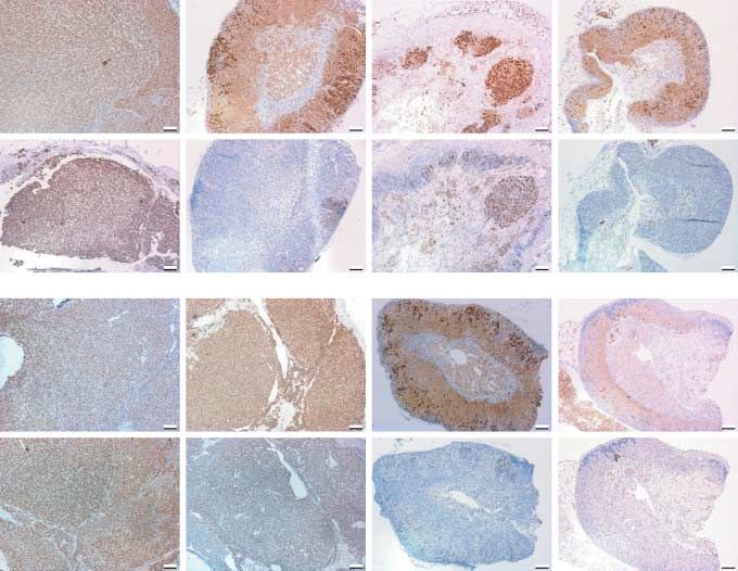
Figure 4 Histopathological analysis of the male (A) and female combined treatments of H-CGb-c and GnRH-a, the
(B) adrenal tumor after different treatments. (A) Hematoxylin– staining pattern was more scattered and very few
eosin staining of male adrenal tumors after treatments: control cells were stained. The same phenomenon could be
(1), Hecate only (2), Hecate-CGb conjugate (3), Hecate-CGb
conjugate and GnRH antagonist (4), GnRH antagonist only (5), seen in females in the case of GnRH-a treatments
and Hecate and GnRH antagonist (6). (B) Hemotoxylin–eosin (Fig. 5). The abundance of Ki-67- and p53-positive
staining of female adrenal tumors after treatments: control (1), cells after E2 treatment supports lower treatment
Hecate-CGb conjugate (2), GnRH antagonist (3), Hecate-CGb
conjugate and GnRH antagonist (4), E2 (5), and Hecate-CGb efficacy by E2 compared with GnRH-a in the females
conjugate and E (6). (data not shown).
Downloaded from Bioscientifica.com at 09/15/2021 10:40:17PM
via free access
www.endocrinology-journals.org 557S Vuorenoja et al.: Treatment strategies for adrenocortical tumors
Table 2 Results of morphometrical analysis in percentagesGS.E.M.
Healthy Tumor Fibrosis Cysts
Treatment (males)
Wild-type 100 0 0 0
Control 1.3G1.3a 62.6G5.8a 19G11.9a 17.1G5.8a
HecateCGnRH antagonist 50.4G5.5b 34.2G10.4ab 15.4G5.5a 0b
Hecate-CGb conjugateCGnRH antagonist 44.1G14b 11.5G5.3b 44.4G17.9a 0b
Hecate 0.5G0.5a 76.6G13.1ab 2.8G2.8a 20G9.7a
Hecate-CGb conjugate 91.3G4b 6.7G4.6b 2G1.4a 0b
GnRH antagonist 48.3G12.5b 34.7G2.9ab 17G11.6a 0b
Treatment (females)
Wild-type 100 0 0 0
Control 0.8G0.3a 57.7G35.6a 4.6G4.6a 36.9G31.2a
HecateCGnRH antagonist 24.4G12.1bc 36.6G23.1a 33.6G16.9a 5.5G5.5a
Hecate-CGb conjugateCGnRH antagonist 66G9.4b 29.4G9.9a 4.6G0.5a 0G0a
Hecate-CGb conjugate 4G2.6ac 65.2G16.9a 5.4G3.1a 25.4G11.2a
GnRH-antagonist 38.4G11.2bc 35.9G6.9a 25.7G14.9a 0a
Estradiol 31.3G7.4bc 55.3G6.5a 8.5G3.4a 4.8G4.4a
EstradiolCHecate-CGb conjugate 39.6G8.3bc 38.8G2.8a 21.6G5.9a 0a
Different letters next to the value indicate that the difference between them is statistically significant (P!0.05).
Reciprocal expression of GATA-4 and GATA-6 in abundant GATA-6 (Fig. 6A and C) but no GATA-4
healthy and adrenal tumor tissue expression (Fig. 6D and F), whereas an opposite
observation was made with the control adrenals
We finally analyzed the expression patterns of GATA-4 (Fig. 6B and E). GATA-4 could be detected in the
and GATA-6 proteins by IHC in the healthy (WT), nodulus formations of E2-treated female tumors, and
tumorous (control), and treated adrenal tissues. In those treated with GnRH-a alone in both sexes,
males, the WT and H-CGb-c-treated tumors had whereas GATA-6 expression was equal in all treatment
Figure 5 Immunohistochemistry for Ki-67 (upper panel) and p53 (lower panel) for both males and females. The order from the left:
control (c), Hecate-CGb conjugate (conj), GnRH antagonist (g), and Hecate-CGb conjugate and GnRH antagonist (conjCg).
Downloaded from Bioscientifica.com at 09/15/2021 10:40:17PM
via free access
558 www.endocrinology-journals.orgEndocrine-Related Cancer (2009) 16 549–564
the treatment efficacy of the TG female mice. This
improved treatment strategy could be helpful for
possible future applications in humans, as women are
more prone to develop adrenocortical tumors (Schulick
& Brennan 1999a,b). We tested two approaches in
order to improve the treatment efficacy by reducing the
circulating LH levels, i.e., by GnRH-a and E2
treatment, expecting that they would augment binding
of the H-CGb-c to Lhcgr following reduced compe-
tition by endogenous LH.
In clinics, GnRH-a are generally and successfully
used in IVF protocols, where a fast blockage of
endogenous gonadotropins are required (Detti et al.
2008, Lainas et al. 2008, Huhtaniemi et al. 2009). The
effectiveness of GnRH-a in cancer treatment, namely
in prostate cancer and mammary gland tumors, was
first established in animal models already 25 years ago
(Redding et al. 1982, Redding & Schally 1983, Schally
et al. 1983). The first studies with the cetrorelix (used
in this study) were performed in the 1990s (Srkalovic
et al. 1990, Szende et al. 1990, Korkut et al. 1991).
There are also reports showing the effectiveness of
Figure 6 Immunohistochemistry for GATA-6 (left panel) and GnRH-a in benign prostate hyperplasia treatment
GATA-4 (right panel). Wild-type (WT) (A and D), control
(B and E), and Hecate-CGb conjugate (C and F) in male (Lepor 2006) and they have been recently clinically
adrenal tumor sections. tested in the treatment of human prostate cancer
(Gittelman et al. 2008, Klotz et al. 2008, Huhtaniemi
groups with effective treatment response (data not et al. 2009). The GnRH agonist, leuprolide acetate, has
shown). These reciprocal expression patterns of been successfully used for treating gonadotropin-
GATA-4 in tumor and GATA-6 in healthy adrenal dependent Lhcgr-bearing adrenal adenoma/Cushing
tissues are in line with the mRNA data (Fig. 3) and also syndromes (Lacroix et al. 1999) and the findings of the
with an earlier publication (Kiiveri et al. 1999). present study support the idea of treating the
gonadotropin-dependent adrenal tumors in females by
GnRH-a.
We have shown earlier that prepubertally gonad-
Discussion ectomized inha/Tag TG mice develop large adrenal
H-CGb-c has been shown to provide a strong and tumors by the age of 6 months (Rahman et al. 2004).
tumor cell-specific antineoplastic effect towards the The tumorigenesis is apparently induced by combined
Lhcgr-bearing endocrine tumors (Hansel et al. 2001, action of the oncogene SV40/Tag and elevated LH
Gawronska et al. 2002, Leuschner et al. 2003, Bodek levels. In the present study, we found that GnRH-a in
et al. 2005b, Vuorenoja et al. 2008). In our previous both sexes could either alone or together with H-CGb-c
study (Vuorenoja et al. 2008), following a 1-month diminish the tumor weight almost by 95% compared
treatment with H-CGb-c adrenal tumor weight could with the control treatment. In TG males H-CGb-c itself
be reduced by an average of two-thirds in inha/Tag TG was as effective as GnRH-a. No significant additive
males compared with Hecate treatment, whereas in TG effect of GnRH-a to H-CGb-c or vice versa could be
females the reduction rate was only a non-significant, observed, even though the progesterone levels were
18%. There was no good explanation for this sex equally decreased in the treatment groups and the cell
difference in treatment effects, but as for unknown proliferation markers Ki-67 and p53 showed better
reason a higher Lhcgr expression was found in the male response after the combination treatment of GnRH-a
tumors, a plausible explanation could be higher Lhcgr and H-CGb-c than H-CGb-c alone. However, in
concentration in the tumor mass attracting larger histopathological evaluation and further quantitative
amounts of H-CGb-c, with a consequently more severe morphometric analysis, GnRH-a in males caused a
lytic effect (Vuorenoja et al. 2008). Owing to negative remarkable fibrotic and necrotic response in the
results in females, it was found important to improve tumorous adrenals, whereas after H-CGb-c treatment
Downloaded from Bioscientifica.com at 09/15/2021 10:40:17PM
via free access
www.endocrinology-journals.org 559S Vuorenoja et al.: Treatment strategies for adrenocortical tumors
the histology appeared more intact and more healthy also been shown to be dramatically downregulated
cells could be seen. The fibrotic/necrotic tissue that along with the adrenal tumor formation and pro-
filled the adrenal structure could possibly explain gression (Kiiveri et al. 2005). Here, we showed the
the decreased Ki-67 and p53 appearance of the novel phenomenon of the reappearance of GATA-6
combination treatment. expression after the tumor treatment with H-CGb-c in
In TG females, the tumor reduction by GnRH-a was males or with combinations with GnRH-a or E2. This
95% and by E2 90%, in comparison with control or observation on GATA-6 may become an additional
H-CGb-c treatment. H-CGb-c did not improve the prognostic marker for adrenocortical tumorigenesis
outcome as compared with GnRH-a, and the pro- and treatment response along with GATA-4 and
gesterone and mRNA levels for Lhcgr and GATA-4 Lhcgr.
after the treatment groups were equal. However, Taken together, we conclude that H-CGb-c is an
histopathologic and morphometric analyses in females efficient treatment in inha/Tag TG males due to its
showed more healthy tissue and less fibrosis after selective efficacy in killing tumor cells bearing Lhcgr
GnRH-a and H-CGb-c combination, and also the IHC with no significant destruction of the normal tissue
for the proliferation markers was less prominent after structure. GnRH-a combined to H-CGb-c caused
the combination therapy compared with GnRH-a alone severe damage to the histological structure of the
in females. As a whole, GnRH-a in females did not adrenals and GnRH-a treatment left still some Lhcgr-
cause destruction to adrenal structure as it did in males bearing cells. In TG females, however, only gonado-
– at the moment, there is no explanation to this. In E2 tropin suppression by GnRH-a or E2 was effective,
treatment TG female group, although the tumor weight since both treatments reduced the tumor size signi-
was significantly reduced, some tumor nodules could ficantly and did not affect severely the adrenal
still be found in the histopathological analysis and structure. H-CGb-c combined with GnRH-a did not
significantly more Lhcgr mRNA was expressed after
improve the tumor reduction. Our findings support the
E2 than GnRH-a. E2 probably worked through
idea of treating the gonadotropin-dependent adrenal
negative feedback by blocking the gonadotropin
tumors in females by GnRH-a. In fact, GnRH agonist
secretion, causing similar effect as occurred after
(leuprolide acetate) has already been successfully used
GnRH-a. The weaker response to E2 could also be
for treating gonadotropin-dependent Lhcgr-bearing
monitored by the hormonal status where GnRH-a
adrenal adenoma/Cushing syndromes (Lacroix et al.
blocked gonadotropin secretion but after E2 treatment
1999). E2 treatment was also shown to be rather
the gonadotropin blockage was less pronounced; E2
effective in TG females, although not as good as
was not either able to block FSH secretion. These
GnRH-a. However, the side effects (increased risk for
results altogether suggest that E2 treatment was not as
effective as GnRH-a, where the near-total ablation of thromboembolia, uterine cancer, and in some cases
LH severely suppressed the tumor progression in TG increased incidence of breast cancer) of prolonged E2
females. treatment are of concern, while considering it as
It is known that GATA-4 is expressed in fetal mouse treatment in human. Our present data showed a novel
and human adrenals but disappears soon after birth phenomenon against the dogma of SV40/Tag-induced
(Kiiveri et al. 2002). However, GATA-4 expression is tumorigenesis that even severe adrenal tumorigenesis
upregulated again upon adrenocortical tumor forma- co-induced by the oncogene SV40/Tag could be
tion, e.g. in inha/Tag TG mice, where it is visible reversible. Our data further showed that in inha/Tag
already 3 months after gonadectomy along with Lhcgr TG mice adrenal tumorigenesis, not only the tumor
expression and correlates with adrenocortical tumor- ontogeny, which have been shown earlier (Kananen
igenesis (Kiiveri et al. 1999, Rahman et al. 2004). We et al. 1997), but also the tumor progression is
now found that GATA-4 and Lhcgr were upregulated gonadotropin dependent, which could be affected by
in the tumor cells, but H-CGb-c in males, GnRH-a, E2 GnRH-a treatment. Finally, we hereby have observed
and their combinations downregulated their two different mechanisms of action underlying the
expression significantly. This result is in line with treatment in adrenocortical tumors. As shown before,
our former data, where we showed that H-CGb-c H-CGb-c caused tumor destruction by selective killing
specifically eradicated Lhcgr expressing tumor cells the tumor cells bearing Lhcgr (Hansel et al. 2001,
overexpressing GATA-4 (Vuorenoja et al. 2008). Gawronska et al. 2002, Leuschner et al. 2003, Bodek
GATA-6 expression has been shown in fetal and in et al. 2005b, Vuorenoja et al. 2008). GnRH-a inhibited
the adult adrenal with a specific role in regulating the tumor growth by blocking the gonadotropin-dependent
adrenal steroidogenesis (Kiiveri et al. 2005) and it has tumor progression through the systemic effects.
Downloaded from Bioscientifica.com at 09/15/2021 10:40:17PM
via free access
560 www.endocrinology-journals.orgEndocrine-Related Cancer (2009) 16 549–564
Declaration of interest Bodek G, Vierre S, Rivero-Muller A, Huhtaniemi I,
Ziecik AJ & Rahman NA 2005b A novel targeted
We declare that there is no conflict of interest that would
therapy of Leydig and granulosa cell tumors through
prejudice this manuscript’s impartiality.
the luteinizing hormone receptor using a hecate-
chorionic gonadotropin beta conjugate in transgenic
Funding mice. Neoplasia 7 497–508.
Bourdeau I, D’Amour P, Hamet P, Boutin JM & Lacroix A
This project was supported by the grants from Academy of 2001 Aberrant membrane hormone receptors in inciden-
Finland (I H, J T, N R), the Finnish Cultural Foundation at tally discovered bilateral macronodular adrenal hyper-
Varsinais-Suomi (S V), Ida Montin Foundation (S V), Turku plasia with subclinical Cushing’s syndrome. Journal of
University Foundations of Gerda and Ella Saarinen and Aili Clinical Endocrinology and Metabolism 86 5534–5540.
Salo (S V), Finnish Cancer Organizations (S V), the Finnish van Casteren JI, Schoonen WG & Kloosterboer HJ 2000
Medical Foundation (S V), Sigrid Juselius Foundation (J T, Development of time-resolved immunofluorometric
I H), and Turku University Hospital (J T). assays for rat follicle-stimulating hormone and luteinizing
hormone and application on sera of cycling rats. Biology
of Reproduction 62 886–894.
Acknowledgements Cattanach BM, Iddon CA, Charlton HM, Chiappa SA &
We thank Dr Harry Kujari for valuable advice with Fink G 1977 Gonadotrophin-releasing hormone
morphometrical analysis, Tero Vahlberg for the fruitful deficiency in a mutant mouse with hypogonadism.
discussions with statistical matters and Juho-Antti Mäkelä Nature 269 338–340.
with qRT-PCR analyses. The authors would also like to thank Chen HM, Chan SC, Lee JC, Chang CC, Murugan M &
Taija Leinonen and Taina Kirjonen for skillful technical Jack RW 2003 Transmission electron microscopic
assistance. Heli Niittymäki, Nina Messner, and the personnel observations of membrane effects of antibiotic cecropin
of Turku University Animal Facility are acknowledged for B on Escherichia coli. Microscopy Research and
their help with mice. Technique 62 423–430.
Detti L, Ambler DR, Yelian FD, Kruger ML, Diamond MP &
Puscheck EE 2008 Timing and duration of use of GnRH
References antagonist down-regulation for IVF/ICSI cycles have no
impact on oocyte quality or pregnancy outcomes. Journal of
Arrowood MJ, Jaynes JM & Healey MC 1991a Hemolytic
properties of lytic peptides active against the sporozoites Assisted Reproduction and Genetics 25 177–181.
of Cryptosporidium parvum. Journal of Protozoology 38 Fassnacht M, Hahner S, Polat B, Koschker AC, Kenn W,
161S–163S. Flentje M & Allolio B 2006 Efficacy of adjuvant
Arrowood MJ, Jaynes JM & Healey MC 1991b In vitro radiotherapy of the tumor bed on local recurrence of
activities of lytic peptides against the sporozoites of adrenocortical carcinoma. Journal of Clinical Endo-
Cryptosporidium parvum. Antimicrobial Agents and crinology and Metabolism 91 4501–4504.
Chemotherapy 35 224–227. Feelders RA, Lamberts SW, Hofland LJ, van Koetsveld PM,
Baghian A, Kousoulas K, Truax R & Storz J 1996 Specific Verhoef-Post M, Themmen AP, de Jong FH, Bonjer HJ,
antigens of Chlamydia pecorum and their homologues in Clark AJ, van der Lely AJ et al. 2003 Luteinizing
C. psittaci and C. trachomatis. American Journal of hormone (LH)-responsive Cushing’s syndrome: the
Veterinary Research 57 1720–1725. demonstration of LH receptor messenger ribonucleic acid
Barbosa AS, Giacaglia LR, Martin RM, Mendonca BB & in hyperplastic adrenal cells, which respond to chorionic
Lin CJ 2004 Assessment of the role of transcript for gonadotropin and serotonin agonists in vitro. Journal of
GATA-4 as a marker of unfavorable outcome in Clinical Endocrinology and Metabolism 88 230–237.
human adrenocortical neoplasms. BMC Endocrine Gawronska B, Leuschner C, Enright FM & Hansel W 2002
Disorders 4 3. Effects of a lytic peptide conjugated to beta HCG on
Bodek G, Rahman NA, Zaleska M, Soliymani R, Lankinen H, ovarian cancer: studies in vitro and in vivo. Gynecological
Hansel W, Huhtaniemi I & Ziecik AJ 2003 A novel Oncology 85 45–52.
approach of targeted ablation of mammary carcinoma Gittelman M, Pommerville PJ, Persson BE, Jensen JK &
cells through luteinizing hormone receptors using hecate- Olesen TK 2008 A 1-year, open label, randomized phase
CGb conjugate. Breast Cancer Research and Treatment II dose finding study of degarelix for the treatment of
79 1–10. prostate cancer in North America. Journal of Urology 180
Bodek G, Kowalczyk A, Waclawik A, Huhtaniemi I & 1986–1992.
Ziecik AJ 2005a Targeted ablation of prostate carcinoma Givens JR, Andersen RN, Wiser WL, Donelson AJ &
cells through LH receptor using Hecate-CGb conjugate: Coleman SA 1975 A testosterone-secreting, gonadotropin-
functional characteristic and molecular mechanism of responsive pure thecoma and polycystic ovarian disease.
cell death pathway. Experimental Biology and Medicine Journal of Clinical Endocrinology and Metabolism 41
230 421–428. 845–853.
Downloaded from Bioscientifica.com at 09/15/2021 10:40:17PM
via free access
www.endocrinology-journals.org 561S Vuorenoja et al.: Treatment strategies for adrenocortical tumors
Goodarzi MO, Dawson DW, Li X, Lei Z, Shintaku P, Rao CV Kananen K, Markkula M, El-Hefnawy T, Zhang FP, Paukku T,
& Van Herle AJ 2003 Virilization in bilateral macro- Su JG, Hsueh AJ & Huhtaniemi I 1996b The mouse
nodular adrenal hyperplasia controlled by luteinizing inhibin alpha-subunit promoter directs SV40 T-antigen to
hormone. Journal of Clinical Endocrinology and Leydig cells in transgenic mice. Molecular and Cellular
Metabolism 88 73–77. Endocrinology 119 135–146.
Haapasalo H, Collan Y, Montironi R, Pesonen E & Atkin NB Kananen K, Rilianawati , Paukku T, Markkula M, Rainio
1990 Consistency of quantitative methods in ovarian EM & Huhtanemi I 1997 Suppression of gonadotropins
tumor histopathology. International Journal of Gyneco- inhibits gonadal tumorigenesis in mice transgenic for
logical Pathology 9 208–216. the mouse inhibin alpha-subunit promoter/simian
Haavisto AM, Pettersson K, Bergendahl M, Perheentupa A, virus 40 T-antigen fusion gene. Endocrinology 138
Roser JF & Huhtaniemi I 1993 A supersensitive 3521–3531.
immunofluorometric assay for rat luteinizing hormone. Kero J, Poutanen M, Zhang FP, Rahman N, McNicol AM,
Endocrinology 132 1687–1691. Nilson JH, Keri RA & Huhtaniemi IT 2000 Elevated
Hanahan D 1989 Transgenic mice as probes into complex luteinizing hormone induces expression of its receptor
systems. Science 246 1265–1275. and promotes steroidogenesis in the adrenal cortex.
Hansel W, Leuschner C, Gawronska B & Enright F Journal of Clinical Investigation 105 633–641.
2001 Targeted destruction of prostate cancer cells Kiiveri S, Siltanen S, Rahman N, Bielinska M, Lehto VP,
and xenografts by lytic peptide-b LH conjugates. Huhtaniemi IT, Muglia LJ, Wilson DB & Heikinheimo M
Reproductive Biology 1 20–32. 1999 Reciprocal changes in the expression of transcrip-
Hansel W, Leuschner C & Enright F 2007a Conjugates of tion factors GATA-4 and GATA-6 accompany adreno-
lytic peptides and LHRH or bCG target and cause cortical tumorigenesis in mice and humans. Molecular
necrosis of prostate cancers and metastases. Molecular Medicine 5 490–501.
and Cellular Endocrinology 269 26–33. Kiiveri S, Liu J, Westerholm-Ormio M, Narita N, Wilson DB,
Hansel W, Enright F & Leuschner C 2007b Destruction of Voutilainen R & Heikinheimo M 2002 Differential
breast cancers and their metastases by lytic peptide expression of GATA-4 and GATA-6 in fetal and adult
conjugates in vitro and in vivo. Molecular and Cellular mouse and human adrenal tissue. Endocrinology 143
Endocrinology 260–262 183–189. 3136–3143.
Henk WG, Todd WJ, Enright FM & Mitchell PS 1995 Kiiveri S, Liu J, Arola J, Heikkila P, Kuulasmaa T, Lehtonen E,
The morphological effects of two antimicrobial peptides, Voutilainen R & Heikinheimo M 2005 Transcription factors
hecate-1 and melittin, on Escherichia coli. Scanning GATA-6, SF-1, and cell proliferation in human adrenocor-
Microscopy 9 501–507. tical tumors. Molecular and Cellular Endocrinology 233
Hogan B, Beddington R, Constantini F & Lacy E 1994 47–56.
Manupulating the Mouse Embryo. A Laboratory Klotz L, Boccon-Gibod L, Shore ND, Andreou C, Persson BE,
Manual. Cold Spring Harbor, NY: Cold Spring Harbor Cantor P, Jensen JK, Olesen TK & Schroder FH 2008 The
Laboratory. efficacy and safety of degarelix: a 12-month, comparative,
Howard CV & Reed MG 1998 Unbiased Stereology: Three- randomized, open-label, parallel-group phase III study
Dimensional Measurement in Microscopy. Oxford: BIOS in patients with prostate cancer. BJU International 102
Scientific Publishers Limited. 1531–1538.
Huhtaniemi I, White R, McArdle CA & Persson BE 2009 Korkut E, Bokser L, Comaru-Schally AM, Groot K &
Will GnRH antagonists improve prostate cancer Schally AV 1991 Inhibition of growth of experimental
treatment? Trends in Endocrinology and Metabolism 20 prostate cancer with sustained delivery systems
43–50. (microcapsules and microgranules) of the luteinizing
Kananen K, Markkula M, Rainio E, Su JG, Hsueh AJ & hormone-releasing hormone antagonist SB-75. PNAS 88
Huhtaniemi IT 1995 Gonadal tumorigenesis in 844–848.
transgenic mice bearing the mouse inhibin alpha-subunit Kuruba R & Gallagher SF 2008 Current management of
promoter/simian virus T-antigen fusion gene: adrenal tumors. Current Opinion in Oncology 20 34–46.
characterization of ovarian tumors and establishment of Lacroix A, Hamet P & Boutin JM 1999 Leuprolide acetate
gonadotropin- responsive granulosa cell lines. Molecular therapy in luteinizing hormone-dependent Cushing’s
Endocrinology 9 616–627. syndrome. New England Journal of Medicine 341
Kananen K, Markkula M, Mikola M, Rainio EM, McNeilly A 1577–1581.
& Huhtaniemi I 1996a Gonadectomy permits adrenocor- Lainas TG, Sfontouris IA, Papanikolaou EG, Zorzovilis JZ,
tical tumorigenesis in mice transgenic for the mouse Petsas GK, Lainas GT & Kolibianakis EM 2008
inhibin alpha-subunit promoter/simian virus 40 T-antigen Flexible GnRH antagonist versus flare-up GnRH agonist
fusion gene: evidence for negative autoregulation of the protocol in poor responders treated by IVF: a
inhibin alpha-subunit gene. Molecular Endocrinology 10 randomized controlled trial. Human Reproduction 23
1667–1677. 1355–1358.
Downloaded from Bioscientifica.com at 09/15/2021 10:40:17PM
via free access
562 www.endocrinology-journals.orgEndocrine-Related Cancer (2009) 16 549–564
de Lange WE, Pratt JJ & Doorenbos H 1980 A gonadotrophin hormone on preovulatory maturation and ovulation of
responsive testosterone producing adrenocortical ade- mouse graafian follicles. Molecular Endocrinology 19
noma and high gonadotrophin levels in an elderly woman. 2591–2602.
Clinical Endocrinology 12 21–28. Rahman NA, Kananen Rilianawati K, Paukku T, Mikola M,
Larson BA, Vanderlaan WP, Judd HL & McCullough DL Markkula M, Hamalainen T & Huhtaniemi IT 1998
1976 A testosterone-producing adrenal cortical adenoma Transgenic mouse models for gonadal tumorigenesis.
in an elderly woman. Journal of Clinical Endocrinology Molecular and Cellular Endocrinology 145 167–174.
and Metabolism 42 882–887. Rahman NA, Kiiveri S, Siltanen S, Levallet J, Kero J, Lensu T,
Leinonen P, Ranta T, Siegberg R, Pelkonen R, Heikkila P & Wilson DB, Heikinheimo MT & Huhtaniemi IT 2001
Kahri A 1991 Testosterone-secreting virilizing adrenal Adrenocortical tumorigenesis in transgenic mice: the role
adenoma with human chorionic gonadotrophin receptors of luteinizing hormone receptor and transcription factors
and 21-hydroxylase deficiency. Clinical Endocrinology GATA-4 and GATA-61. Reproductive Biology 1 5–9.
34 31–35. Rahman NA, Kiiveri S, Rivero-Muller A, Levallet J, Vierre S,
Lepor H 2006 The role of gonadotropin-releasing hormone Kero J, Wilson DB, Heikinheimo M & Huhtaniemi I 2004
antagonists for the treatment of benign prostatic hyper- Adrenocortical tumorigenesis in transgenic mice expres-
plasia. Reviews in Urology 8 183–189. sing the inhibin alpha-subunit promoter/simian virus
Leuschner C & Hansel W 2004 Membrane disrupting lytic 40 T-antigen transgene: relationship between ectopic
peptides for cancer treatments. Current Pharmaceutical expression of luteinizing hormone receptor and trans-
Design 10 2299–2310. cription factor GATA-4. Molecular Endocrinology 18
Leuschner C & Hansel W 2005 Targeting breast and prostate 2553–2569.
cancers through their hormone receptors. Biology of Redding TW & Schally AV 1983 Inhibition of mammary
Reproduction 73 860–865. tumor growth in rats and mice by administration of
Leuschner C, Enright FM, Melrose PA & Hansel W agonistic and antagonistic analogs of luteinizing
2001 Targeted destruction of androgen-sensitive and hormone-releasing hormone. PNAS 80 1459–1462.
insensitive prostate cancer cells and xenografts
Redding TW, Coy DH & Schally AV 1982 Prostate
through luteinizing hormone receptors. Prostate 46
carcinoma tumor size in rats decreases after adminis-
116–125.
tration of antagonists of luteinizing hormone-releasing
Leuschner C, Enright FM, Gawronska B & Hansel W 2003
hormone. PNAS 79 1273–1276.
Membrane disrupting lytic peptide conjugates destroy
Rilianawati, Paukku T, Kero J, Zhang FP, Rahman N,
hormone dependent and independent breast cancer cells
Kananen K & Huhtaniemi I 1998 Direct luteinizing
in vitro and in vivo. Breast Cancer Research and
hormone action triggers adrenocortical tumorigenesis in
Treatment 78 17–27.
castrated mice transgenic for the murine inhibin alpha-
Mijnhout GS, Danner SA, van de Goot FR & van Dam EW
subunit promoter/simian virus 40 T-antigen fusion gene.
2004 Macronodular adrenocortical hyperplasia in a
Molecular Endocrinology 12 801–809.
postmenopausal woman. Netherlands Journal of
Rilianawati, Kero J, Paukku T & Huhtaniemi I 2000 Long-
Medicine 62 454–455.
term testosterone treatment prevents gonadal and adrenal
Mikola M, Kero J, Nilson JH, Keri RA, Poutanen M &
Huhtaniemi I 2003 High levels of luteinizing hormone tumorigenesis of mice transgenic for the mouse inhibin-
analog stimulate gonadal and adrenal tumorigenesis in alpha subunit promoter/simian virus 40 T-antigen fusion
mice transgenic for the mouse inhibin-alpha-subunit gene. Journal of Endocrinology 166 77–85.
promoter/Simian virus 40 T-antigen fusion gene. Saner-Amigh K, Mayhew BA, Mantero F, Schiavi F, White PC,
Oncogene 22 3269–3278. Rao CV & Rainey WE 2006 Elevated expression
Miyamura N, Taguchi T, Murata Y, Taketa K, Iwashita S, of luteinizing hormone receptor in aldosterone-
Matsumoto K, Nishikawa T, Toyonaga T, Sakakida M & producing adenomas. Journal of Clinical Endocrinology
Araki E 2002 Inherited adrenocorticotropin-independent and Metabolism 91 1136–1142.
macronodular adrenal hyperplasia with abnormal cortisol Schally AV, Redding TW & Comaru-Schally AM 1983
secretion by vasopressin and catecholamines: detection of Inhibition of prostate tumors by agonistic and antagonistic
the aberrant hormone receptors on adrenal gland. analogs of LH-RH. Prostate 4 545–552.
Endocrine 19 319–326. Schulick RD & Brennan MF 1999a Adrenocortical carci-
Pabon JE, Li X, Lei ZM, Sanfilippo JS, Yussman MA & noma. World Journal of Urology 17 26–34.
Rao CV 1996 Novel presence of luteinizing Schulick RD & Brennan MF 1999b Long-term survival after
hormone/chorionic gonadotropin receptors in human complete resection and repeat resection in patients with
adrenal glands. Journal of Clinical Endocrinology adrenocortical carcinoma. Annals of Surgical Oncology 6
and Metabolism 81 2397–2400. 719–726.
Pakarainen T, Zhang FP, Nurmi L, Poutanen M & Sheeler LR 1994 Cushing’s syndrome and pregnancy.
Huhtaniemi I 2005 Knockout of luteinizing hormone Endocrinology and Metabolism Clinics of North America
receptor abolishes the effects of follicle-stimulating 23 619–627.
Downloaded from Bioscientifica.com at 09/15/2021 10:40:17PM
via free access
www.endocrinology-journals.org 563You can also read