Green nanotechnology synthesized silver nanoparticles: Characterization and testing its antibacterial activity
←
→
Page content transcription
If your browser does not render page correctly, please read the page content below
Green Processing and Synthesis 2021; 10: 518–528
Research Article
Najla AlMasoud*, Hajar Alhaik, Malak Almutairi, Asmaa Houjak, Khlood Hazazi,
Fatema Alhayek, Sarah Aljanoubi, Ahad Alkhaibari, Asma Alghamdi, Dina A. Soliman,
Taghrid S. Alomar*, and Manal A. Awad
Green nanotechnology synthesized silver
nanoparticles: Characterization and testing its
antibacterial activity
https://doi.org/10.1515/gps-2021-0048 Furthermore, AgNPs and aqueous leaf extracts of F. carica
received April 11, 2021; accepted July 14, 2021 and rosemary were examined for their antimicrobial activ-
Abstract: The green plant-mediated synthesis of silver ities against different Gram-positive and Gram-negative
(GPS-Ag) nanoparticles (NPs) has been increasingly pop- bacteria strains. The results indicated that the AgNPs,
ular due to its eco-friendliness, availability, cost-effec- derived from F. carica, have more antibacterial activities
tiveness, and the fact that it can be safely handled and than others and inhibited bacterial growth.
possesses a broad variability of metabolites, such as anti- Keywords: green synthesis, AgNPs, Ficus carica, Salvia
oxidant and antimicrobial activities. In this current study, rosmarinus, antimicrobial activities
the synthesis of AgNPs has been demonstrated using aqu-
eous extracts of fresh leaves of Ficus carica and Salvia
rosmarinus (rosemary) that reduced aqueous silver nitrate.
This procedure made the synthesis of NPs possible, which 1 Introduction
was characterized by numerous analytical techniques such
as ultraviolet-visible (UV-Vis) spectrophotometry, Fourier Nanotechnology refers to the science, engineering, and
transform infrared spectroscopy, transmission electron technology carried out at the nanoscale from 1–100 nm. The
microscopy (TEM), energy-dispersive X-ray spectroscopy, use of this advanced technology extends to a number of
and dynamic light scattering studies. The visual observa- fields such as chemistry, materials science, and other similar
tion indicated that the colour of aqueous silver nitrate disciplines. Moreover, nanotechnology has been used in var-
turned brownish yellow after treatment with the fresh ious applications such as energy, catalysis, food science,
leaf extracts and was confirmed by UV-Vis spectra. In biomedicine, wastewater treatment, and electronics [1–3].
addition, the TEM analysis showed that the synthesized Green synthesis methods (green chemistry) are known
NPs well dispersed with average sizes less than 22 nm. to be safer in comparison to chemical methods. This is
because the former is known to be more sustainable and
offer an environmentally safer approach during the synth-
* Corresponding author: Najla AlMasoud, Department of Chemistry,
esis of nanoparticles (NPs) [4]. Some of the novel green
College of Science, Princess Nourah Bint Abdulrahman University, chemistry methods such as burgeoning green nanotech-
Riyadh 11451, Saudi Arabia, e-mail: nsalamsoud@pnu.edu.sa nology have proven to be important in newer nanoparticle
* Corresponding author: Taghrid S. Alomar, Department of synthesis techniques. These alternative methods utilize
Chemistry, College of Science, Princess Nourah Bint Abdulrahman microorganisms and plant-based extracts and have shown
University, Riyadh 11451, Saudi Arabia,
to be more effective in the synthesis of NPs [5,6].
e-mail: tsalomar@pnu.edu.sa
Hajar Alhaik, Malak Almutairi, Asmaa Houjak, Khlood Hazazi, Several research studies carried out using silver nano-
Fatema Alhayek, Sarah Aljanoubi, Ahad Alkhaibari, Asma Alghamdi: particles (AgNPs) reported improved properties, including
Department of Chemistry, College of Science, Princess Nourah Bint good electrical and thermal properties, chemical stability
Abdulrahman University, Riyadh 11451, Saudi Arabia as well as catalytic and antimicrobial properties [7–10].
Dina A. Soliman: Department of Botany and Microbiology, Faculty of
Besides, the physical, mechanical, and structural proper-
Science, King Saud University, Riyadh 11459, Saudi Arabia
Manal A. Awad: King Abdullah Institute for Nanotechnology,
ties of AgNPs with various sizes and shapes can be modi-
King Saud University, Riyadh 11451, Saudi Arabia, fied by the addition of biological agents such as bacteria,
e-mail: mawad@ksu.edu.sa fungi, and algae along with their enzymes. Researchers
Open Access. © 2021 Najla AlMasoud et al., published by De Gruyter. This work is licensed under the Creative Commons Attribution 4.0
International License.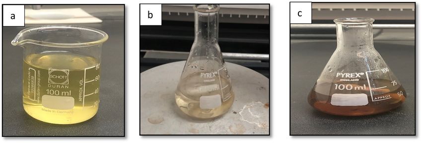
Green nanotechnology synthesized silver nanoparticles 519
used an isolated bacterium to synthesize AgNPs (30–60 nm) electron microscopy, energy-dispersive X-ray spectroscopy
biologically [11–14], with improved catalytic activity and a (EDX), ultraviolet-visible absorption spectroscopy (UV-Vis),
stable hydrazine oxidation reaction. Another study carried and transmission electron microscopy (TEM). The antibac-
out by Abdel-Raouf involved a rapid biogenic process using terial potentials of the green synthesized AgNPs have been
Laurencia catarinensis, a marine red alga [15,16]. More evaluated against various strains of bacteria.
recent studies have employed several plant-based extracts
for the synthesis of AgNPs, including extracts of blackberry
fruit [17], Sacha inchi shell biomass and leaf [18,19], natural 2 Materials and methods
rubber latex, aloe vera [20], rambutan peel [21], clove [22],
coffee, green tea [23], and leaves of Coccina grandis [24].
Some of the more recent uses of natural extracts for nano- 2.1 Preparation of NPs using Ficus carica and
particle synthesis include rosemary extracts; the use of this Salvia rosmarinus leaves extracts
plant and others resulted in the approval of the use of
R. officinalis by the European Union as an efficient natural The leaves of Ficus carica and Salvia rosmarinus (rosemary)
food preservative [25]. R. officinalis has proven to be bene- were collected from well-grown trees in the farms in Saudi
ficial for a number of medical conditions, for example useful Arabia, Riyadh. The collected leaves were washed thor-
as an anti-inflammatory and hepatoprotective [26]. Rosemary oughly with tap water, until no residual impurities were
was used to acquire aqueous extracts as reductants for the left; then they were washed with deionized water and air-
synthesis of AgNPs [27] and the MgO nanoflowers (MgONFs) dried. About 10 g from each dried Ficus carica and rosemary
[28]. The successful green synthesis of AgNPs through the leaves were macerated separately in 100 mL of boiling dis-
dried fruit extract of Ficus carica was described previously tilled water. The mixtures were kept overnight, and the
[29] and it was shown that the synthesis was reasonably non- extracts were filtered using filter papers.
toxic, and thus, it can be employed as a capable anticancer Roughly 5 mL from the aqueous extracts were mixed
agent. Ficus carica is known to be used as a healing agent in with 50 mL of AgNO3 (1.0 mM) separately; both mixtures
many medical applications as a cardiovascular, respiratory, were then placed on an electric heater and magnetic
and as an anti-inflammatory agent [30]. stirrer at 60°C until brownish silver colloid was observed,
The chemical profile of Salvia rosmarinus was carried indicating the formation of AgNPs (Figures 1 and 2),
out by Leporini et al. [31]. The chemical composition of which were stable for several weeks in glass conical
the essential oil from Salvia rosmarinus was studied by flasks covered with foil at room temperature.
modern analytical techniques such as gas chromato- The precise mechanism involved in the reduction and
graphy (GC) and gas chromatography-mass spectrometry stability of silver ions has not been clearly understood
(GC-MS). 1,8-Cineole, α-pinene, camphor, and trans-caryo- until now because biomolecules vary from plant to plant
phyllene were the most present compounds. In addition, [34]. As a result, more research is needed into the synth-
phytochemical studies on F. carica revealed the presence esis of AgNPs with plant extracts in order to identify the
of numerous bioactive compounds such as phenolic com- right biomolecules that serve as the capping and stabi-
pounds, phytosterols, organic acids, anthocyanin, triter- lizing agents. Nonetheless, the trapping of AgNPs ions on
penoids, coumarins, and volatile compounds such as the protein surface due to electrostatic interactions between
hydrocarbons, aliphatic alcohols, and a few other classes silver ions and proteins in plant material extract has been
of secondary metabolites from different parts of F. carica. proposed as a probable mechanism implicated in the bio-
Most species of F. carica contain phenolic compounds, reduction of Ag. Proteins decrease AgNPs ions, causing
organic acids, and volatile compounds. Phenolic acids them to change the secondary structure and generate silver
such as 3-O- and 5-O-caffeoylquinic acids, ferulic acid, nuclei. Silver nuclei are formed by further reducing Ag ions
quercetin-3-O-glucoside, quercetin-3-O-rutinoside, psor- and their build-up at the nucleus, resulting in the produc-
alen, bergapten, and organic acids (oxalic, citric, malic, tion of AgNPs [35].
quinic, shikimic, and fumaric acids) have been isolated
from the water extract of the leaves of F. carica L. [32,33].
This study summarizes and investigates the use of 2.2 Characterization of the formation of
Ficus carica and rosemary-based extracts to reduce Ag+ nanoparticles
ions to synthesize AgNPs. The synthesized AgNPs were
analyzed to confirm and study their characteristics using A variety of analytical techniques were used to examine the
Fourier transform infrared spectroscopy (FTIR), scanning formation of NPs. UV 2450 Spectrophotometer (Shimadzu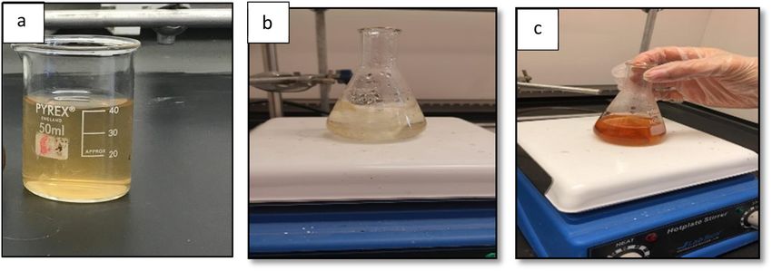
520 Najla AlMasoud et al.
Figure 1: Visual observation of (a) the Ficus carica extract, (b) after 5 min of mixing the Ficus carica extract with silver nitrate solution, and
(c) the synthesis of AgNPs (after 15 min the colour changed to brown).
Corporation, Kyoto, Japan) was employed to carry out ATCC 43300, and Bacillus cereus ATCC 11778 (clinical iso-
UV-Vis spectral analysis in a range of 100 to 800 nm. late), which were obtained from King Khalid University
Furthermore, the particle size distribution of biosynthe- Hospital, Riyadh, Saudi Arabia. In this work, the antimicro-
sized AgNPs was obtained using the dynamic light scat- bial activity of the produced AgNPs was tested using a well-
tering (DLS) method using a Zetasizer (Nano series, HT cut diffusion method. About 14 g of nutrient agar was dis-
Laser, ZEN3600 Malvern Instruments, Malvern, UK). The solved in distilled water (500 mL) and autoclaved at 121°C
synthesized AgNPs and functional groups of the extracts and 15 psi for 45 min. The solid medium was punched with a
were also studied using FTIR. The aim of the study was to sterile cork borer to make a well (4 wells in each plate). The
analyse and evaluate the interaction between the extracts leaf extract was used as a control, and three different con-
and AgNPs in the wavenumber range of 400–4000 cm−1. centrations of AgNPs were used (2, 4, and 6 μL), followed by
Moreover, the TEM (Zeiss, Germany, EM 10C-200 kV) was a gradual addition until the hole was filled and incubated at
utilized to observe the shape, size, and morphology of 37°C for 24 h. Then, the zone was measured after the incuba-
the synthesized NPs. Elemental analysis was performed tion and expressed in millimeters of the diameter.
using EDX coupled with a JEM-2100F transmission elec-
tron microscope; this was carried out to confirm the pre-
sence of silver in the suspension.
3 Results and discussion
2.3 Antimicrobial activity 3.1 UV analysis
Three different types of bacteria were used in this study, Initial stages of the reduction illustrated a change in the
including Escherichia coli ATCC35218, Staphylococcus aureus colour of the mixture from almost colourless to brown
Figure 2: Visual observation of (a) the rosemary extract, (b) after 5 min of mixing the rosemary extract with silver, and (c) the synthesis of
AgNPs (15 min after mixing) the colour changed to brown.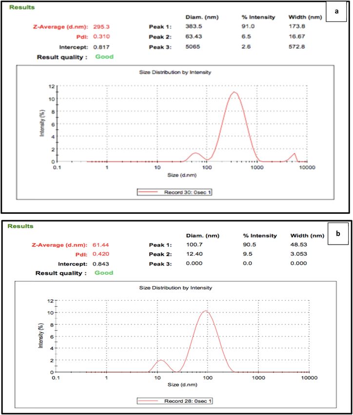
Green nanotechnology synthesized silver nanoparticles 521
when the silver nitrate solution was mixed with the aqu- were found to be 295 and 0.31 nm, and 61.44 and 0.42 nm,
eous extract of the leaf, which indicated the formation respectively. The measured z-average and PdI of the
of AgNPs due to the surface plasmon resonance (SPR) of synthesized NPs illustrate that these NPs, are monodis-
AgNPs [36,37], which might be the primary signature of perse, verifying the data presented by the producer and
the nanoparticle. In this work, different analytical methods UV-Vis spectroscopy [42].
were used to study the characterization of AgNPs. One of
the main methods to detect and evaluate the formation of
NPs in an aqueous solution is UV-Vis spectroscopy. The
confirmation and stability of the synthesized AgNPs was 3.3 Fourier infrared spectroscopy analysis
by UV-Vis spectroscopy since the plasmon band of Ag is
sensitive to the size and shape of the formed NPs [38]. The FTIR techniques were performed to demonstrate the func-
silver ions are reduced to silver atoms due to the compo- tional collections of the green synthesized NPs [43] and the
nents in the plant extract [39]. Figure 3 shows the UV-Vis leaves extracts of Ficus carica and rosemary. Figures 5 and 6
spectra of the silver nanoparticle formation for both illustrate the FTIR spectra of the synthesized NPs by using
F. carica and rosemary extracts and were measured from an aqueous extract of Ficus carica leaves and the Ficus
200 to 800 nm. Generally, broad peaks at higher wave- carica leaf extract, respectively.
lengths indicate particle size growth, whereas a narrow FTIR was employed to investigate the synthesis of
peak at a shorter wavelength confirms the formation of AgNPs using rosemary leaves and to detect the possible
smaller-sized AgNPs. Figure 2a and b shows sharp and biomolecules accountable for the reduction of Ag+ ions
intense surface plasmon resonance (SPR) bands at 450 nm, and capping the bio-reduced AgNPs synthesized by the
corresponding to the characteristic SPR of synthesized AgNPs plant extract. The absorption bands in the FTIR spectrum
using both plants extracts and this result is in agreement (Figures 5 and 6) indicate the presence of active func-
with the previously reported results that AgNPs having wave- tional groups in the synthesized AgNPs. To obtain a
length (λmax) values in the range between 400 and 500 nm good signal/noise ratio, the FTIR transmission spectra
[40] indicate the formation of good quality AgNPs [41]. were recorded in the region 400–4,000 cm−1.
FTIR analysis of the synthesized AgNPs (Figure 5a)
shows major peaks at 1,057, 1,386, 1,620, and 3,417 cm−1.
The peak at 1,075 cm−1 is attributed to primary alcohols,
3.2 DLS analysis 1,386 cm−1 arises due to NO3 and may be due to C–N
stretching vibrations of aliphatic and aromatic amines.
The size distribution and the average size of the synthe- In addition, the peak at 1,620 cm−1 is due to the nitro
sized AgNPs were carried out using the DLS technique as compounds, whereas the peak at 3,417 cm−1 is attributed
shown in Figure 4. From Figure 4, it can be noted that the to OH stretching in alcohol and phenolic compounds. The
average size and polydispersity index (PdI) of the synthe- FTIR spectrum shows the presence of biomolecules of
sized AgNPs using F. carica and rosemary leaves extracts Ficus carica leaves in the solution of AgNPs (Figure 5b).
Figure 3: UV-Vis absorption spectra of AgNPs synthesized by an aqueous (a) F. carica and (b) rosemary leaves extracts.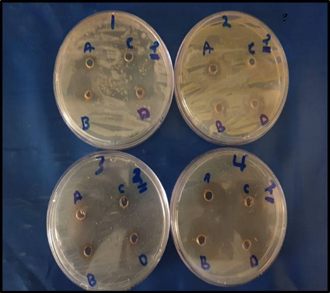
522 Najla AlMasoud et al.
Figure 4: DLS analysis of the synthesized AgNPs using (a) Ficus carica and (b) rosemary extracts.
On the other hand, in the spectrum of rosemary aqueous 3.4 TEM and EDX analysis
extract (Figure 6b), peaks at 1,610, 1,384, and 1,057 cm−1
have been attributed to enzymes, amides, and proteins, The shape and size of the synthesized AgNPs extracted
which appear to be responsible for the decrease of metal from F. carica and rosemary fresh leaves were carried out
ions when using vegetable materials for the synthesis of using TEM analysis (Figure 7a and b). From Figure 6, it
metal NPs [38]. Some IR bands common to the rosemary can be noted that the shape of the NPs is spherical with a
aqueous extract appeared in the synthesized AgNP sample few agglomerations. The obtained results are in line with
(Figure 6a), but the transmittance level of the plant extract previous methods (UV and DLS). The UV results display a
bands was weakened after interaction with AgNPs and wide SPR band due to the adsorption of compounds in
shifted to 3,431 cm−1 (O–H stretching), 2,366 cm−1 (alkyls the extract of leaves on the surface of NPs. In addition,
C–H stretching), 1,630 cm−1 (assigned to amide I, arising the DLS results illustrate the monodispersity index of the
due to carbonyl stretch in proteins) and 1,058 cm−1 corre- synthesized AgNPs using Ficus carica and rosemary; this
sponding to C–O, C–N stretching vibrations of the aliphatic finding shows that the synthesized particles vary in size
amines or alcohols/phenols, representing the presence of and display no agglomeration [46,47]. Furthermore, the
polyphenols in the rosemary extract [44,45]. EDX spectrometry was carried out and provided both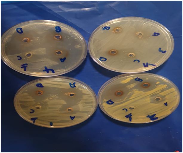
Green nanotechnology synthesized silver nanoparticles 523
a
2340.68 609.39
2367.93
2930.82 1057.33
1620.99
1386.17
%T 3417.52
b 2370.48
618.43
2929.68 1114.06
1400.55
1266.83
1602.56
4400.0 4000 3000 2000 1500 1000 500 400.0
cm-1
Figure 5: FTIR spectra of (a) the synthesized NPs by using an aqueous extract of Ficus carica leaves and (b) the Ficus carica leaf extract.
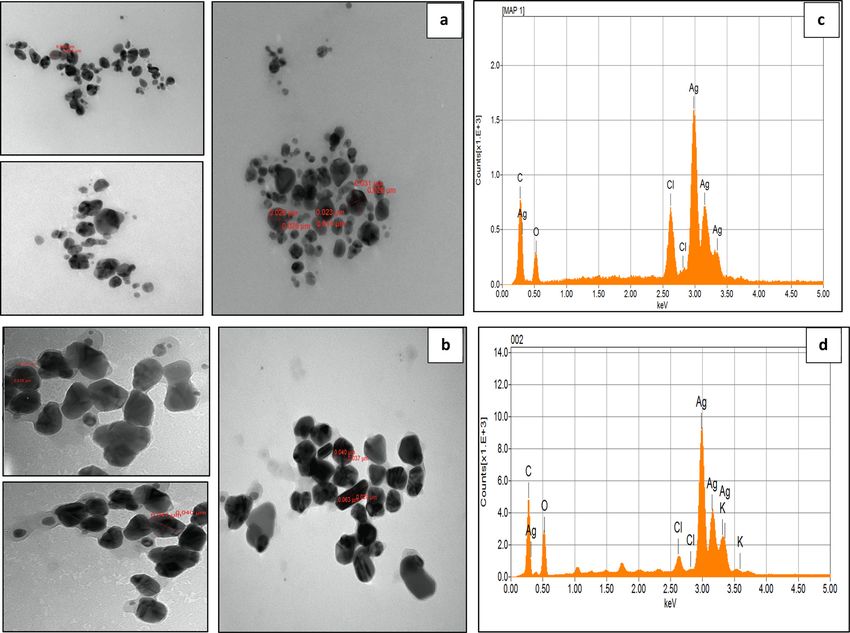
quantitative and qualitative details about the elements that 3 keV because of the presence of Ag (Figure 7c and d) respec-
were used in the formation of the NPs. Metallic silver nano- tively. On the other hand, the signals of oxygen, carbon,
crystals generally display peaks around 3 keV because of potassium, and chlorine atoms were detected in both synthe-
their surface plasmon resonance [48], and the synthesized sized AgNPs. These elements could have acted as capping
NPs using F. carica and rosemary displayed greater counts at organic agents attached to the surface of AgNPs [49].
A
a
3778.31
2366.98 488.28
776.52
2924.46
1630.19
1384.35 1058.78
%T 3431.51
b 863.62
830.33
3752.02
619.04
2930.03
1610.05 1384.66 1057.15
4400.0 4000 3000 2000 1500 1000 500 400.0
cm-1
Figure 6: FTIR spectra of (a) the synthesized NPs by using an aqueous extract of rosemary leaves and (b) the rosemary leaf extract.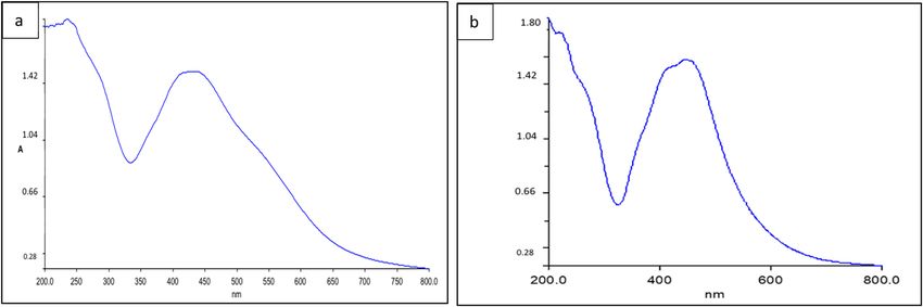
524 Najla AlMasoud et al.
3.5 Antimicrobial efficacy analysis (Figure 8a and b). Similarly, three concentrations (A – 2 μL,
B – 4 μL, and D – 6 μL) of the synthesized AgNPs using
The antimicrobial effects of the synthesized NPs and the the rosemary extract also exhibited the potent antibac-
extracts of Ficus carica and rosemary were studied against terial activity against E. coli, B. cereus, and S. aureus with
four different types of pathogenic bacteria including Gram- zones of inhibition of 10, 16, and 22 mm, 12, 22, and 30 mm,
positive Staphylococcus aureus (ATCC 43300), Bacillus cereus and 35, 40, and 48 mm, respectively (Figure 9a and b).
(ATCC 11778), and Gram-negative bacteria Escherichia coli The Ficus carica extract displays activity against
(ATCC35218). E. coli and S. aureus with zones of inhibition of 10 and
As presented in Figures 7 and 8, the synthesized 12 mm and for B. cereus there was no zone of inhibition;
AgNPs from the Ficus carica extract show greater anti- while for rosemary extract, it shows activity against
bacterial activity than the synthesized AgNPs using rosemary E. coli, B. cereus, and S. aureus with the diameter of
extract, and poor activity of both extracts against the tested zones of inhibition being 6, 3, and 15 mm as shown in
bacterial strains. These results corroborate those obtained by Figures 8 and 9.
Acay et al. [50] and Logaranjan et al. [51]. AgNPs from extracts exhibit remarkable antibacterial
Significant activity was seen at three concentrations activities and could be clarified by the fact that very
(A – 2 μL, B – 4 μL, and D – 6 μL) of the synthesized AgNPs explicit phytoactive compounds covered the AgNPs from
using the Ficus carica extract against E. coli, B. cereus, the plant extract. These phytochemicals that might per-
and S. aureus with zones of inhibition of 10, 16, and 22 mm, form as reducing and/or capping agents would be favour-
12, 22, and 30 mm, and 35, 40 and 48 mm, respectively ably involved in the reduction of AgNPs ions into silver
Figure 7: TEM micrographs and EDX spectrum analysis of AgNPs synthesized using F. carica (a and c) and rosemary (b and d) extracts.Green nanotechnology synthesized silver nanoparticles 525
a 60 a 60
Zone of inhibition (mm)
Zone of inhibition (mm) 50 50
40
40
30
30 20
20 10
0
10
E. co
oli B. cereus S. aurous
0 2 uL 4 uL 6 uL
L Contrrol (6 uL)
oli
E. co B. ccereus S.. aurous
2 uL 4 uL
u 6 uL Contro
ol (6 uL) b E. coli B. cereus
E. Coli B. ceereus
b
E. faecalis S. aurous
Figure 9: (a) The graphical of the values of inhibition zones (mm);
(b) the antibacterial activity of produced AgNPs at A (2 μL), B (4 μL),
E. faecaalis S. au
urous
D (6 μL), and C (rosemary extract as a control, 6 μL) concentrations
against E. coli, B. cereus, and S. aureus.
Figure 8: (a) The graphical of the values of inhibition zones (mm);
(b) the antibacterial activity of produced AgNPs at A (2 μL), B (4 μL),
D (6 μL), and C (Ficus carica extract as a control, 6 μL) concentra-
tions against E. coli, B. cereus, and S. aureus.
4 Conclusion
Biological silver nanoparticle synthesis (AgNPs), which is
an environmentally friendly method, could be carried out
metal and the phytochemical-assisted synthesis of AgNPs in a variety of applications involving medical treatments.
from the plant extract for nucleation, stabilization, and The current study established the non-hazardous mate-
capping [52]. rials, eco-friendly, and facile production of AgNPs from
Consequently, the synthesized AgNPs improved the plant extracts. The colour shift of the aqueous silver nitrate
antimicrobial activity owing to the particle size and the from colourless to brown with the addition of plant extracts
aggregation of NPs [50]. In addition, previous studies provided the first confirmation of the synthesis of AgNPs.
show that the antibacterial activity depends on the dose Other analytical methods were employed to study the char-
of the synthesized AgNPs. In addition, previous studies acterization of NPs, including UV-Vis spectroscopy, FTIR,
show that the antibacterial activity depends on the dose TEM, DLS, and EDX analysis. These confirmed the suc-
of the synthesized AgNPs [53–55]. From Figures 7 and 8, cessful synthesis of spherical AgNPs and verified the role
it can be noted that the high concentration of AgNPs of extracts’ phytochemicals as reducing, stabilizing, and
causes more antibacterial activity and bacterial cell death, capping agents in the green synthesis of AgNPs.
as a result of the interaction between nanoparticles and Furthermore, the biological evaluation of AgNPs obtained
the protein of bacteria [56,57]. through green synthesis exposed good bactericidal properties
Overall, the interaction between the synthesized NPs against E. coli, B. cereus, and S. aureus, all of which are patho-
and bacteria can be explained as follows: gens generally involved in infectious skin diseases. Thus, the
(1) the interface among positive charges on the bacteria wide variety of green synthesized AgNPs as bioactive com-
surface and negative charges in NPs, pounds makes them an ideal agent for controlling infectious
(2) the expulsion of intracellular material that instigates agents and useful in other pharmaceutical areas.
cell death due to physicochemical modifications in
the bacterial cell wall, resulting in the NPs stopping Funding information: This research was funded by the
DNA replication and respiration of bacteria [58,59]. Deanship of Scientific Research at Princess Nourah bint526 Najla AlMasoud et al.
Abdulrahman University through the Fast-Track Research [9] Srivastava S, Usmani Z, Atanasov AG, Singh VK, Singh NP,
Funding Program. Abdel-Azeem AM, et al. Biological nanofactories using living
forms for metal nanoparticle synthesis. Mini-Rev Med Chem.
2021;21(2):245–65.
Author contributions: Najla AlMasoud, Taghrid S. Alomar,
[10] Roya S, Das TK, Maiti GP, Basu U. Microbial biosynthesis of
and Manal A. Awad: writing – original draft, writing – nontoxic gold nanoparticles. Mater Sci Eng B Adv.
review and editing, conception, and design of the study. 2016;203:41–51.
Hajar Alhaik, Malak Almutairi, Asmaa Houjak, Asmaa [11] Şimşek B, Sevgili İ, Ceran ÖB, Korucu H. Tools and techniques
Houjak, Khlood Hazazi, Fatema Alhayek, Sarah Aljanoubi, for purification of water using nano materials. in nanotech-
nology characterization tools for environment, health, and
Ahad Alkhaibari, and Asma Alghamdi: writing – original
safety. Berlin, Heidelberg: Springer; 2019. p. 285–322.
draft, methodology, analysis, and interpretation of data; [12] Dosoky WM, Fouda MM, Alwan AB, Abdelsalam NR, Taha AE,
Dina A. Soliman: writing – original draft, and methodology. Ghareeb RY, et al. Dietary supplementation of silver-silica
nanoparticles promotes histological, immunological, ultra-
Conflict of interest: The authors state no conflict of structural, and performance parameters of broiler chickens.
Sci Rep UK. 2021;11(1):1–15.
interest.
[13] Rostami H, Khosravi F, Mohseni M, Rostami AA. Biosynthesis
of Ag nanoparticles using isolated bacteria from contaminated
sites and its application as an efficient catalyst for hydrazine
electrooxidation. Int J Biol Macromol. 2018;107:343–8.
[14] Abdel-Raouf N, Alharbi RM, Al-Enazi NM, Alkhulaifi MM,
References Ibraheem IBM. Rapid biosynthesis of silver nanoparticles
using the marine red alga Laurencia catarinensis and their
[1] Hossain MI, Soliman MM, El-Naggar ME, Sultan MZ, Kechi A, characterization. Beni-Suef Univ J Basic Appl Sci.
Abdelsalam NR, et al. Synthesis and characterization of gra- 2018;7:150–7.
phene oxide-ammonium ferric sulfate composite for the [15] Bhattacharya D, Gupta RK. Nanotechnology and potential of
removal of dyes from tannery wastewater. J Mater Res Technol. microorganisms. Crit Rev Biotechnolo. 2005;25:199–204.
2021;12:1715–27. [16] Mohanpuria P, Rana NK, Yadav SK. Biosynthesis of nanopar-
[2] El-Naggar ME, Abdelsalam NR, Fouda MM, Mackled MI, ticles: technological concepts and future applications.
Al-Jaddadi MA, Ali HM, et al. Soil application of nano silica on J Nanopart Res. 2008;10:507–17.
maize yield and its insecticidal activity against some stored [17] Kumar B, Smita K, Cumbal L, Debut A. Green synthesis of silver
insects after the post-harvest. Nanomaterials. nanoparticles using Andean blackberry fruit extract. Saudi
2020;10(4):739. J Biol Sci. 2017;24:45–50.
[3] Al Saqr A, Khafagy ES, Alalaiwe A, Aldawsari MF, [18] Kumar B, Smita K, Cumbal L, Debut A. Synthesis of silver
Alshahrani SM, Anwer MK, et al. Synthesis of gold nanoparti- nanoparticles using Sacha inchi (Plukenetia volubilis L.) leaf
cles by using green machinery. characterization and in vitro extracts. Saudi J Biol Sci. 2014;21:605–9.
toxicity. Nanomaterials (Basel). 2021;11(3):808. [19] Sandhya J, Kalaiselvam S. Biogenic synthesis of magnetic iron
[4] Rónavári A, Igaz N, Adamecz DI, Szerencsés B, Molnar C, oxide nanoparticles using inedible borassus flabellifer seed
Kónya Z, et al. Green silver and gold nanoparticles. Biological coat: characterization, antimicrobial, antioxidant activity and
synthesis approaches and potentials for biomedical applica- in vitro cytotoxicity analysis. Mater Res Express.
tions. Molecules. 2021;26(4):844. 2020;7(1):015045.
[5] Mosa WF, Ali HM, Abdelsalam NR. The utilization of tryptophan [20] Rónavári A, Igaz N, Adamecz DI, Szerencsés B, Molnar C,
and glycine amino acids as safe alternatives to chemical Kónya Z, et al. Green silver and gold nanoparticles. Biological
fertilizers in apple orchards. Environ Sci Pollut R. synthesis approaches and potentials for biomedical applica-
2021;28(2):1983–91. tions. Molecules. 2021;26(4):844.
[6] Ghareeb RY, Alfy H, Fahmy AA, Ali HM, Abdelsalam NR. [21] Kumar B, Smita K, Cumbal L, Angulo Y. Fabrication of silver
Utilization of Cladophora glomerata extract nanoparticles as nanoplates using Nephelium lappaceum (Rambutan) peel:a
eco-nematicide and enhancing the defense responses of sustainable approach. J Mol Liq. 2015;211:476–80.
tomato plants infected by Meloidogyne javanica. Sci Rep UK. [22] Vijayaraghavan K, Kamala SP, Udaya PN, Madhankumar D.
2020;10(1):1–15. Biomimetic synthesis of silver nanoparticles by aqueous
[7] Anand K, Kaviyarasu K, Muniyasamy S, Roopan SM, extract of Syzygium aromaticum. Mater Lett. 2012;75:33–5.
Gengan RM, Chuturgoon AA. Bio-synthesis of silver nano- [23] Nadagouda MN, Varma RS. Green synthesis of silver and pal-
particles using agroforestry residue and their catalytic degra- ladium nanoparticles at room temperature using coffee and
dation for sustainable waste management. J Clust Sci. tea extract. Green Chem. 2008;10:859–62.
2017;28(4):2279–91. [24] Arunachalam R, Dhanasingh S, Kalimuthu B, Uthirappan M,
[8] Deepika S, Selvaraj CI, Roopan SM. Screening bioactivities of Rose C, Mandal AB. Phytosynthesis of silver nanoparticles
Caesalpinia pulcherrima L. swartz and cytotoxicity of extract using Coccinia grandis leaf extract and its application in
synthesized silver nanoparticles on HCT116 cell line. Mater Sci the photocatalytic degradation. Colloid Surface B.
Eng. 2020;106:110279. 2012;94:226–30.Green nanotechnology synthesized silver nanoparticles 527
[25] Aguilar F, Autrup H, Barlow S, Castle L, Crebelli R, Dekant W, [39] Pantidos N, Horsfall LE. Biological synthesis of metallic
et al. Use of rosemary extracts as a food additive scientific nanoparticles by bacteria, fungi and plants. J Nanomed
opinion of the panel on food additives, flavourings, processing Nanotechno. 2014;5:5.
aids and materials in contact with food. EFSA Journal. [40] Sastry M, Mayya K, Bandyopadhyay K. pH dependent changes
2008;6:721. in the optical properties of carboxylic acid derivatized silver
[26] Gonçalves GA, Corrêa RC, Barros L, Dias MI, Calhelha RC, colloidal particles. Colloid Surf A Physicochem Eng Asp.
Correa VG, et al. Effects of in vitro gastrointestinal digestion 1997;127:221–8.
and colonic fermentation on a rosemary (Rosmarinus offici- [41] Nazeruddin G, Prasad N, Waghmare S, Garadkar K, Mulla I.
nalis L) extract rich in rosmarinic acid. Food chem. Extracellular biosynthesis of silver nanoparticle using
2019;271:393–400. Azadirachta indica leaf extract and its anti-microbial activity.
[27] González‐Rivera J, Duce C, Ierardi V, Longo I, Spepi A, Tine MR, J Alloy Compd. 2014;583:272–7.
et al. Fast and eco–friendly microwave‐assisted synthesis of [42] Xu J, Han X, Liu H, Hu Y. Synthesis and optical properties of
silver nanoparticles using rosemary essential oil as renewable silver nanoparticles stabilized by gemini surfactant. Colloid
reducing agent. Chem Select. 2017;2:2131–8. Surf A. 2006;273:179–83.
[28] Abdallah Y, Ogunyemi SO, Abdelazez A, Zhang M, Hong X, [43] Najla AL, Taghrid SAL, Manal A, Maha F, Din A. Multifunctional
Ibrahim E, et al. The green synthesis of MgO nano-flowers green silver nanoparticles in pharmaceutical and biomedical
using Rosmarinuso cinalis L. (Rosemary) and the antibacterial applications. Green Chem Lett Rev. 2020;13:316–27.
activities against xanthomonas oryzaepv. oryzae. Bio Med Res [44] Podila R, Chen R, Ke PC, Brown JM, Rao AM. Effects of surface
Int. 2019;56:209–89. functional groups on the formation of nanoparticle-protein
[29] Justin PS, SivaPrasad VL, Sivasankar S, Muralidharan P. corona. Appl phys Lett. 2012;101:263701–4.
Biosynthesis of silver nanoparticles using dried fruit extract of [45] He Y, Du Z, Lv H, Jia Q, Tang Z, Zheng X, et al. Green synthesis
Ficus carica – screening for its anticancer activity and toxicity of silver nanoparticles by Chrysanthemum morifolium Ramat.
in animal models. Food Chem Toxicol. 2017;109:951–6. extract and their application in clinical ultrasound gel. Int J
[30] Fouda MM, Abdelsalam NR, Gohar IMA, Hanfy AE, Othman SI, Nanomed. 2013;8:1809–15.
Zaitoun AF, et al. Utilization of High throughput microcrystal- [46] Mubayi A, Chatterji S, Rai PM, Watal G. Evidence based green
line cellulose decorated silver nanoparticles as an eco-nema- synthesis of nanoparticles. Adv Mat Lett. 2012;3:519–25.
ticide on root-knot nematodes. Colloid Surf B. [47] Krishnaraj C, Jagan E, Rajasekar S, Selvakumar P,
2020;188:110805. Kalaichelvan P, Mohan N. Synthesis of silver nanoparticles
[31] Leporini M, Bonesi M, Loizzo MR, Passalacqua NG, Tundis R. using Acalypha indica leaf extracts and its antibacterial
The essential oil of Salvia rosmarinus Spenn. from Italy as a activity against water borne pathogens. Colloid Surf B.
source of health-promoting compounds: chemical profile and 2010;76:50–6.
antioxidant and cholinesterase inhibitory activity. Plants [48] Banerjee P, Satapathy M, Mukhopahayay A, Das P. Leaf extract
(Basel). 2020;9:798–6. doi: 10.3390/plants9060798. mediated green synthesis of silver nanoparticles from widely
[32] Oliveira AP, Valentão P, Pereira JA, Silva BM, Tavares F, available Indian plants: synthesis, characterization, antimicrobial
Andrade PB. Ficus carica L: metabolic and biological property and toxicity analysis. Bioresour Bioproc. 2014;1:1–10.
screening. Food Chem Toxicol. 2009;47(11):2841–6. [49] Mallikarjunaa K, Sushmab JN, Narasimhac G. Phytochemical
[33] Gibernau M, Buser HR, Frey JE, Hossaert-McKey M. Volatile fabrication and characterization of silver nanoparticles by
compounds from extracts of figs of Ficus carica’. using pepper leaf broth. Arab J Chem. 2014;7:1099–103.
Phytochemistry. 1997;46(2):241–4. [50] Acay H. Biosynthesis and characterization of silver nanopar-
[34] Das RK, Satinder KB. ‘Plant mediated green synthesis: modi- ticles using fig (ficus carica) leaves: a potential antimicrobial
fied approaches’. Nanoscale. 2013;5(21):10155–62. activity. Appl Ecol Env Res. 2019;17:13793–802.
[35] Zhao X, Wang K, Ai C, Yan L, Jiang C, Shi J. Improvement of [51] Logaranjan K, Devi S, Pandian K. Biogenic synthesis of
antifungal and antibacterial activities of food packages using silver nanoparticles using fruit extract of ficus carica and
silver nanoparticles synthesized by iturin A. Food Packag Shelf study its antimicrobial activity. Nano Biomed Eng.
Life. 2021;28:100669. 2012;4:177–82.
[36] Aziz SB, Hussein G, Brza MA, Mohammed JS, Abdulwahid TR, [52] Awad MA, Alkhulaifi MM, Aldosari NS, Alzahly S, Aldalbahi A.
Raza Saeed S, et al. Fabrication of interconnected plasmonic Novel eco-synthesis of PD silver nanoparticles: characteriza-
spherical silver nanoparticles with enhanced localized surface tion, assessment of its antimicrobial and cytotoxicity proper-
plasmon resonance (LSPR) peaks using quince leaf extract ties. Materials. 2019;12:3890.
solution. Nanomaterials. 2019;9:15–57. [53] Devipriya D, Roopan SM. Cissus quadrangularis mediated
[37] Fouda MM, Abdelsalam NR, El-Naggar ME, Zaitoun AF, ecofriendly synthesis of copper oxide nanoparticles and its
Salim BM, Bin-Jumah M, et al. Impact of high throughput green antifungal studies against Aspergillus niger, Aspergillus
synthesized silver nanoparticles on agronomic traits of onion. flavus. Mater Sci Eng. 2017;80:38–44.
Int J Biol Macromol. 2020;149:1304–17. [54] Kumar DA, Palanichamy V, Roopan SM. Photocatalytic action
[38] Asimuddin M, Mohammed RS, Syed FA, Mohammed R, of AgCl nanoparticles and its antibacterial activity. J Photoch
Siddiqui H, Abdulrahman AL, et al. Azadirachta indica based Photo B. 2014;138:302–6.
biosynthesis of silver nanoparticles and evaluation of their [55] Roopan SM, Madhumitha G, Rahuman AA, Kamaraj C,
antibacterial and cytotoxic effects. Journal of King Saud Bharathi A, Surendra TV. Low-cost and eco-friendly phyto-
University – Science. 2020;32:648–56. synthesis of silver nanoparticles using Cocos nucifera coir528 Najla AlMasoud et al.
extract and its larvicidal activity. Ind Crop Prod. [58] Espoir K, Kambale A, Christian I, Nkanga AB, Blaise-Pascal I,
2013;43:631–5. Mutonkole A, et al. Memvanga green synthesis of antimicro-
[56] Magudapatty P, Gangopadhyayrans P, Panigrahi BK, Nair KG, bial silver nanoparticles using aqueous leaf extracts from
Dhara S. Electrical transport studies of Ag nano- three Congolese plant species (Brillantaisia patula,
clusters embedded in glass matrix. Physica B. Crossopteryx febrifuga and Senna siamea). Heliyon.
2001;299:142–6. 2020;6:e04493.
[57] Femi-Adepoju AG, Dada AO, Otun KO, Adepoju AO, Fatoba OP. [59] Feng QL, Wu J, Chen GQ, Cui FZ, Kim TN, Kim JO. A mechanistic
Green synthesis of silver nanoparticles using terrestrial fern study of the antibacterial effect of silver ions on Escherichia
(Gleichenia Pectinata (Willd.) C. Presl) characterization and coli and taphylococcus aureus. J Biomed Mater Res.
antimicrobial studies. Heliyon. 2019;5:01543. 2000;52:662–8.You can also read