Gastrointestinal foreign bodies in Dogs and Cats: (2018-2020) 32 Cases
←
→
Page content transcription
If your browser does not render page correctly, please read the page content below
DOI: https://doi.org/10.52973/rcfcv-e32097                                    Revista Cientifica, FCV-LUZ / Vol. XXXII, rcfcv-e32097, 1 - 8

                     Gastrointestinal foreign bodies in Dogs and Cats:
                                   (2018–2020) 32 Cases
             Cuerpos extraños gastrointestinales en perros y gatos: (2018-2020) 32 casos

           Cafer Tayer İşler¹*      , Ömer Kırgız¹ , Mehmet Zeki Yılmaz Deveci¹ , İbrahim Alakuş¹             , Halil Alakuş¹   ,
                                               Ziya Yurtal¹ and Muhammed Enes Altuğ¹

                    1
                        Department of Veterinary Surgery, University of Hatay Mustafa Kemal University. Hatay, Turkey.
                                                       Email: cafer.isler@gmail.com

                            ABSTRACT                                                                 RESUMEN

Gastrointestinal foreign bodies (GFB) in cats and dogs are                   Los cuerpos extraños gastrointestinales (CEG) en perros
among the life-threatening surgical diseases that require invasive        y gatos se encuentran entre las enfermedades quirúrgicas
surgery. This study aimed to evaluate the cases of GFB in 32              potencialmente mortales que requieren cirugía invasiva. Este
cats and dogs diagnosed and treated in Hatay Mustafa Kemal                estudio tuvo como objetivo evaluar los casos de CEG en 32 gatos
University Veterinary Health Practice and Research Hospital.              y perros diagnosticados y tratados en el Hospital de Investigación
Information regarding the type, breed, age, sex, clinical symptoms,       y Práctica de Salud Veterinaria de la Universidad Hatay Mustafa
characteristics of foreign bodies (FB) localization, prognosis,           Kemal. Se recogió información sobre el tipo, raza, edad, sexo,
type of treatment administered, and conditions determined in              clínica, características de los cuerpos extraños (CE), localización
postoperative controls was collected. The cases were aged                 del CE, pronóstico, tipo de tratamiento administrado y condiciones
between 1 and 7 years and adult animals were also included.               determinadas en los controles postoperatorios. Los casos tenían
The rate of FB incident was the same in male and female cats,             edades comprendidas entre 1 y 7 años y también se incluyeron
whereas male dogs had a higher rate of FB incident. Strings/              animales adultos. La tasa de incidentes con CE fue la misma en
ropes and metallic objects were the most common foreign objects           gatos machos y hembras, mientras que los perros machos tuvieron
found in animals. Early diagnosis and treatment were important in         una mayor tasa de incidentes con CE. Las cuerdas / mecates y los
preventing complications in the cases, FB was usually found in the        objetos metálicos eran los objetos extraños más comunes que se
stomach, and vomiting was the most common clinical symptom.               encontraban en los animales. El diagnóstico y tratamiento precoces
Further, surgical intervention (gastrostomy/enterotomy) was the           fueron importantes para prevenir complicaciones en estos casos,
most common treatment method with a rate of 53.13 % for treating          habitualmente se encontraban CE en el estómago y el vómito era
FB in the gastrointestinal system that yielded successful results.        el síntoma clínico más común. Además, la intervención quirúrgica
Thus, to avoid complications and for a good prognosis, owners             (gastrostomía / enterotomía) fue el método de tratamiento más
must be conscious and seek for diagnosis and treatment as soon            común con una tasa del 53,13 % para el tratamiento de CE en el
as they notice the incidence of swallowing a FB, so as to ensure          sistema gastrointestinal que arrojó resultados exitosos. Así, para
early diagnosis and treatment.                                            evitar complicaciones y para un buen pronóstico, los propietarios
                                                                          deben estar conscientes y buscar el diagnóstico y tratamiento tan
Key words: Cat; dog; enterotomy; foreign bodies; gastrostomy
                                                                          pronto como se percaten de la incidencia de la ingestión de un CE,
                                                                          a fin de asegurar un diagnóstico y tratamiento precoces.
                                                                          Palabras clave: Gato; perro; enterotomía; cuerpos extraños;
                                                                                          gastrostomía

Received: 06/10/2021          Acepted: 28/10/2021       Published: 09/02/2022
1 of 8
Gastrointestinal foreign bodies in Dogs and Cats / İşler et al._____________________________________________________________

INTRODUCTION                                                             patient was clinically examined. The presence of FB was detected in
                                                                         24 (75 %) the animals based on the clinical findings. In accordance
   The presence of foreign bodies (FB) in the gastrointestinal tract
                                                                         with the obtained information and clinical findings, X-ray imaging
often poses life-threatening risks in cats (Felis catus) and dogs
                                                                         (Intermedical, Basic 100-30®, Italy) was performed at a dose range
(Canis Familiaris). These FB swallowed by cats and dogs lead
                                                                         of 75-100 kilowatts (kv) 5-10 miliamper second (mAs) to determine
to obstruction or perforation in the digestive system resulting in
                                                                         the topographic anatomy of the FB. FB were visualized by direct
emergency surgical treatment [12]. Gastrointestinal foreign bodies
                                                                         radiography in 20 (62.5 %) animals and indirectly in 12 (37.5 %) the
(GFB) may cause complete or partial obstruction in the digestive
                                                                         animals. For the indirect radiographic procedure, barium sulphate
system. Complete obstruction leads to very severe clinical symptoms
                                                                         (R-X suspension 100 %, Yenişehir Laboratuarı Tic. Ve San. Ltd. Şti,
and a faster deterioration of general condition, whereas partial
                                                                         Turkey) at 50 % concentration was administered to the animals with
obstruction results in chronic digestive problems and malabsorption
                                                                         suspected or known FB and no FB opacity on direct radiography,
[7]. Further, obstructions due to FB in the gastrointestinal system
                                                                         and images were captured at 5, 15, 30, 45, 60, 75, 90, and 120
may lead to gastric dilatation or volvulus [10]. Stones, plastic
                                                                         minutes (min). Overall, 12 (37.5 %) animals were examined by
objects, blunt objects such as erasers, and sharp foreign objects
                                                                         indirect radiography, 9 (28.13 %) by direct radiography, 6 (18.75 %)
such as strings, needles, and wires have been generally reported
                                                                         by both endoscopic examination and direct radiography, and 4
in cats and dogs [1, 2, 4, 9]. The clinical signs generally include
                                                                         (12.5 %) by both physical imaging and direct radiographic imaging.
recurrent vomiting, diarrhea, sudden loss of appetite, abdominal
pain, abdominal swelling, dehydration, and depression [2]. An
                                                                         Treatment and surgical technique
appropriate anamnesis along with clinical and radiographic
examinations is required for the diagnosis of FB. Radiopaque GFB            Surgical intervention was performed in 17 (53.13 %) of 32 animals
can be easily visualized on direct radiographs. Non-radiopaque FB        with FB in the gastrointestinal system, whereas the FB was removed
can be detected by their shape and gas accumulation; however,            with the help of endoscopic imaging (Karl Storz, Model 69045020,
this is not always possible. Positive contrast agents such as barium     Germany) in seven (21.88 %). Further, the FB was removed with
sulfate or iodine are used to detect obstruction or perforation caused   medical treatment in six (18.75 %) and no procedure was performed
by radiolucent FB [11]. FB are localized in the digestive system,        in two (6.25 %) because the treatment was not accepted. All the
esophagus, stomach, and small and large intestines. The most             animals who underwent surgery were administered 30 miligram
common place of obstruction is the jejunum, and the most effective       (mg) / kilogram (kg) dose of cefazolin sodium antibiotics (cefazolin,
treatment is enterotomy [10]. Conservative or surgical intervention      MN Pharmaceutical Co., Turkey) 30 min before the surgery. Fluid
can be used as the treatment method, depending on the localization       therapy was administered to the animals with vomiting and diarrhea
of the FB and the problem it creates [7].                                an hour (h) before the surgery.
  This study aimed to present the localization, nature, diagnosis,         For the surgery, animals were anesthetized by administering 1
and treatment of FB detected in the gastrointestinal systems of          mg·kg-1 2 % xylazine HCl (alfazyne 2 %; Egevet, Turkey), 10 mg·kg-1
cats and dogs of different breeds and sexes brought to Hatay             10 % ketamine (alfamine 10 %; Egevet, Turkey). Subsequently,
Mustafa Kemal University Veterinary Health Practice and Research         inhalation anesthesia (TMS, Model Future FX, Turkey) was started
Hospital between 2018 and 2020.                                          with isoflurane (1 - 3 % inhalation, isoflurane-USP, Piramal Critical
                                                                         Care Inc, USA).
MATERIALS AND METHODS                                                       All the animals were placed on their back, and the abdominal
   The clinical examination, diagnosis, treatment, and prognosis         cavity was accessed through the ventral midline of the abdomen.
of 32 cats and dogs with gastrointestinal obstruction who were           A careful examination of the abdominal cavity was made before
brought to Hatay Mustafa Kemal University Veterinary Health              the organs of the digestive system were intervened. Thereafter, the
Practice and Research Hospital between January 1, 2018 and               topographic anatomy of the FB was determined, the area where
October 30, 2020 were evaluated. The animals were assessed               the object was located was taken out of the operation wound, and
according to routine recording, anamnesis details, examination,          the incision location and shape were determined. Based on the
and treatment protocol. The animals with FB detected in the entire       determined topographic anatomy, enterotomy was performed in 6
gastrointestinal system, i.e., from the mouth to the anus, were          of 13 dogs, and gastrostomy was performed in of them 7. At the
included in the study. Information regarding the type, breed, age,       site, where the object was located, an incision was made from the
sex, clinical symptoms of the animals, the duration of clinical          intact and non-ischemic part of the organ to access the cavity of the
symptoms, material of the FB, FB localization, treatment type,           organ from the back of the object, from the opposite direction to the
and postoperative processes of the animals were followed up and          movement direction of the object in the cranial section to the organ.
recorded. The duration of clinical symptoms was defined as the             The object was moved back and removed from the incision
time elapsed from the time of occurrence of ingestion or the onset       area. In gastrostomy cases, the incision area was closed with an
of clinical symptoms in case no swallowing was observed. The             appositional pattern using double-layer sutures with schmieden
animals in whom the FB were removed from their bodies before             and cushing. In enterotomy cases, the area was closed with an
the diagnosis were excluded from the study.                              appositional pattern using a single layer of suture. Absorbable
                                                                         polydioxanone (Polidiox, Boz Medical Supplies Sanayi ve Ticaret
Diagnosis                                                                A.Ş., Turkey) suture of surgical quality was used for all applied
  Initially, careful and detailed anamnesis was obtained from the        sutures. Prior to the complete closure of the abdomen in each
owners of the animals. Overall, 8 of 32 (25 %) animals were brought      patient, the abdomen was washed several times with saline, and
by the owner with the information that it had swallowed a FB. Each       intraperitoneal penicillin G potassium (Penicillin G Potassium

2 of 8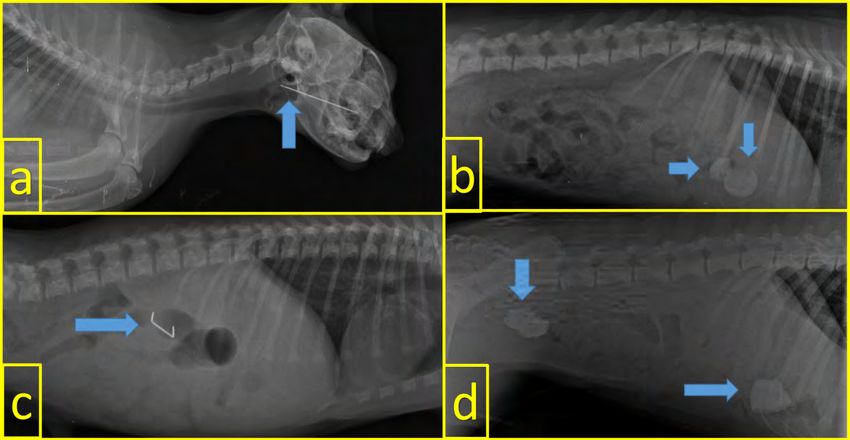
____________________________________________________________Revista Cientifica, FCV-LUZ / Vol. XXXII, rcfcv-e32097, 1 - 8

1,000,000 IU, I.E. ULAGAY İlaç Sanayii TÜRK A.Ş, Turkey) was                                     TABLE I
administered.                                                               Summary of categorical information of foreing body
                                                                                            ingestions cases
Postoperative care and treatment
                                                                                 Information            Cases           Percentage
  No food was provided to the animals for at least 24 h as per the
surgeon’s discretion. Oral intake could be administered in liquid                    Male                 19                59.38
form after the first 24 h. Intravenous fluids were continued until
                                                                                    Female                13                40.63
oral intake was sufficient. Antibiotics and analgesics were normally
continued for at least 3 postoperative days (d). Only one patient                    Acute                23                71.88
was re-operated as a result of swallowing socks again on d 3 after
the surgery. Skin stitches were removed from all the animals within                 Chronic                9                28.13
7–10 d after the surgery                                                            Age < 1               10                31.25
                                                                                   Age < 1-7              17                53.13
RESULTS AND DISCUSSION
  A total of 20 dogs and 12 cats from 16 different breeds were                      Age > 7                5                15.63
included in the study (TABLE I). Only one dog was recorded as a new                   Dog                 20                62.5
case due to swallowing socks again 3 d after the surgery. The clinical
symptoms included vomiting (40.62 %), diarrhea (18.75 %), salivation                  Cat                 12                37.5
(18.75 %), open mouth (18.75 %), anorexia (15.63 %), inability to
defecate (12.5 %), abdominal pain (12.5 %), cough (3.13 %), and
bloody diarrhea (3.13 %). Further, eight (25 %) cases observed by        intestine) in another. Based on the control radiographs following
their owners to swallow the FB had no clinical symptoms when they        the treatment, it was observed that the object was not inside the
were brought to the hospital (TABLE II). Detailed information on the     animals. In all three cases, the FB was removed using forceps
cases with FB is presented in TABLES I and II                            (Kruuse, 45 Degree Curved Forceps 220 mm, Germany) under
   In terms of the distribution of FB by sex, male dogs had a higher     the guidance of imaging with endoscopy. Of 13 dogs, 6 underwent
rate, whereas an equal distribution was observed between sexes           enterotomy and 7 underwent gastrostomy. As one dog swallowed
in cats. Based on the sex distribution of the animals, 12 (60 %) and     a FB 3 d after the surgery, it was evaluated as a new case.
8 (40 %) were male and female dogs, respectively, and 6 (50 %)           Further, three of 13 dogs who underwent surgical intervention had
and 6 (50 %) were female and male cats, respectively. In terms of        complications associated with wound infection due to disruption in
breed, no FB were found in the native breeds, whereas the breeds         post-operative dressing and treatment (TABLE II).
such as American and British ones were more prone to FB incidents.          In terms of time elapsed from the emergence of clinical
  The owners of two of 20 dogs with GFB refused the proposed             symptoms or the observation of swallowing, 27 and 4 animals were
operative intervention after examination and diagnosis. Moreover,        in the acute and subacute phases, respectively. The owners of 8
medical therapy was administered in two animals. Medical treatment       (25 %) animals reported that the complaint was direct FB ingestion
(LAKS ENEMA Iyon Saglik Medikal Ltd. Sti, Turkey) was rectally           and 19 (59 %) were not aware that the problem was associated
administered via enema, and 2 % xylazine HCl (alfazyne 2 %;              with the ingestion of a FB; however, treatment within the first 7 d of
Egevet, Turkey) was administered for vomiting; 10 min after the          the disease revealed that the owners were conscious, which had a
administration, the FB was naturally removed from the body               positive effect on the prognosis of the disease (FIG. 1).
(stomach) by vomiting in one patient and from feces (large

FIGURE 1. Time elapsed (d) from the emergence of clinical symptoms or the observation of swallowing vs the number of cases

                                                                                                                                        3 of 8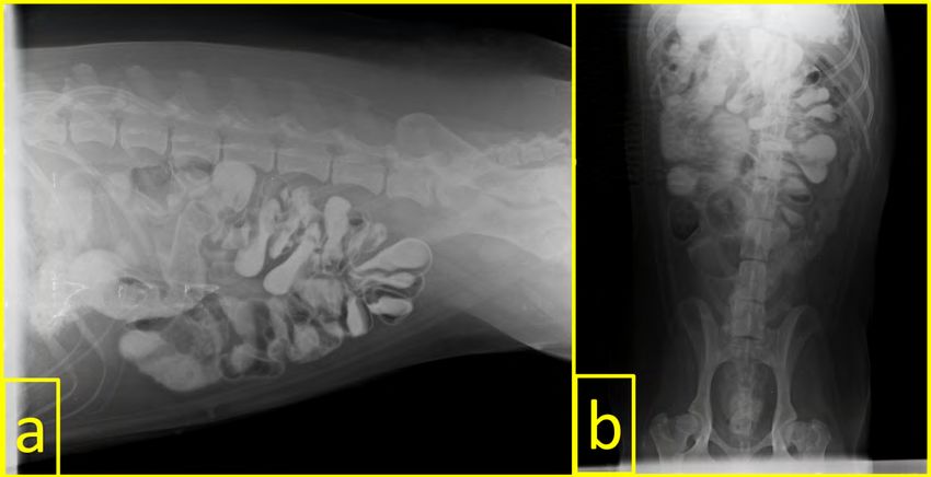
Gastrointestinal foreign bodies in Dogs and Cats / İşler et al._____________________________________________________________

                                                          TABLE II
                 Summary of signalment, disease, and treatment information of foreing body ingestions cases
N° Species      Breed       Sex     Foreign Object          Loc.               Clinical Sign              Treatment             Prognosis
 1       Cat      D-S       M        Sewing Needles          E         Salivation and Opening mouth       Endoscopy                Good

 2       Cat     British     F            Bone               E           Cough and opening mouth          Endoscopy                Good

 3       Cat      D-S       M             Cloth              S          Vomiting, Salivation, Anorexia     Surgical                Good

 4       Cat      D-S       M          LFB - Skein           SI                Abdominal pain              Surgical                Good

 5       Cat      D-S       M           LFB- Skein           SI            Vomiting and diarrhoea          Surgical                Good

 6       Cat      D-S        F       Sewing Needles          E         Salivation and Opening mouth       Endoscopy                Good

 7       Cat      D-S        F            Wood               S             Vomiting and diarrhoea          Surgical             Complicated

 8       Cat      D-S       M           LFB-Skein            SI            Vomiting and diarrhoea          Surgical                Good

 9       Cat      D-S        F        Plastic object         S                 Asymptomatic                Medical                 Good

10       Cat      S-F       M             Cloth              S             Vomiting and anorexia           Surgical                Good

11       Cat      S-F        F       Sewing Needles          E         Salivation and Opening mouth       Endoscopy                Good

12       Cat      S-F        F         Olive seeds           S                 Asymptomatic                Medical                 Good

13       Dog      B-M        F       Metallic object         S                 Asymptomatic                Medical                 Good

14       Dog      C-C       M             Bone               S             Vomiting and Anorexia           Surgical                Good

15       Dog    Cocker      M             Bone               LI            Hemorragic diarrhoea           Endoscopy           Wound infection

16       Dog    Cocker      M             Socks              S                 Asymptomatic                Surgical                Good

17       Dog    Cocker      M             Socks              S                 Asymptomatic                Surgical                Good

18       Dog   Crossbreed   M          LFB - Skein           S                    Vomiting                 Surgical                Good

19       Dog   Crossbreed   M        Metallic object         SI                No clinical Sign            Rejected              Unknown

20       Dog   Crossbreed    F       Metallic object         E         Salivation and Opening mouth       Endoscopy                Good

21       Dog      G-S       M             Cloth              S           Vomiting and no defecation        Surgical                Good

22       Dog      G-S        F        Plastic object         SI            Vomiting and diarrhoea          Surgical                Good

23       Dog      G-R       M             Stone              LI       Abdominal pain and no defecation     Surgical           Wound infection

24       Dog      G-R        F    Slipper and LFB-Skein      SI          Vomiting and no defecation        Surgical    Complicated, Wound infection

25       Dog      G-R       M             Stone              S          Abdominal pain and anorexia        Surgical           Wound infection

26       Dog      F-B        F      Fish hook and line       E         Salivation and Opening mouth       Endoscopy                Good

27       Dog      M-T        F      Fish hook and line       S                 Asymptomatic                Surgical                Good

28       Dog    Pointer      F          Unknown              SI       Vomiting, no defecation, Anorexia    Rejected              Unknown

29       Dog   Pomerian     M        Metallic object         SI                Abdominal pain              Surgical                Good

30       Dog      Pug       M            Rubber              S                 Asymptomatic                Surgical                Good

31       Dog      Pug        F       Electrical cable        SI            Vomiting and diarrhoea          Surgical                Good

32       Dog   Rotweiller   M             Stone           SI and LI        Vomiting and diarrhoea          Surgical                Good

D-S: Domestic Shorthair, S-F: Scottish Fold, B-M: Belgian Malinois, C-C: Cane Corso, G-S: German Shepherd, G-R: Golden Retriever, F-B: French Bulldog,
M-T: Maltese Terrier Loc.: Localization, E: Esophagus, S: Stomach, SI: Small Intestine, LI: Large Intestine, LFB: Linear Foreign Body

4 of 8
____________________________________________________________Revista Cientifica, FCV-LUZ / Vol. XXXII, rcfcv-e32097, 1 - 8
A FB was detected in the gastrointestinal system of 12 cats. Gianella [6] conducted a study on the GFB in 102 dogs in 2009
Further, two of these cats vomited after the administration of 2 % and reported that the dog breeds that experienced the highest
xylazine HCl (alfazyne 2 %; Egevet, Turkey), and the FB was number of FB incidents included West Highland white terrier,
naturally removed from their body. In four cats, the FB was removed Yorkshire terrier, Bernese mountain dog, Labrador retriever,
using forceps under the guidance of imaging with endoscopy. Golden retriever, and German shepherd. In the present study,
Surgical procedures were performed in six cats. Of the 6 cats who three of 20 (15 %) dogs were not of a specific breed and 17 (85 %)
underwent surgical intervention, enterotomy and gastrostomy were belonged to certain breeds [6]. Most of the dogs with FB were
performed in 4 and two, respectively. One of 6 cats who underwent Golden retrievers and Germanic shepherds, consistent with the
surgical intervention had complications associated with wound statement of Gianella [6]. A previous study reported that FB in the
infection (TABLE II). Some of the FB removed during or after the gastrointestinal system were more life threatening in breeds such
surgery are shown in FIG. 2. as Staffordshire bull terriers, English bull terriers, Jack Russell
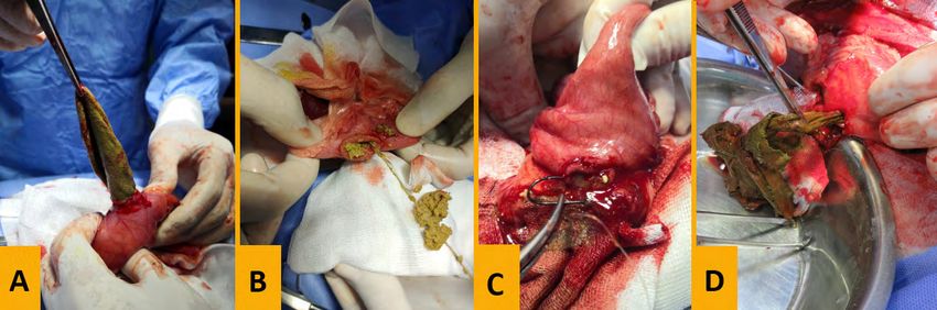
FIGURE 2. Removal of various FB materials from the area of localization during the surgery. A: socks, B: sewing needle and
thread, C: fishing rod and fishing line, and D: a piece of cloth
FB objects TABLE III
Overall, 14 different FB were detected in cats and dogs. As FB Foreing Body materials detected in the
material, five (15.63 %) animals had threads or balls, four (12.5 %) gastrointestinal system of cats and dogs
had metallic objects, three (9.36 %) had stones, three (9.36 %) had Type of Type of
sewing needles, three (9.36 %) had bones, three (9.36 %) had Cat Dog Cat Dog
foreign body foreign body
pieces of cloth, two (6.26 %) had plastic objects, two (6.26 %) had
fishing rod and fishing line, one (6.26 %) had socks at two separate Bonnet 1 2 Rubber - 1
times, one (3.13 %) had an eraser, one (3.13 %) had olive seeds,
one (3.13 %) had slippers, one (3.13 %) had wooden pieces, and Sewing
Cloths 2 1 3 -
one (3.13 %) had electrical cable. The distribution of these FB in needles
cats and dogs is presented in TABLE III. Further, metallic objects,
fishing rod and fishing line, electrical cable, slippers, socks, stones, Electric cable - 1 Slipper - 1
and eraser were detected only in dogs, not in cats. Olive seeds,
plastic objects, and wood fragments were among the FB detected Fish hook
- 2 Socks - 2
only in cats. and line
Metallic
- 4 Stone - 3
FB localization object
The locations of FB in the gastrointestinal system were as follows: Olive seeds 1 - String/rope 3 2
stomach in 14 (43.75 %) animals, small intestine in 10 (31.25 %),
esophagus in six (18.75 %), and large intestine in three (9.38 %).
FB were detected in both small intestine and large intestines only Plastic object 1 - Wood 1 -
in one patient. Some of the FB detected by radiography are shown
in FIGS 3 and 4.
5 of 8Gastrointestinal foreign bodies in Dogs and Cats / İşler et al._____________________________________________________________
FIGURE 3. Visualization of some of the Foreing Body by direct radiography. a: sewing needle in a cat, b: two erasers in the
stomach in a dog, c: metallic object in a dog, and d: two stones in the stomach and large intestine in a dog
FIGURE 4. A typical accordion image on indirect radiography due to swallowing of a rope in a dog. a: Latero-Lateral (LL)
view and b: Ventro-Dorsal (VD) view
terriers, Border collies, and Springer spaniels [7]. These breeds mixed breed, three of the remaining four cats were Scottish fold,
were not present in this study, and FB were life threatening in all and one was British shorthair. More comprehensive studies are
the animals with complete obstruction, regardless of the breed. required for predisposition based on breeds in the distribution of
case numbers.
In cats, FB incidents are common in domestic short-haired cats
that do not belong to any specific breed [77]. Abd Elkader et al. In 2018, Caixeta et al. reported that 31 (62 %) of 50 dogs that
reported that FB were detected in 16 Persian breed cats and 14 swallowed FB were male and 19 (38 %) were female [3]. Hobday
mixed breed cats [1]. In the present study, 8 of 12 cats were of et al. reported that 337 (67.5 %) of 499 dogs that swallowed FB
6 of 8____________________________________________________________Revista Cientifica, FCV-LUZ / Vol. XXXII, rcfcv-e32097, 1 - 8
were male and 162 (32.5 %) were female. In the present study, cassette tapes, needles and threads, fish hooks and lines, plastic
32 cats and dogs (19 male and 13 female) were exposed to GFB bags, blankets, electric cables, peach stones, wooden skewers,
[8]. Further, 12 of 20 dogs were male (60 %) and 8 dogs were potatoes, pine cones, beads, lolly sticks, marble, and glass objects
female (40 %), indicating higher rate of FB in male dogs, which in the digestive system of cats and dogs [3, 6, 7, 10]. Similar FB
is consistent with the results of Caixeta et al. and Hobday et al. were found in the present study; however, no socks, cloth, or olive
[3, 8]. On the other hand, FB were found in six female (50 %) seeds were found in the previous studies.
and six male (50 %) cats in the present study. Therefore, there
Hayes reported that FB were mostly detected in the small
is a need for more comprehensive studies that will demonstrate
intestine [7]. In the present study, FB the gastrointestinal system
the relationship between species, breed, and sex differences,
were mostly detected in the stomach (43.75 %) and small intestines
individuals affinity toward FB and animal behavior.
(31.25 %). The least common regions for FB were esophagus
Fossum reported that exposure to FB generally occurs in dogs (18.75 %) and large intestines (9.38 %). Thus, these results are
when they are puppies [5]. However, Caixeta et al. reported the inconsistent with the results of the studies by Hoffmann, Veeder
age of dogs ingesting FB as 5.8 ± 4.5, Hobday et al. as 4.27 ± and Taylor, Kassem et al., and Caixeta et al. [3, 9, 10, 13]. The
3.51, Hayes as 2.5 ± 3.0, and 1.8 ± 1.8 y in cats [3, 7, 8]. Thus, higher number of stomach cases in the present study can be
the age range of 1.6 ± 1.43 in cats and 4.08 ± 3.98 y in dogs associated with early diagnosis.
identified in the present study is consistent with that reported in
other studies [3, 7, 8] and differs from that reported by Fossum [3, CONCLUSIONS
5, 7, 8]. Moreover, exposure to FB is the most common in adult
cats and dogs. The ingestion of FB is more common in adult cats and dogs
aged between 1 and 7 y, the ingestion of strings/ropes and metallic
FB can be diagnosed and identified by methods such as physical objects as FB is more common, and it is important that owners
examination and palpation techniques, radiographic imaging, and notice the incident for early diagnosis and treatment to ensure
endoscopy. Radiographic imaging can consist of a combination of prevention of complications. Thus, this study shows that FB are
direct and indirect methods (contrast radiographs). Endoscopy is a mostly found in the stomach and vomiting is the most common
good method to identify and remove FB from the esophagus and clinical symptom.
stomach [2, 12]. In the present study, direct and indirect radiographic
and endoscopic techniques were used for the diagnosis based
on anamnesis details and physical examination results. The CONFLICT OF INTEREST
use of endoscopic examination for diagnosis and treatment is The authors declare that they have no conflicts of interest in
advantageous. Indirect radiography is an important diagnostic the research.
method for detecting obstruction caused by radiolucent objects
without contrast images on direct radiography.
BIBLIOGRAPHIC REFERENCES
The common clinical symptoms include anorexia, vomiting,
[1] ABD ELKADER, NA; EMAM, IA; FARGHALI, HA; SALEM,
dehydration, depression, diarrhea, and abdominal pain [7, 8,
NY. Oesophageal FB in cats: Clinical and anatomic findings.
11]. In the present study, vomiting (40.62 %), diarrhea (18.75 %),
Plos One. 15: 1-15. 2020.
salivation (18.75 %), anorexia (15.63 %), abdominal pain (12.5 %),
and bloody diarrhea (3.13 %) were detected. Further, the clinical [2] BEBCHUK, TN. Feline gastrointestinal foreign bodies.
findings not reported in other studies were keeping the mouth Veterinary Clinics. J. Small Anim. Pract. 32: 861-880. 2002.
open (18.75 %), inability to defecate (12.5 %), and cough (3.13 %)
[3] CAIXETA, ACF; ALVES, EGL; COELHO, NGD; SOUZA, ACF;
in cats that ingested needles. In addition, eight (25 %) animals
TORRES, RCS; NEPOMUCENO, AC. FB in the gastrointestinal
were brought to the hospital without any clinical symptom after
tract of dogs: A retrospective study. Ars. Vet. 34: 20-24. 2018.
their owners witnessed them to swallow foreign bodies. Thus, the
incidence of clinical findings in the present study is consistent with [4] DEN HERTOG, E. Endoscopic removal of FB from cats or
that reported in other studies. dogs. Tijdschr Diergeneeskd. 128: 434-439. 2003.
Kassem et al. reported that complications are rare in cases of [5] FOSSUM, TW. Surgery of the Digestive System. Cap 20.
FB, whereas Gianella reported a complication rate of 12.74 %, and Small Animal Surgery. Fossum, TW. (Ed), 4th Ed. Elsevier
Hayes reported a complication rate of 80 % [6, 7, 10]. Yurdakul Health Sciences, St Louis, US. Pp. 386-583. 2014.
reported that intestinal hyperemia, congestion, and intestinal
[6] GIANELLA, P; PFAMMATTER, NS; BURGENER, IA.
contents flowed into the abdominal cavity in cases of FB [14]. Hayes
Oesophageal and gastric endoscopic FB removal: complications
reported that in cases of 14 d or more, the prognosis was poor, the
and follow - up of 102 dogs. J. Small Anim. Pract. 50: 649-654.
risk of peritonitis increased, and in some cases, the euthanasia
2009.
option was used [7]. In the present study, as most of the animals
were treated in the subacute and acute phases, no complications [7] HAYES, G. Gastrointestinal FB in dogs and cats: a retrospective
were observed. Only 3 of 23 (9.68 %) animals had wound infection study of 208 cases. J. Small Anim. Pract. 50: 576-583. 2009.
due to the lack of post-operative dressing and antibiotic treatment.
Thus, the prognosis was overall good. [8] HOBDAY, MM; PACHTINGER, GE; DROBATZ, KJ; SYRING,
RS. Linear versus non - linear gastrointestinal FB in 499 dogs:
Some studies have reported the presence of FB such as latex clinical presentation, management and short - term outcome.
teat, plastic/rubber objects, string/rope/fishing lines, stones, balls, J. Small Anim. Pract. 55: 560-565. 2014.
underwear/nappy, corn-cob, leather, metallic objects/coins, bones,
7 of 8Gastrointestinal foreign bodies in Dogs and Cats / İşler et al._____________________________________________________________
[9] HOFFMANN, KL. Sonographic signs of gastroduodenal linear [12] TYRRELL, D; BECK, C. Survey of the use of radiography vs.
FB in 3 dogs. Vet. Radiol. Ultrasound. 44: 466-469. 2003. ultrasonography in the investigation of gastrointestinal FB in
small animals. Vet. Radiol. Ultrasound. 47: 404-408. 2006.
[10] KASSEM, MM; EL-KAMMAR, MH; EL-MENSHAWEY, MF.
Surgical Managment of FB in Stomach and Intestine of Some [13] VEEDER, CL; TAYLOR, DK. Injury related to environmental
Foregin Breed Dogs. Alexandria J. Vet. Sci. 42: 11-15. 2014. enrichment in a dog (Canis familiaris): Gastric foreign body.
J. Am. Assoc. Lab. Anim. Sci. 48: 76-78. 2009.
[11] PAPAZOGLOU, LG; PATSIKAS, MN; RALLIS, T. Intestinal FB
in dogs and cats. Compend. Contin. Educ. Vet. 25: 830-845. [14] YURDAKUL, I. Intestinal Perforation Due to Gunshot Injury
2003. in a Kangal Dog. CUSBED. 2: 1-8. 2017.
8 of 8You can also read


