Extracellular vesicles isolated from dsRNA sprayed barley plants exhibit no growth inhibition or gene silencing in Fusarium graminearum
←
→
Page content transcription
If your browser does not render page correctly, please read the page content below
Extracellular vesicles isolated from dsRNA sprayed barley plants exhibit no growth inhibition or gene silencing in Fusarium graminearum Timo Schlemmer Justus Liebig Universitat Giessen https://orcid.org/0000-0003-4034-445X Richard Lischka Justus Liebig Universitat Giessen Dagmar Biedenkopf Justus Liebig Universitat Giessen Aline Koch ( aline.koch@uni-hohenheim.de ) University of Hohenheim https://orcid.org/0000-0003-3536-5344 Short Report Keywords: Extracellular vesicles, plant EVs, barley, Fusarium graminearum, RNAi, RNA biopesticides, spray-induced gene silencing, RNAi-based plant protection, dsRNA, siRNA Posted Date: September 30th, 2021 DOI: https://doi.org/10.21203/rs.3.rs-944619/v1 License: This work is licensed under a Creative Commons Attribution 4.0 International License. Read Full License
1   Extracellular vesicles isolated from dsRNA sprayed barley plants exhibit no growth

 2   inhibition or gene silencing in Fusarium graminearum

 3   Timo Schlemmer1,2, Richard Lischka1, Dagmar Biedenkopf1, Aline Koch2
     1
 4       Institute of Phytopathology, Centre for BioSystems, Land Use and Nutrition, Justus Liebig

 5   University, Heinrich-Buff-Ring 26, D-35392, Giessen, Germany
     2
 6       Institute of Phytomedicine, University of Hohenheim, Otto-Sander-Strasse 5, D-70599,

 7   Stuttgart, Germany

 8   Corresponding author: Aline Koch

 9   Email: aline.koch@uni-hohenheim.de

10

11   Abstract

12   Incorporating a double-stranded RNA (dsRNA)-expressing transgene into plants or applying

13   dsRNA by spraying it onto plant leaves successfully protects plants against invading pathogens

14   with RNA interference (RNAi). How dsRNAs or siRNAs are transferred between donor host

15   cells and recipient fungal cells is largely unknown It is speculated that plant extracellular

16   vesicles (EVs) function as RNA shuttles between plants and their interacting pathogens.

17   Recently, we found that EVs isolated from HIGS or SIGS plants contained dsRNA-derived

18   siRNAs. In this study, we evaluated whether isolated EVs from RNA-sprayed barley (Hordeum

19   vulgare) plants affected the growth of the phytopathogenic ascomycete Fusarium graminearum

20   (Fg). Encouraged by our previous finding that dropping barley-derived EVs on Fg cultures

21   caused fungal stress phenotypes, we conducted an in vitro growth experiment in microtiter

22   plates where we co-cultivated Fg with plant EVs isolated from dsRNA-sprayed barley leaves.

23   We observed that co-cultivation of Fg macroconidia with barley EVs did not affect fungal

24   growth. Furthermore, plant EVs containing SIGS-derived siRNA appeared not to affect Fg

25   growth and showed no gene silencing activity on FgCYP51 genes. We conclude that either the

                                                    1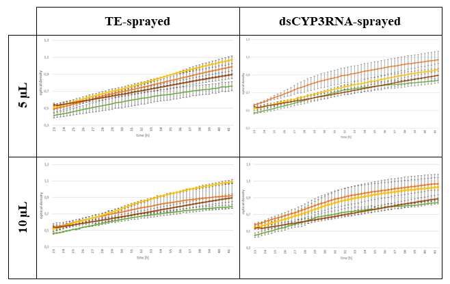
26   amount of spray-derived sRNA was insufficient to induce target gene silencing (SIGS) in Fg

27   or Fg uptake of plant EVs from liquid cultures was inefficient or impossible.

28   Keywords

29   Extracellular vesicles, plant EVs, barley, Fusarium graminearum, RNAi, RNA biopesticides,

30   spray-induced gene silencing, RNAi-based plant protection, dsRNA, siRNA

31   Background

32   Research on plant extracellular vesicles (EVs) has exploded since the first reports in the 1970s,

33   and the number of scientific studies on them has quintupled over the last decade. Plant EVs

34   attract immense scientific interest due to the anti-inflammatory, anticancer, antioxidative and

35   antisenescence properties of EVs derived or isolated from, e.g., Panax ginseng (Cho et al.,

36   2021), Asparagus cochinchinensis (He, 2021), Aloe vera (Kim et al., 2021), Allium sativum

37   (Özkan et al., 2021), bitter melon (Yang et al., 2021), grapefruit (Stanly et al., 2020), strawberry

38   (Perut et al., 2021), carrot (Kim and Rhee, 2021) and honey (Chen et al., 2021). Their strong

39   bioactivity and biocompatibility together with efficient uptake by human cells have raised the

40   possibility of exploiting them as novel drug delivery vehicles (Bokka et al., 2020; Niu et al.,

41   2021; You et al., 2021). The bioengineering of plant EVs to develop next-generation therapeutic

42   tools in nanomedicine has also inspired plant biotechnologists who continuously seek

43   sustainable and eco-friendly solutions in agriculture and plant protection. Thus, using EVs as

44   natural blueprints may stimulate and further boost the development of nanocarrier-based

45   technologies that facilitate the efficient delivery of CRISPR/Cas components or systems in the

46   future (Alghuthaymi et al., 2021). However, applied plant EV research is a more recent

47   development that has undergone rapid progress in the last three years, with more than 260

48   studies published1.

     1
         PubMed (nih.gov)
                                                      2
49   Although plant EVs were first described in the apoplast in 1967 (Halperin and Jensen, 1967), it

50   was almost half a century before they were separated from plant apoplastic fluids and then

51   visualized with transmission electron microscopy (TEM) (Regente et al., 2009, 2017; Rutter

52   and Innes, 2017). These pioneering works have laid the foundation for studies demonstrating

53   the pivotal role of EVs in plant-pathogen interactions (Cai et al., 2021; Stotz et al., 2021). For

54   example, plant- or pathogen-derived EVs contribute bidirectionally to this highly specialized

55   interspecies communication through the release of lipids, proteins and small RNAs (sRNAs)

56   that regulate and deregulate defence and offence responses (Cai et al., 2020; Liu et al., 2020;

57   De Palma et al., 2020; Cavaco et al., 2021; Woith et al., 2021). In particular, the identification

58   of plant EV-derived sRNAs furnished strong evidence for their role as shuttles in interspecies

59   communication and mechanistic insights into plant antifungal defence responses (Cai et al.,

60   2018; Baldrich et al., 2019; Roth et al., 2019; Schlemmer et al. 2020; 2021a; 2021b).

61   Conversely, fungal pathogens secrete sRNAs to dampen plant immunity (Weiberg et al., 2013;

62   Kusch et al., 2018; Dunker et al., 2020; Werner et al., 2021). This sRNA-based crosstalk, also

63   known as cross-species RNA interference (RNAi), was first described in 2013, in a paper

64   demonstrating that the fungal pathogen Botrytis cinerea produces sRNAs that mimic plant

65   sRNAs and bind to Arabidopsis AGO1 to antagonistically silence important plant immunity

66   genes (Weiberg et al., 2013). Similar to plant EV-mediated sRNA transport, it is proposed that

67   fungal sRNA delivery is facilitated by EVs (Kwon et al., 2020). To support this hypothesis,

68   EVs isolated from different fungal pathogens, such as Ustilago maydis (Kwon et al., 2021),

69   Zymoseptoria tritici (Hill and Solomon, 2020), Fusarium oxysporum (Bleackley et al., 2020;

70   Garcia-Ceron et al., 2021) and Fusarium graminearum (Fg) (Schlemmer et al. 2020), were

71   described for subsequent RNA content analysis.

72   In agriculture, RNAi technologies attract immense scientific and political interest as powerful

73   substitutes for conventional chemical pesticides to reach the EU’s sustainability goals (Taning

74   et al., 2021). Currently, RNAi-based plant protection relies on two strategies that differ in the
                                                     3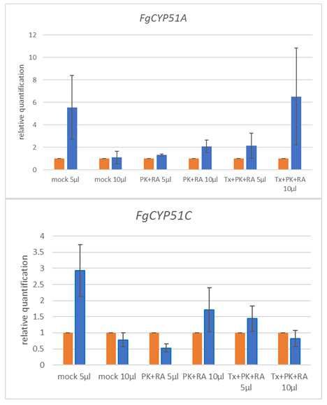
75   origin of dsRNA. First, endogenous dsRNA formation mediated by transgene expression is

76   designated as host-induced gene silencing (HIGS). The second strategy is based on exogenous,

77   foliar dsRNA application known as spray-induced gene silencing (SIGS). Notably, the principle

78   of cross-species RNAi was biotechnologically used (HIGS) before its naturally occurring

79   equivalent was discovered (Nowara et al., 2010). We previously demonstrated that a transgene-

80   derived CYP3RNA (a dsRNA designed to target CYP51A, CYP51B and CYP51C genes in Fg),

81   as well as foliar application of CYP3RNA, induced CYP51 target gene silencing in Fg.

82   Remarkably, HIGS-or SIGS-mediated FgCYP51 downregulation conferred strong Fg disease

83   resistance in Arabidopsis thaliana (HIGS) and Hordeum vulgare (HIGS and SIGS) (Koch et

84   al., 2013, 2016, 2019; Biedenkopf et al., 2020; Höfle et al., 2020).

85   Recently, we showed that EVs isolated from CYP3RNA-expressing Arabidopsis plants contain

86   CYP3RNA-derived siRNAs (Schlemmer et al 2021a). Notably, subsequent differential

87   digestive treatments of EVs with RNase, protease and a detergent revealed that, compared with

88   that in the apoplastic fluid of Arabidopsis, the amount of intravesicular sRNA was surprisingly

89   low (Schlemmer et al. 2021a). In addition, we observed a loss of CYP3RNA/HIGS-mediated

90   Fg disease resistance in Arabidopsis with a mutated endosomal sorting complex required for

91   transport III (ESCRT-III), which is necessary for proper EV biogenesis (Schlemmer et al.

92   2021a). Moreover, ESCRT-III mutants were impaired in EV production and no CYP3RNA-

93   derived siRNAs were detected in either the apoplastic fluid or inside the EVs (Schlemmer et al.

94   2021a). EVs isolated from CYP3RNA-sprayed barley plants revealed CYP3RNA-derived

95   siRNAs; however, their abundance was even lower compared with Arabidopsis EVs

96   (Schlemmer et al 2021b). This difference might be due to the various dsRNA origins in HIGS

97   and SIGS approaches, whereby SIGS-induced RNAs must be taken up by plant cells before

98   being packed into plant EVs (Koch and Wassenegger, 2021). CYP3RNA uptake into plant cells

99   and its systemic spread via the phloem have been previously reported as well as its apoplastic

                                                     4
100 transport in the xylem (Koch et al., 2016; Biedenkopf et al., 2020). However, whether EVs are
101 required for the delivery of exogenously applied dsRNA to induce SIGS in Fg remains unclear.
102 To address this question, we assessed whether EVs isolated from SIGS plants can induce
103 FgCYP51 target gene silencing and Fg growth inhibition. We performed in vitro treatments of
104 Fg with EVs isolated from CYP3RNA sprayed barley plants. Remarkably, we found no effects
105 on FgCYP51 expression or Fg growth, further underlining the importance of clarifying whether
106 EV-mediated sRNA transport is required during SIGS-barley–Fg interaction.
107 Results
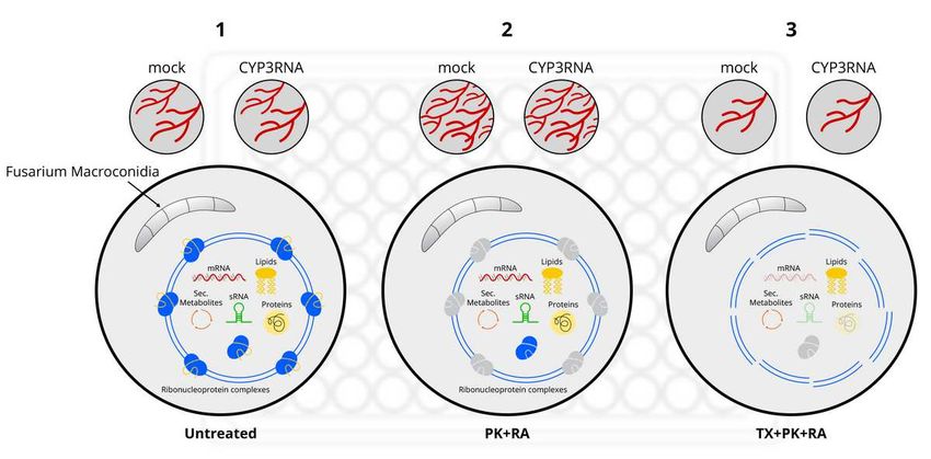
108 To test the possibility of plant EV uptake by Fg in vitro, we isolated EVs from control-
109 (Tris-EDTA buffer) and CYP3RNA-sprayed barley leaves using a modified protocol as
110 described (Rutter and Innes, 2017; Schlemmer et al 2020). In our recent studies, we observed
111 that state-of-the-art EV purification from apoplastic fluids leads to impure EV isolates
112 containing additional co-purified apoplastic substances (Schlemmer et al., 2021a). This finding
113 aligns with recent debates discussing the pitfalls of and the standardization needs in plant EV
114 research, e.g., the contamination risks of different plant EV separation and characterization
115 methods (Rutter and Innes, 2020; Mammadova et al., 2021; Pinedo et al., 2021). To avoid such
116 pitfalls, we performed a stringent digestive treatment of EV isolates to degrade extravesicular
117 proteins and RNAs before in vitro treatment of Fg with plant EVs. Each EV isolate was derived
118 from 80 barley leaves and EVs were ultimately resuspended in 190 µl PBS. We reserved 40 µl
119 for quality control measurements, TEM and nanoparticle trafficking analysis (NTA). The
120 remaining resuspension was divided into three equal fractions (Fig. 1). To degrade
121 extravesicular proteins, RNAs and ribonucleoprotein complexes, one fraction of EV isolates
122 were treated with proteinase K and RNase A (PK+RA). In addition to PK+RA, the next fraction
123 was treated with triton X (TX+PK+RA) to break up EVs and degrade extravesicular and
124 intravesicular proteins, RNAs and ribonucleoprotein complexes (Fig. 1). One fraction remained
5125 untreated to evaluate whether the observed effects resulted from EVs or co-purified apoplastic
126 fluid proteins or RNAs. Finally, EVs were co-inoculated with Fg macroconidia and fungal
127 growth was determined, after 20 hours of pre-incubation, by optical density measurements (OD)
128 every 20 minutes for a further 24 hours.
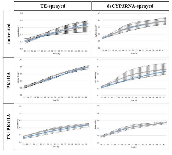
129 To assess whether the effects depended on the investigated volumes, we used two
130 different amounts of resuspended EV solution. We tested untreated EVs isolated from TE- or
131 CYP3RNA-sprayed barley leaves and EVs treated with PK+RA and TX+PK+RA. We added 5
132 µl or 10 µl of each EV fraction to Fg macroconidia. Regardless of whether EVs were derived
133 from CYP3RNA- or TE-sprayed barley leaves or how EVs were treated after purification, no
134 differences in Fg growth were observed between treatment volumes (Fig. 2). At the beginning
135 of the measurement period, 23 hours post-inoculation (hpi), all samples showed an OD value
136 around 0.5. At 42 hpi, the OD had increased for untreated EVs and PK+RA-treated EVs up to
137 0.9–1.1, while the OD of TX+PK+RA-treated EVs only rose to 0.7–0.9.
138 As we observed no difference in the effect on fungal growth induced by different EV
139 volumes, we next assessed the effect of EV treatments after EV isolation on Fg growth. As a
140 control, we used EV-free PBS, which was also used for EV resuspension after isolation. We
141 compared the fungal growth over the measured time among the different EV treatments and the
142 investigated amount of EVs. Focusing on Fg growth co-cultivated with EVs from TE-sprayed
143 barley leaves independent of the applied volume of EVs, we observed that PK+RA-treated EVs
144 promoted Fg growth compared with PBS-treated Fg cultures (Fig. 3; Fig. 1), possibly triggered
145 by simplified nutrient uptake via the degraded proteins and RNAs the enzymatic treatment
146 created or by the destruction of proteins that usually inhibit Fg. However, we did not observe
147 growth promotion when Fg was fed with untreated EVs. The same observation was made when
148 we focused on EVs from CYP3RNA-sprayed barley leaves, where no difference in the fungal
149 growth was visible when differently prepared EVs were applied to Fg. Regardless of whether
150 EVs originated from TE- or CYP3RNA-sprayed barley leaves and whether 5µl or 10 µl were
6151 applied, Fg co-cultivated with TX+PK+RA-treated EVs was more inhibited than Fg co-
152 cultivated with PBS, untreated EVs, or PK+RA-treated EVs (Fig. 3). We, therefore, tested the
153 detergents’ effects on Fg. We mixed TX, PK, RA, PK+RA and TX+PK+RA with PBS,
154 incubated them under the same conditions as the plant EVs and tested the mixtures in our
155 growth assay. We observed no difference in the growth behaviour of Fg treated with PK, RA
156 or a combination of both (Fig. 4). Notably, TX or TX with PK+RA led to a clear growth
157 reduction compared with the PBS control, indicating a clear effect of TX on fungal growth
158 independent of plant EVs (Fig. 4; Fig. 1). To avoid misinterpreting the effect of TX as the effect
159 of the investigated CYP3RNA, we calculated the relative growth per EV treatment to compare
160 the effects of TE- and CYP3RNA-sprayed EVs. Remarkably, we found no growth inhibition
161 caused by the CYP3RNA spray application independently of how EVs were treated after
162 isolation (Fig. 5). To verify this result and determine whether the unimpaired fungal growth
163 could be explained by a lack of FgCYP51 gene silencing, we isolated RNA from the Fg cultures
164 grown in microtiter plates and performed FgCYP51 gene expression analysis. Supporting our
165 previous assumption, we found no gene silencing activity in Fg after co-cultivation with EVs
166 isolated from CYP3RNA-sprayed barley leaves (Fig. 6).
167
168 Discussion
169 The more than 50 studies that demonstrate RNAi-based control of fungal pathogens with
170 an average plant disease resistance of about 60% (Koch and Wassenegger, 2021) reflect the
171 enormous potential of RNAi technologies to meet the socio-political demand to halve the use
172 of chemical pesticides by 2030 (European Commission, 2021). However, our mechanistic
173 knowledge of HIGS and SIGS is still incomplete, although researchers hope to transition testing
174 from the lab to the field soon (Rank and Koch 2021). Towards this goal, unravelling the routes
175 by which dsRNAs and siRNAs are delivered into fungal cells is key to further improve cellular
176 uptake and systemic distribution and therefore increase the stability and efficacy of sprayed
7177 RNA biopesticides. RNA uptake and transport essentially serve as effective RNA protection,
178 preventing RNA degradation. Besides RNA stabilization with RNA ribonucleoprotein
179 complexes or lipoproteins, EVs encapsulate RNAs (Lasser et al 2011) thus sheltering them from
180 RNases or degradation in general during short (cell-to-cell) or long-distance (systemic)
181 movement (Valadi et al 2007, Hunter et al 2008). Previously, we found that barley EVs led to
182 stress-related discolouration of Fg colonies (Schlemmer et al., 2020) and that CYP3RNA-
183 sprayed barley leaves, which confer Fg disease resistance (Koch et al., 2016) contained
184 CYP3RNA-derived sRNAs (Schlemmer et al., 2021b). However, as the amount of spray-
185 derived sRNA in barley EVs was low, questions about their role and relevance in SIGS-barley–
186 Fg interaction arise. To assess this further, we treated Fg with EVs isolated from sprayed barley
187 plants in vitro. The impurity of plant EV isolates raised concerns about the reliability of findings
188 and their interpretation (Rutter and Innes, 2020; Mammadova et al., 2021), thus, we performed
189 rigorous digestive treatments of EV isolates before Fg in vitro testing. Encouraged by our
190 previous finding that drop inoculation of barley EVs on Fg cultures grown on solid agar plates
191 caused an increase in purple pigmentation, indicative of the stress-induced premature formation
192 of fruiting bodies (Schlemmer et al., 2020), we expected to observe the effects of barley EVs
193 on Fg in liquid in vitro cultures. Interestingly, another recent study demonstrated the antifungal
194 activity of EVs derived from root exudates of tomato plants against Fusarium oxysporum,
195 Botrytis cinerea and Alternaria alternata (De Palma et al., 2020) underlining the validity of in
196 vitro EV–fungal spore interaction tests. Surprisingly, we found that neither wild-type barley
197 EVs nor EVs isolated from CYP3RNA-sprayed barley leaves affected Fg growth (Fig. 5). In
198 addition, different EV volumes (5 µl or 10 µl EV suspension) did not affect fungal growth (Fig.
199 2). In contrast, on solid agar plates, 40 µl of EV solution derived from 80 barley leaves was
200 drop-inoculated onto Fg, suggesting that the tested volumes of 5 µl and 10 µl might be too low.
201 We also did not observe a CYP3RNA-dependent effect on Fg growth (Fig. 5). Based on these
202 results, we hypothesized two possibilities: first, Fg is unable to take up EVs in vitro, and second,
8203 the amount of spray-derived sRNA in EVs is insufficient to induce SIGS. To test the second
204 possibility, we performed FgCYP51 gene expression analysis on Fg cultures after EV treatment,
205 which is a more sensitive way to test CYP3RNA effects on Fg than determining the OD of
206 liquid fungal cultures.
207 Consistent with our results finding no Fg growth inhibition, we measured no gene-
208 silencing activity in Fg co-cultivated with EVs from CYP3RNA barley leaves (Fig. 6).
209 However, this could still be explained by the inability of Fg to take up plant EVs in vitro.
210 However, this could be still due to the inability of Fg to take up plant EVs in vitro. Notably,
211 plant-derived EVs were shown to contain stress response proteins and lipids (Rutter and Innes,
212 2017; De Palma et al., 2020; Liu et al., 2020; Cavaco et al., 2021; Schlemmer et al., 2021a) and
213 exhibit antifungal activity (Schlemmer et al. 2020; De Palma et al., 2020). Given this, it is
214 surprising that we found no inhibitory effects of barley-derived EVs. This raises the question
215 of whether EVs or EV content are stable in liquid media, able to overcome the membrane or
216 cellular barriers of Fg and able to reach a defined threshold to activate the distinct RNAi
217 machinery in Fg. While another study convincingly demonstrated sunflower-derived EV uptake
218 by the ascomycete Sclerotinia sclerotiorum through reduced hyphae growth and spore
219 germination (Regente et al., 2017), whether this holds true for other fungi or other pathosystems
220 remains unknown. Notably, the latest studies demonstrated in vitro uptake of plant-derived
221 (ginger, grapefruit, pineapple and paprika) EVs in human and rat cells (Garaeva et al., 2021;
222 Ito et al., 2021; Man et al., 2021), which is of great scientific interest due to their therapeutic
223 potential in nanomedicine (Alfieri et al., 2021). Studies on CYP3RNA-expressing Arabidopsis
224 (HIGS) plants revealed a loss of CYP3RNA-mediated Fg resistance in ESCRT-III mutants
225 (Schlemmer et al., 2021a). Additionally, EV purification from these mutants revealed no or
226 aberrant EVs with no CYP3RNA-derived sRNA, indicating the potential role of EVs in HIGS-
227 Arabidopsis–Fg interaction. However, HIGS in Arabidopsis and SIGS in barley are not
228 mechanistically comparable.
9229 In summary, we found no Fg growth inhibition after treatment of Fg in vitro cultures
230 with CYP3RNA-spray-derived barley EVs. Subsequently, we found no FgCYP51 target gene
231 silencing, raising the question of whether Fg is unable to take up EVs from liquid culture or
232 whether EV-contained CYP3RNA-spray-derived siRNAs are physiologically inactive.
233 However, further research is required to differentiate between the possibility of improper EV
234 uptake and the possibility that the amount of spray-derived sRNA was insufficient to induce
235 FgCYP51 gene silencing (SIGS) by elucidating the role and relevance of EVs for SIGS.
236
237 Conclusion
238 RNA biopesticides represent a powerful alternative to chemical pesticides. To make
239 future field applications reliable and realistic for agriculture, we require mechanistic knowledge
240 of RNA uptake and interspecies (plant–fungus) sRNA transfer. Identification and
241 characterization of plant and fungal EV content, as well as the mechanisms of loading and
242 release, have begun (He et al., 2021; Woith et al., 2021) but remain limited, unless required
243 exploit EVs as bioagents to confer disease resistance in a more natural context. Importantly,
244 fungal uptake of plant-derived EVs may offer potential routes to cure fungal diseases in
245 humans, based on the emerging evidence that plant-derived EVs exhibit great potential for
246 human health applications (Alfieri et al., 2021). We have just begun to understand the enormous
247 potential underlying natural compounds and delivery routes or compartments as we seek
248 sustainable, biocompatible and biodegradable alternatives to conventional treatments in
249 agriculture as well as medicine.
250
10251 Methods
252 Differential EV treatments
253 EVs of TE- and CYP3RNA-sprayed barley leaves were isolated as described in Schlemmer et
254 al. (2021b). EV isolation was performed in three technical replicates. Each isolation included
255 80 barley leaves per spray application. EVs were resuspended in 190 µl PBS (8mM NaH2PO4,
256 150mM NaCl, 3mM KCl and 2mM KH2PO4; pH 7.4) and subdivided into three groups after
257 isolation. The first group was untreated and served as a positive control (Tab. 1). The second
258 group was treated with proteinase K and RNaseA (PK+RA) and the third group with triton X,
259 proteinase K and RNaseA (TX+PK+RA) (Tab. 1). All groups were incubated for 30 minutes at
260 37°C and then added to Fusarium graminearum (Fg) macroconidia. Table1 Components of the
261 digestive EV treatments for eliminating intravesicular and apoplastic co-purified proteins and
262 RNAs.
TE sprayed barley dsCYP3RNA sprayed barley
group 1 2 3 1 2 3
EV solution 50 µl 50 µl 50 µl 50 µl 50 µl 50 µl
RNase - 1,2 µl 1,2 µl - 1,2 µl 1,2 µl
PK - 3 µl 3 µl - 3 µl 3 µl
Triton X - - 5,8 µl - - 5,8 µl
PBS 10 µl 5,8 µl - 10 µl 5,8 µl -
total 60 µl 60 µl 60 µl 60 µl 60 µl 60 µl
263 Investigated concentrations: Proteinase K (20 ng/µl) (Thermo Fisher Scientific); RNase A (20
264 ng/µl) (Thermo Fisher Scientific); 10 % Triton X-100 (Sigma)
265 Plant EV – Fg co-culture assay
266 Plant EV–Fg co-culture assays were performed in transparent 96-well plates with flat
267 bottoms. ½ PDB (potato dextrose broth, Formedium) was used as a carbon source. Each well
268 had 5440 macroconidia, 5µl or 10µl treated EV suspension and PBS added (Tab. 2). 96-well
269 plates were pre-incubated on the lab bench for 20 hours before they were put into a plate reader
11270 (CLARIOstar, BMG Labtech) for another 24-hour incubation at 25°C with 60 rpm shaking
271 where optical density (OD600) was measured every 20 minutes. To exclude microbial
272 contamination from EV isolates and misinterpretation of optical density due to this microbial
273 growth, one control (C) contained no macroconidia (C1) (Tab. 3). Hygromycin was added to
274 inhibit microbial growth and allow changes in optical density to be attributed to fungal growth
275 (C2). C3 contained no PBS but rather an additional 0.5 PDB. C4 contained no EVs. C3 and C4
276 were used to estimate the effect of the PBS on the optical density and growth behaviour of Fg.
277 As a reference for different EV treatments, the effects of EV treatment detergents were
278 determined by incubating EV-free PBS with PK+RA (C5), TX+PK+RA (C6), PK (C7), RA
279 (C8) and TX (C9) and used during the co-culture assay (Tab. 4). PBS was added to compensate
280 for volume differences resulting from differences in the inserted amounts of EVs. The co-
281 cultivation was then performed according to the plant EV–Fg cultivation method described in
282 Tab. 5.
283 Table 2 Well composition for microtiter well co-cultivation of differentially treated plant EVs
284 with Fg.
group 1 1 2 2 3 3
investigated vol. 5 µl 10 µl 5 µl 10 µl 5 µl 10 µl
Fg 20 µl 20 µl 20 µl 20 µl 20 µl 20 µl
0,5 PDB 125 µl 125 µl 125 µl 125 µl 125 µl 125 µl
PBS 10 µl 5 µl 10 µl 5 µl 10 µl 5 µl
total 160 µl 160 µl 160 µl 160 µl 160 µl 160 µl
285
286 Table 3 Overview of tested controls and their well composition.
controls C1 C2 C3 C4
Fg 20 µl 20 µl 20 µl
0,5 PDB 160 µl 125 µl 140 µl 125 µl
12PBS 15 µl
Hygromycin 15 µl
total 160 µl 160 µl 160 µl 160 µl
287
288 Table 4 Components of the digestive EV treatments to measure the effects of treatment reagents
289 on fungal growth.
controls C5 C6 C7 C8 C9
PBS 55,8 µl 50 µl 57 µl 58,8 µl 54,2 µl
RNase 1,2 µl 1,2 µl 1,2 µl
PK 3 µl 3 µl 3 µl
Triton X 5,8 µl 5,8 µl
total 60 µl 60 µl 60 µl 60 µl 60 µl
290
291 Table 5 Well composition for microtiter well co-cultivation of EV-free detergent reagents to
292 estimate treatment-dependent effects.
controls C5 C5 C6 C6 C7 C7 C8 C8 C9 C9
invest. vol. 5 µl 10 µl 5 µl 10 µl 5 µl 10 µl 5 µl 10 µl 5 µl 10 µl
Fg 20 µl 20 µl 20 µl 20 µl 20 µl 20 µl 20 µl 20 µl 20 µl 20 µl
0,5 PDB 125 125 125 125 125 125 125 125 125 µl 125
µl µl µl µl µl µl µl µl µl
PBS 10 µl 5 µl 10 µl 5 µl 10 µl 5 µl 10 µl 5 µl 10 µl 5 µl
total 160 160 160 160 160 160 160 160 160 µl 160
µl µl µl µl µl µl µl µl µl
293
294 FgCYP51 gene silencing analysis
295 Technical replicates per plate were collected after 44 h of incubation. RNA extraction, cDNA
296 synthesis and qRT-PCR for transcript analysis of FgCYP51A and FgCYP51C were performed
297 as described (Koch et al., 2013, 2016).
13298 Figure legends
299 Fig. 1 Schematic overview above the investigated EV treatments and their potential effect on
300 EV and EVs cargo. Fraction one (1) contains untreated EVs from mock or CYP3RNA sprayed
301 barley leaves and cause average fungal growth. EVs of fraction two (2) were treated with
302 proteinase K (PK) and RNase A (RA) to degrade extravesicular ribonuclear complexes. In
303 fraction three (3) EVs were broken up by triton x (TX) treatment and cargo was degraded by
304 PK and RA treatment.
305 Fig. 2 5 µl (light blue cross) and 10 µl (grey triangle) of purified EVs from control (Tris-EDTA)
306 and CYP3RNA-sprayed barley leaves were added to Fg liquid culture. Growth was determined
307 by optical density measurements between 23 and 42 hpi.
308 Fig. 3 Purified barley EVs were differentially treated with RNase A and Protease K (yellow
309 square) or Triton-X 100, RNase A and Protease K (green rhombus) after isolation and co-
310 inoculated with Fg. Additionally, untreated (orange circle) and EV-free PBS (brown cross)
311 were co-inoculated as positive and negative controls.
312 Fig. 4 The effects of investigated enzymes and detergents were evaluated by co-cultivating
313 without barley EVs. 5 µl and 10 µl were added per enzyme, detergent or combination of both.
314 PBS (negative control: EV-free and enzyme or detergent free; red line) is shown as a reference.
315 Fig. 5 The relative growth was calculated from co-culture assays with differently treated barley
316 EVs compared with the EV-free cultivation using the enzymes and detergents used for EV
317 treatment. Control (TE: Tris-EDTA): circle; CYP3RNA: triangle.
318 Fig. 6 42 hpi of EV–Fg co-cultivation, Fg suspension was harvested and technical triplicates
319 for each well were combined before RNA isolation. Transcriptional analyses were performed
320 and FgCYP51A and FgCYP51C expression was calculated with the Delta Delta CT method
321 using the elongation factor 1 α as the reference gene. Relative quantification was determined
14322 for the equivalently co-incubated Fg cultures with EVs derived from control-sprayed and
323 equally treated EV fractions.
324
325
326
327 Funding
328 This work was supported by the Deutsche Forschungsgemeinschaft, Research Training Group
329 (RTG) 2355 (project number 325443116) to A.K. and T.S.
330 Acknowledgements
331 We thank Christina Birkenstock for plant cultivation. We thank Georg Petschenka for providing
332 access to the plate reader and Anja Betz for technical support during optical density
333 measurements.
334 Author contributions
335 "Conceptualization, A.K. and T.S.; Methodology, T.S., R.L and D.B; Software, T.S. and R.L;
336 Validation, A.K., T.S. and R.L; Formal Analysis, T.S., R.L and D.B; Investigation, T.S. and
337 R.L; Data Curation, T.S. and R.L; Writing – Original Draft Preparation, A.K. and T.S.; Writing
338 – Review & Editing, A.K.; Visualization, T.S. and R.L.; Supervision, A.K.; Project
339 Administration, A.K.; Funding Acquisition, A.K.”
340 Data Availability Statement
341 All relevant data is contained within the article. The original contributions presented in the
342 study are included in the article material, further inquiries can be directed to the corresponding
343 author.
15344 Conflict of interests
345 The authors declare no conflict of interest. The authors declare no competing financial interests.
346
347
348 References
349
350 Alfieri, Mariaevelina; Leone, Antonietta; Ambrosone, Alfredo (2021): Plant-Derived Nano and
351 Microvesicles for Human Health and Therapeutic Potential in Nanomedicine. In:
352 Pharmaceutics 13 (4). DOI: 10.3390/pharmaceutics13040498.
353 Alghuthaymi, Mousa A.; Ahmad, Aftab; Khan, Zulqurnain; Khan, Sultan Habibullah; Ahmed,
354 Farah K.; Faiz, Sajid et al. (2021): Exosome/Liposome-like Nanoparticles: New Carriers for
355 CRISPR Genome Editing in Plants. In: International journal of molecular sciences 22 (14).
356 DOI: 10.3390/ijms22147456.
357 Baldrich, Patricia; Rutter, Brian D.; Karimi, Hana Zand; Podicheti, Ram; Meyers, Blake C.;
358 Innes, Roger W. (2019): Plant Extracellular Vesicles Contain Diverse Small RNA Species and
359 Are Enriched in 10- to 17-Nucleotide "Tiny" RNAs. In: The Plant cell 31 (2), S. 315–324. DOI:
360 10.1105/tpc.18.00872.
361 Biedenkopf, D.; Will, T.; Knauer, T.; Jelonek, L.; Furch, Alexandra Charlotte Ursula; Busche,
362 T.; Koch, A. (2020): Systemic spreading of exogenous applied RNA biopesticides in the crop
363 plant Hordeum vulgare. In: ExRNA 2 (1). DOI: 10.1186/s41544-020-00052-3.
364 Bleackley, Mark R.; Samuel, Monisha; Garcia-Ceron, Donovan; McKenna, James A.; Lowe,
365 Rohan G. T.; Pathan, Mohashin et al. (2019): Extracellular Vesicles From the Cotton Pathogen
366 Fusarium oxysporum f. sp. vasinfectum Induce a Phytotoxic Response in Plants. In: Frontiers
367 in plant science 10, S. 1610. DOI: 10.3389/fpls.2019.01610.
368 Bokka, Ramesh; Ramos, Anna Paulina; Fiume, Immacolata; Manno, Mauro; Raccosta,
369 Samuele; Turiák, Lilla et al. (2020): Biomanufacturing of Tomato-Derived Nanovesicles. In:
370 Foods (Basel, Switzerland) 9 (12). DOI: 10.3390/foods9121852.
16371 Cai, Qiang; He, Baoye; Wang, Shumei; Fletcher, Stephen; Niu, Dongdong; Mitter, Neena et al.
372 (2021): Message in a Bubble: Shuttling Small RNAs and Proteins Between Cells and
373 Interacting Organisms Using Extracellular Vesicles. In: Annual review of plant biology 72, S.
374 497–524. DOI: 10.1146/annurev-arplant-081720-010616.
375 Cai, Qiang; He, Baoye; Weiberg, Arne; Buck, Amy H.; Jin, Hailing (2019): Small RNAs and
376 extracellular vesicles: New mechanisms of cross-species communication and innovative tools
377 for disease control. In: PLoS pathogens 15 (12), e1008090. DOI:
378 10.1371/journal.ppat.1008090.
379 Cai, Qiang; Qiao, Lulu; Wang, Ming; He, Baoye; Lin, Feng-Mao; Palmquist, Jared et al.
380 (2018a): Plants send small RNAs in extracellular vesicles to fungal pathogen to silence
381 virulence genes. In: Science (New York, N.Y.) 360 (6393), S. 1126–1129. DOI:
382 10.1126/science.aar4142.
383 Cai, Qiang; Qiao, Lulu; Wang, Ming; He, Baoye; Lin, Feng-Mao; Palmquist, Jared et al.
384 (2018b): Plants send small RNAs in extracellular vesicles to fungal pathogen to silence
385 virulence genes. In: Science 360 (6393), S. 1126–1129. DOI: 10.1126/science.aar4142.
386 Cavaco, Ana Rita; Matos, Ana Rita; Figueiredo, Andreia (2021): Speaking the language of
387 lipids: the cross-talk between plants and pathogens in defence and disease. In: Cell. Mol. Life
388 Sci. 78 (9), S. 4399–4415. DOI: 10.1007/s00018-021-03791-0.
389 Chen, Xingyi; Liu, Baolong; Li, Xingzhi; An, Thuy T.; Zhou, You; Li, Gang et al. (2021):
390 Identification of anti-inflammatory vesicle-like nanoparticles in honey. In: Journal of
391 extracellular vesicles 10 (4), e12069. DOI: 10.1002/jev2.12069.
392 Cho, Eun-Gyung; Choi, Suh-Yeon; Kim, Hyoseon; Choi, Eun-Jeong; Lee, Eun-Jeong; Park,
393 Phil-Jun et al. (2021): Panax ginseng-Derived Extracellular Vesicles Facilitate Anti-Senescence
394 Effects in Human Skin Cells: An Eco-Friendly and Sustainable Way to Use Ginseng
395 Substances. In: Cells 10 (3). DOI: 10.3390/cells10030486.
396 De Palma, Monica; Ambrosone, Alfredo; Leone, Antonietta; Del Gaudio, Pasquale; Ruocco,
397 Michelina; Turiák, Lilla et al. (2020): Plant Roots Release Small Extracellular Vesicles with
398 Antifungal Activity. In: Plants (Basel, Switzerland) 9 (12), S. 1777.Dunker, Florian;
399 Trutzenberg, Adriana; Rothenpieler, Jan S.; Kuhn, Sarah; Pröls, Reinhard; Schreiber, Tom et
400 al. (2020): Oomycete small RNAs bind to the plant RNA-induced silencing complex for
401 virulence. In: eLife 9. DOI: 10.7554/eLife.56096.
17402 Garaeva, Luiza; Kamyshinsky, Roman; Kil, Yury; Varfolomeeva, Elena; Verlov, Nikolai;
403 Komarova, Elena et al. (2021): Delivery of functional exogenous proteins by plant-derived
404 vesicles to human cells in vitro. In: Scientific reports 11 (1), S. 6489. DOI: 10.1038/s41598-
405 021-85833-y.
406 Garcia-Ceron, Donovan; Dawson, Charlotte S.; Faou, Pierre; Bleackley, Mark R.; Anderson,
407 Marilyn A. (2021): Size-exclusion chromatography allows the isolation of EVs from the
408 filamentous fungal plant pathogen Fusarium oxysporum f. sp. vasinfectum (Fov). In:
409 Proteomics 21 (13-14), e2000240. DOI: 10.1002/pmic.202000240.
410 Halperin, Walter; Jensen, William A. (1967): Ultrastructural changes during growth and
411 embryogenesis in carrot cell cultures. In: Journal of Ultrastructure Research 18 (3-4), S. 428–
412 443. DOI: 10.1016/S0022-5320(67)80128-X.
413 He, Baoye; Cai, Qiang; Qiao, Lulu; Huang, Chien-Yu; Wang, Shumei; Miao, Weili et al.
414 (2021): RNA-binding proteins contribute to small RNA loading in plant extracellular vesicles.
415 In: Nature plants 7 (3), S. 342–352. DOI: 10.1038/s41477-021-00863-8.
416 Hill, Erin H.; Solomon, Peter S. (2020): Extracellular vesicles from the apoplastic fungal wheat
417 pathogen Zymoseptoria tritici. In: Fungal biology and biotechnology 7, S. 13. DOI:
418 10.1186/s40694-020-00103-2.
419 Höfle, L.; Biedenkopf, D.; Werner, B. T.; Shrestha, A.; Jelonek, L.; Koch, A. (2020): Study on
420 the efficiency of dsRNAs with increasing length in RNA-based silencing of the Fusarium
421 CYP51 genes. In: RNA biology 17 (4), S. 463–473. DOI: 10.1080/15476286.2019.1700033.
422 Hunter, Melissa Piper; Ismail, Noura; Zhang, Xiaoli; Aguda, Baltazar D.; Lee, Eun Joo; Yu,
423 Lianbo et al. (2008): Detection of microRNA expression in human peripheral blood
424 microvesicles. In: PloS one 3 (11), e3694. DOI: 10.1371/journal.pone.0003694.
425 Ito, Yuko; Taniguchi, Kohei; Kuranaga, Yuki; Eid, Nabil; Inomata, Yosuke; Lee, Sang-Woong;
426 Uchiyama, Kazuhisa (2021): Uptake of MicroRNAs from Exosome-Like Nanovesicles of
427 Edible Plant Juice by Rat Enterocytes. In: International journal of molecular sciences 22 (7).
428 DOI: 10.3390/ijms22073749.
429 Kim, Do Kyung; Rhee, Won Jong (2021): Antioxidative Effects of Carrot-Derived
430 Nanovesicles in Cardiomyoblast and Neuroblastoma Cells. In: Pharmaceutics 13 (8). DOI:
431 10.3390/pharmaceutics13081203.
18432 Kim, Min Kang; Choi, Young Chan; Cho, Seung Hee; Choi, Ji Suk; Cho, Yong Woo (2021):
433 The Antioxidant Effect of Small Extracellular Vesicles Derived from Aloe vera Peels for
434 Wound Healing. In: Tissue engineering and regenerative medicine 18 (4), S. 561–571. DOI:
435 10.1007/s13770-021-00367-8.
436 Koch, Aline; Biedenkopf, Dagmar; Furch, Alexandra; Weber, Lennart; Rossbach, Oliver;
437 Abdellatef, Eltayb et al. (2016): An RNAi-Based Control of Fusarium graminearum Infections
438 Through Spraying of Long dsRNAs Involves a Plant Passage and Is Controlled by the Fungal
439 Silencing Machinery. In: PLoS pathogens 12 (10), e1005901. DOI:
440 10.1371/journal.ppat.1005901.
441 Koch, Aline; Höfle, Lisa; Werner, Bernhard Timo; Imani, Jafargholi; Schmidt, Alexandra;
442 Jelonek, Lukas; Kogel, Karl-Heinz (2019): SIGS vs HIGS: a study on the efficacy of two
443 dsRNA delivery strategies to silence Fusarium FgCYP51 genes in infected host and non-host
444 plants. In: Molecular plant pathology 20 (12), S. 1636–1644. DOI: 10.1111/mpp.12866.
445 Koch, Aline; Kumar, Neelendra; Weber, Lennart; Keller, Harald; Imani, Jafargholi; Kogel,
446 Karl-Heinz (2013): Host-induced gene silencing of cytochrome P450 lanosterol C14α-
447 demethylase-encoding genes confers strong resistance to Fusarium species. In: Proceedings of
448 the National Academy of Sciences of the United States of America 110 (48), S. 19324–19329.
449 DOI: 10.1073/pnas.1306373110.
450 Koch, Aline; Wassenegger, Michael (2021): Host-induced gene silencing - mechanisms and
451 applications. In: The New phytologist 231 (1), S. 54–59. DOI: 10.1111/nph.17364.
452 Kusch, Stefan; Frantzeskakis, Lamprinos; Thieron, Hannah; Panstruga, Ralph (2018): Small
453 RNAs from cereal powdery mildew pathogens may target host plant genes. In: Fungal biology
454 122 (11), S. 1050–1063. DOI: 10.1016/j.funbio.2018.08.008.
455 Kwon, Seomun; Rupp, Oliver; Brachmann, Andreas; Blum, Christopher Frederik; Kraege,
456 Anton; Goesmann, Alexander; Feldbrügge, Michael (2021): mRNA Inventory of Extracellular
457 Vesicles from Ustilago maydis. In: Journal of fungi (Basel, Switzerland) 7 (7). DOI:
458 10.3390/jof7070562.
459 Kwon, Seomun; Tisserant, Constance; Tulinski, Markus; Weiberg, Arne; Feldbrügge, Michael
460 (2020): Inside-out: from endosomes to extracellular vesicles in fungal RNA transport. In:
461 Fungal Biology Reviews 34 (2), S. 89–99. DOI: 10.1016/j.fbr.2020.01.001.
462 Lässer, Cecilia; Alikhani, Vesta Seyed; Ekström, Karin; Eldh, Maria; Paredes, Patricia
463 Torregrosa; Bossios, Apostolos et al. (2011): Human saliva, plasma and breast milk exosomes
19464 contain RNA: uptake by macrophages. In: J Transl Med 9 (1), S. 9. DOI: 10.1186/1479-5876-
465 9-9.
466 Liu, Ning-Jing; Bao, Jing-Jing; Wang, Ling-Jian; Chen, Xiao-Ya (2020): Arabidopsis leaf
467 extracellular vesicles in wound-induced jasmonate accumulation. In: Plant signaling &
468 behavior 15 (12), S. 1833142. DOI: 10.1080/15592324.2020.1833142.
469 Mammadova, Ramila; Fiume, Immacolata; Bokka, Ramesh; Kralj-Iglič, Veronika; Božič,
470 Darja; Kisovec, Matic et al. (2021): Identification of Tomato Infecting Viruses That Co-Isolate
471 with Nanovesicles Using a Combined Proteomics and Electron-Microscopic Approach. In:
472 Nanomaterials (Basel, Switzerland) 11 (8). DOI: 10.3390/nano11081922.
473 Man, Fulong; Meng, Chen; Liu, Yang; Wang, Yuchen; Zhou, Yun; Ma, Jinqian; Lu, Rong
474 (2021): The Study of Ginger-Derived Extracellular Vesicles as a Natural Nanoscale Drug
475 Carrier and Their Intestinal Absorption in Rats. In: AAPS PharmSciTech 22 (6), S. 206. DOI:
476 10.1208/s12249-021-02087-7.
477 Niu, Wenbo; Xiao, Qian; Wang, Xuejiao; Zhu, Junqiao; Li, Jinheng; Liang, Xiaomei et al.
478 (2021): A Biomimetic Drug Delivery System by Integrating Grapefruit Extracellular Vesicles
479 and Doxorubicin-Loaded Heparin-Based Nanoparticles for Glioma Therapy. In: Nano letters
480 21 (3), S. 1484–1492. DOI: 10.1021/acs.nanolett.0c04753.
481 Nowara, Daniela; Gay, Alexandra; Lacomme, Christophe; Shaw, Jane; Ridout, Christopher;
482 Douchkov, Dimitar et al. (2010): HIGS: host-induced gene silencing in the obligate biotrophic
483 fungal pathogen Blumeria graminis. In: The Plant cell 22 (9), S. 3130–3141. DOI:
484 10.1105/tpc.110.077040.
485 Özkan, İrem; Koçak, Polen; Yıldırım, Merve; Ünsal, Naz; Yılmaz, Hazal; Telci, Dilek; Şahin,
486 Fikrettin (2021): Garlic (Allium sativum)-derived SEVs inhibit cancer cell proliferation and
487 induce caspase mediated apoptosis. In: Scientific reports 11 (1), S. 14773. DOI:
488 10.1038/s41598-021-93876-4.
489 Perut, Francesca; Roncuzzi, Laura; Avnet, Sofia; Massa, Annamaria; Zini, Nicoletta;
490 Sabbadini, Silvia et al. (2021): Strawberry-Derived Exosome-Like Nanoparticles Prevent
491 Oxidative Stress in Human Mesenchymal Stromal Cells. In: Biomolecules 11 (1). DOI:
492 10.3390/biom11010087.
493 Pinedo, Marcela; La Canal, Laura de; Marcos Lousa, Carine de (2021): A call for Rigor and
494 standardization in plant extracellular vesicle research. In: Journal of extracellular vesicles 10
495 (6), e12048. DOI: 10.1002/jev2.12048.
20496 Rank AP and Koch A (2021) Lab-to-Field Transition of RNA Spray Applications – How Far
497 are We? Invited Special Issue in Frontiers in Plant Science on the topic ‘Advances and
498 Challenges of RNAi Based Technologies for Plants - Volume 2’ Front. Plant Sci. in press
499 Regente, Mariana; Corti-Monzón, Georgina; Maldonado, Ana María; Pinedo, Marcela; Jorrín,
500 Jesús; La Canal, Laura de (2009): Vesicular fractions of sunflower apoplastic fluids are
501 associated with potential exosome marker proteins. In: FEBS letters 583 (20), S. 3363–3366.
502 DOI: 10.1016/j.febslet.2009.09.041.
503 Regente, Mariana; Pinedo, Marcela; San Clemente, Hélène; Balliau, Thierry; Jamet, Elisabeth;
504 La Canal, Laura de (2017): Plant extracellular vesicles are incorporated by a fungal pathogen
505 and inhibit its growth. In: Journal of experimental botany 68 (20), S. 5485–5495. DOI:
506 10.1093/jxb/erx355.
507 Roth, Ronelle; Hillmer, Stefan; Funaya, Charlotta; Chiapello, Marco; Schumacher, Karin; Lo
508 Presti, Libera et al. (2019): Arbuscular cell invasion coincides with extracellular vesicles and
509 membrane tubules. In: Nature plants 5 (2), S. 204–211. DOI: 10.1038/s41477-019-0365-4.
510 Rutter, Brian D.; Innes, Roger W. (2017): Extracellular Vesicles Isolated from the Leaf
511 Apoplast Carry Stress-Response Proteins. In: Plant physiology 173 (1), S. 728–741. DOI:
512 10.1104/pp.16.01253.
513 Rutter, Brian D.; Innes, Roger W. (2020): Growing pains: addressing the pitfalls of plant
514 extracellular vesicle research. In: The New phytologist 228 (5), S. 1505–1510. DOI:
515 10.1111/nph.16725.
516 Schlemmer, Timo; Barth, Patrick; Weipert, Lisa; Preußer, Christian; Hardt, Martin; Möbus,
517 Anna et al. (2021b): Isolation and Characterization of Barley (Hordeum vulgare) Extracellular
518 Vesicles to Assess Their Role in RNA Spray-Based Crop Protection. In: International journal
519 of molecular sciences 22 (13). DOI: 10.3390/ijms22137212.
520 Schlemmer, Timo; Lischka, Richard; Koch, Aline (2020): Elucidating the role of extracellular
521 vesicles in the Barley-Fusarium interaction. In: TEV 2 (1), S. 28–35. DOI:
522 10.47184/tev.2020.01.03.
523 Schlemmer, Timo; Weipert, Lisa; Barth, Patrick; Werner, Bernhard Timo; Preußer, Christian;
524 Hardt, Martin et al. (2021a): Host-induced gene silencing involves Arabidopsis ESCRT-III
525 pathway for the transfer of dsRNA-derived siRNA.
21526 Stanly, Christopher; Alfieri, Mariaevelina; Ambrosone, Alfredo; Leone, Antonietta; Fiume,
527 Immacolata; Pocsfalvi, Gabriella (2020): Grapefruit-Derived Micro and Nanovesicles Show
528 Distinct Metabolome Profiles and Anticancer Activities in the A375 Human Melanoma Cell
529 Line. In: Cells 9 (12). DOI: 10.3390/cells9122722.
530 Stotz, Henrik U.; Brotherton, Dominik; Inal, Jameel (2021): Communication is key:
531 Extracellular vesicles as mediators of infection and defence during host-microbe interactions in
532 animals and plants. In: FEMS microbiology reviews. DOI: 10.1093/femsre/fuab044.
533 Taning, Clauvis Nji Tizi; Mezzetti, Bruno; Kleter, Gijs; Smagghe, Guy; Baraldi, Elena (2021):
534 Does RNAi-Based Technology Fit within EU Sustainability Goals? In: Trends in biotechnology
535 39 (7), S. 644–647. DOI: 10.1016/j.tibtech.2020.11.008.
536 Valadi, Hadi; Ekström, Karin; Bossios, Apostolos; Sjöstrand, Margareta; Lee, James J.; Lötvall,
537 Jan O. (2007): Exosome-mediated transfer of mRNAs and microRNAs is a novel mechanism
538 of genetic exchange between cells. In: Nat Cell Biol 9 (6), S. 654–659. DOI: 10.1038/ncb1596.
539 Weiberg, Arne; Wang, Ming; Lin, Feng-Mao; Zhao, Hongwei; Zhang, Zhihong; Kaloshian,
540 Isgouhi et al. (2013): Fungal small RNAs suppress plant immunity by hijacking host RNA
541 interference pathways. In: Science (New York, N.Y.) 342 (6154), S. 118–123. DOI:
542 10.1126/science.1239705.
543 Werner, Bernhard Timo; Koch, Aline; Šečić, Ena; Engelhardt, Jonas; Jelonek, Lukas;
544 Steinbrenner, Jens; Kogel, Karl-Heinz (2021): Fusarium graminearum DICER-like-dependent
545 sRNAs are required for the suppression of host immune genes and full virulence. In: PloS one
546 16 (8), e0252365. DOI: 10.1371/journal.pone.0252365.
547 Woith, Eric; Guerriero, Gea; Hausman, Jean-Francois; Renaut, Jenny; Leclercq, Céline C.;
548 Weise, Christoph et al. (2021): Plant Extracellular Vesicles and Nanovesicles: Focus on
549 Secondary Metabolites, Proteins and Lipids with Perspectives on Their Potential and Sources.
550 In: International journal of molecular sciences 22 (7). DOI: 10.3390/ijms22073719.
551 Yang, Meng; Luo, Qingqiong; Chen, Xu; Chen, Fuxiang (2021): Bitter melon derived
552 extracellular vesicles enhance the therapeutic effects and reduce the drug resistance of 5-
553 fluorouracil on oral squamous cell carcinoma. In: Journal of nanobiotechnology 19 (1), S. 259.
554 DOI: 10.1186/s12951-021-00995-1.
555 You, Jae Young; Kang, Su Jin; Rhee, Won Jong (2021): Isolation of cabbage exosome-like
556 nanovesicles and investigation of their biological activities in human cells. In: Bioactive
557 materials 6 (12), S. 4321–4332. DOI: 10.1016/j.bioactmat.2021.04.023.
22558 Zhang, Lei; He, Fengjun; Gao, Lina; Cong, Minghui; Sun, Juan; Xu, Jialu et al. (2021):
559 Engineering Exosome-Like Nanovesicles Derived from Asparagus cochinchinensis Can Inhibit
560 the Proliferation of Hepatocellular Carcinoma Cells with Better Safety Profile. In: IJN 16, S.
561 1575–1586. DOI: 10.2147/IJN.S293067.
562
563
23Figures Figure 1 Schematic overview above the investigated EV treatments and their potential effect on EV and EVs cargo. Fraction one (1) contains untreated EVs from mock or CYP3RNA sprayed barley leaves and cause average fungal growth. EVs of fraction two (2) were treated with proteinase K (PK) and RNase A (RA) to degrade extravesicular ribonuclear complexes. In fraction three (3) EVs were broken up by triton x (TX) treatment and cargo was degraded by PK and RA treatment.
Figure 2

5 μl (light blue cross) and 10 μl (grey triangle) of puri ed EVs from control (Tris-EDTA) and CYP3RNA-
sprayed barley leaves were added to Fg liquid culture. Growth was determined by optical density
measurements between 23 and 42 hpi.
Figure 3

Puri ed barley EVs were differentially treated with RNase A and Protease K (yellow square) or Triton-X
100, RNase A and Protease K (green rhombus) after isolation and co-inoculated with Fg. Additionally,
untreated (orange circle) and EV-free PBS (brown cross) were co-inoculated as positive and negative
controls.
Figure 4

The effects of investigated enzymes and detergents were evaluated by co-cultivating without barley EVs.
5 μl and 10 μl were added per enzyme, detergent or combination of both. PBS (negative control: EV-free
and enzyme or detergent free; red line) is shown as a reference.
Figure 5

The relative growth was calculated from co-culture assays with differently treated barley EVs compared
with the EV-free cultivation using the enzymes and detergents used for EV treatment. Control (TE: Tris-
EDTA): circle; CYP3RNA: triangle.
Figure 6

42 hpi of EV–Fg co-cultivation, Fg suspension was harvested and technical triplicates for each well were
combined before RNA isolation. Transcriptional analyses were performed and FgCYP51A and FgCYP51C
expression was calculated with the Delta Delta CT method using the elongation factor 1 α as the
reference gene. Relative quanti cation was determined for the equivalently co-incubated Fg cultures with
EVs derived from control-sprayed and equally treated EV fractions.
You can also read