Evaluation of Non-Neoplastic Pathology in Tumour Nephrectomy Specimens
←
→
Page content transcription
If your browser does not render page correctly, please read the page content below

Saudi Journal of Pathology and Microbiology
                                                                                                    Abbreviated Key Title: Saudi J Pathol Microbiol
                                                                                               ISSN 2518-3362 (Print) |ISSN 2518-3370 (Online)
                                                                                     Scholars Middle East Publishers, Dubai, United Arab Emirates
                                                                                                    Journal homepage: https://saudijournals.com

                                                                                                             Original Research Article

Evaluation of Non-Neoplastic Pathology in Tumour Nephrectomy
Specimens
Dr. Sana Fatima1*, Dr. Annapoorna Sireesha2, Dr. Bhanupriya Kakarala3
1
  Assistant Professor, Bowring & Lady Curzon Medical College, 1, Lady Curzon Rd, Shivaji Nagar, Bengaluru, Karnataka 560001,
India
2
  Associate Professor, Osmania Medical College, 5-1-876, Turrebaz Khan Rd, Troop Bazaar, Koti, Hyderabad, Telangana 500095,
India
3
 Consultant Pathologist, Medall Health Care, Vijayawada, Andhra Pradesh, India

DOI: 10.36348/sjpm.2021.v06i01.009                                 | Received: 10.01.2021 | Accepted: 22.01.2021 | Published: 28.01.2021
*Corresponding author: Dr. Sana Fatima

    Abstract
Renal neoplasms are one of the most common cancers contributing to significant morbidity and mortality. In patients
undergoing nephrectomy for renal neoplasm, the structural integrity and function of contralateral kidney assumes
extreme importance. Non-neoplastic renal tissue accompanying the tumour provides an opportunity to recognize non
neoplastic pathological changes and to identify patients at risk for progressive renal disease after nephrectomy. The
purpose of this study is to evaluate the spectrum of non-neoplastic lesions in tumour nephrectomy specimens. We
reviewed the hematoxylin and eosin stained slides of 100 tumour nephrectomy specimens with our emphasis on studying
the non-neoplastic renal parenchyma. Our study revealed significant non-neoplastic lesions in 76 of the total 100
specimens evaluated with Diabetic Nephropathy being the most common (36%). Identification of renal non-neoplastic
pathology may lead to initiation of medical intervention and can facilitate early preventive and treatment measures
ensuring better quality of life for the patient.
Keywords: Tumour nephrectomy, chronic kidney disease, renal cell carcinoma, diabetic nephropathy, chronic
pyelonephritis.
Copyright © 2021 The Author(s): This is an open-access article distributed under the terms of the Creative Commons Attribution 4.0 International
License (CC BY-NC 4.0) which permits unrestricted use, distribution, and reproduction in any medium for non-commercial use provided the original
author and source are credited.

                                                                                 dialysis. There is increasing evidence of a strong
INTRODUCTION                                                                     association of renal malignancy with alcohol
         Renal malignancies are one of the most                                  consumption. Few analgesics and occupational
common carcinomas diagnosed worldwide with an                                    exposure have been associated with an increased risk of
increase in incidence rate in recent years. The most                             renal cell carcinoma.
frequent type of renal malignancy is Renal Cell
Carcinoma (RCC), which accounts for approximately                                          The recent years have witnessed rapid rise in
85% of all renal malignancies [1]. The 5 year relative                           the burden of various non-communicable diseases,
survival rate is 92% for localized tumours [2].                                  which can adversely impact the kidney. India leads the
                                                                                 world with largest number of diabetic cases; the number
         Renal cell carcinoma arises from the renal                              of people with diabetes in India is currently around 40.9
parenchyma, particularly from the cells of the nephron                           million, which is expected to rise to 69.9 million by
and can be further specified into histological subtypes.                         2025 [3]. The important diseases in this category
Clear cell RCC (70%) which arises from the proximal                              include diabetes mellitus, hypertension, connective
convoluted tubule, is the most common histological                               tissue disorders like Systemic Lupus Erythematosus,
subtype, followed by papillary carcinoma.                                        amyloidosis etc. Primary reasons for rise in non-
                                                                                 communicable diseases in India are nutrition and
         The most common risk factors for renal                                  lifestyle changes. So it is not surprising that many
malignancies are cigarette smoking, obesity and                                  patients with renal neoplasms may harbour co-existing
hypertension. Acquired cystic kidney disease is also a                           non-neoplastic pathology.
significant risk factor, especially in patients undergoing

Citation: Sana Fatima et al (2021). Evaluation of Non-Neoplastic Pathology in Tumour Nephrectomy Specimens. Saudi J Pathol Microbiol,           46
6(1): 46-52.
Sana Fatima et al; Saudi J Pathol Microbiol, Jan, 2021; 6(1): 46-52

          Pathological changes in non-neoplastic renal                    worsening of renal          function    6    months     after
parenchyma of tumour nephrectomy specimens and the                        nephrectomy [8].
significance of these changes with regard to the
outcome of contralateral kidney function has not                                   The aim of this cross-sectional study was to
assumed much importance in earlier studies. The                           evaluate the spectrum of non-neoplastic kidney lesions
functioning of contralateral kidney has utmost                            in tumour nephrectomy specimens with an objective of
importance in the follow up of the patient.                               recognizing patients at risk for progressive renal disease
Nephrectomized patients are at an increased risk for                      after nephrectomy.
postoperative decline in renal function potentially
because of pre-existing renal parenchymal changes,                        MATERIALS & METHODS
secondary to age-related changes or comorbid medical                                This cross-sectional, retrospective study was
conditions. The histopathological evaluation of tumour                    performed at Gandhi Medical College, Secunderabad in
nephrectomy specimens has always been focused                             the state of Telangana, India. The study was performed
entirely on the renal mass with analysis of parameters                    in the Departments of Pathology and Urology for a
like size, margin status, capsule or renal vein invasion.                 period of 18 months from January 2015 to June 2016.
This focus has led to chronic kidney disease being                        All tumour nephrectomy cases with adequate clinical
overlooked during reporting. Therefore a thorough                         history were included and nephrectomy cases indicated
preoperative clinical assessment and histopathological                    for non-neoplastic conditions were excluded from the
study of the non-neoplastic portion of the resected                       study.
tumour nephrectomy specimens may provide valuable
diagnostic and prognostic information for appropriate                              Detailed clinical history was acquired. The
management.                                                               specimens were fixed in 10% formalin for 24 hours and
                                                                          then meticulously grossed. Representative bits were
         Diabetes is an established risk factor for                       taken from the specimens and submitted for processing.
developing RCC [4, 5] and is present in approximately                     The tissue bits were routinely processed and sections of
10% to 20% of RCC patients [6, 7]. Pathological                           3-4 micron thickness were cut and stained with
features of Diabetic Nephropathy are seen in up to 20%                    Haematoxylin and Eosin stains. The sections were then
of tumour nephrectomy specimens [8, 9]. Diabetic                          studied under microscopy and the results were
nephropathy demonstrates a constellation of                               recorded. This study was approved by the institutional
histopathological changes affecting all four anatomic                     Human Ethics Committee.
compartments of the kidney. Initially the glomeruli
become enlarged. Diffuse thickening of both the tubular                   STATISTICAL ANALYSIS
and glomerular basement membranes gradually                                         Non-neoplastic     pathology     in    tumour
develops. Diffuse mesangial matrix deposition is found                    nephrectomy specimens was considered as primary
and may be difficult to identify in the early stages.                     outcome variable. Age, gender, type of renal tumour,
Additional features of Diabetic Nephropathy are                           renal function parameters, past and family history, any
hyalinosis or insudative lesions (fibrin caps and                         history of chronic diseases were considered as
capsular drops) that represent localized collections of                   explanatory variables. The data was analysed by using
plasma proteins.                                                          Statistical Package for Social Sciences (SPSS) version
                                                                          21. Appropriate graphical representation of the data was
           Arterionephrosclerosis        (also       called               made using pie charts, bar charts and box plots.
hypertensive nephropathy/nephrosclerosis) is another
common finding in adult tumour nephrectomies. The
pathological diagnosis of arterionephrosclerosis is                       RESULTS
based on a constellation of nonspecific histopatholgical                            Out of the 100 subjects studied, 64 (64%) were
features. The gross appearance of kidney shows                            known to have diabetes mellitus and 78 (78%) were
granularity of the capsular surface, which corresponds                    known hypertensives. Our results showed that among
to     the     light   microscopic      glomerular     and                the different types of neoplasms, Clear Cell Renal Cell
tubulointerstitial scarring due to vascular injury.                       Carcinoma was highest (30%) followed by Papillary
Additional light microscopic features include                             RCC (18%). Out of the 100 tumour nephrectomy
proliferative and fibrotic intimal thickening with                        specimens examined, 24 (24%) specimens showed no
narrowing of the arteries with replication of the internal                significant non-neoplastic pathology. The most
elastic lamina. In the absence of an immune complex-                      common non-neoplastic pathology observed was
mediated injury, the combination of global                                Diabetic Nephropathy (36%) followed by Chronic
glomerulosclerosis, interstitial fibrosis, tubular atrophy                Pyelonephritis (20%). Hypertensive nephrosclerosis
and arteriosclerosis is consistent with the diagnosis of                  was noted in 12 (12%) of the specimens. Out of the 64
hypertensive nephropathy. Global glomerulosclerosis                       known diabetic patients, Diabetic Nephropathy was
involving more than 20% of glomeruli is predictive of                     observed in 36 specimens and of the 78 known
                                                                          hypertensives, hypertensive nephrosclerosis was
                                                                          observed in 12 specimens.

© 2021 |Published by Scholars Middle East Publishers, Dubai, United Arab Emirates                                                   47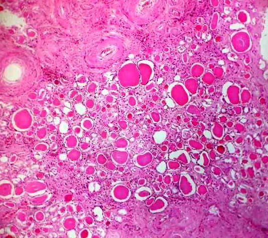
Sana Fatima et al; Saudi J Pathol Microbiol, Jan, 2021; 6(1): 46-52
Table-1: Descriptive analysis of Past History (N=100)
Parameter Frequency Percentage
Diabetes Mellitus
Yes 64 64.0
No 36 36.0
Hypertension
Yes 78 78.0
No 22 22.0
Table-2: Descriptive analysis of Neoplasm (N=100)
Parameter Frequency Percentage
Clear cell RCC 60 60.0
Papillary RCC 18 18.0
Renal Oncocytoma 6 6.0
Sarcomatoid RCC 6 6.0
Transitional cell carcinoma 4 4.0
Nephroblastoma 4 4.0
Chromophobe RCC 2 2.0
Fig-1: Bar chart of different types of neoplasm in the study population (N=100)
Table-3: Analysis of changes in non-neoplastic renal parenchyma (N=100)
Non neoplastic types Frequency Percentage
Normal 24 24.0%
Abnormal 76 76.0%
Diabetes Mellitus Nephropathy 36 36
Chronic Pyelonephritis 20 20
Hypertensive Nephrosclerosis 12 12
Immunoglobulin A nephropathy 4 4
Amyloidosis 4 4
Table-4: Analysis of non-neoplastic pathology with respect to neoplasm
Tumor type DM HTN IgA nephropathy Amyloidosis CPN No change
Clear Cell RCC 22 6 2 0 18 12
Papillary RCC 6 0 0 2 0 10
Chromophobe RCC 0 2 0 0 0 0
Renal Oncocytoma 2 2 0 0 0 2
Sarcomatoid RCC 2 0 2 0 2 0
© 2021 |Published by Scholars Middle East Publishers, Dubai, United Arab Emirates 48
Sana Fatima et al; Saudi J Pathol Microbiol, Jan, 2021; 6(1): 46-52

       Fig-2: Pie chart showing Analysis of Diabetic
      Nephropathy in clinically diabetic subjects (64)

                                                                           Fig-6: Histopathology of Diabetic Nephropathy showing
                                                                          glomerular enlargement with mild mesangial expansion(H
                                                                                                 & E -40X)

    Fig-3: Pie chart showing Analysis of Hypertensive
   Nephropathy in clinically hypertensive subjects (78)

                                                                           Fig-7: Histopathology of Diabetic Nephropathy showing
                                                                           insudative glomerular lesion FIBRIN CAP, which is the
                                                                            result of accumulation of plasma proteins (hyalinosis)
                                                                             between glomerular endothelium and the glomerular
                                                                                       basement membrane (H & E-10X)

    Fig-4: Microscopy of Clear Cell RCC (H&E -40x)

                                                                           Fig-8: Histopathology of Diabetic Nephropathy showing
                                                                            diffuse widening of mesangium with increased matrix
                                                                           deposition and mild hypercellularity (H & E Stain-10X)

     Fig-5: Microscopy of Papillary RCC (H&E-40x)

© 2021 |Published by Scholars Middle East Publishers, Dubai, United Arab Emirates                                                   49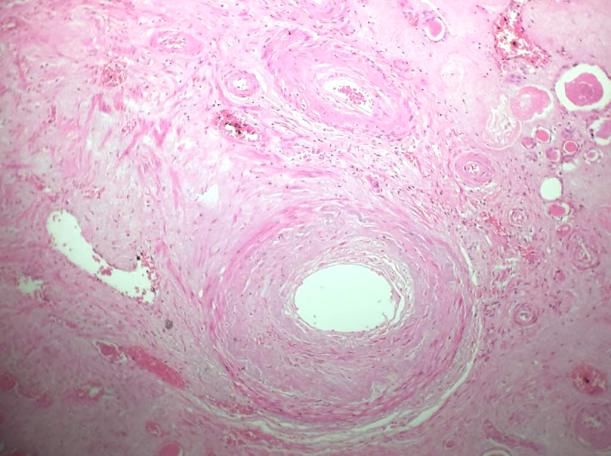
Sana Fatima et al; Saudi J Pathol Microbiol, Jan, 2021; 6(1): 46-52

Fig-9: Histopathology of Chronic Pyelonephritis showing
 atrophic tubules and dense mononuclear inflammatory
       cell infiltrate in the interstitium (H&E-10X)
                                                                            Fig-12: Histopathology of Hypertensive Nephropathy
                                                                          showing sclerotic glomeruli- GLOMERULOSCLEROSIS,
                                                                              thickening and hyalinization of wall of arterioles-
                                                                               HYALINE ARTERIOSCLEROSIS (H&E10X)

Fig-10: Histopathology of Chronic Pyelonephritis showing
thyroidization (colloid in the lumen) of atrophic tubules(H                 Fig-13: Histopathology of Hypertensive Nephropathy
                          &E-10X)                                           showing deposition of hyaline thrombi in the lumen of
                                                                               glomerular capillary loops (arrows) (H&E-10X)

                                                                          DISCUSSION
                                                                                   Progressive renal disease is an important
                                                                          public health problem showing increasing prevalence
                                                                          throughout the world leading to significant morbidity
                                                                          and mortality [10]. International studies in Canada,
                                                                          United States of America and Europe have
                                                                          demonstrated an increasing incidence in Renal Cell
                                                                          Carcinoma (RCC) with increase in rates by 20% to 30%
                                                                          in recent times [11]. The concerns after nephrectomy
                                                                          for RCC patients involve not only the oncological
                                                                          outcome, but also chronic kidney disease progression
                                                                          because of the large amount of nephron tissue loss. The
                                                                          adverse renal outcomes in patients undergoing
                                                                          nephrectomy for RCC were determined by population
  Fig-11: Histopathology of Hypertensive Nephropathy                      based analysis. Such obstacles have also been
  showing thickening and hyalinization of wall of small
                                                                          investigated in living recipients of kidney [12, 13].
           arteries and arterioles- HYALINE
  ARTERIOLOSCLEROSIS, which represents plasma                             Therefore, early recognition of patients at risk for
      proteins pushed into vessel walls (H&E-10X)                         chronic kidney disease and prompt institution of
                                                                          treatment may be of considerable importance [14].

© 2021 |Published by Scholars Middle East Publishers, Dubai, United Arab Emirates                                                   50
Sana Fatima et al; Saudi J Pathol Microbiol, Jan, 2021; 6(1): 46-52

          Types of neoplasm were assessed in the                          3.        Mohan, V., Sandeep, S., Deepa, R., Shah, B., &
current study with Clear cell RCC (60%) found to be                                 Varghese, C. (2007). Epidemiology of type 2
the highest and Chromophobe RCC (2%) was the                                        diabetes: Indian scenario. The Indian journal of
lowest. Remaining were Papillary RCC (18%), Renal                                   medical research, 125(3), 217-30.
Oncocytoma (6%), Sarcomatoid RCC (6%),                                    4.        Lindblad, P., Chow, W. H., Chan, J., Bergström,
Transitional cell carcinoma (4%) and Nephroblastoma                                 A., Wolk, A., Gridley, G., ... & Adami, H. O.
(4%) respectively. Similar results were seen in a study                             (1999). The role of diabetes mellitus in the
carried out by Amin et al., [15] where Clear cell RCC                               aetiology           of          renal          cell
was highest (63%) followed by Papillary RCC (18.5%)                                 cancer. Diabetologia, 42(1), 107-112.
and Chromophobe RCC (5.9%) was the lowest. Patard                         5.        Zucchetto, A., Dal Maso, L., Tavani, A.,
JJ et al., [16] also conducted a similar study and their                            Montella, M., Ramazzotti, V., Talamini, R., ... &
results were also close to those obtained in the current                            La Vecchia, C. (2007). History of treated
study, where Clear cell RCC, Papillary RCC and                                      hypertension and diabetes mellitus and risk of
Chromophobe RCC were 87.7%, 9.7% and 2.5%                                           renal cell cancer. Annals of Oncology, 18(3),
respectively.                                                                       596-600.
                                                                          6.        Hepps, D., & Chernoff, A. (2006, September).
         Our findings of non-neoplastic pathology in                                Risk of renal insufficiency in African-Americans
tumour nephrectomy specimens corroborate well with                                  after radical nephrectomy for kidney cancer.
the observations made by other researchers in earlier                               In Urologic Oncology: Seminars and Original
studies [13-19]. In our study, Diabetic Nephropathy was                             Investigations (Vol. 24, No. 5, pp. 391-395).
observed in 36% of the cases while Henriksen KJ et al.,                             Elsevier.
[18] reported Diabetic Nephropathy in 20% of the cases                    7.        Huang, W. C., Levey, A. S., Serio, A. M.,
and Salvatore SP et al., [19] reported the same in 7.34%                            Snyder, M., Vickers, A. J., Raj, G. V., ... &
of the cases.                                                                       Russo, P. (2006). Chronic kidney disease after
                                                                                    nephrectomy in patients with renal cortical
         Out of the total 100 specimens studied, 76%                                tumours: a retrospective cohort study. The lancet
showed significant non-neoplastic pathology which                                   oncology, 7(9), 735-740.
emphasizes the importance of medical follow up of the                     8.        Bijol, V., Mendez, G. P., Hurwitz, S., Rennke, H.
patient with regular laboratory investigations to assess                            G., & Nosé, V. (2006). Evaluation of the
renal function. Early institution of preventive and                                 nonneoplastic pathology in tumor nephrectomy
therapeutic management can delay progress of chronic                                specimens: predicting the risk of progressive
kidney disease. This can lead to prevention of End                                  renal failure. The American journal of surgical
Stage Renal Disease (ESRD) thereby improving the                                    pathology, 30(5), 575-584.
quality of life of nephrectomy patients.                                  9.        Henriksen, K. J., Meehan, S. M., & Chang, A.
                                                                                    (2007). Non-neoplastic renal diseases are often
CONCLUSION                                                                          unrecognized in adult tumor nephrectomy
         Chronic kidney disease is a major cause of                                 specimens: a review of 246 cases. The American
morbidity and mortality leading to significant burden on                            journal of surgical pathology, 31(11), 1703-1708.
health care system and low quality of life for the                        10.       Shrivastava, U., Misra, A., Mohan, V.,
patient. Given the relatively favourable 5-year survival                            Unnikrishnan, R., & Bachani, D. (2017). Obesity,
rates for renal cell carcinomas, accurate evaluation of                             diabetes and cardiovascular diseases in India:
the non-neoplastic kidney parenchyma is imperative.                                 public health challenges. Current diabetes
The practicing surgical pathologist should be aware of                              reviews, 13(1), 65-80.
the importance of both correctly classifying the renal                    11.       Williamson, T. J., Pearson, J. R., Ischia, J.,
neoplasm and the concomitant non-neoplastic kidney                                  Bolton, D. M., & Lawrentschuk, N. (2016).
disease that may be present but often overlooked.                                   Guideline of guidelines: follow‐ up after
Regular medical follow up and care with appropriate                                 nephrectomy for renal cell carcinoma. BJU
management of the underlying renal medical disorder is                              international, 117(4), 555-562.
crucial in preventing or delaying onset of end stage                      12.       Sejima, T., Honda, M., & Takenaka, A. (2015).
renal disease (ESRD).                                                               Renal parenchymal histopathology predicts life‐
                                                                                    threatening chronic kidney disease as a result of
REFERENCES                                                                          radical nephrectomy. International Journal of
1.     Tavani, A., & La, C. V. (1997). Epidemiology of                              Urology, 22(1), 14-21.
       renal-cell          carcinoma. Journal         of                  13.       Sejima, T., Yumioka, T., Yamaguchi, N.,
       nephrology, 10(2), 93-106.                                                   Iwamoto, H., Masago, T., Morizane, S., ... &
2.     Kabaria, R., Klaassen, Z., & Terris, M. K. (2016).                           Takenaka, A. (2016). Characterization of mild
       Renal       cell     carcinoma:     links     and                            and severe post-radical nephrectomy renal
       risks. International journal of nephrology and                               functional          deterioration        utilizing
       renovascular disease, 9, 45-52.                                              histopathological evaluation of non-neoplastic

© 2021 |Published by Scholars Middle East Publishers, Dubai, United Arab Emirates                                                   51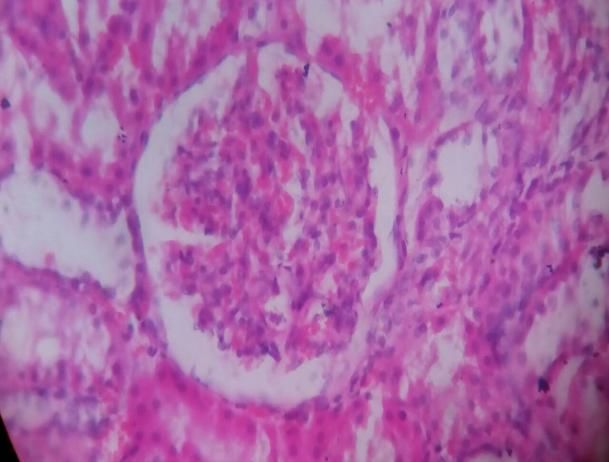
Sana Fatima et al; Saudi J Pathol Microbiol, Jan, 2021; 6(1): 46-52

       nephrectomized renal parenchyma. International                               multicenter experience. Journal of Clinical
       journal of clinical oncology, 21(3), 588-594.                                Oncology, 23(12), 2763-2771.
14.    Wee, J. W., Kang, H. R., Kwon, S. H., Jeon, J. S.,                 17.       Sarsık, B., Şimşir, A., Yılmaz, M., Yörükoğlu,
       Han, D. C., Jin, S. Y., ... & Noh, H. (2016).                                K., & Şen, S. (2013). Spectrum of nontumoral
       Clinical value of pathologic examination of non-                             renal pathologies in tumor nephrectomies:
       neoplastic kidney in patients with upper urinary                             nontumoral renal parenchyma changes. Annals
       tract malignancies. The Korean Journal of                                    of diagnostic pathology, 17(2), 176-182.
       Internal Medicine, 31(4), 739-749.                                 18.       Henriksen, K. J., Meehan, S. M., & Chang, A.
15.    Amin, M. B., Amin, M. B., Tamboli, P., Javidan,                              (2009). Nonneoplastic kidney diseases in adult
       J., Stricker, H., Venturina, M. D. P., ... & Menon,                          tumor nephrectomy and nephroureterectomy
       M. (2002). Prognostic impact of histologic                                   specimens:       common,        harmful,    yet
       subtyping of adult renal epithelial neoplasms: an                            underappreciated. Archives of pathology &
       experience of 405 cases. The American journal                                laboratory medicine, 133(7), 1012-1025.
       of surgical pathology, 26(3), 281-291.                             19.       Salvatore, S. P., Cha, E. K., Rosoff, J. S., &
16.    Patard, J. J., Leray, E., Rioux-Leclercq, N.,                                Seshan, S. V. (2013). Nonneoplastic renal
       Cindolo, L., Ficarra, V., Zisman, A., ... &                                  cortical scarring at tumor nephrectomy predicts
       Pantuck, A. J. (2005). Prognostic value of                                   decline in kidney function. Archives of
       histologic subtypes in renal cell carcinoma: a                               Pathology & Laboratory Medicine, 137(4), 531-
                                                                                    540.

© 2021 |Published by Scholars Middle East Publishers, Dubai, United Arab Emirates                                                   52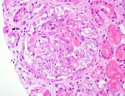
You can also read