Engineering CAR- NK cells to secrete IL- 15 sustains their anti- AML functionality but is associated with systemic toxicities
←
→
Page content transcription
If your browser does not render page correctly, please read the page content below
Open access Original research
Engineering CAR-NK cells to secrete IL-
J Immunother Cancer: first published as 10.1136/jitc-2021-003894 on 12 December 2021. Downloaded from http://jitc.bmj.com/ on July 1, 2022 by guest. Protected by copyright.
15 sustains their anti-AML functionality
but is associated with systemic toxicities
Ilias Christodoulou,1,2 Won Jin Ho,1 Andrew Marple,1 Jonas W Ravich,1 Ada Tam,1
Ruyan Rahnama,1,3 Adam Fearnow,1 Cambrynne Rietberg,4 Sean Yanik,4
Elena E Solomou,2 Ravi Varadhan,1 Michael A Koldobskiy,1,3
Challice L Bonifant 1,3
To cite: Christodoulou I, Ho WJ, ABSTRACT aggressive clinical course in both adults and
Marple A, et al. Engineering Background The prognosis of patients with recurrent/ children.1 2 Intensive chemotherapeutic regi-
CAR-NK cells to secrete IL- refractory acute myelogenous leukemia (AML) remains poor
15 sustains their anti-AML
mens with consolidative hematopoietic cell
and cell-based immunotherapies hold promise to improve transplantation (HCT) remain the standard
functionality but is associated
outcomes. Natural Killer (NK) cells can elicit an antileukemic of care, but these treatments can cause signif-
with systemic toxicities. Journal
for ImmunoTherapy of Cancer response via a repertoire of activating receptors that bind
icant short- term and long- term toxicities.
2021;9:e003894. doi:10.1136/ AML surface ligands. NK-cell adoptive transfer is safe but thus
far has shown limited anti-AML efficacy. Here, we aimed to Moreover, a subset of patients fail to respond
jitc-2021-003894
overcome this limitation by engineering NK cells to express to initial treatment or relapse after chemo-
►► Additional supplemental chimeric antigen receptors (CARs) to boost their anti-AML therapy±HCT. For this reason, targeted thera-
material is published online only. activity and interleukin (IL)-15 to enhance their persistence. pies with non-overlapping toxicity profiles are
To view, please visit the journal Methods We characterized in detail NK-cell populations aggressively being developed. The alpha chain
online (http://dx.d oi.org/10. expressing a panel of AML (CD123)-specific CARs and/or of the interleukin (IL)-3 receptor (CD123)
1136/j itc-2021-0 03894). IL-15 in vitro and in AML xenograft models. is highly expressed on both AML blasts and
Results CARs with 2B4.ζ or 4-1BB.ζ signaling domains leukemic stem cells,3 4 and has been shown
Accepted 14 November 2021 demonstrated greater cell surface expression and endowed
to be a safe target in clinical trials of immu-
NK cells with improved anti-AML activity in vitro. Initial in vivo
testing revealed that only 2B4.ζ Chimeric Antigen Receptor
notherapeutic agents.5–7 Leukemic stem cells
(CAR)-NK cells had improved anti-AML activity in comparison to are often resistant to chemotherapy and may
untransduced (UTD) and 4-1BB.ζ CAR-NK cells. However, the be most responsible for disease initiation and
benefit was transient due to limited CAR-NK-cell persistence. relapse.3 Thus, CD123 targeting therapies
Transgenic expression of secretory interleukin (sIL)-15 in 2B4.ζ could serve as valuable adjunct treatment
CAR and UTD NK cells improved their effector function in the modalities to achieve and/or sustain remis-
setting of chronic antigen simulation in vitro. Multiparameter sion in high-risk patients with AML.
© Author(s) (or their
flow analysis after chronic antigen exposure identified the Adoptive cell transfer is a form of anti-
employer(s)) 2021. Re-use
expansion of unique NK-cell subsets. 2B4.ζ/sIL-15 CAR
permitted under CC BY-NC. No cancer immunotherapy that has promise, and
commercial re-use. See rights and sIL-15 NK cells maintained an overall activated NK-cell
has been successful in the form of chimeric
and permissions. Published by phenotype. This was confirmed by transcriptomic analysis,
BMJ. which revealed a highly proliferative and activated signature in antigen receptor (CAR)–T cell infusions used
1
Sidney Kimmel Comprehensive these NK-cell groups. In vivo, 2B4.ζ/sIL-15 CAR-NK cells had to treat relapsed/refractory Acute Lympho-
Cancer Center, Johns Hopkins potent anti-AML activity in one model, while 2B4.ζ/sIL-15 CAR blastic Leukemia (ALL).8 CAR-T cells are also
University School of Medicine, and sIL-15 NK cells induced lethal toxicity in a second model. being tested against AML in several clinical
Baltimore, Maryland, USA
2
Conclusion Transgenic expression of CD123-CARs trials.9 CAR-T cell therapies have associated
Department of Internal and sIL-15 enabled NK cells to function in the setting severe toxicities including cytokine release
Medicine, University of Patras
of chronic antigen exposure but was associated with syndrome (CRS), immune effector cell associ-
School of Health Sciences,
systemic toxicities. Thus, our study provides the impetus ated neurotoxicity syndrome (ICANS),10 and
Patras, Western Greece, Greece
3 to explore inducible and controllable expression systems
Department of Pediatrics, Johns CAR- associated hemophagocytic lymphohis-
Hopkins University School of to provide cytokine signals to AML-specific CAR-NK cells
before embarking on early-phase clinical testing. tiocytosis.11 All Food and Drug Administration-
Medicine, Baltimore, Maryland,
USA
approved CAR-T cell therapies originate from
4
Department of Pediatrics, autologous hematopoietic starting material.
University of Michigan, Ann INTRODUCTION The manufacturing of patient-derived T-cell
Arbor, Michigan, USA Acute myelogenous leukemia (AML) is a products in the setting of a highly prolifera-
Correspondence to neoplastic disorder characterized by the accu- tive disease can be challenging.12 13 Delay in
Dr Challice L Bonifant; mulation of malignant myeloid precursor therapy associated with the time required for
cbonifa2@jh.e du cells in the bone marrow. AML has an per-patient CAR-T manufacturing may not be
Christodoulou I, et al. J Immunother Cancer 2021;9:e003894. doi:10.1136/jitc-2021-003894 1
Open access
J Immunother Cancer: first published as 10.1136/jitc-2021-003894 on 12 December 2021. Downloaded from http://jitc.bmj.com/ on July 1, 2022 by guest. Protected by copyright.
possible in the setting of uncontrolled AML. In addition, METHODS
intensive chemotherapy regimens, such as those admin- Details about cell lines, determination of vector copy
istered for AML treatment, are associated with poor ex number (VCN), cytotoxicity assays, cytokine secretion
vivo T-cell expansion.14 Donor T cells can be considered measurement, and RNAseq library preparation and align-
as a product source; however, the infusion of allogeneic T ment are provided in online supplemental methods.
cells is associated with the serious risk of graft-versus-host
disease (GVHD).15 CAR generation
NK cells are immune effector cells that play a pivotal CAR transgenes were designed using the CD123-specific
single chain variable fragment (scFv, 26292)25 sequence
role as first-line defenders against virally infected or
and the hinge (H), transmembrane (TM), and IC
tumor-transformed cells. Though they act with similar
domains indicated in figure 1A. Sequences were synthe-
cytotoxic mechanisms, NK- cell activation and func-
sized (GeneArt, Thermo Fisher Scientific) and subcloned
tion are distinct from those of T cells. NK cells express into pSFG retroviral vectors. All sequences were validated
a variety of activating receptors (eg, NKG2D, NKp30, by Sanger sequencing (Johns Hopkins Genetic Resources
NKp46, 2B4 and CD16)16 17 that directly interact with Core Facility).
cell surface ligands.16 18 These activating receptors asso-
ciate with coreceptors (ex. DAP10, CD3ζ and FcεRIγ) to CAR-NK-cell production
heighten directed cytotoxicity.17 The use of NK cells for Healthy donor peripheral blood mononuclear cells
immunotherapy has major advantages over the use of T (Anne Arundel Medical Blood Donor Center, Annapolis,
cells. NK cells do not directly cause GVHD, though they Maryland, USA; Carter Bloodcare, Woodway, Texas, USA)
may contribute inflammatory mediators that potentiate were isolated by Ficoll density gradient centrifugation
pre-existing pathology.15 19 Thus, they have the potential and depleted of T cells using CD3 microbeads (Militenyi
to be used as an off-the-shelf cellular product that can be Biotec, Cologne, Germany). The remaining cells were
manufactured on a large scale and can be readily available stimulated on day (D)0 with lethally irradiated K562
to patients. This has the advantage of decreasing produc- feeder cells26 expressing membrane bound IL- 15 and
tion costs and preventing manufacturing associated treat- 4-1BB ligand at a 1:1 ratio. Cells were maintained in SCGM
ment delays. Adoptive NK-cell transfer has been shown media (CellGenix, Freiburg, Germany) supplemented
with 10% fetal bovine serum, 2 mmol/L GlutaMAX
to be safe in clinical trials, without associated CRS or
(Thermo Fisher), and 200 IU/mL human interleukin
ICANS.20 Because of this safety profile and the expression
(hIL)-2 (Biological Resources Branch Preclinical Biore-
of NK activating ligands on AML cells,18 NK-cell therapy
pository, National Cancer Institute, Frederick, Maryland,
has been tested in clinical trials. However, when used as USA). NK- cell purity was verified with flow cytometry
AML treatment, NK-cell adoptive transfer induces only using fluorophore conjugated antibodies against CD56
transient remission, and additional therapies are needed and CD3 (online supplemental table 1). NK cells were
to achieve durable responses.21–24 transduced on D4 of culture using transiently produced
In this study, we engineered NK cells to express CARs replication incompetent RD114 pseudotyped retroviral
with intracellular (IC) domains rationally designed as particles immobilized on RetroNectin (Clontech Labora-
those predicted to enhance NK- cell antitumor func- tories, Palo Alto, California, USA).
tionality. We demonstrate that NKs expressing CARs
targeting CD123 show potent antigen-dependent acti- Flow cytometry
vation and cytotoxicity. We find CAR expression to Antibodies for NK and cancer cell identification targeted
be stable and high across a panel of tested receptors CD56 and CD33 (AML cell lines) or CD19 (Raji) markers.
incorporating NK-specific activating and costimulatory A detailed list of all antibodies, including those used for the
molecules, with optimal functionality associated with evaluation of immune cell phenotype is in online supple-
2B4.ζ and 4-1BB.ζ containing CARs. CAR-NK cells did mental table 1. CAR expression analysis was performed using
not demonstrate improved expansion or persistence incubation with His- tagged recombinant CD123 protein
(SinoBiological, Beijing, China) and secondary staining with
when compared with unmodified NK cells in vivo. We
αHis-PE or αHis-APC (BioLegend, San Diego, California,
therefore added constitutive IL- 15 secretion to our
USA). Dead cells were excluded from analysis using LIVE/
CAR-NK cells and observed that this supported an acti-
DEAD Fixable Viability Stains 780 or 575V (BD Biosciences,
vated phenotype that led to enhanced expansion and Franklin Lakes, New Jersey, USA). Cell enumeration was
antitumor cytotoxicity. NK-cell activation translated to performed with CountBright counting beads (Thermo
improved NK-cell persistence and expansion in vivo. Fisher). Human Fc receptors were blocked using Human
However, constitutive cytokine secretion was also associ- TruStain FcX (BioLegend). In samples stained with multiple
ated with severe toxicity in one animal model. Prior to BD Horizon Brilliant reagents, Brilliant Stain Buffer Plus
clinical translation, alternate strategies to activate cyto- (BD Horizon) was used. Compensation was performed with
kine signaling in CAR-NK cells should be investigated UltraComp eBeads Compensation Beads (Thermo Fisher).
with a focus to include safety. Cell surface antigens were quantified using microspheres of
2 Christodoulou I, et al. J Immunother Cancer 2021;9:e003894. doi:10.1136/jitc-2021-003894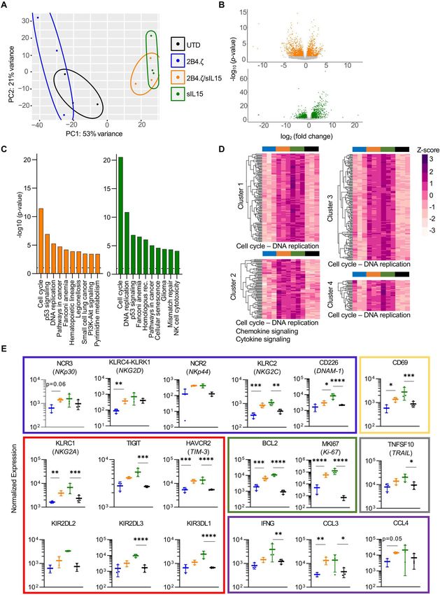
Open access
J Immunother Cancer: first published as 10.1136/jitc-2021-003894 on 12 December 2021. Downloaded from http://jitc.bmj.com/ on July 1, 2022 by guest. Protected by copyright.
Figure 1 NK cells engineered with anti-CD123 CARs have antigen-specific functionality. (A) Schema of CAR design. All CARs
bind CD123 via an extracellular scFv. The H, TM and IC domains of the CARs are as indicated. Colored boxes represents each
particular CAR with colors carried through each figure. (B) Percentage (%) of CAR(+) NK cells detected on D8 and D18. (C)
Bar plot comparing the percentage of CAR(+) NK cells with indicated TM domains on D8. (D) Absolute number of target cells
measured after 72 hours of coculture with indicated NK cells. Initial target cell count was 100 000 in 1:1 and 250 000 in 1:5 E:T
ratio conditions. Each bar representative of the mean±SEM; each dot is representative of individual NK cell donor. n=3 donors.
*P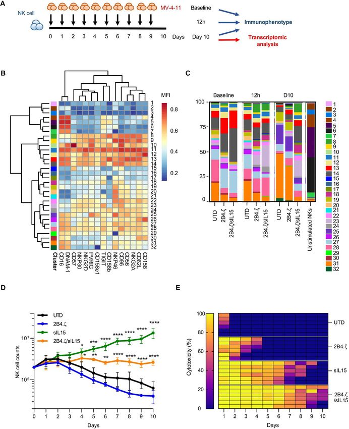
Open access
J Immunother Cancer: first published as 10.1136/jitc-2021-003894 on 12 December 2021. Downloaded from http://jitc.bmj.com/ on July 1, 2022 by guest. Protected by copyright.
the Quantum APC Molecules of Equivalent Soluble Fluoro- bioluminescence (BL) was measured using IVIS Spec-
chrome kit (Bangs Laboratories, Fisher, Indiana, USA). All trum (In Vivo Imaging System). Data were analyzed using
samples were acquired on FACSCelesta or FACSymphony Living Image Software V.4.7.3 (PerkinElmer, Waltham,
Cell Analyzers (BD Biosciences) and analyzed with FlowJo Massachusetts, USA). When indicated, peripheral blood
software V.10.6.1 and V.10.7.2. Cell sorting was performed on (PB) was drawn via facial vein; red blood cells were lysed
FACSMelody (BD Biosciences). with eBioscience RBC Lysis Buffer (Thermo Fisher); and
the remaining cells were analyzed with flow cytometry.
Immunophenotype analysis Bone marrow and spleen were harvested, and tissues were
Two different panels (A and B) were used for evaluation of analyzed with flow cytometry. Analysis of PB for cytokines
NK-cell receptors and one panel (C) for receptor ligands. (hIL-15, hTumor Necrosis Factor (TNF)-α, mouse inter-
Data analysis of the multiparameter panels A and B was
leukin (mIL)-6, and mIL-1β) was performed with ELISA
performed in R V.3.6.2. The median marker intensities
(R&D Systems). Mice were euthanized when they exhib-
were transformed using arcsinh (inverse hyperbolic sine)
ited >20% weight loss, hind limb paralysis, or moribund
with cofactor 150.27 Non-linear dimensionality reduction on
state as per protocol guidelines.
randomly selected 500 data points per sample of each panel
was performed using uniform manifold approximation and
projection (UMAP).28 NK-cell clusters were identified with Statistical analysis
the FlowSOM V.1.18.0 algorithm, and 40 different metaclus- All statistical analyses was performed using GraphPad
ters were generated per panel.29 Subsequently, we manually Prism Software V.9.2.0. Our comparisons included more
merged hierarchically neighboring clusters similar in biology than three groups and ordinary one-way or two-way anal-
and median marker intensities. Panel A clusters do not ysis of variance (ANOVA) corrected using the method of
correlate with the ones in panel B. Bonferroni. Data with variance of several logs of magni-
tude were log transformed (Y=log(Y)) before analysis with
Serial stimulation assay ANOVA. Survival of mice was estimated by the Kaplan-
NK cells were stimulated daily with MV-4-11 cells at a 1:1 Meier method, and differences in survival between groups
effector:target (E:T) ratio in G-rex plates (Wilson Wolf, were calculated by log-rank (Mantel-Cox) test.
New Brighton, Minnesota, USA) for a total of 10 days.
NK-cell proliferation and cytotoxicity was measured using
flow cytometric analysis. Percent (%) cytotoxicity was
calculated based on the target cell numbers on the day of RESULTS
(Y) and the day after (X) stimulation using the formula CD123-CARs are highly expressed on the NK-cell surface
100×(X−Y)/(Y). Cell phenotype was evaluated at base- We considered NK- cell biology in our design of eight
line, on the 1st (12 hours) and the 10th (D10) days. different NK-tailored CARs (figure 1A) to complement the
common 4-1BB.ζ CAR.33 34 All CARs are composed of an
RNA sequencing extracellular scFv targeting CD123.25 The H, TM, and IC
On D10 of serial stimulation, coculture was depleted portion of our CARs consisted of different combinations
first of dead cells using the Dead Cell Removal Kit and of activating coreceptors DAP10 and FcεRIγ, the costimu-
next of leukemia cells with CD33 microbeads (Militenyi). latory receptor 2B4, and the ζ chain of the T-cell receptor
NK- cell purity was verified with flow cytometry using (figure 1A). All CARs were expressed stably on the surface
fluorophore- conjugated CD56 and CD33 antibodies. of primary human NK cells for at least 2 weeks in culture,
RNA was extracted from NK cells using the RNeasy
with transduction efficiencies ranging from 21% to 98%
Mini Kit (Qiagen, Hilden, Germany) and RNAseq was
(figure 1B). Representative flow cytometric plots are shown
performed (online supplemental methods). Differential
in online supplemental figure 1A). CARs encoding 2B4
expression analysis and statistical testing were performed
or CD8α TM domains demonstrated higher transduc-
using DESeq2 software.30Mice were injected with 1×10⁶
tion efficiencies (medians 89 (range 53%–98%) and 84
MV-4-11 cells modified
(range 75%–90%), respectively) than constructs containing
Xenograft mouse model FcεRIγ or DAP10 TM (medians 62 (range 21%–77%) and
All animal studies were carried out under protocols 39 (24%–64%), respectively; figure 1B,C). 2B4 and CD8α
approved by the Johns Hopkins Institutional Animal Care TM domains also conferred optimal CAR surface density
and Use Committee. NSG (NOD.Cg-PrkdcscidIl2rgtm1Wjl/ as estimated by comparative mean fluorescence intensi-
SzJ) mice 6–8 weeks old were obtained from an internal ties (MFIs±SEM: 2B4-TM 2158±242, CD8α-TM 3254±970,
colony that originated from the Jackson Laboratory (Bar FcεRIγ-TM 827±151, DAP10-TM 366±23; 2B4 and CD8α vs
Harbor, Maine). Mice were injected with 1×10⁶ MV-4-11 DAP10: p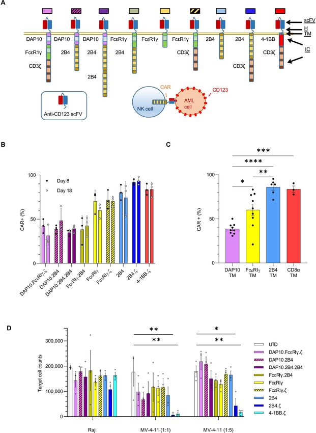
Open access
J Immunother Cancer: first published as 10.1136/jitc-2021-003894 on 12 December 2021. Downloaded from http://jitc.bmj.com/ on July 1, 2022 by guest. Protected by copyright.
CD123-CAR NK cells have antigen-specific anti-AML activity accomplish this, we cloned a sequence encoding human
in vitro IL-15 downstream of an Internal Ribosome Entry Site
We evaluated the target specificity of our CAR-NK cells (IRES) element into our 2B4.ζ CAR vector. We simultane-
using the CD123- positive MV-4-
11 and CD123- negative ously generated a second retroviral vector encoding IL-15
Raji cell lines. When challenged with MV-4-11 in cocul- and the fluorescent molecule mOrange as a control with
ture assays, all CAR-NK cells responded with enhanced IL-15, but not CAR expression (figure 3A). We verified
cytokine secretion above that seen when using unmodi- transduction by measuring VCN per cell (median: 2B4.ζ
fied NK cells under identical conditions (mean percent 10 (range 5.8–14), 2B4.ζ/sIL- 15 4.1 (range 3.8–4.7),
(%) change of interferon gamma (IFN-γ) secretion; sIL-15/mOrange 16.6 (range 4.8–19.2); figure 3B). We
CAR- NKs (range): 65%–313% vs unmodified: 14%; found no significant difference in VCN/cell or in CAR
online supplemental figure 2). There was no difference expression between NK cells transduced with 2B4.ζ or
in cytokine production of CAR(+) and CAR(−) NK cells 2B4.ζ/sIL-15 encoding retroviral vectors (range CAR(+):
after coculture with CD123(−) targets (online supple- 2B4.ζ vs 2B4.ζ/sIL-15 70%–97% vs 81%–96%, p>0.99,
mental figure 2). Next, we assessed CAR-NK-cell cytotox- MFI±SEM: 2B4.ζ vs 2B4.ζ/sIL-15 2566±531 vs 2089±424;
icity against CD123(+) target cells in 72-hour coculture p>0.99; figure 3C and online supplemental figure 4A).
assays. We found that CARs with 2B4 or 4-1BB costimu- Expression of IL- 15 was measured with quantitative
latory and TCRζ signaling domains endowed NK cells reverse transcription (qRT)- PCR (online supplemental
with the greatest cytolytic activity against MV-4-11 at both figure 4B) and IL-15 secretion was confirmed by ELISA
1:1 (mean % cytotoxicity±SEM: 2B4.ζ 93.8±5%, 4-1BB.ζ (figure 3D).
89.9±6.8%) and 1:5 E:T ratios (82.9±11.6% and 93±2.6%, We evaluated short-term cytotoxicity of 2B4.ζ/sIL-15
respectively; figure 1D). CD123(−) Raji cells were again CAR-NKs against the CD123(+) MV-4-11, MOLM-13, and
used as controls. There was no difference between Raji.CD123 cell lines. The parental CD123(−) Raji line
CAR-NK versus unmodified NK cell-mediated cytotoxicity was used as a negative control. When compared with
against Raji cells, confirming specificity (figure 1D). 2B4.ζ, the 2B4.ζ/sIL- 15 CAR- NKs had higher cytotox-
icity against CD123+ targets (figure 3E–G). Both 2B4.ζ
CD123-2B4.ζ CAR-NK cells have limited anti-AML efficacy in and 2B4.ζ/sIL- 15 demonstrated antigen- specific cyto-
vivo toxicity as Raji.CD123 were more effectively killed than
Given the superior anti-AML activity of 2B4.ζ and 4-1BB.ζ parental (CD123−) Raji cells (figure 3G). Target cell
CAR-NK cells in our in vitro assays, we next evaluated CD123 and IL-15Rα expressions were quantified in order
their antitumor activity in a xenograft model of human to evaluate for any effect of CD123 surface density or
AML. NSG mice were first engrafted with MV-4-11.ffLuc IL-15 trans presentation on NK-cell cytotoxicity (online
cells,31 then treated with CAR-NK or unmodified NK cells supplemental figure 5A). Differences in measured CD123
on D7 (figure 2A). Leukemic growth was measured with surface density did not correlate with observed short-
serial BL imaging. 2B4.ζ CAR-NK-cell postinjection had term cytotoxicity, underlining the existence of additional
transient anti-AML activity, which translated into a signif- complex mechanisms affecting NK-cell activation. Simi-
icant survival advantage in comparison to other experi- larly, differences observed in IL-15Rα expression did not
mental groups (median survival in days: 2B4.ζ vs 4-1BB.ζ correlate with cytotoxicity (online supplemental figure
63 vs 56, p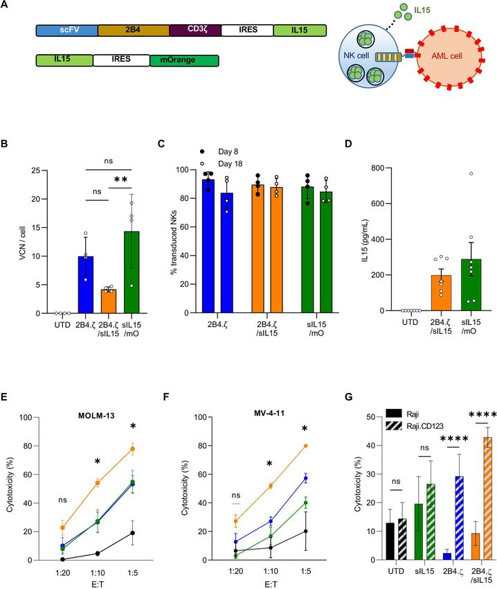
Open access
J Immunother Cancer: first published as 10.1136/jitc-2021-003894 on 12 December 2021. Downloaded from http://jitc.bmj.com/ on July 1, 2022 by guest. Protected by copyright.
Figure 2 Anti-CD123.2B4.ζ CAR-NKs have transient anti-AML activity in vivo. (A) Schematic of MV-4-11 xenograft model.
On D0, NSG mice were injected via tail vein with 1×10⁶ CD123(+) MV-4-11 cells that express ffLuc (MV-4-11.ffLuc cells). In
treatment groups, 10×106 NK cells were administered on D7. Cohorts: UTD/unmodified, 4-1BB.ζ CAR-NK, and 2B4.ζ CAR-NK.
(B) Leukemia proliferation was monitored with bioluminescence imaging and was recorded as photons/s/cm²/sr; n=8–12 mice
per group. Magnification of D7–D21 shown. (C) Representative images of three mice per condition. Minimum and maximum
values of color scale are depicted at the top (min–max). (D) Kaplan-Meier survival analysis of MV-4-11 xenografts (n=8–12 mice
per condition). (E) Mouse PB collected at indicated time points and analyzed via flow cytometry. Each dot represents a single
mouse. Solid line: median. At later time points, NK-cell count was undetectable for all groups and is not plotted. *POpen access
J Immunother Cancer: first published as 10.1136/jitc-2021-003894 on 12 December 2021. Downloaded from http://jitc.bmj.com/ on July 1, 2022 by guest. Protected by copyright.
Figure 3 Simultaneous 2B4.ζ CAR expression and IL-15 secretion strengthens NK-cell cytotoxicity. (A) Schema of vectors
and IL-15 secretion from CAR-NK cells. (B) Quantification of retroviral VCN in transduced NK cells. UTD NK cells served as
negative controls (n=4 donors). (C) Percentage (%) of transduced NK cells in cultures on D8 (black circle) and D18 (white
circle, n=4 donors). (D) NK-cell supernatant was used for quantification of IL-15 by ELISA (n=7 biological replicates using four
donors). BL-based cytotoxicity assays were performed using the CD123(+) AML cell lines MV-4-11(E) and MOLM-13 (F) with
ffLuc expression (n=4 donors). Asterisks indicate 2B4.ζ/sIL-15 versus 2Β4.ζ comparison. (G) Bar graph comparing percent
(%) cytotoxicity of NK cells against Raji (CD123(−): solid), and Raji (CD123 (CD123(+): diagonal stripe) cancer cell lines at 1:10
E:T ratio (n=4 donors). (B-G) Mean±SEM represented. *POpen access
J Immunother Cancer: first published as 10.1136/jitc-2021-003894 on 12 December 2021. Downloaded from http://jitc.bmj.com/ on July 1, 2022 by guest. Protected by copyright.
Figure 4 IL-15 maintains NK cell-activated phenotype in a model of chronic antigen stimulation. (A) Schematic representation
of our serial stimulation assay. D0 was the day of the initial seeding of the coculture; D1 was the first; and D10 was the last
day of cell quantification. Immunophenotypical analysis of effector and target cells performed at baseline (before coculture),
12 hours after first stimulation, and on D10 (n=1 donor). Transcriptomic analysis performed on D10 (n=3 donors). (B) Heatmap
of flow cytometry data showing expression of 15 different NK-cell surface markers. Heatmap coloring represents arcsinh
transformed median marker intensity. (C) Bar plots of relative abundance of the 32 population subsets found in each sample.
(D) NK-cell counts over a period of 10 days. Initial seeding count was two million NK cells (mean±SEM, n=3 donors). Asterisks
indicate 2B4.ζ/sIL-15 versus 2Β4.ζ and sIL-15 versus UTD comparison. (E) Heat map of the percent (%) NK-cell cytotoxicity.
Each column represents the specific day and each row a unique biological replicate (n=3 donors). D, day; IL, interleukin; MFI,
mean fluorescence intensity; NK, natural killer; sIL, secretory interleukin; UTD, untransduced.
8 Christodoulou I, et al. J Immunother Cancer 2021;9:e003894. doi:10.1136/jitc-2021-003894Open access
J Immunother Cancer: first published as 10.1136/jitc-2021-003894 on 12 December 2021. Downloaded from http://jitc.bmj.com/ on July 1, 2022 by guest. Protected by copyright.
changes in population density of these subsets were NK cells were isolated, and RNA libraries were prepared
observed on D10 (figure 4B,C). The unique 2Β4.ζ/sIL-15 and used for RNAseq analysis. Samples clustered by IL-15
immunophenotype is highlighted by comparison of the secretion, with overlap between 2B4.ζ/sIL-15 and sIL-15
MultiDimensional Scaling (MDS, global) and UMAP conditions. These clearly separated from NK cells that did
(cluster-specific) plots in online supplemental figure 8. not secrete IL-15 (UTD and 2B4.ζ, figure 5A). Differential
Clustering of NK cells based on expression of markers gene expression analysis (DESeq2) of 2B4.ζ/sIL-15 versus
included in our second receptor panel (panel B) again 2B4.ζ and sIL-15 versus UTD NKs identified in the IL-15-
supported the maintained NK-cell activation to D10 in secreting NK-cell differences in expression of genes in the
2B4.ζ/sIL- 15 CAR- NK cells (predominant clusters 16, pathways of DNA replication, cell cycle progression, and
20, and 26 and minor clusters 11, 13, 14, 15, 21, and NK cell-mediated cytotoxicity (figure 5B,C). Kyoto Ency-
23; online supplemental figures 6–9). Differences were clopedia of Genes and Genomes (KEGG) enrichment
again observed in 2B4.ζ and unmodified NK cells on D10 analysis revealed cell cycle progression as the top-ranked
compared with earlier time points (increasing percentage pathway for both comparisons (figure 5C). No biologically
of cells populating clusters 2, 4, 17, and 18, and decreasing relevant pathways were found to be significantly enriched
percentages of 14, 16, 20, nd 26; online supplemental when comparing IL-15-secreting CAR-NK cells to non-
figure 6). Clusters 14, 16, 20, and 26 expressed higher CAR sIL-15 NKs (online supplemental figures 13A,B).
levels of LFA-1, CD69, TRAIL, TIM-3, NKG2A, and KLRG1 Hierarchical clustering of differentially expressed genes
compared with clusters 2, 4, 17, and 18 (online supple- also revealed upregulation of genes involved predomi-
mental figure 9). Negligible differences were observed in nantly in cell cycle progression, chemokine, and cytokine
PD-1, LAG-3, FASL, 2B4, and NKG2C expression. Taken signaling in IL- 15-
secreting NK conditions (figure 5D
together, 2B4.ζ/sIL-15 CAR-NKs had a higher percentage and online supplemental table 2). We used this dataset to
of NK cells populating clusters defined by higher surface evaluate differential expression of molecules of biological
expression of LFA- 1 (adhesion/activation receptor), relevance. 2B4.ζ/sIL-15 and sIL-15 NK cells had higher
CD69 (activation marker), TRAIL (death receptor), expression of genes encoding for NK activating recep-
and TIM-3 (commonly upregulated after NK-cell activa- tors (NCR2 (NKp44), NCR3 (NKp30), KLRC2 (NKG2C),
tion), as well as NKG2A and KLRG1 (inhibitory recep- KLRC4-KLRK1 (NKG2D), CD226 (DNAM- 1), and
tors; figure 4B,C, and online supplemental figures 6–10). FCGR3A (CD16)), adaptor molecules (FCER1G (FcεRIγ)),
Overall, the data from both antibody panels suggest that death receptor ligands (TNFSF10 (TRAIL)), granzyme
with continuous antigen stimulation, IL-15 preserves an (granzyme A (GZMA), proinflammatory cytokines and
activated CAR- NK-cell phenotype associated with high chemokines (IFNG (IFN-γ), CCL1, CCL3, CCL4, XCL1,
cytotoxicity. We also analyzed the expression of NK-cell XCL2, and CCL3L3), activation markers (CD69), prolifer-
activating and inhibitory ligands. AML cells expressed ation markers (MKI67 (Ki-67)), antiapoptosis regulators
high levels of MICA/MICB (NKG2D ligands), CD112 and (BCL2), and adhesion molecules (ITGB2 (integrin-β2),
CD155 (DNAM-1, PVRIG, CD96, and TIGIT ligands) and CD2, and CD53; figure 5E and online supplemental figure
PDL1 (PD-1 ligand), but not ULBP1 (NKG2D ligand) or 13C). We also observed upregulation of select inhibitory
galectin-9 (TIM-3 ligand, online supplemental figure 11). receptors (NKG2A, CEACAM1, and LILRB1) including
Next, we evaluated NK-cell cytotoxicity and persistence inhibitory killer Ig- like receptors (KIRs; KIR2DL1,
in our model of chronic antigenic stimulation (figure 4A). KIR2DL2, KIR2DL3, KIR2DL4, and KIR3DL1) as well as
While unmodified and 2B4.ζ CAR-NK cells sharply dimin- ‘checkpoint’ molecules (HAVCR2 (TIM- 3) and TIGIT,
ished in number over the course of this assay, NK cells figure 5E and online supplemental figure 13C). KIRs
genetically engineered to express IL- 15 (2B4.ζ/sIL-15 are subject to extensive regulatory splicing and there is
and sIL-15) survived throughout the experiment (n=3, an inability to distinguish KIR with identical extracel-
figure 4D). As predicted by the short-term cytotoxicity lular domains but functionally disparate IC tails by flow
assay, 2B4.ζ CAR- NK cells had high initial anti- AML cytometry, making transcriptional analysis necessary and
activity. However, their killing potential decreased as complementary for study of KIR expression. Analysis of
2B4.ζ CAR- NK- cell counts declined. Cytotoxicity was the chemokine receptor expression showed upregulation
completely abrogated by D5 (figure 4E). IL-15 activation of CCRL2, CCR1, CCR5, and CCR6 with similar or lower
alone (without CAR expression) also had an early anti- expression of CX3CR1, CXCR3, and CXCR4 in IL- 15-
tumor effect that was not sustained. Only the combination secreting NKs (online supplemental figure 13C).
of CAR expression and IL-15 secretion led to continued
NK cell-mediated anti-AML cytotoxicity (figure 4E and Constitutive IL-15 expression improves NK cell in vivo
online supplemental figure 12). persistence but causes lethal toxicity
We next evaluated IL-15-secreting NK cells in two AML
NK cells secreting IL-15 exhibit a highly proliferative and xenograft models. In our first experiment, we used our
activated transcriptomic signature after chronic antigen MV-4-11 xenograft model (figure 2A) and our previous
stimulation NK-cell dosing regimen (online supplemental figure 14A).
We evaluated the transcriptional programs of our NK Surprisingly, MV-4-11 engrafted mice treated with 10×106
cells on the 10th day of continuous antigen stimulation. IL-
15-secreting NK cells had early mortality (median
Christodoulou I, et al. J Immunother Cancer 2021;9:e003894. doi:10.1136/jitc-2021-003894 9Open access
J Immunother Cancer: first published as 10.1136/jitc-2021-003894 on 12 December 2021. Downloaded from http://jitc.bmj.com/ on July 1, 2022 by guest. Protected by copyright.
Figure 5 IL-15 stimulation of NK cells upregulates genes important to cell cycle progression, NK-cell activation, and
cytotoxicity. (A) PCA of NK-cell transcriptome on D10 of the chronic antigen stimulation. Each dot represents a unique NK-cell
donor (n=3 donors). (B) Volcano plots representing differentially regulated genes in 2B4.ζ/sIL-15 compared with 2B4.ζ (orange)
and sIL-15 compared with unmodified cells (green). Gray dots are those not meeting criteria: p value ≥0.05; fold change > 2
orOpen access
J Immunother Cancer: first published as 10.1136/jitc-2021-003894 on 12 December 2021. Downloaded from http://jitc.bmj.com/ on July 1, 2022 by guest. Protected by copyright.
survival (days): 2B4.ζ/sIL-15: 25, sIL-15: 21; n=5 mice with 2B4.ζ CAR-NK cells as compared with those treated
each group; online supplemental figure 14B). Circulating with 4-1BB.ζ CAR-NK cells, which suggests a potential
human IL-15 and NK-cell counts both increased in the additive effect of 2B4 and CD3ζ signaling. However,
3 weeks from NK-cell injection to death (online supple- this in vivo effect was short-lived and was accompanied
mental figure 14C,D). Therefore, in our next experi- by circulating NK-cell decline. We thus engineered our
ment we decreased our treatment dose of infused NK 2B4.ζ CAR-NK cells with an IL-15 transgene to promote
cells in order to evaluate whether we could mitigate the IL-15-
mediated activation, proliferation, and survival.
observed treatment associated mortality. We compared a Secretion of IL- 15 stimulated CAR- NK-cell expansion
single decreased dose of 2B4.ζ/sIL-15 CAR-NK cells with both in vitro and in vivo, and enhanced short-term and
multiple doses of 2B4.ζ CAR-NK cells (figure 6A). Infu- long-term anti-AML cytotoxicity. This bolstered activation
sion of a single dose of 3×10⁶ 2B4.ζ/sIL-15 CAR-NK and profile was confirmed by immunophenotypical and tran-
three doses of 3×10⁶ 2B4.ζ CAR-NK cells both had tran- scriptomic analysis in the setting of chronic stimulation.
sient AML control (figure 6B,C). Three doses of 2B4.ζ However, in one in vivo AML model, constitutive IL-15
CAR- NKs prolonged survival, as compared with mice secretion caused dramatic NK-cell expansion and high
in all other treatment groups (median survival in days; levels of circulating human proinflammatory cytokines,
2B4.ζ vs UTD and untreated; 71 vs 48 and 49, pOpen access
J Immunother Cancer: first published as 10.1136/jitc-2021-003894 on 12 December 2021. Downloaded from http://jitc.bmj.com/ on July 1, 2022 by guest. Protected by copyright.
Figure 6 IL-15 secreting CAR-NK cells can cause lethal toxicity in an AML xenograft model. (A) Schematic of NK-cell dosing
in MV-4-11.ffLuc model. (B) MV-4-11 proliferation was monitored with BLI. Representative images of mice. The minimum and
maximum values of the color scale are indicated (min–max). (C) BL representative of leukemia proliferation was recorded as
photons/s/cm²/sr . Dotted lines: individual mice; solid lines: mean (n=5–7 mice per group; two experiments performed for UTD
and 2Β4.ζ cohorts). (D) Kaplan-Meier survival analysis. (E) Mouse PB was collected at indicated time points and analyzed
via flow cytometry. NK-cell numbers per microlitre of mouse PB were tracked starting on D13 of the experiment. Each dot
represents cell numbers from a single mouse; line is at median. Asterisks indicate 2B4.ζ/sIL-15 versus 2Β4.ζ comparison.
(F) Human IL-15 from PB of MV-4-11 engrafted mice drawn at the indicated time points was quantified (pg/mL) with ELISA.
Asterisks indicate 2B4.ζ/sIL-15 versus 2Β4.ζ comparison. (G) Schematic of NK-cell dosing in MOLM-13.ffLuc model. (H)
MOLM-13 proliferation was monitored using BLI (n=5 mice per group). Representative images, (I) radiance, and (J) Kaplan-
Meier survival analysis. *POpen access
J Immunother Cancer: first published as 10.1136/jitc-2021-003894 on 12 December 2021. Downloaded from http://jitc.bmj.com/ on July 1, 2022 by guest. Protected by copyright.
safety, immediate availability once manufactured and human or murine T cells in our NSG model. However,
stored, and reduced manufacturing costs as compared the possibility of lethal toxicity due to NK-cell alloreac-
with per-patient manufacture of autologous cell therapy tivity against mouse cells cannot be excluded.
products.12 13 Historically, one challenge facing PB-NK- We observed that IL-15-secreting CAR-NK cells both
cell engineering was that of poor viral and non- viral prolonged and shortened survival in two xenograft
genetic modification.42 With our method, we are able models of AML. Notably, the median survival of mice
to achieve high levels of PB-NK transduction. All of our treated with the same dose of 2B4.ζ/sIL-15 NK cells was
CARs were stably expressed on the surface of primary NK similar in each model (MOLM-13: 27 days, MV-4-11: 26
cells, though inter-CAR variability in surface density was days). This could potentially be explained by the aggres-
observed. siveness of the MOLM- 13 model, as these mice died
NK cells have a natural life span of approximately 2 of widespread disease at roughly the same time as we
weeks in humans. The success of adoptively transferred recorded the onset of treatment-associated mortality. Our
cellular therapies for cancer is determined, in part, by observation of unexpected toxicity begs for expanded
effector cell persistence. The use of systemic cytokine study of novel ways to stimulate CAR-NK-cell cytokine
supplementation is one common strategy employed to receptor pathways while circumventing systemic cytokine
support prolongation of NK-cell survival. The short half- delivery. Localization of IL-15 with membrane tethering55
life of infused IL-15 necessitates frequent or continuous or targeted delivery through the use of oncolytic viruses56
administration, and systemic toxicity is common.43–46 IL-15 are alternative therapeutic strategies. Controllable cyto-
super agonists, like N-803 (formerly known as ALT-803), kine expression using engineered inducible systems and
have a longer half-life and can mimic physiological IL-15 safety switches also holds promise. Specific activation of
trans presentation, but administration can cause inflam- intrinsic gamma-cytokine receptor signaling without the
matory toxicities.47 The administration of N-803 has been use of a pharmacological agent is another strategy that
effective at promoting NK- cell proliferation and anti- has the potential to sustain NK- cell function with an
tumor efficacy against hematological malignancies, with improved safety profile.
expected associated fever, chills, and injection site rashes In conclusion, our data underscore the critical nature
observed.48–51 Another strategy that has been successful of IL-15 signaling as a stimulant of CAR-NK-cell long-
in specifically supporting in vivo CAR-NK-cell survival is term survival and anti- AML functionality. Our in vivo
engineering constitutive activating cytokine expression.52 data highlight the limitations in murine modeling and
This approach has been shown to be safe in a clinical trial the necessity for caution if the use of IL-15 secreting NK
using CD19-CAR NK cells against CD19+ lymphoid malig- cells is planned in human trials of CAR-NK-cell adoptive
nancies.53 The demonstrated safety profile motivated transfer. Continued therapeutic development is impera-
us to also test transgenic IL-15 expression with a goal of tive, to include testing of alternate and potentially safer
enhanced in vivo CAR-NK cell persistence. We found NK methods of cytokine pathway activation.
cells subject to activation with constantly available IL-15
exhibited enhanced and sustained in vitro and in vivo Acknowledgements We thank Dr Stephen Gottschalk and Dr Peter Chockley for
helpful discussion, review, and advice. We also thank the Johns Hopkins University
functionality. Though we concluded this to be resultant Genetic Resources Core Facility for DNA sequencing, the Experimental and
from specific activation above baseline, it is possible that Computational Genomics Core for RNAseq data generation, and the Bloomberg-
our NK cells had been rendered ‘cytokine addicted’54 due Kimmel Institute Cytometry Center for multiparameter flow cytometry data
to ex vivo culture conditions that include supplemental acquisition.
IL-2. In this case, the cohorts of cells without engineered Contributors IC and CB conceived of the work, developed the methodology,
IL-15 secretion may have become dysfunctional once interpreted the data, and wrote the manuscript. IC, AM, JWR, AT, RR, AF, CR, SY, EES,
and CB performed the experiments. IC, WJH, MAK, and CB analyzed the data. IC,
removed from a state of trophic cytokine availability. RV, and CB performed statistical analysis. All authors revised the manuscript. CB is
In our study, treatment with IL-15 secreting CAR-NK responsible for overal manuscript content as guarantor.
cells caused early death in mice engrafted with MV-4-11 Funding This work was supported by the Emerson Collective Cancer Research
AML. A likely cause of the observed systemic toxicity is Fund (CLB and IC).
severe inflammation due to the dramatic NK-cell prolifer- Competing interests CLB and IC have pending patent applications describing
ation associated with high levels of circulating IL-15 and the use of engineered natural killer cells to enhance tumor targeting. WJH is a
other proinflammatory cytokines. A CRS-like syndrome coinventor of patents with potential for receiving royalties from Rodeo Therapeutics/
Amgen, is a consultant for Exelixis, and receives research funding from Sanofi.
triggered by murine monocytes or other immune cells is
unlikely due to our inability to detect common murine Patient consent for publication Not applicable.
proinflammatory cytokines. IL- 15 stimulation of accel- Ethics approval No human samples were collected for this study. Human cells and
erated leukemic growth was not observed. Clinical signs cell lines were deidentified and used under a secondary use protocol approved by
the institutional review board. All animal studies were carried out under protocols
associated with hyperinflammation (such as weight loss approved by the Johns Hopkins Institutional Animal Care and Use Committee.
and hunching) are also seen in GVHD and have been
Provenance and peer review Not commissioned; externally peer reviewed.
observed in our premorbid mice. NK cells have the
Data availability statement Data are available in a public, open access repository.
potential to exacerbate subclinical T cell-mediated acute Data are available upon reasonable request. Data and materials are available upon
GVHD.19 We believe that classically defined GVHD is a less request. The bulk RNAseq data generated for this study are available at Gene
likely cause of our observed toxicity due to the absence of Expression Omnibus repository under the accession number: GSE184552.
Christodoulou I, et al. J Immunother Cancer 2021;9:e003894. doi:10.1136/jitc-2021-003894 13Open access
J Immunother Cancer: first published as 10.1136/jitc-2021-003894 on 12 December 2021. Downloaded from http://jitc.bmj.com/ on July 1, 2022 by guest. Protected by copyright.
Supplemental material This content has been supplied by the author(s). It has 19 Shah NN, Baird K, Delbrook CP, et al. Acute GVHD in patients
not been vetted by BMJ Publishing Group Limited (BMJ) and may not have been receiving IL-15/4-1BBL activated NK cells following T-cell-depleted
peer-reviewed. Any opinions or recommendations discussed are solely those stem cell transplantation. Blood 2015;125:784–92.
of the author(s) and are not endorsed by BMJ. BMJ disclaims all liability and 20 Bachiller M, Battram AM, Perez-Amill L, et al. Natural killer cells in
immunotherapy: are we nearly there? Cancers 2020;12 doi:10.3390/
responsibility arising from any reliance placed on the content. Where the content cancers12113139
includes any translated material, BMJ does not warrant the accuracy and reliability 21 Nguyen R, Wu H, Pounds S, et al. A phase II clinical trial of adoptive
of the translations (including but not limited to local regulations, clinical guidelines, transfer of haploidentical natural killer cells for consolidation
terminology, drug names and drug dosages), and is not responsible for any error therapy of pediatric acute myeloid leukemia. J Immunother Cancer
and/or omissions arising from translation and adaptation or otherwise. 2019;7:81.
22 Shaffer BC, Le Luduec J-B, Forlenza C, et al. Phase II study of
Open access This is an open access article distributed in accordance with the haploidentical natural killer cell infusion for treatment of relapsed or
Creative Commons Attribution Non Commercial (CC BY-NC 4.0) license, which persistent myeloid malignancies following allogeneic hematopoietic
permits others to distribute, remix, adapt, build upon this work non-commercially, cell transplantation. Biol Blood Marrow Transplant 2016;22:705–9.
and license their derivative works on different terms, provided the original work is 23 Choi I, Yoon SR, Park S-Y, et al. Donor-Derived natural killer cell
properly cited, appropriate credit is given, any changes made indicated, and the use infusion after human leukocyte Antigen-Haploidentical hematopoietic
is non-commercial. See http://c reativecommons.org/licenses/by-nc/4.0 /. cell transplantation in patients with refractory acute leukemia. Biol
Blood Marrow Transplant 2016;22:2065–76.
ORCID iD 24 Lee DA, Denman CJ, Rondon G, et al. Haploidentical natural
Challice L Bonifant http://orcid.org/0000-0001-9028-1683 killer cells infused before allogeneic stem cell transplantation for
myeloid malignancies: a phase I trial. Biol Blood Marrow Transplant
2016;22:1290–8.
25 Du X, Ho M, Pastan I. New immunotoxins targeting CD123, a
stem cell antigen on acute myeloid leukemia cells. J Immunother
2007;30:607–13.
26 Fujisaki H, Kakuda H, Shimasaki N, et al. Expansion of highly
REFERENCES cytotoxic human natural killer cells for cancer cell therapy. Cancer
1 Gorman MF, Ji L, Ko RH, et al. Outcome for children treated for
Res 2009;69:4010–7.
relapsed or refractory acute myelogenous leukemia (rAML): a
27 Nowicka M, Krieg C, Crowell HL, et al. CyTOF workflow: differential
therapeutic advances in childhood leukemia (TACL) Consortium
discovery in high-throughput high-dimensional cytometry datasets.
study. Pediatr Blood Cancer 2010;55:421–9.
F1000Res 2017;6:748.
2 Döhner H, Estey E, Grimwade D, et al. Diagnosis and management
28 Becht E, McInnes L, Healy J. Dimensionality reduction for visualizing
of AML in adults: 2017 ELN recommendations from an international
expert panel. Blood 2017;129:424–47. single-cell data using UMAP. Nat Biotechnol 2018 doi:10.1038/
3 Yanagisawa B, Perkins B, Karantanos T, et al. Expression of putative nbt.4314
leukemia stem cell targets in genetically-defined acute myeloid 29 Van Gassen S, Callebaut B, Van Helden MJ, et al. FlowSOM: using
leukemia subtypes. Leuk Res 2020;99:106477. self-organizing maps for visualization and interpretation of cytometry
4 Jordan CT, Upchurch D, Szilvassy SJ, et al. The interleukin-3 data. Cytometry A 2015;87:636–45.
receptor alpha chain is a unique marker for human acute 30 Love MI, Huber W, Anders S. Moderated estimation of fold change
myelogenous leukemia stem cells. Leukemia 2000;14:1777–84. and dispersion for RNA-seq data with DESeq2. Genome Biol
5 Frankel A, Liu J-S, Rizzieri D, et al. Phase I clinical study of diphtheria 2014;15:550.
toxin-interleukin 3 fusion protein in patients with acute myeloid 31 Krawczyk E, Zolov SN, Huang K, et al. T-cell activity against AML
leukemia and myelodysplasia. Leuk Lymphoma 2008;49:543–53. improved by dual-targeted T cells stimulated through T-cell and IL7
6 He SZ, Busfield S, Ritchie DS, et al. A phase 1 study of the safety, receptors. Cancer Immunol Res 2019;7:683–92.
pharmacokinetics and anti-leukemic activity of the anti-CD123 32 Bonifant CL, Szoor A, Torres D, et al. CD123-engager T cells as
monoclonal antibody CSL360 in relapsed, refractory or high-risk a novel immunotherapeutic for acute myeloid leukemia. Mol Ther
acute myeloid leukemia. Leuk Lymphoma 2015;56:1406–15. 2016;24:1615–26.
7 Uy GL, Aldoss I, Foster MC, et al. Flotetuzumab as salvage 33 Zolov SN, Rietberg SP, Bonifant CL. Programmed cell death protein
immunotherapy for refractory acute myeloid leukemia. Blood 1 activation preferentially inhibits CD28.CAR-T cells. Cytotherapy
2021;137:751–62. 2018;20:1259–66.
8 Greenbaum U, Mahadeo KM, Kebriaei P, et al. Chimeric antigen 34 Imai C, Mihara K, Andreansky M, et al. Chimeric receptors with
receptor T-cells in B-acute lymphoblastic leukemia: state of the art 4-1BB signaling capacity provoke potent cytotoxicity against acute
and future directions. Front Oncol 2020;10:1594. lymphoblastic leukemia. Leukemia 2004;18:676–84.
9 Fiorenza S, Turtle CJ. CAR-T cell therapy for acute myeloid leukemia: 35 Imai C, Iwamoto S, Campana D. Genetic modification of primary
preclinical rationale, current clinical progress, and barriers to natural killer cells overcomes inhibitory signals and induces specific
success. BioDrugs 2021;35:281–302. killing of leukemic cells. Blood 2005;106:376–83.
10 Lee DW, Santomasso BD, Locke FL, et al. ASTCT consensus 36 Gurney M, Stikvoort A, Nolan E, et al. CD38 knockout natural killer
grading for cytokine release syndrome and neurologic toxicity cells expressing an affinity optimized CD38 chimeric antigen receptor
associated with immune effector cells. Biol Blood Marrow Transplant successfully target acute myeloid leukemia with reduced effector cell
2019;25:625–38. fratricide. Haematologica 2020 doi:10.3324/haematol.2020.271908
11 Hines MR, Keenan C, Maron Alfaro G, et al. Hemophagocytic 37 Salman H, Pinz KG, Wada M, et al. Preclinical targeting of human
lymphohistiocytosis-like toxicity (carHLH) after CD19-specific CAR acute myeloid leukemia using CD4-specific chimeric antigen
T-cell therapy. Br J Haematol 2021;194:701–7. receptor (CAR) T cells and NK cells. J Cancer 2019;10:4408–19.
12 Köhl U, Arsenieva S, Holzinger A, et al. CAR T cells in trials: 38 Tang X, Yang L, Li Z, et al. First-in-man clinical trial of CAR NK-
recent achievements and challenges that remain in the production 92 cells: safety test of CD33-CAR NK-92 cells in patients with
of modified T cells for clinical applications. Hum Gene Ther relapsed and refractory acute myeloid leukemia. Am J Cancer Res
2018;29:559–68. 2018;8:1083–9.
13 Papathanasiou MM, Stamatis C, Lakelin M, et al. Autologous CAR 39 Tangye SG, Cherwinski H, Lanier LL, et al. 2B4-mediated activation
T-cell therapies supply chain: challenges and opportunities? Cancer of human natural killer cells. Mol Immunol 2000;37:493–501.
Gene Ther 2020;27:799–809. 40 Gütgemann SA, Sandusky MM, Wingert S, et al. Recruitment
14 Das RK, Vernau L, Grupp SA, et al. Naïve T-cell deficits at diagnosis of activating NK-cell receptors 2B4 and NKG2D to membrane
and after chemotherapy impair cell therapy potential in pediatric microdomains in mammalian cells is dependent on their
cancers. Cancer Discov 2019;9:492–9. transmembrane regions. Eur J Immunol 2015;45:1258–69.
15 Ferrara JLM, Levine JE, Reddy P, et al. Graft-versus-host disease. 41 Kim S, Poursine-Laurent J, Truscott SM, et al. Licensing of natural
Lancet 2009;373:1550–61. killer cells by host major histocompatibility complex class I
16 Chester C, Fritsch K, Kohrt HE. Natural killer cell immunomodulation: molecules. Nature 2005;436:709–13.
targeting activating, inhibitory, and co-stimulatory receptor signaling 42 Boissel L, Betancur M, Lu W, et al. Comparison of mRNA and
for cancer immunotherapy. Front Immunol 2015;6:601. lentiviral based transfection of natural killer cells with chimeric
17 Lanier LL. Up on the tightrope: natural killer cell activation and antigen receptors recognizing lymphoid antigens. Leuk Lymphoma
inhibition. Nat Immunol 2008;9:495–502. 2012;53:958–65.
18 Verheyden S, Demanet C. NK cell receptors and their ligands in 43 Conlon KC, Lugli E, Welles HC, et al. Redistribution,
leukemia. Leukemia 2008;22:249–57. hyperproliferation, activation of natural killer cells and CD8 T cells,
14 Christodoulou I, et al. J Immunother Cancer 2021;9:e003894. doi:10.1136/jitc-2021-003894Open access
J Immunother Cancer: first published as 10.1136/jitc-2021-003894 on 12 December 2021. Downloaded from http://jitc.bmj.com/ on July 1, 2022 by guest. Protected by copyright.
and cytokine production during first-in-human clinical trial of 50 Xu W, Jones M, Liu B, et al. Efficacy and mechanism-of-action of a
recombinant human interleukin-15 in patients with cancer. J Clin novel superagonist interleukin-15: interleukin-15 receptor αSu/Fc
Oncol 2015;33:74–82. fusion complex in syngeneic murine models of multiple myeloma.
44 Conlon KC, Potter EL, Pittaluga S, et al. Il15 by continuous Cancer Res 2013;73:3075–86.
intravenous infusion to adult patients with solid tumors in a phase 51 Foltz JA, Hess BT, Bachanova V, et al. Phase I trial of N-803, an
I trial induced dramatic NK-cell subset expansion. Clin Cancer Res IL15 receptor agonist, with rituximab in patients with indolent non-
2019;25:4945–54. Hodgkin lymphoma. Clin Cancer Res 2021;27:3339–50.
45 Cooley S, He F, Bachanova V, et al. First-in-human trial of rhIL-15 and 52 Liu E, Tong Y, Dotti G, et al. Cord blood NK cells engineered
haploidentical natural killer cell therapy for advanced acute myeloid to express IL-15 and a CD19-targeted CAR show long-
leukemia. Blood Adv 2019;3:1970–80. term persistence and potent antitumor activity. Leukemia
46 Miller JS, Morishima C, McNeel DG, et al. A first-in-human phase 2018;32:520–31.
I study of subcutaneous outpatient recombinant human IL15 53 Liu E, Marin D, Banerjee P, et al. Use of CAR-Transduced natural
(rhIL15) in adults with advanced solid tumors. Clin Cancer Res killer cells in CD19-Positive lymphoid tumors. N Engl J Med
2018;24:1525–35. 2020;382:545–53.
47 Knudson KM, Hodge JW, Schlom J, et al. Rationale for IL-15 54 Duke RC, Cohen JJ. IL-2 addiction: withdrawal of growth factor
superagonists in cancer immunotherapy. Expert Opin Biol Ther activates a suicide program in dependent T cells. Lymphokine Res
2020;20:705–9. 1986;5:289–99.
48 Romee R, Cooley S, Berrien-Elliott MM, et al. First-in-human phase 55 Imamura M, Shook D, Kamiya T, et al. Autonomous growth and
1 clinical study of the IL-15 superagonist complex ALT-803 to treat increased cytotoxicity of natural killer cells expressing membrane-
relapse after transplantation. Blood 2018;131:2515–27. bound interleukin-15. Blood 2014;124:1081–8.
49 Rosario M, Liu B, Kong L, et al. The IL-15-based ALT-803 complex 56 Ma R, Lu T, Li Z, et al. An oncolytic virus expressing IL15/
enhances FcγRIIIa-Triggered NK cell responses and in vivo clearance IL15Rα combined with off-the-shelf EGFR-CAR NK cells targets
of B cell lymphomas. Clin Cancer Res 2016;22:596–608. glioblastoma. Cancer Res 2021;81:3635–48.
Christodoulou I, et al. J Immunother Cancer 2021;9:e003894. doi:10.1136/jitc-2021-003894 15You can also read