Early View Original research article
←
→
Page content transcription
If your browser does not render page correctly, please read the page content below
Early View

Original research article

Pneumomediastinum in COVID-19: a phenotype of
severe COVID-19 pneumonitis? The results of the
United Kingdom (POETIC) survey

James Melhorn, Andrew Achaiah, Francesca M. Conway, Elizabeth M. F. Thompson, Erik W.
Skyllberg, Joseph Durrant, Neda A. Hasan, Yasser Madani, Prasheena Naran, Bavithra Vijayakumar,
Matthew J. Tate, Gareth E. Trevelyan, Irfan Zaki, Catherine A. Doig, Geraldine Lynch, Gill Warwick,
Avinash Aujayeb, Karl A. Jackson, Hina Iftikhar, Jonathan H. Noble, Anthony Y. K. C. Ng, Mark
Nugent, Philip J. Evans, Robert A. Hastings, Harry R. Bellenberg, Hannah Lawrence, Rachel L. Saville,
Nikolas T. Johl, Adam N. Grey, Huw C. Ellis, Cheng Chen, Thomas L. Jones, Nadeem Maddekar,
Shahul Leyakathali Khan, Ambreen Iqbal Muhammad, Hakim Ghani, Yadee Maung Maung Myint,
Cecillia Rafique, Benjamin J. Pippard, Benjamin R. H. Irving, Fawad Ali, Viola H. Asimba, Aqeem
Azam, Eleanor C. Barton, Malvika Bhatnagar, Matthew P. Blackburn, Kate J. Millington, Nicholas J.
Budhram, Katherine L. Bunclark, Toshit P. Sapkal, Giles Dixon, Andrew J. E. Harries, Mohammad Ijaz,
Vijayalakshmi Karunanithi, Samir Naik, Malik Aamaz Khan, Karishma Savlani, Vimal Kumar, Beatriz
Lara Gallego, Noor A. Mahdi, Caitlin Morgan, Neena Patel, Elen W. Rowlands, Matthew S. Steward,
Richard S. Thorley, Rebecca L. Wollerton, Sana Ullah, David M. Smith, Wojciech Lason, Anthony J
Rostron, Najib M Rahman, Rob J Hallifax

Please cite this article as: Melhorn J, Achaiah A, Conway FM, et al. Pneumomediastinum in
COVID-19: a phenotype of severe COVID-19 pneumonitis? The results of the United Kingdom
(POETIC) survey. Eur Respir J 2022; in press (https://doi.org/10.1183/13993003.02522-2021).

This manuscript has recently been accepted for publication in the European Respiratory Journal. It is
published here in its accepted form prior to copyediting and typesetting by our production team. After
these production processes are complete and the authors have approved the resulting proofs, the article
will move to the latest issue of the ERJ online.

Copyright ©The authors 2022. This version is distributed under the terms of the Creative Commons
Attribution Non-Commercial Licence 4.0. For commercial reproduction rights and permissions contact
permissions@ersnet.org
Pneumomediastinum in COVID-19: a phenotype of severe COVID-19

pneumonitis? The results of the United Kingdom (POETIC) survey

James Melhorn1,2,3, Andrew Achaiah 3,4, Francesca M. Conway5, Elizabeth M. F.

Thompson6, Erik W. Skyllberg7, Joseph Durrant7, Neda A. Hasan8, Yasser Madani8,

Prasheena Naran 9, Bavithra Vijayakumar5,10, Matthew J. Tate11, Gareth E.

Trevelyan12, Irfan Zaki12, Catherine A. Doig13, Geraldine Lynch14, Gill Warwick15 ,

Avinash Aujayeb 16 , Karl A. Jackson16, Hina Iftikhar 17, Jonathan H. Noble17, Anthony

Y. K. C. Ng18, Mark Nugent19, Philip J. Evans19, Robert A. Hastings20, Harry R.

Bellenberg20, Hannah Lawrence21, Rachel L. Saville21, Nikolas T. Johl22, Adam N.

Grey22, Huw C. Ellis23, Cheng Chen23, Thomas L. Jones24, Nadeem Maddekar 25,

Shahul Leyakathali Khan25, Ambreen Iqbal Muhammad26, Hakim Ghani26, Yadee

Maung Maung Myint26, Cecillia Rafique27, Benjamin J. Pippard27, Benjamin R. H.

Irving28, Fawad Ali29, Viola H. Asimba30, Aqeem Azam31, Eleanor C. Barton32,

Malvika Bhatnagar33, Matthew P. Blackburn34, Kate J. Millington35, Nicholas J.

Budhram35, Katherine L. Bunclark36, Toshit P. Sapkal36, Giles Dixon 37, Andrew J. E.

Harries 38, Mohammad Ijaz39, Vijayalakshmi Karunanithi40, Samir Naik40, Malik Aamaz

Khan41, Karishma Savlani 41, Vimal Kumar 42, Beatriz Lara Gallego43, Noor A. Mahdi44,

Caitlin Morgan45, Neena Patel46, Elen W. Rowlands47, Matthew S. Steward48,

Richard S. Thorley49, Rebecca L. Wollerton49, Sana Ullah50, David M. Smith51,

Wojciech Lason 1, Anthony J Rostron51, Najib M Rahman2,52 and Rob J Hallifax1,3
1 Nuffield Department of Medicine, John Radcliffe Hospital, University of Oxford,

United Kingdom

2 National Institute for Health Research (NIHR) Oxford Biomedical Research Centre,

Oxford, United Kingdom

3 John Radcliffe Hospital, Oxford University Hospitals NHS Foundation Trust,

Oxford, United Kingdom

4 MRC Human Immunology Unit, Weatherall Institute of Molecular Medicine,

University of Oxford, United Kingdom

5 Royal Brompton Hospital, National Heart and Lung Institute, London, United

Kingdom

6 St Bartholomew's Hospital, Barts Health NHS Trust, London, United Kingdom

7 Newham University Hospital, Barts Health NHS Trust, London, United Kingdom

8 Wexham Park Hospital, Frimley Health NHS Foundation Trust, Slough, United

Kingdom

9 Royal Free Hospital, Royal Free London NHS Foundation Trust, London, United

Kingdom

10 Chelsea and Westminster Hospital, National Heart and Lung Institute, Imperial

College London, United Kingdom

11 Queen Elizabeth University Hospital, NHS Greater Glasgow and Clyde, Glasgow,

Scotland, United Kingdom

12 Royal Berkshire Hospital, Royal Berkshire NHS Foundation Trust, Reading,

United Kingdom
13 Southend University Hospital, Mid and South Essex NHS Foundation Trust,

Southend, United Kingdom

14 Prince of Wales Hospital, Cwm Taf Morgannwg University Health Board,

Bridgend, Wales, United Kingdom

15 The Royal Gwent Hospital, Aneurin Bevan Health Board, Newport, Wales, United

Kingdom

16 Northumbria Specialist Emergency Care Hospital, Northumbria Healthcare NHS

Foundation Trust, Cramlington, United Kingdom

17 Gloucester Royal Hospital, Gloucestershire Hospitals NHS Foundation Trust,

Gloucester, United Kingdom

18 Addenbrookes Hospital, Cambridge University Hospitals NHS Trust, Cambridge,

United Kingdom

19 Glangwilli General Hospital, Hywel Dda University Health Board, Carmarthen,

Wales, United Kingdom

20 Barnet General Hospital, Royal Free London NHS Foundation Trust, London,

United Kingdom

21 Royal Derby Hospital , University Hospitals of Derby and Burton NHS Foundation

Trust, Derby, United Kingdom

22 University Hospital of Wales, Cardiff and Vale University Health Board, Cardiff,

Wales, United Kingdom

23 Stoke Mandeville Hospital, Buckinghamshire Healthcare NHS Trust, Stoke

Mandeville, United Kingdom
24 Basingstoke and North Hampshire Hospital, Hampshire Hospitals NHS

Foundation Trust, Basingstoke, United Kingdom

25 Royal Stoke University Hospital, University Hospitals of North Midlands NHS

Trust, Stoke, United Kingdom

26 Watford General Hospital, West Hertfordshire Hospitals NHS Trust, Watford,

United Kingdom

27 University Hospital of North Tees, North Tees and Hartlepool NHS Foundation

Trust, Hartlepool, United Kingdom

28 Queen Alexandra Hospital, Portsmouth Hospitals University NHS Trust,

Portsmouth, United Kingdom

29 Bedford Hospital, Bedfordshire Hospitals NHS Foundation Trust, Bedford, United

Kingdom

30 Nottingham University Hospital, Nottingham University Hospitals NHS Trust,

Nottingham, United Kingdom

31 Royal Blackburn Teaching Hospital, East Lancashire Hospitals NHS Trust,

Blackburn, United Kingdom

32 The Grange University Hospital, Aneurin Bevan Health Board, Cwmbran, Wales,

United Kingdom

33 Darlington Memorial Hospital, County Durham and Darlington NHS Foundation

Trust, Durham, United Kingdom

34 Southport and Ormskirk District General Hospital, Southport and Ormskirk

Hospital NHS Trust, Southport, United Kingdom
35 Great Western Hospital, Great Western Hospital NHS Foundation Trust,

Swindon, United Kingdom

36 Norwich and Norfolk University Hospital, Norfolk and Norwich University Hospitals

NHS Foundation Trust, Norwich, United Kingdom

37 Royal United Hospitals Bath, The Royal United Hospitals Bath NHS Foundation

Trust, Bath, United Kingdom

38 Royal Glamorgan Hospital, Llantrisant, Cwm Taf University Health Board, W ales,

United Kingdom

39 Wythenshawe Hospital, Manchester, Manchester University NHS Foundation

Trust, United Kingdom

40 The Princess Alexandra Hospital, The Princess Alexandra Hospital NHS Trust,

Harlow, United Kingdom

41 Luton & Dunstable University Hospital, Bedfordshire Hospitals NHS Foundation

Trust, Luton, United Kingdom

42 Kettering General Hospital, Kettering General Hospital NHS Foundation Trust,

Kettering, United Kingdom

43 University Hospital, University Hospitals Coventry and Warwickshire NHS Trust,

Coventry, United Kingdom
44 Lister Hospital, East and North Hertfordshire NHS Trust Stevenage, United

Kingdom

45 Musgrove Park Hospital, Somerset NHS Foundation Trust, Taunton, United

Kingdom

46 Whipps Cross Hospital, Barts Health NHS Trust, London, United Kingdom

47 Neville Hall Hospital, Aneurin Bevan University Health Board, Abergavenny,

Wales, United Kingdom

48 Royal Devon & Exeter Hospital, Royal Devon and Exeter NHS Foundation Trust,

Exeter, United Kingdom

49 The Royal Cornwall Hospital, Royal Cornwall Hospitals NHS Trust, Truro, United

Kingdom

50 Ysbyty Glan Clwyd Hospital, Betsi Cadwaladr University Health Board, Rhyl,

Wales, United Kingdom

51 Integrated Critical Care Unit, Sunderland Royal Hospital, South Tyneside and

Sunderland NHS Foundation Trust, Sunderland, United Kingdom

52 Oxford Centre for Respiratory Medicine, Nuffield Department of Medicine,

University of Oxford, Oxford, United Kingdom

Correspondence and requests for reprints should be addressed to James Melhorn,

Respiratory Medicine Unit, Nuffield Department of Medicine, John Radcliffe Hospital,
University of Oxford, United Kingdom, OX3 9DU, UK. E-mail:

james.melhorn@ndm.ox.ac.uk

This article has supplementary material available from erj.ersjournals.com

JM is the guarantor and takes responsibility for the integrity of the work from

inception to published article.

Author Contributions

J.M conceived and designed the study, collated and analysed data and drafted the

manuscript. A.A and R.J.H contributed data, contributed to study design and data

analysis and drafted the manuscript. F.C, E.T, E.S, N. P, J.D, P.N, B.V, M.T, C.D,

G.L, V.A, T.J, A.A, B.I, A.H, V.K, E.R, A.Y.K.C.N, S.U, M.S, N.M, P.E, M.N, F.A,

G.D, M.B, C.M, M.I, M.B, N.H, Y.M, R.H, H.B, A.A, K.J, G.T, I.Z, D.S, N.M, S.K, H.I,

J.N, H.W, C.C, H.L, R.S, N.J, A.G, T.S, K.B, C.R, B.P, E.B, G.W, R.W, R,T, K.M,

N.B, V.K, S.N, K.S, M.A.K, A.I.M, Y.M.M.M, H.G, B.L & A.R, contributed data and

revised the manuscript for important intellectual content, W.L designed the figures

and carried out statistical analyses and N.M.R helped design the study and revised

the manuscript for important intellectual content

Funding: The research was funded by the National Institute for Health Research

(NIHR) Oxford Biomedical Research Centre (BRC)

Manuscript word count: 2985
‘Take Home Message’ / ERS Social Media

Roughly 0.6% of patients admitted with COVID-19 have pneumomediastinum

identified. The finding is associated with severe COVID-19 and high mortality.

Plain Language Summary

Pneumomediastinum is air around the heart and structures in the middle of the

chest. This survey of hospitals from around the UK found that roughly 1 in every 160

patients (0.6%) admitted to hospital with COVID-19 had pneumomediastinum

identified. Most of these patients were not mechanically ventilated when

pneumomediastinum was diagnosed. However, by the end of their admissions more

than three quarters (76.5%) of all patients with COVID-19 and pneumomediastinum

who were eligible for mechanical ventilation had been mechanically ventilated. Half

of all patients with COVID-19 and pneumomediastinum died. Pneumomediastinum

occurs in patients with severe COVID-19 pneumonitis but it is not clear if

pneumomediastinum is a contributory factor to the high death rate. There was no

difference in outcome associated with removing patients from CPAP treatment after

pneumomediastinum was identified.
Abstract

Background

There is an emerging understanding that coronavirus disease 2019 (COVID-19) is

associated with increased incidence of pneumomediastinum. We aimed to determine

its incidence among patients hospitalized with COVID-19 in the United Kingdom and

describe factors associated with outcome.

Methods

A structured survey of pneumomediastinum and its incidence was conducted from

September 2020 to February 2021. United Kingdom-wide participation was solicited

via respiratory research networks. Identified patients had SARS-CoV-2 infection and

radiologically proven pneumomediastinum. The primary outcomes were to determine

incidence of pneumomediastinum in COVID-19 and to investigate risk factors

associated with patient mortality.

Results

377 cases of pneumomediastinum in COVID-19 were identified from 58,484

inpatients with COVID-19 at 53 hospitals during the study period, giving an incidence

of 0.64%. Overall 120-day mortality in COVID-19 pneumomediastinum was 195/377

(51.7%). Pneumomediastinum in COVID-19 was associated with high rates of

mechanical ventilation. 172/377 patients (45.6%) were mechanically ventilated at the

point of diagnosis. Mechanical ventilation was the most important predictor of
mortality in COVID-19 pneumomediastinum at the time of diagnosis and thereafter

(p < 0.001) along with increasing age (p < 0.01) and diabetes mellitus (p = 0.08).

Switching patients from continuous positive airways pressure support to oxygen or

high flow nasal oxygen after the diagnosis of pneumomediastinum was not

associated with difference in mortality.

Conclusions

Pneumomediastinum appears to be a marker of severe COVID-19 pneumonitis. The

majority of patients in whom pneumomediastinum was identified had not been

mechanically ventilated at the point of diagnosis.

Abstract Word Count: 242

Keywords: Pneumomediastinum, Mediastinal Emphysema, COVID-19,

Subcutaneous Emphysema, Pneumothorax, Barotrauma

Glossary of terms

ARDS = acute respiratory distress syndrome; BiPaP = conscious non-invasive bi-

level positive airways pressure ventilation; COVID-19 = coronavirus 2019 infection;

CPAP = continuous positive airways pressure; CT = computed tomography; ECMO =

extracorporeal membrane oxygenation; FiO2 = fraction of inspired oxygen; HFNO =

high flow nasal oxygen; Mechanical ventilation = invasive mechanical ventilation;

PEEP = positive end expiratory pressure; PTM = pneumomediastinum; PPV =

positive pressure ventilation; UK = United Kingdom
Introduction

Pneumomediastinum (PTM) is the abnormal presence of air or gas in the

mediastinum. Spontaneous PTM is rare, appearing in approximately 1 in 33,000

hospital admissions [1]. PTM has a higher reported incidence among patients

receiving positive pressure ventilation (PPV), particularly those with acute respiratory

distress syndrome (ARDS) [2, 3].

The COVID-19 pandemic has seen a remarkable increase in the number of patients

receiving PPV within a given period with many patients with COVID-19 pneumonitis

meeting ARDS criteria [4, 5]. The publication of several case reports and small

series of PTM in patients with COVID-19 could be viewed in this context [5-8]. There

have however, been a number of reports of PTM occurring in COVID-19 pneumonitis

without positive pressure ventilation [9-11]. The true incidence of PTM in COVID-19

and its relationship to PPV remains unclear. In addition, whether management

should be altered after the identification of PTM is not known.

We report a multi-centre observational study of 377 cases of COVID-19 PTM from

53 hospitals in the United Kingdom between September 2020 and February 2021.

We describe the incidence and risk factors associated with PTM in COVID-19 and

associations with mortality.
Materials and methods

Study population

The study recruited across the United Kingdom (UK). It was advertised via national

and regional trainee research networks including Pulmonary Research Inter-Site

Matrix (PRISM) and North West Collaborative Respiratory Research (NCORR).

Participating institutions contributed cases of PTM in inpatients with COVID-19

identified between 01/09/2020 to 31/01/2021. The diagnosis of PTM was based on a

computed tomography (CT) or plain radiograph of chest and the diagnosis of

COVID-19 was based on a positive SARS-CoV-2 PCR result or evidence of COVID-

19 pneumonitis on CT imaging and a clear clinical history. All participating

institutions searched radiology reports using the keywords ‘pneumomediastinum’,

‘pneumothorax’ or ‘subcutaneous emphysema’, and patient lists from medical and

respiratory wards and intensive care units to ensure all cases were identified.

Anonymized data were collected for each case. These included; demographics; past

medical history; radiological findings; clinical outcomes and respiratory settings from

all respiratory support prior to and after diagnosis of PTM. Follow up at 120 days or

more was recorded for all patients and all cases of interhospital transfer were cross-

checked to ensure no duplication. In order to accurately estimate incidence data

were collected on the total numbers of patients admitted during this period who were

coronavirus positive on SARS-CoV-2 PCR testing and the proportion who underwent

CT imaging of chest at each institution.
All data pertaining to fraction of inspired oxygen (FiO2) were normalized to a uniform

scale prior to analyses. This is described in online supplementary table S2 (e.g., all

patients receiving 15L of oxygen via a non-rebreather mask with reservoir were

assigned an inspired FiO2 of 90%). A variety of devices were used to deliver high

flow nasal oxygen (HFNO) and continuous positive airways pressure (CPAP).

Positive end expiratory pressure (PEEP) and FiO2 for patients receiving CPAP were

normalized to values based on data from the Association of Respiratory Technology

and Physiology (2020). PEEP for patients on HFNO was estimated and normalized

based on published physiological data from Groves & Tobin (2007) [12]. Details of

this can be found in online supplementary Tables S3a and S3b and supplementary

Figure S3c.

Ethical approval was obtained from the Oxford University Hospitals NHS Foundation

Trust (UK) audit committee with additional ethical approval from local NHS Trusts

where relevant. All data were collected retrospectively and entered by local

physicians in anonymized fashion without linkage to patient identifiers.

Statistical methods

Patient data are presented as frequencies and percentages for categorical data and

mean (±SD) for continuous data. Where data were not normally distributed the

median and interquartile range (IQR) are presented. Differences in categorical data

are presented using the chi-square test. For comparisons of continuous normally

distributed data 2-sided independent t-tests were used. All outcomes quoted are

outcomes at 120 days. Variable entry into regression models was performed
backward stepwise. Analyses were performed using SPSS 28 (SPSS, Chicago, IL,

USA). Figures were created in R version 4.0.3.

We present the following article in accordance with the STROBE reporting checklist.
Results

A total of 377 cases of pneumomediastinum were detected of whom 98.4% had a

positive SARS-CoV-2 PCR and the remainder were diagnosed clinically. The

diagnosis of PTM was made or confirmed on CT-scan of chest in 318 cases (84.4%).

For 147/318 (46.2%) of the cases diagnosed by CT scan, PTM had not been visible

on a preceding chest radiograph. Outcome data were obtained for all patients and

incidence data from all included hospitals. All other data was ≥ 95% complete for all

parameters.

Incidence

There were 58,484 PCR positive inpatient admissions for the period 1 st September

2020 to 31st January 2021 within the 53 participating hospitals (mean 1103 ± 611

COVID-19 inpatients per hospital). The incidence of PTM was 0.64% (95% CI

0.58%-0.71%) per COVID-19 inpatient admission with a mean number of PTM cases

of 7.1 ± 4.8 per hospital. 12,703 of the 58,484 PCR positive inpatients (21.7%)

underwent CT imaging of chest during admission with a mean number of CT scans

performed per hospital of 240 ± 200. The relationship between the number of total

inpatient admissions, use of CT imaging of chest and the number of cases of PTM

across hospitals is presented in online supplementary Figure S4.
Demographics

The median age was 60 years (IQR 52-78). Male patients were over-represented

with 277 (73.5%) male to 100 (26.5%) female patients. The most prevalent medical

comorbidities were: hypertension (32.4%), diabetes mellitus (21.5%), asthma

(19.4%), obesity (10.6%), ischaemic heart disease / left ventricular systolic

dysfunction (8.8%) and chronic kidney disease (4.5%). Three patients were

pregnant. The median duration of symptoms prior to admission to hospital was 7

days (IQR 5-10) while the median duration from admission to the identification of

PTM on imaging was 7 days (IQR 5-12.3). Chest pain was a feature of the

presenting complaint in 11.9% of patients.

315/377 (83.6%) patients were considered eligible for mechanical ventilation should

it be required. Eligibility for mechanical ventilation was a clinical decision recorded in

the notes. Treatment of the remaining 62 patients was limited to CPAP support

should it be required. Given the differences in management between these two

groups they are considered separately in our regression analyses examining factors

linked to patient outcome.

Management

241/315 (76.5%) of patients considered eligible for escalation to mechanical

ventilation were mechanically ventilated at some point during their admission. 172 of

these 241 (71.4%) patients were mechanically ventilated prior to the diagnosis of

PTM. PTM was detected within 24 hours following intubation in 38/241 (15.8%) of

these patients. 24/241 (10.0%) of these patients went on to receive extracorporeal
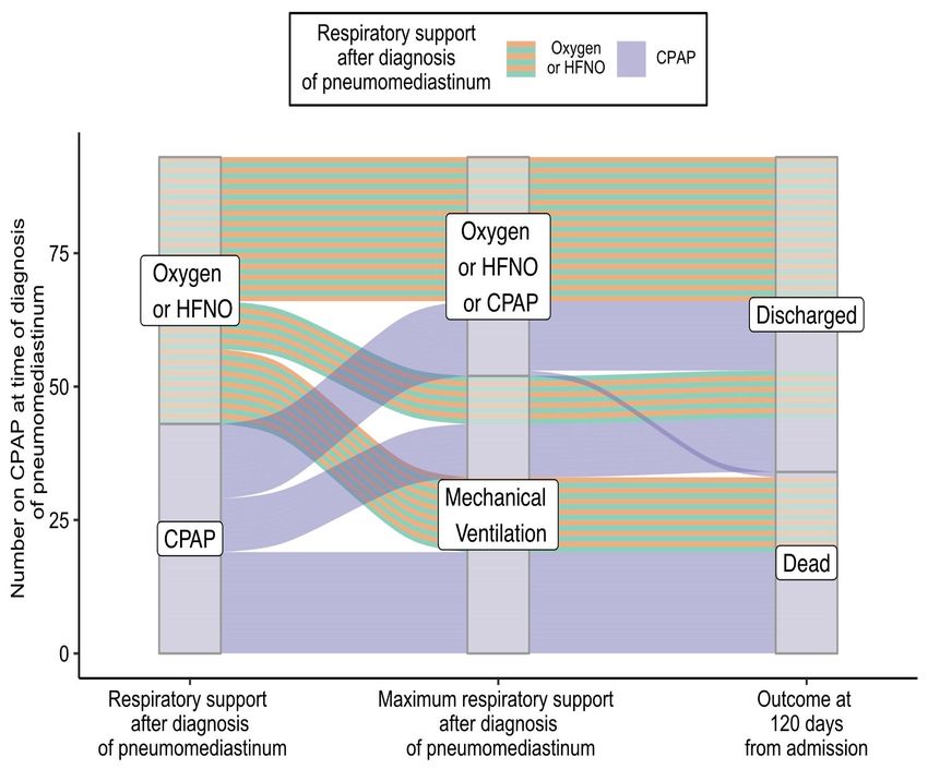
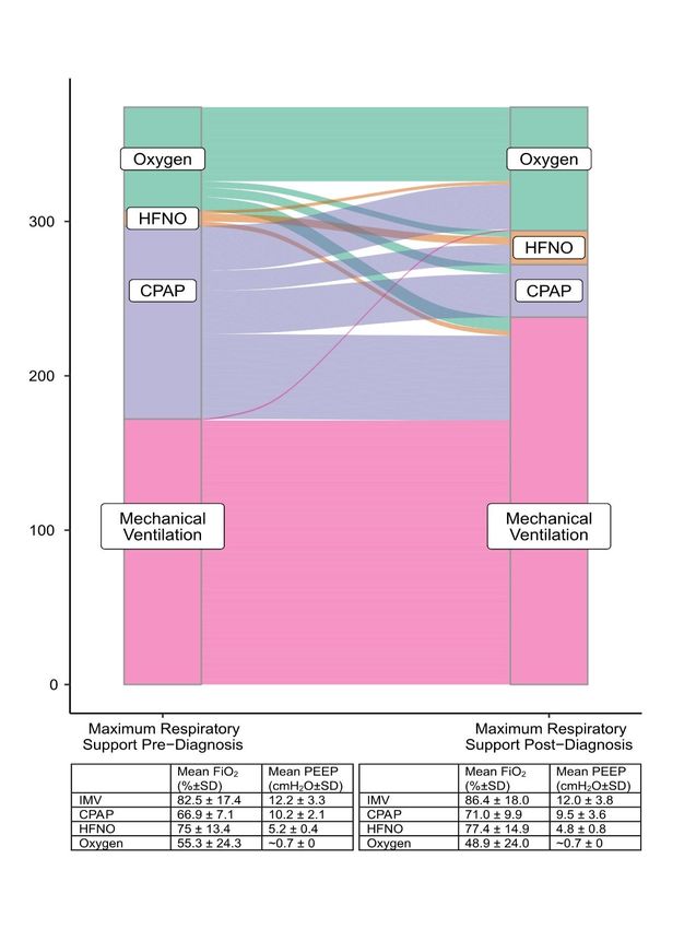
membrane oxygenation (ECMO). Of the 377 patients eligible to receive CPAP

256/377 (67.9%) had received CPAP prior to the diagnosis of PTM. Conscious non-

invasive bi-level positive airways pressure ventilation (BiPaP) was used at some

point in the admissions of 9/377 (2.4%) patients, other than its use in weaning

patients from mechanical ventilation. Four of these nine patients were in type two

respiratory failure. The indication for use of BiPaP for the other five patients was not

clear. Given the few patients who received BiPaP and the variation in its use we

have excluded it from our analyses.

The maximum respiratory support provided to all patients before and after diagnosis

of PTM is described in Figure 1. Four patients whose treatment was limited to non

invasive respiratory support were switched from CPAP to Oxygen at the point of

diagnosis of PTM as part of a decision to initiate palliative treatment. Two patients

were managed on room air throughout.

Alteration of respiratory support at the time of diagnosis is illustrated in Figure 1.

Most patients whose respiratory support was changed after diagnosis of PTM were

on CPAP. At the point of diagnosis of PTM 93 patients eligible for mechanical

ventilation were on CPAP. Fifty (53.8%) of these patients were switched immediately

on diagnosis of PTM to either Oxygen or HFNO therapy creating two subgroups

amenable to analysis; the 50 switched to Oxygen or HFNO and the 43 continuing on

CPAP. These two subgroups were retrospectively well matched at the point of

diagnosis by age (CPAP mean age 57.0 years vs Oxygen or HFNO 55.6 years, p =

0.51), by the maximum FiO2 they had received (CPAP mean FIO2 66% vs Oxygen
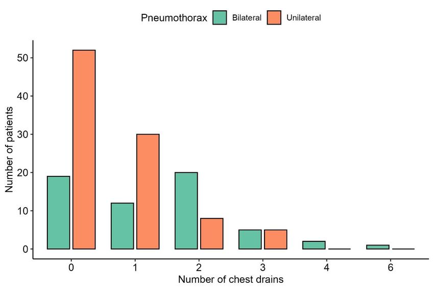
or HFNO 68%, p = 0.15) or by the maximum PEEP they had received (CPAP mean

PEEP 10.4cmH20 vs Oxygen or HFNO 9.8cmH20, p = 0.19). The subsequent

trajectory of these two subgroups is illustrated in Figure 2. Associations of change in

mode of respiratory support and mortality for these patients was examined by

ANOVA. There was no significant main effect of switching support from CPAP to

Oxygen or HFNO on outcome. There was however, a main effect of mechanical

ventilation as a factor associated with mortality for both subgroups (p < 0.001).

Co-occurrence of pneumothorax, subcutaneous emphysema and complications

associated with pneumomediastinum

Pneumothorax was seen concurrently in 154/377 patients (40.8%) and

subcutaneous emphysema was seen in 280/377 (74.3%) of patients. The co-

occurrence of pneumomediastinum with pneumothorax, subcutaneous emphysema

and tension phenomena and the use of intercostal drains are displayed in Figure 3.

The number and frequencies of intercostal chest drains inserted are presented in

online supplementary Figure S5. In cases associated with subcutaneous

emphysema, subcutaneous drains were employed in 6 (1.6%) cases. In 5 of these 6

cases subcutaneous drains were inserted for threatened or actual tension

subcutaneous emphysema. There were 4 (1.1%) instances of mediastinal drains

being used. In 1 of these 4 cases the mediastinal drain was inserted as an

emergency bedside procedure for suspected tension PTM and tension

subcutaneous emphysema. In the other 3/4 cases the mediastinal drain was inserted

to obviate possible tension PTM. These four mediastinal drains were inserted in

patients at four different hospitals, each without on-site cardiothoracic services.
There were 14 cases of tension pneumothorax. During 10 cases of suspected

tension phenomena bilateral intercostal drains were inserted as an emergency

procedure. Seven of these 10 cases were performed without prior radiographic

evidence of pneumothorax.

The development of pneumothorax was not associated with increased risk of death

for our cohort (Table 1) including the subset of 16 patients who were mechanically

ventilated before pneumothorax developed [11/116 (9.5%) were among those

patients who subsequently died while 5/56 (8.9%) were among those discharged, p =

0.9] There were two cases of pneumoperitoneum. Both of these cases were in

mechanically ventilated patients.

In 8 cases PTM appeared following an interventional procedure that could potentially

represent a separate mechanism for occurrence e.g., tracheostomy, and these

cases are included in the final analysis. Analyses were performed excluding these

cases without any statistically significant deviation from the results presented.

Mortality

At 120 days from admission 175/377 (46.4%) patients had been discharged and

195/377 (51.7%) patients had died. Of the seven patients still in hospital at 120 days,

at time of writing one patient had died on day 162 of their admission. Three patients

remained mechanically ventilated on days 131, 146 and 150 of admission. One

patient had been extubated but remained within intensive care on day 132 of
admission. These 5 patients were categorized with those who had died at 120 days

in all outcome analyses. The remaining two patients were medically fit for discharge

to rehab facilities at days 137 and 149 of admission. They were categorized with

patients discharged at 120 days in all outcome analyses. A breakdown of mortality is

provided in online supplementary Tables S6a and S6b according to whether patients

were eligible for mechanical ventilation or limited to CPAP support.

Factors of the presentation and association with outcome are presented for all

patients in Table 1. All factors significantly associated with mortality in univariate

analyses were entered into binary regression prediction models with the exception of

the use of ECMO which, was excluded as the direction of association for this

variable was in favour of discharge rather than death. The variable ‘radiographic

progression of pneumomediastinum’ was excluded where the model was conducted

from the point of diagnosis.

A regression model comparing the predictive utility of variables for mortality at 120

days from the point of diagnosis for patients eligible for all treatment is presented in

Table 2. Further models looking at the predictive utility of the same variables for

mortality across the duration of hospital admission are presented for patients eligible

for all treatment in online supplementary Table S7 and for those limited to CPAP in

online supplementary Table S8.
Discussion

These data comprise the largest series of PTM in COVID-19 to date. In comparison

with other series we sought to accurately represent the incidence of PTM in COVID-

19 during the period of the survey – the United Kingdom’s ‘second wave’ of the

pandemic. Hospital records and radiology reports were systematically reviewed in

each centre. Hospitals that did not observe cases of PTM but provided accurate

incidence data were included. However, hospital participation was sought via trainee

research networks and this may have resulted in inclusion bias.

Our estimate of incidence is also subject to diagnostic biases. We identified cases

through radiology reports which, may not always reference a relevant finding. The

main mode of diagnosis of PTM was CT imaging and there was considerable

variation in the use of CT by participating hospitals (online supplementary Figure

S4). Many CT scans of chest were pulmonary angiogram studies assaying for

pulmonary emboli, not for PTM. For 46.2% of the patients diagnosed with PTM on

thoracic CT the PTM was not visible on their preceding chest radiograph. As only

21.7% of our total denominator population of 58,484 COVID-19 positive inpatients

had thoracic CT imaging performed during their admissions, there is likely to be a

number of undetected cases of PTM in our denominator population. These unknown

cases may have had a more benign disease trajectory than the cases identified.

With these caveats these data demonstrate an incidence of PTM in COVID-19 of

0.64% per inpatient admission and 3.0% per COVID-19 inpatients undergoing
thoracic CT. This incidence is similar to rates reported by two other studies of PTM in

hospitalized COVID-19 populations from Brazil and Romania, of 0.51% and 0.67%

respectively [13,14]. The incidence of ‘spontaneous’ PTM in COVID-19 in this cohort

i.e., without any PPV via mechanical ventilation or CPAP, was 77/58,484 (0.13%).

This is much higher than estimated background rates of non-COVID-19

‘spontaneous’ PTM. The largest study of non-COVID-19 ‘spontaneous’ PTM in the

literature with a defined denominator population, identified 41 cases of PTM from

1,824,967 emergency department admissions over 16 years (0.00002%) [15].

The mean age of the cohort (59.1 years) is consistent with inpatient international

COVID-19 PTM cohorts from Brazil, Romania, Turkey, Pakistan and the USA [13,

16-19]. It is somewhat younger than the mean age of general COVID-19 inpatients in

the UK, according to the largest epidemiological study (70.4 years) [20]. There could

be pathophysiological reasons why COVID-19 inpatients who develop PTM are

younger than the hospital population average (we note that background rates of non-

COVID-19 PTM typically occur in younger adults) [1, 14-15]. It could reflect bias

towards more frequent imaging in younger patients who are usually eligible for all

treatments, with an artificial reduction in the identification of PTM in older patient

groups. A younger mean age is also representative of trends in patients hospitalized

with COVID-19 during the ‘second wave’ in the UK [21].

Pneumothorax was found to co-exist with PTM in 40.3% of cases. This compares to

reported rates of between 20.0% and 72.7% in other series with more than 10

patients [6, 13, 16-18, 22]. There was no finding of an effect on mortality of
pneumothorax within this cohort, nor specifically for those patients who were

mechanically ventilated when pneumothorax occurred. This contrasts with the

findings of Marciniak et al [23] who report an increased risk of mortality with COVID-

19 pneumothorax in a large dataset of UK inpatients, and Chopra et al [24] who

found increased mortality in mechanically ventilated patients across four intensive

care units in the USA. As concurrent PTM was not reported in the Marciniak et al

study and was relevant to 30% of the patients in the Chopra et al study it is not clear

how comparable these patient groups are to our cohort. The extent to which

pneumothorax and PTM are manifestations of barotrauma in COVID-19 underwritten

by a pathophysiological process and the extent to which they are distinct entities

remains to be determined.

Subcutaneous emphysema was seen in 77.9% of COVID-19 PTM patients.

Subcutaneous emphysema has been documented at rates of between 63.6% and

90.5% in other COVID-19 PTM series with more than 10 patients [13, 16-17]. This

result is in keeping with high reported rates of subcutaneous emphysema in

spontaneous non-COVID PTM of up to 100% [1] and in excess of lower rates of co-

occurrence between subcutaneous emphysema and non-COVID-19 pneumothorax

of up to 20% [25]. It would suggest that subcutaneous emphysema is a feature

strongly associated with PTM and not specifically to COVID-19 PTM. It is

acknowledged however, that co-occurrence of subcutaneous emphysema and PTM

may be subject to diagnostic bias with patients presenting with subcutaneous

emphysema more likely to have CT imaging and subsequent revealing of a

diagnosis of PTM.
It is not possible to determine the effect of different ventilatory strategies on outcome

within an observational study such as this. However, we examined this for those

patients eligible for mechanical ventilation who were on CPAP when PTM was

diagnosed. The role of CPAP in patients with PTM is a clinically important question:

Analysis of changes in respiratory support after diagnosis of PTM permits an

exploration of physician preferences regarding respiratory support, and by inference

use of PEEP, in PTM. Those patients who remained on CPAP immediately after

diagnosis of PTM were retrospectively well matched with those patients who were

switched immediately to Oxygen or HFNO by age, maximum FiO2 and maximum

PEEP. There was no difference in survival at 120 days between these subgroups.

Thus, the current data do not support a policy of taking patients off CPAP when PTM

is diagnosed, although we acknowledge potential confounders.

The 120-day mortality rate for patients with COVID-19 PTM of 51.7% is in keeping

with reported mortality rates of 47.7% - 72.2% in other COVID-19 PTM cohorts [13,

16-17]. The severity of COVID-19 illness is demonstrated by the high mean levels of

FiO2 and PEEP before and after the diagnosis of PTM was made (Figure 1). Only

two patients (0.5%) were managed on room air throughout admission. The number

of patients who were mechanically ventilated at some point during their admission

was remarkable at 76.5% of those eligible, in comparison to the UK average for

mechanical ventilation of COVID-19 inpatients of 8.8% [20]. Mechanical ventilation

was unsurprisingly an important prognostic factor and dominant variable in outcome

prediction models (Table 2). It is a ubiquitous event in the trajectory of a deteriorating

patient eligible for this support. Only one eligible patient in our cohort died without

having been mechanical ventilated.
High rates of mechanical ventilation in COVID-19 PTM have been reported in other

general hospital inpatient COVID-19 studies [13,16]. This may reflect a confounding

relationship between more severe illness and higher rates of CT scanning and

detection in high-care environments. It may also indicate an important role for

mechanical ventilation in the development of PTM in COVID-19. However, the

majority of this cohort, 205/377 patients (54.4%), were not mechanically ventilated at

the point the diagnosis of PTM was made. Mechanical ventilation was therefore not a

sufficient or necessary mechanism of PTM for the majority of patients.

Different mechanisms of PTM are described in the literature, including posterior

membrane tracheal lesion or rupture due to coughing [26]. The ‘Macklin effect’ [27]

describes PTM secondary to the rupture of marginal alveoli due to a steeply

increased pressure gradient between the alveolus and the interstitial space. After

rupture of the alveolus air dissects centripetally along the sheaths of the broncho-

vascular bundles into the mediastinum. Depending on volume and pressure, air can

be decompressed along cervical fascial planes into the subcutaneous tissues of the

chest wall, neck or face. Air may rupture the relatively thin mediastinal pleura to

enter the pleural space causing unilateral or bilateral pneumothorax and /or

pneumopericardium / pneumoperitoneum. Macklin and Macklin believed the effect

could be benign or result in circulatory collapse if air directly compressed the

pericardium or venous return – a tension PTM or pneumothorax . Air in the broncho-

vascular bundles could also have a pernicious splinting effect leading to

hyperinflation and low compliance with vascular compression and poor gas

exchange, ‘malignant interstitial emphysema’. The ‘Macklin effect’ was inspired by

physician descriptions of ‘pulmonary interstitial emphysema’ in patients suffering
severe respiratory illness during the 1918-20 influenza pandemic, [28, 29] the

pathophysiology of which may bear comparison with the COVID-19 pandemic.

The ‘Macklin effect’ offers a plausible mechanism for PTM in COVID-19 whereby the

pneumonitis creates an altered diathesis for the rupture of alveoli and the emergence

of PTM. The proposition that COVID-19 PTM patients have severe pneumonitis is

supported by cohort studies that describe high radiological scores of pneumonitis in

COVID-19 PTM [10,16-18]. The complimentary findings in our cohort of; high levels

of respiratory support (taken to represent severe pneumonitis); high rates of

subcutaneous emphysema; episodes of tension phenomena, and low rates of chest

pain (compared to spontaneous PTM) support the Macklin effect as the likely

mechanism of PTM in COVID-19. The previous 2002-4 SARS epidemic also saw an

increase in case reports of PTM [30] and this may reflect similar pathophysiology.

Future studies among mechanically ventilated patients with COVID-19 may elucidate

whether strategies which modify trans-alveolar pressure have any association with

development or progression of PTM. We notice that the 40/377 (10.6%) patients in

our cohort with obesity were not at increased risk of death compared to other

patients with PTM and speculate whether this could relate to mass loading around

the chest wall and/or abdomen with reduction of alveolar compliance and/or trans-

alveolar gradients. Propensity matched cohort analyses may address whether the

development of PTM confers increased mortality risk, beyond severe pneumonitis, or

whether development of PTM is affected by disease modifying drugs such as
dexamethasone (standard of care for our cohort) or different variants of coronavirus

2019.

In summary this study is the largest reported series of PTM in COVID-19 disease.

PTM appears to be a marker of severe pneumonitis, and not necessarily as a result

of the use of PPV. There was no evidence of increased harm by continuing CPAP in

COVID-19 patients who developed PTM.

Acknowledgements

We would like to thank Pallav Shah, Lupei Cai, Muhammad Tariq, Benjamin Jones ,

and Emma Helm for their help with data collection, Maria Tsakok and Nick Tessier

for assistance with imaging and Simon Couillard and Sanjay Ramakrishnan for

review of the manuscript.

The research was funded by the National Institute for Health Research (NIHR)

Oxford Biomedical Research Centre (BRC). The views expressed are those of the

authors and not necessarily those of the NIHR or the Department of Health and

Social Care.
Data Sharing

De-identified participant data from the study will be made available with publication

to medical researchers on a not for profit basis by email request to the corresponding

author for the purposes of propensity matching or meta-analysis.

Declaration of Interest

The authors declare no conflicts of interest.
References

1. Mondello B, Pavia R, Ruggeri P, Barone M, Barresi P, Monaco M. Spontaneous

pneumomediastinum: experience in 18 adult patients. Lung 2007;185:9-14.

2. Anzueto A, Frutos-Vivar F, Esteban A, et al. Incidence, risk factors and outcome

of barotrauma in mechanically ventilated patients. Intensive Care Med

2004;30:612-9.

3. Talmor D, Sarge T, Malhotra A, et al. Mechanical ventilation guided by

esophageal pressure in acute lung injury. N Engl J Med 2008;359:2095-104.

4. Fan E, Beitler JR, Brochard L, et al. COVID-19-associated acute respiratory

distress syndrome: is a different approach to management warranted? The Lancet

Respiratory Medicine 2020;8:816-21.

5. Lemmers DHL, Abu Hilal M, Bnà C, et al. Pneumomediastinum and subcutaneous

emphysema in COVID-19: barotrauma or lung frailty? ERJ Open Res

2020;6(4):00385-2020.

6. Belletti A, Palumbo D, Zangrillo A, et al. Predictors of

Pneumothorax/Pneumomediastinum in Mechanically Ventilated COVID-19 Patients.

J Cardiothorac Vasc Anesth 2021;35(12):3642-3651.

7. Udi J, Lang CN, Zotzmann V, et al. Incidence of Barotrauma in Patients With

COVID-19 Pneumonia During Prolonged Invasive Mechanical Ventilation - A Case-

Control Study. J Intensive Care Med 2021;36:477-83.

8. Wong K, Kim DH, Iakovou A, et al. Pneumothorax in COVID-19 Acute Respiratory

Distress Syndrome: Case Series. Cureus 2020;12(11):e11749.

9. Diaz A, Patel D, Sayedy N, et al. COVID-19 and Spontaenous

Pneumomediastinum: A case series. Heart & Lung 2021;50:202-205
10. Manna S, Maron SZ, Cedillo MA, et al. Spontaneous subcutaneous emphysema

and pneumomediastinum in non-intubated patients with COVID-19. Clin Imaging

2020;67:207-

11. Tucker L, Patel S, Vatsis C, et al. Pneumothorax and Pneumomediastinum

Secondary to COVID-19 Disease Unrelated to Mechanical Ventilation. Case Rep Crit

Care 2020;2020:6655428.

12. Groves N, Tobin A. High flow nasal oxygen generates positive airway pressure in

adult volunteers. Aust Crit Care 2007;20:126-31.

13. Cut TG, Tudoran C, Lazureanu VE, Marinescu AR, Dumache R, Tudoran M.

Spontaneous Pneumomediastinum, Pneumothorax, Pneumopericardium and

Subcutaneous Emphysema-Not So Uncommon Complications in Patients with

COVID-19 Pulmonary Infection-A Series of Cases. J Clin Med 2021;10(7):1346.

14. Asma M, Nesrine F, Ahmed BS, Sameh J, Saoussen CM, Naceur R.

Spontaneous pneumomediastinum: Experience in 13 patients. Respir Med Case

Rep 2019;28:100946.

15. Macia I, Moya J, Ramos R, Morera R, Escobar I, Saumench J, Perna V, Rivas F,

Spontaneous pneumomediastinum: 41 cases, European Journal of Cardio-Thoracic

Surgery 2007; 31:6,1110–1114

16. Brito J, Gregório P, Mariani A, et al. Pneumomediastinum in COVID-19 disease:

Outcomes and relation to the Macklin effect. Asian Cardiovasc Thorac Ann

2021;29:541-8.

17. Kangas-Dick A, Gazivoda V, Ibrahim M, et al. Clinical Characteristics and

Outcome of Pneumomediastinum in Patients with COVID-19 Pneumonia. J

Laparoendosc Adv Surg Tech A 2021;31:273-8.
18. Ozsoy IE, Tezcan MA, Guzeldag S, Ozdemir AT. Is Spontaneous

Pneumomediastinum a Poor Prognostic Factor in Covid-19? Journal of the College

of Physicians and Surgeons--Pakistan : JCPSP 2021;31:132-7.

19. Sethi SM, Ahmed AS, Hanif S, Aqeel M, Zubairi ABS. Subcutaneous

emphysema and pneumomediastinum in patients with COVID-19 disease; case

series from a tertiary care hospital in Pakistan. Epidemiol Infect 2021;149:e37.

20. Docherty AB, Mulholland RH, Lone NI, et al. Changes in in-hospital mortality in

the first wave of COVID-19: a multicentre prospective observational cohort study

using the WHO Clinical Characterisation Protocol UK. The Lancet Respiratory

Medicine 2021;9(7):773-785.

21. Doidge JC, Gould DW, Ferrando-Vivas P, et al. Trends in Intensive Care for

Patients with COVID-19 in England, Wales, and Northern Ireland. Am J Respir Crit

Care Med 2021;203:565-74.

22. Martinelli AW, Ingle T, Newman J, et al. COVID-19 and pneumothorax: a

multicentre retrospective case series. Eur Respir J 2020;56(5):2002697.

23. Marciniak SJ, Farrell J, Rostron A, et al. COVID-19 Pneumothorax in the United

Kingdom: a prospective observational study using the ISARIC WHO clinical

characterisation protocol. Eur Respir J 2021;58(3)2100929

24. Chopra A, Al-Tarbsheh AH, Shah NJ, et al. Pneumothorax in critically ill patients

with COVID-19 infection: Incidence, clinical characteristics and outcomes in a case

control multicenter study. Respir Med. 2021 Aug;184:106464.

25. Melhorn J, Davies HE. The Management of Subcutaneous Emphysema in

Pneumothorax: A Literature Review. Current Pulmonology Reports 2021;10:92-

97.26. Akkas M, Tiambeng C, Aksu NM, Onur R. Tracheal rupture as a result of

coughing. Am J Emerg Med. 2018 Nov;36(11):2133.
27. Madge Thurlow Macklin and Charles C Macklin. Malignant interstitial

emphysema of the lungs and mediastinum as an important occult complication in

many respiratory diseases and other conditions: An interpretation of the clinical

literature in light of laboratory experiment. Medicine 1944;23(4)281-358.

28. Torrey RG. Acute Pulmonary Emphysema observed during the epidemic of

influenza pneumonia at Camp Hancock, Georgia American Journal of the Medical

Sciences 1919:170 - 81.

29. Longcope WT. Survey of the epidemic of influenza in the american expeditionary

forces. Journal of the American Medical Association. 1919;73:189-91

30. Chu CM, Leung YY, Hui JYH et al. Spontaneous pneumomediastinum in patients

with severe acute respiratory syndrome. Eur Respir J 2004;23(6): 802-804.
Figure Legends

Figure 1. Sankey Plot charting the maximum respiratory support given to all patients

four hours before the diagnosis of Pneumomediastinum and then following the

diagnosis of Pneumomediastinum (n = 374). The mean fraction of inspired oxygen

(FiO2) and positive end expiratory pressure (PEEP) received on these levels of

support is given in the tables below. HFNO = high flow nasal oxygen; CPAP =

continuous positive airways pressure.

Figure 2. Alluvial Plot describing the trajectory of 93 patients eligible for mechanical

ventilation who were on continuous positive airways pressure (CPAP) at the point of

diagnosis of pneumomediastinum. At point of diagnosis there was no statistical

difference in age, maximum fraction of inspired oxygen (FiO2) or maximum positive

end expiratory pressure (PEEP) received between those patients subsequently

maintained on CPAP and those subsequently switched to Oxygen or high flow nasal

oxygen (HFNO).

Figure 3. UpSet Plot illustrating the co-occurrence of pneumomediastinum with

subcutaneous emphysema, pneumothorax and tension phenomena and the use of

intercostal chest drains (n = 377). Bilateral pneumothorax was ascribed to

pneumothoraces occurring on both sides of the thorax within the same admission
Tables

Table 1. Univariate analyses: Factors of the presentation and their association with outcome at 120 days (n = 377). HFNO = high

flow nasal oxygen; CPAP = continuous positive airways pressure; ECMO = extracorporeal membrane oxygenation; MV =

mechanical ventilation

                                   Outcome                Outcome                 N         Univariant analysis:       p

                                   Dead                   Discharged                        Odds ratio

                                                                                            for death

                                                                                            with 95% CI
                                   Mean value (±SD) or    Mean values (±SD)

                                   N and %                or N and %

Mechanically ventilated              160 / 161 (99.8)       81 / 154 (52.6)       315       144.2 (19.7 – 1056)      < 0.001

at any time (of those eligible)
Respiratory support at time of diagnosis: Oxygen (ref group) 24 / 199 (12.1) 44 / 176 (25.0) 375 HFNO 4 / 199 (2.0) 6 / 176 (3.4) 1.2 (0.3 – 4.8) 0.77 CPAP 55 / 199 (27.6) 70 / 176 (39.8) 1.4 (0.8 – 2.7) 0.24 Mechanical ventilation 116 / 199 (58.3) 56 / 176 (31.8) 3.8 (2.1 – 6.9) < 0.001 Age (mean years) 62.1 (11.4) 55.8 (13.1) ---- < 0.001 Subcutaneous emphysema 167 / 200 (83.5) 113 / 177 (63.8) 2.9 (1.8 – 4.6) < 0.001
Ischaemic heart disease or left    26 / 200   (13.0)   7   / 177 (4.0)          3.6   (1.5 – 8.6)   < 0.01

ventricular systolic dysfunction

Hypertension                       76 / 200   (38.0)   46 / 177 (26.0)          1.9   (1.2 – 2.9)    0.01

Diabetes mellitus                  53 / 200   (26.5)   28 / 177 (15.8)          1.9   (1.2 – 3.2)    0.01

Radiographic progression           58 / 200   (29.0)   31 / 177 (17.5)          1.9   (1.2 – 3.2)    0.01

of pneumomediastinum

ECMO (those eligible)              11 / 160   (6.9)    13 / 81   (16.0)   241   0.4   (0.2 – 0.9)    0.03
Male sex                     140 / 200   (70.0)   137 / 177 (77.4)          0.7   ( 0.4 – 1.1)   0.11

Chest pain at presentation   19 / 193    (9.8)    26 / 172 (15.1)     365   0.6   (0.3 – 1.2)    0.13

Chronic kidney disease       11 / 200    (6.0)    5   / 177 (2.8)           1.7   (0.6 – 4.6)    0.21

Asthma                       43 / 200    (21.5)   30 / 177 (16.9)           1.3   (0.8 – 2.3)    0.27

Tension phenomena            13 / 200    (6.5)    7   / 177   (4.0)         1.7   (0.7 – 4.3)    0.28
Obesity (BMI ≥35)           23 / 200   (11.5)   17 / 177   (9.6)   1.2   (0.6 - .4)    0.55

Pneumothorax (at anytime)   84 / 200   (42.0)   70 / 177 (39.5)    1.1   (0.7 – 4.7)   0.63
Table 2.Binary Logistic Regression Model of factors predictive of death at 120 days from the point of diagnosis of

pneumomediastinum (all patients eligible for mechanical ventilation, n=315). All variables significantly associated with mortality in

univariate analyses were entered into the model stepwise, backwards. The model produces prediction accuracy for outcome of

68.4% versus a 51.1% default accuracy.

                                B(SE)                       Odds Ratio or % increase per unit                     p

                                                            (95% CI)

Mechanically ventilated         1.40    (0.26)                   4.0                        (2.4 - 6.7)           < 0.001

(at diagnosis)

Age                             -0.38   (0.12)                   3.7 % per year              (1.4 - 5.9)          < 0.01
Diabetes mellitus 0.59 (0.34) 1.8 (0.9 - 3.5) 0.08 Model R2 = 0.251 Nagelkerke χ2 (6) = 65.2 p < .001. Constant B(SE) = 2.38 (0.79) Variables in the regression but not listed, subcutaneous emphysema (p = .12), Hypertension (p = .35), ischaemic heart disease / left ventricular systolic dysfunction (p = .50).
Figures

Figure 1.
Figure 2.
Figure 3.
Pneumomediastinum in COVID-19: a phenotype of severe COVID-19
pneumonitis? The results of the United Kingdom (POETIC) survey

                           Online Data Supplement
                                  (S1 – S9)

James Melhorn, Andrew Achaiah, Francesca M. Conway, Elizabeth M. F.
Thompson, Erik W. Skyllberg, Joseph Durrant, Neda A. Hasan, Yasser Madani,
Prasheena Naran, Bavithra Vijayakumar, Matthew J. Tate, Gareth E. Trevelyan, Irfan
Zaki, Catherine A. Doig, Geraldine Lynch, Gill Warwick , Avinash Aujayeb , Karl A.
Jackson, Hina Iftikhar, Jonathan H. Noble, Anthony Y. K. C. Ng, Mark Nugent, Philip
J. Evans, Robert A. Hastings, Harry R. Bellenberg, Hannah Lawrence, Rachel L.
Saville, Nikolas T. Johl, Adam N. Grey, Huw C. Ellis, Cheng Chen, Thomas L.
Jones, Nadeem Maddekar, Shahul Leyakathali Khan, Ambreen Iqbal Muhammad,
Hakim Ghani, Yadee Maung Maung Myint, Cecillia Rafique, Benjamin J. Pippard,
Benjamin R. H. Irving, Fawad Ali, Viola H. Asimba, Aqeem Azam, Eleanor C. Barton,
Malvika Bhatnagar, Matthew P. Blackburn, Kate J. Millington, Nicholas J. Budhram,
Katherine L. Bunclark, Toshit P. Sapkal, Giles Dixon, Andrew J. E. Harries,
Mohammad Ijaz, Vijayalakshmi Karunanithi, Samir Naik, Malik Aamaz Khan,
Karishma Savlani, Vimal Kumar, Beatriz Lara Gallego, Noor A. Mahdi, Caitlin
Morgan, Neena Patel, Elen W. Rowlands, Matthew S. Steward, Richard S. Thorley,
Rebecca L. Wollerton, Sana Ullah, David M. Smith, Wojciech Lason, Anthony J
Rostron, Najib M Rahman and Rob J Hallifax
Glossary of terms used ARDS = acute respiratory distress syndrome BiPaP = conscious non-invasive bi-level positive airways pressure COVID-19 = coronavirus 2019 infection CPAP = continuous positive airways pressure CT = computed tomography CXR = chest radiograph ECMO = extracorporeal membrane oxygenation FiO2 = fraction of inspired oxygen IHD = ischaemic heart disease HFNO = high flow nasal oxygen LVSD = left ventricular systolic dysfunction MV = invasively mechanically ventilated PEEP = positive end expiratory pressure PPV = positive pressure ventilation PTM = pneumomediastinum UK = United Kingdom
S1

Prior to performing the current study we conducted a literature search of evidence on the subject. We searched MEDLINE and
PubMed for original peer-reviewed cohort studies describing the incidence of pneumomediastinum in COVID-19 between March
2020 and June 2021. Search terms were “Pneumomediastinum” AND “COVID-19” OR “Barotrauma” AND “COVID-19” OR
“Pneumothorax” AND “COVID-19”. Only reports published in English that included at least 5 cases and with estimates of a
background population were included. Our search yielded 15 studies. These are detailed in table S1 below

Table S1. Previously published cohort studies with ≥5 cases of pneumomediastinum (PTM) and an identified denominator
population.

Study and      Single or   Date      Number % cases        PTM with   PTM with   Number of   Denominator         % cases in
country of     Multicentre Published of     diagnosed      concurrent concurrent denominator patient             denominator
origin                               cases  by thoracic    PTX        SCE        population  population          PCR
                                     of PTM CT                                                                   positive

McGuiness      Single     Nov 2020   59        CXR         Not stated   Not stated   601          Mechanically   100%
et al (USA)1                                   diagnoses                                          Ventilated

Kangas-Dick    Single     Mar 2021   34        CXR         35.3%        Not stated   346          Mechanically   Unclear
et al2 (USA)                                   diagnoses                                          Ventilated

Housman et     Single     Sep 2020   29        CXR         6.9 - 24%    100%         171          Mechanically   100%**
al3 (USA)                                      diagnoses                                          Ventilated
Wong et al4 Multi (2) Nov 2020 27 CXR 100% Not stated 1822 ARDS Unclear (USA)* diagnoses Chopra et al5 Multi (4) May 2021 24 Not stated 100% Not stated 842 Critical Care 100% (USA)* (594 MV) Lemmers et Single Sep 2020 23 CXR/CT 0% 100% 169 ARDS 100% al6 (Italy) Rajdev et al7 Single May 2021 21 CXR/CT Not stated Not stated 353 Oxygen / PPV 100% (USA) (121 MV) Brito et al8 Single April 2021 21 100% 33% 90.5% 4087 Hospital 100%** (Brazil) Inpatients Martinelli et9 Multi (16) Sep 2020 17 CXR/CT 35.3% Not stated 6574 Hospital Clinical al (UK) Inpatients Diagnoses Belletti et al10 Single Feb 2021 13 38.5% 53.8% Not stated 116 ARDS Criteria 100% (Italy) Cut et al11 Single Mar 2021 11 100% 72.7% 63.6% 1648 Hospital Unclear (Romania) Inpatients Edwards et Single Nov 2020 10 CXR 20% 90% 574 Mechanically 100% al12 (USA) diagnoses Ventilated
Talan et al13   Single   Dec 2020   7         71.4%        57.1%      57.1%       161           Critical Care   Unclear
(Turkey)                                                                                        (96 MV)

Udi et al14     Single   Aug 2020   5         Not stated   40%        40%         20            ARDS            100%
(Germany)

Eperjesiova     Single   Jul 2020   5         Not stated   20%        80%         976           Hospital        Unclear
et al15 (USA)                                                                                   Inpatients

Studies marked with an asterix (*) focused on identifying COVID-19 pneumothorax (PTX) rather than COVID-19
pneumomediastinum (PTM) therefore all cases were PTM/PTX overlap with likely underestimation of incidence of COVID-19 PTM.
Studies marked with (**) describe „confirmed COVID-19 infection‟ rather than SARS-CoV-2 PCR positivity.
S2

Hospitals within the POETIC consortium were a representative mix of secondary and
tertiary hospitals throughout the UK including those within areas of high index of
multiple deprivation. They are listed below in alphabetical order:

Addenbrooke's Hospital, Cambridge
Andover War Memorial Hospital
Barnet Hospital
Basingstoke and North Hampshire Hospital, Basingstoke
Bedford Hospital
Burnley General Teaching Hospital
Chelsea and Westminster Hospital
Glangwilli Hospital, Carmarthen
Gloucester Royal Hospital
Grange University Hospital, Newport
Great Western Hospital, Swindon
John Radcliffe Hospital
Kettering General Hospital
Lister Hospital, Stevenage
Luton & Dunstable University Hospital, Luton
Musgrove Park Hospital, Taunton
Neville Hall Hospital, Abergavenny
Newham University Hospital, London
Norfolk and Norwich University Hospital, Norwich
Northumbria Specialist Emergency Care Hospital
Nottingham City Hospital
Prince Philip Hospital, Llanelli
Prince of Wales Hospital, Bridgend
Princess Alexandria Hospital, Harlow
Queen Alexandra Hospital, Portsmouth
Queen Elizabeth University Hospital, Glasgow
Royal Berkshire Hospital, Reading
Royal Blackburn Teaching Hospital
Royal Brompton Hospital, London
Royal Cornwall Hospital, Truro
Royal Derby Hospital , Derby
Royal Devon & Exeter Hospital
Royal Free Hospital, London
Royal Glamorgan Hospital, Llantrisant
Royal Gwent Hospital, Newport
Royal Hampshire County Hospital, Winchester
Royal London Hospital
Royal Stoke University Hospital
Royal United Hospitals, Bath
Saint Bartholomew's Hospital, London
Southend University Hospital
Southport & Ormskirk District General Hospital
Stoke Mandeville Hospital
You can also read