Diagnostic challenges in systemic amyloidosis: a case report with clinical and laboratorial pitfalls - Autopsy and ...
←
→
Page content transcription
If your browser does not render page correctly, please read the page content below
Clinical Case Report
Diagnostic challenges in systemic amyloidosis: a case report with
clinical and laboratorial pitfalls
Angelina Maria Martins Lino1 , Jussara Bianchi Castelli2,3 ,
Roberta Shcolnik Szor4 , Fabio Fernandes5 , Vera Demarchi Aiello2
How to cite: Lino AMM, Castelli JB, Szor RS, Fernandes F, Aiello VD. Diagnostic challenges in systemic amyloidosis: a case
report with clinical and laboratorial pitfalls. Autops Case Rep [Internet]. 2021;11:e2021326. https://doi.org/10.4322/acr.2021.326
ABSTRACT
Currently, there is growing evidence in the literature warning of misdiagnosis involving amyloidosis and chronic
inflammatory demyelinating polyneuropathy (CIDP). Although inducing clinical manifestations outside the peripheral
nervous system, light chain and transthyretin amyloidosis may initially present with peripheral neuropathy, which can be
indistinguishable from CIDP, leading to a delay in the correct diagnosis. Besides, the precise identification of the amyloid
subtype is often challenging. This case report exemplifies clinical and laboratory pitfalls in diagnosing amyloidosis and
subtyping amyloid, exposing the patient to potentially harmful procedures.
Keywords
Polyradiculoneuropathy, Chronic Inflammatory Demyelinating; Diagnostic Errors; Amyloidosis, Familial; Light Chain
Immunoglobulin Amyloidosis; Paraproteinemias.
INTRODUCTION
Misdiagnosis is a well-known problem in medical diseases.1 Atypical CIDP is a heterogeneous group in
practice, delaying the correct diagnosis and introducing which distal acquired demyelinating symmetric (DADS)
the best therapeutic approach. In Neurology practice, neuropathy accounts for 5-13% of cases, presenting
peripheral neuropathy (PN) may present with varied with sensory ataxia, distal motor involvement and
clinical phenotypes, with or without associated fulfilling demyelination criteria of the European
systemic diseases, easily inducing misdiagnosis. In this Federation of Neurological Societies and the Peripheral
setting, amyloidosis and the clinical spectrum of CIDP Nerve Society (EFNS/PNS).2-5 Having refractoriness to
are examples. immunotherapy and preferential involvement of men
The agreement rate among experts for typical from the 6 th to the 8 th decade of life, monoclonal
and atypical CIDP is 97% and 60%, respectively, gammopathy of undetermined significance (MGUS)
and initial alternative diagnosis is more likely in is found in two-thirds patients with DADS, 60%
atypical CIDP due to the concomitance of systemic associated with immunoglobulin (Ig) M, 30% IgG,
1
Universidade de São Paulo (USP), Hospital das Clínicas, Department of Neurology, Clinical Peripheral Nerve Group, São Paulo, SP, Brasil
2
Universidade de São Paulo (USP), Instituto do Coração, Laboratory of Pathology, São Paulo, SP, Brasil
3
Grupo Fleury, Department of Pathology, São Paulo, SP, Brasil
4
Universidade de São Paulo (USP), Instituto do Câncer do Estado de São Paulo, São Paulo, SP, Brasil
5
Universidade de São Paulo (USP), Instituto do Coração, Cardiomiopathy Group, São Paulo, SP, Brasil
Copyright © 2021 The Author(s). This is an Open Access article distributed under the terms of the Creative
Commons Attribution License, which permits unrestricted use, distribution, and reproduction in any medium,
provided the original work is properly cited.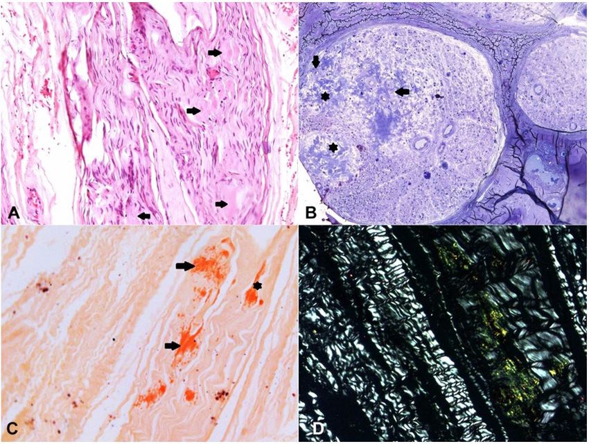
Diagnostic challenges in systemic amyloidosis: a case report with clinical and laboratorial pitfalls

and 10% IgA.6,7 MGUS can progress to light-chain                              CASE REPORT
amyloidosis (AL), representing a differential diagnosis
in a previously diagnosed MGUS-associated CIDP.                                     A previously healthy 67-year-old Caucasian male
                                                                              was admitted to the outpatient service of Neurology
     Likewise, amyloidosis is a heterogeneous group in
                                                                              Division in April 2015, complaining of numbness
which different patterns of PN can be observed. In AL,
                                                                              of hands and feet, and inability to walk that had
PN may be present at the disease onset in up to 20%
                                                                              started two years before with progressive worsening,
of cases and represent the main clinical feature in some
                                                                              associated with weight loss of 14kg. He denied
inherited variants of transthyretin (TTR) amyloidosis                         dry mouth and eyes or sweating, gastrointestinal
(ATTRv).8,9 The clinical overlapping between AL and                           or sexual dysfunction, previous diseases, and any
late-onset ATTRv may hinder the recognition of the                            relevant familial medical history. No abnormalities
amyloid subtype.                                                              were found on general clinical examination, but
     Since AL is the most common subtype of systemic                          skinny complexion. At neurological assessment,
amyloidosis with incidence up to 10 cases/million/                            Medical Research Council scale (MRC) was grade 5 in
year, hereditary forms might be misdiagnosed as AL                            all muscles, except grade 1 for flexion and extension
due to absence of familial history, same age range,                           of the feet; slight atrophy was found in feet intrinsic
the common finding of MGUS in the elderly, or false-                          muscle added to global areflexia, positive Romberg
positive immunohistochemistry staining.8,10,11 In a                           sign, superficial hypoesthesia at toes, severe loss
series of 350 presumptive AL cases, 9.7% harbored a                           of vibratory sensation up to elbows and knees.
hereditary cause, being TTR mutation found in 38%                             Blood pressure and heart rate were 130x82 mmHg
                                                                              and 83 bpm, respectively, on supine position, and
of them.12 TTR variants are the worldwide commonest
                                                                              125x80 mmHg and 92 bpm, respectively, after
cause of hereditary systemic amyloidosis, in which
                                                                              3 minutes on standing position; cranial nerves were
the nervous system and heart involvement may be
                                                                              preserved; 51 points on Neuropathy Impairment Score
indistinguishable from those in AL. In the late-onset
                                                                              (NIS); score 2 for upper (U) and 2 for lower (L) limbs (L)
ATTRv, presenting symptoms occurs in (i) patients over
                                                                              on Overall Neurological Limitation Scale (ONLS). Except
50 years, (ii) with male predominance, (iii) not striking                     for IgA lambda-MG in serum immunofixation, a slightly
family history due to low gene penetrance rate, (iv)                          elevated serum beta-2 microglobulin of 2.0 mg/mL
loss of all sensory modalities rather than unmyelinated                       (RR = 1.0-1.7 mg/mL) and mild anemia (hemoglobin
fibers at beginning, (v) relatively mild autonomic                            of 11,5 g/dL, RR = 13-18 g/dL), other laboratory
dysfunction, and (vi) more organ involvement (heart,                          tests were normal, including complete blood count,
kidney, eye).9,11,13                                                          vitamin B12 and folic acid serum levels, serum kappa
     The most common TTR mutation, Methionine                                 and lambda free light-chains ratio, liver, renal and
for Valine at position 30 (Val30Met), shows clinical                          thyroid functions, blood glucose, lactic dehydrogenase,
and electrophysiological heterogeneity, depending on                          calcium, serology for infectious diseases (HIV, B, and C
the (i) patient’s geographical origin (endemic versus                         hepatitis, VDRL), antinuclear and rheumatoid factors,
non-endemic area), (ii) ethnic group, (iii) incomplete                        antineutrophil cytoplasmic antibodies, anti-SSA/SSB,
                                                                              total complement and fractions, anti-cardiolipin,
penetration of gene mutation, and (iv) age of disease
                                                                              prostatic specific antigen, carcinoembryonic antigen,
onset. 9,11,13 Misdiagnosis of ATTRv as CIDP ranges
                                                                              alpha fetus protein, and urinalysis with protein
from 20 to 61%.10,14 Adding challenges to diagnosis,
                                                                              electrophoresis and immunofixation. Computed
cytoalbuminologic dissociation in the cerebral spinal
                                                                              tomography (CT) of chest, abdomen, and pelvis were
fluid (CSF) and electroneuromyographic (ENMG)
                                                                              normal, and 99mTc-MDP scintigraphy (SC) did not show
features obeying demyelinating criteria for CIDP were                         abnormal uptake in bones. Bone marrow aspirate was
found in 23% and 39%, respectively, in symmetrical                            normal. The CSF showed 1 cell/mm3 and 88 mg/dL (RR
and asymmetrical neuropathies in late-onset ATTRv.14-17                       < 45mg/dL) of protein with 16% gamma (RR < 14%).
     Herein, a clinical case exemplifies the diagnostic                       Anti-nerve antibodies detection was not available.
challenges in systemic amyloidosis.                                           An external ENMG showed longer motor distal and

2-11                                                                                                   Autops Case Rep (São Paulo). 2021;11:e2021326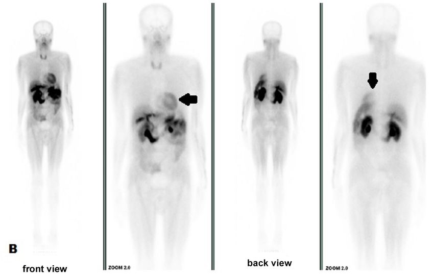
Lino AMM, Castelli JB, Szor RS, Fernandes F, Aiello VD

F wave latencies, moderately reduced motor nerve                          Despite the negative SC biopsy result, AL was
conduction velocity, no conduction block or abnormal                still suspected associated with the IgA lambda
temporal dispersion, slightly reduced motor amplitude               component. Due to the marked motor worsening (NIS
data, and was interpreted as a primarily demyelinating              88.5, ONLS 3 UL and 5 LL) and the onset of pain and
process with slight secondary axonal degeneration                   other autonomic complaints (orthostatic intolerance
fulfilling demyelination criteria of EFNS/PNS.2                     and voiding dysfunction), Fludarabine (100 mg/iv/
      DADS with IgA lambda-MGUS was diagnosed,                      monthly) was prescribed from December 2016 to April
and the patient started on monthly pulse therapy with               2017, resulting in slight improvement (NIS 83.5, ONLS
intravenous (iv) methylprednisolone (MP, 1000 mg/                   3 UL and 4 LL) and disappearance of bone uptake in
day/3days) from May to October 2015. No clinical-
laboratory evidence of any systemic disease arose, NIS
remained unchanged, but worsening in ONLS (2 UL and
3 LL) lead to the addition of monthly cyclophosphamide
(1g/iv/month) to MP from December 2015 to May
2016, and then changed to oral methotrexate (15 mg/
week) for 7 months.
    In March 2016, under immunosuppressive
therapy, the patient started with sexual distress
complaints. Except for the IgA lambda-MG, extensive
serum reinvestigation and thoracic and abdominal
CT remained normal. The ENMG (Table 1) was
remade and showed unexpected and marked axonal
impairment.
     Autonomic evaluation disclosed the absence of
cutaneous sympathetic reflex in all limbs and reduced
cardiac variability at rest and with a deep breath and
Valsalva maneuver (RR interval = 901.2 ± 4.6 ms).
The uptake in fifth and seventh ribs observed in
99m
   TC-MDP SC (Figure 1) was not confirmed in 99mTC-MIBI
SC (Figure 2). No plasmacytosis or other alterations were
seen in bone marrow aspirate, and a subcutaneous (SC)               Figure 1. Bone scintigraphy with 99mTc-MDP in May
tissue biopsy resulted negative for amyloid deposition              2016 showing abnormal bone uptake in 5th, 6th and 7th
under Congo red staining.                                           right coastal arches (arrow).

Table 1. Nerve conduction study (2016)
                                                     Motor                                                 Sensory
     Nerve           CMAP (mV)
                                       CV (m/s) L/R      L (ms) L/R        FWL (ms) L/R SNAP (µV) L/R             CV (m/s) L/R
                        L/R
Median                  1.2/1.7          34.1/33.5           5.5/5.8          ND/ND                U/U                  U/U
Ulnar                   1.7/2.3          32.5/35.1           4.7/4.8         52.2/47.8             U/U                  U/U
Peroneal                  U/U                   U/            U/U
Tibial                    U/U                   U/            U/U
Femoral                 6.0/4.6            5.6/5.8
 Sural                                                                                       U/U               U/U
CMAP: compound muscle action potential; CV: conduction velocity; L: distal latency; FWL: F-wave latency; L: latency;
m/s: meter per second; ms: millisecond; mV: milli Volt; µV: microVolt; ND: not done; SNAP: sensory nerve action potential;
U: unexcitable nerve

Autops Case Rep (São Paulo). 2021;11:e2021326                                                                                   3-11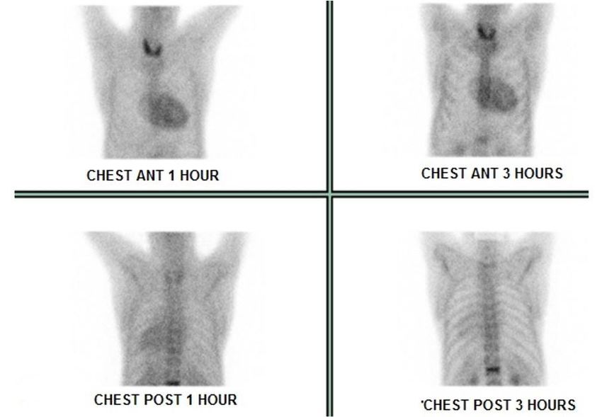
Diagnostic challenges in systemic amyloidosis: a case report with clinical and laboratorial pitfalls

Figure 2. Normal bone uptake with slight uptake in the myocardium (arrow) at bone scintigraphy with 99mTc-MIBI
in November 2016.

coastal arches but showing distension of urinary tract
(Figure 3).
     A sural nerve biopsy (February 2018) revealed
amyloid deposition (Figures 4) with IgA positivity
and non-specific lambda light-chain staining by
immunohistochemistry (IHC) (Figures 5 A-C). Kidney
dysfunction started in 2018 with a protein/creatinine
ratio of 1,59 (RR = 0.06 – 0.35). Imaging studies
showed renal parenchyma with normal thickness and
echotexture associated with dilatation of urinary tract
and thickened and trabeculated bladder walls (data
not shown).
     Still denying any suspected familial history, the
patient brought us in October 2018 a photograph of
his mother at the age of 60 years old showing a very
lean lady with atrophic hands, who was bedridden
for several years and died at 82 years old due to a
non-confirmed bone malignancy. This information
led us to search for a mutation in the TTR gene to
find a heterozygous Val30Met mutation. At the
age of 69 years, a liver transplant was not indicated
due to the risk associated with age, low body mass                            Figure 3. Normal bone uptake with 99mTc-MDP in October
index, and renal dysfunction. Tafamidis was also not                          2017, showing distension of urinary tract (arrows).

4-11                                                                                                   Autops Case Rep (São Paulo). 2021;11:e2021326
Lino AMM, Castelli JB, Szor RS, Fernandes F, Aiello VD
Figure 4. Optic microscopy of sural nerve biopsy. A – Extracellular eosinophilic deposits in the endoneuro (arrows)
(H&E, X200); B – Semithin section (araldite-embedded) showing nerve fascicles with severe loss of thick and thin
myelinated fibers and large amyloid deposits in the endoneuro (asterisk) and around endoneural blood vessels
(arrows) (Toluidine blue, X200); C – Congo red staining showing deposits in the endoneuro (arrows) and epineuro
(arrowhead) (X100) showing endoneural apple-green birefringence under polarized light in D (X200).
indicated due to familial amyloidosis staging system in another medical service where the autopsy was not
score of II (ambulatory but require assistance) and IIIb performed.
at polyneuropathy disability score (walking with the Being performed in January 2021, under research
help of two sticks). Only in 2019 was it possible to settings, laser microdissection and mass spectrometry
carry out an IHC staining for TTR in the stored nerve analysis (LC/MS) showed only TTR in amyloid on nerve
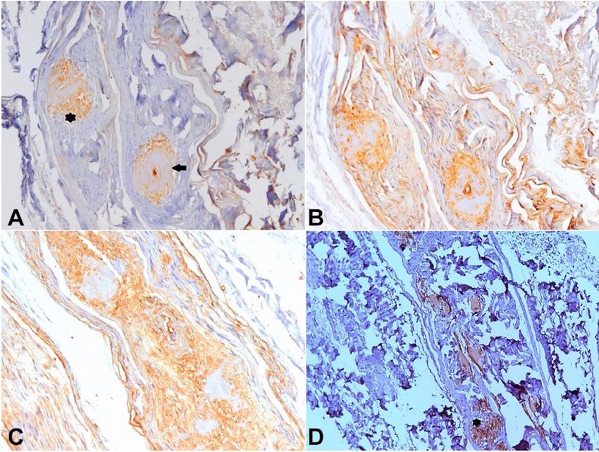
tissue biopsy, which showed positive staining at the biopsy (Figure 7).
same sites of previous IgA deposition (Figure 5D).
Tc99m-pyrophosphate SC showed strong diffuse DISCUSSION
uptake in ventricular walls in February 2019 (Figure 6),
with heart/contralateral area ratio of 1.9 and 1.7 This case report exemplifies some challenging
after 1 and 3 hours, respectively, highly suggestive of aspects that lead to misdiagnosis of systemic amyloidosis
ATTR. At the last follow-up in April 2019, the patient in a patient who consecutively received a diagnosis of
reported a stable neurological condition, sporadic pain DADS, AL, and finally ATTRv.
in lower limbs, and denied any autonomic symptoms; Early and severe sensory ataxia, dissociated CSF,
NIS and ONLS had not changed since 2017. The findings of demyelination in the first ENMG, presence
patient deceased in July 2019 of undetermined cause of MG, the initial absence not only of autonomic
Autops Case Rep (São Paulo). 2021;11:e2021326 5-11Diagnostic challenges in systemic amyloidosis: a case report with clinical and laboratorial pitfalls
Figure 5. Brown immunolabeling with anti-IgA antibody (A) showing two large positive areas in the endoneuro
(asterisk) and around an epineural blood vessel (arrow). Intense non-specific background immunolabeling was
seen with anti-lambda (B) and anti-kappa (C) light-chains antibodies [Horseradish peroxidase, X200]. D – Brown
immunolabeling with anti-Transthyretin antibody showing large positive amyloid deposits in the epineuro at the
same endoneural area showed in 5A (asterisk) [Horseradish peroxidase, X100].
symptoms but also of positive family history, and an multi-symptom involvement or, in sporadic patients,
IHC staining showing two amyloid types in nerve biopsy combinations of PN with cardiac manifestations or
were the main pitfalls in our patient. severe and progressive carpal tunnel syndrome (CTS) in
Amyloidosis should be hypothesized as a cause of a association with other systemic manifestations.9-11 Often
PN when early marked symptoms and signs of autonomic bilateral, CTS can predate other symptoms in ATTRv
dysfunction and/or severe neuropathic pain with a and AL by a mean period of 4-6 years and 1 year,
length-dependent pattern are inaugural manifestations respectively.3,18 Having overlapping clinical manifestations,
or appear during the clinical course in a neuropathy the cardiovascular, gastrointestinal, and genitourinary
initially attributed to another cause.9,11,13,18 In our case, autonomic system are affected in up to 65% of patients
the emergence of marked autonomic dysfunction only with AL and 75% of patients with ATRRv.18
after two years of follow-up worked as a red flag that Considering the whole-body nuclear imaging with
drove the clinical reasoning for amyloid deposition, bone tracers, amyloidosis can be suspected when there
initially thought associated with IgA lambda-MG, later is uptake in unexpected areas, particularly in the heart.
confirmed due to mutated TTR. Currently, 99mTc-PYP has been used in distinguishing
Other important clues for amyloidosis are a ATTR from AL in advanced cardiac amyloidosis, as in
combination of hereditary neuropathy with general our patient. In the absence of MG, a positive cardiac
6-11 Autops Case Rep (São Paulo). 2021;11:e2021326Lino AMM, Castelli JB, Szor RS, Fernandes F, Aiello VD
Figure 6. Strong diffuse uptake in ventricular walls at 99mTc-pyrophosphate scintigraphy in May 2019.
99m
Tc-PYP shows 99% sensitivity and 86% specificity
but lacks specificity for ATTR in patients who have
concomitant MG. 19 Other 99mTc-labelled phosphate
derivatives, such as 99mTC-DPD and 99mTc-HMDP, can be
uptake due to high calcium compounds in the amyloid
with variable sensitivity.20,21 Despite being criticized in
searching multiple myeloma lesions, as used in our
case, 99mTc-MIBI imaging normally shows relatively
high uptake in normal metabolically active tissues,
such as myocardium and has no utility in cardiac
amyloid detection.22-26 Tracing sympathetic innervation,
123
I-MIBGF seems to be useful for early diagnosis in
cardiac amyloidosis, specifically in ATTRv.20,21 99mTc-
Figure 7. Mass spectrometry data of the sural nerve. After Aprotinin and 123I-SAP are potential amyloid tracers,
selecting the amyloid deposit area, tissue microdissection but do not make a distinction between the different
and laser capture, the analysis of the peptide fragments amyloid subtypes.20,21 Conventional cardiac magnetic
revealed the presence of three proteins: transthyretin as
resonance imaging is most helpful in establishing or
the amyloidogenic protein and two other constituents
confirming the diagnosis in later stages.19,26 However,
common to all types of amyloidosis, apolipoprotein E and
serum amyloid P-component. The protein recognition functional assessment with mitral inflow peak
occurs when 3 or more peptide fragments of a specific velocities, deceleration times, and other parameters
protein are identified in the sample [spectra acquired can assist in early diagnosis. Also, atrial gadolinium
by Q-Exactive HF-X spectrometer and analised using uptake along with abnormal gadolinium kinetics
MaxQuant, version 1.6.15.0]. should make aware of cardiac amyloidosis.26
Autops Case Rep (São Paulo). 2021;11:e2021326 7-11Diagnostic challenges in systemic amyloidosis: a case report with clinical and laboratorial pitfalls
An important pitfall was the initial absence of amyloid types.33 All nine cases showed ATTR as one of the
familial history in our case. In ATTRv, the absence amyloid types, being mutated TTR in only one. The second
of family history varies from 33% to 100% and is type of amyloid was Ig-derived in seven, SAA and insulin-
more frequently observed in late-onset patients from derived in one patient each. Regarding tissue distribution,
non-endemic areas.9,12,14,27 However, when a relevant two separated anatomical sites were identified in four
family history exists in patients with PN with prominent patients and in the same tissue sample in five.33
autonomic and/or painful symptoms, hereditary causes Revising our case after LD/MS that only identified
of systemic amyloidosis should be considered. With ATTR in nerve tissue, the peripheral nerve lesion was
autosomal dominant-inheritance in all, the three more imputed to ATTRv, with a false-positive IHC reaction to
important proteins associated with familial amyloid AL due to concomitant IgA lambda-MGUS. However,
polyneuropathy are TTR, by far the most frequent, considering that an autopsy was not performed, AL in
apolypoprotein AI, and gelsolin.10,19 other target organs cannot be ruled out.
The distinction between causal factors and incidental Recognition of amyloidogenic protein is a critical step
findings in a patient with PN and M protein is difficult. for therapeutic decisions. Although IHC is a validated tool
The prevalence of MG in all cases of PN ranges from 3 to with variable sensitivity and specificity, a false positive
5% and can reach up to 10% in neuropathies with no result may occur due to contamination of amyloid
identifiable cause.6,19,28 Moreover, the rate of 1% per year deposits by serum proteins.34,35 On the other hand, a false
of progression to hematologic malignancies, all forms of negative result can be due to epitope loss during tissue
MGUS can progress to AL or cause organ damage due to fixation or by low antibody affinity for cross beta fibrils
immunogenicity of M protein, being the peripheral nerve structure.9,34,36,37 Nowadays, LD/MS is considered the gold
one of the targets. MGUS is associated with a higher standard in amyloid typing, which is essential to guide
relative risk of CIDP and autonomic neuropathy, 5.9 and the therapeutic option.37 By this technique, diagnosis,
3.2, respectively.28 Concurrent presence of MG associated and subtyping of amyloidosis are based on the presence
with misdiagnosis in ATTR patients ranges from 12% to of large spectra of the amyloidogenic precursor protein
49%, being essential to obtain biopsies from different sites and other signature proteins that occur in all amyloid
and organs to proceed the adequate amyloid typing.14,18 types, such as apolipoprotein E, apolipoprotein A IV, and
Data regarding two types of amyloid coexisting serum amyloid P component.19,35
in the same patient is scarce in the literature. The therapeutic approach in our case should also
Koba et al.29 reported the IHC findings of Ig (kappa and be discussed. The international therapeutic consensus
lambda)-derived and keratin-derived amyloid in one was followed since the diagnosis of DADS in the absence
patient with basal cell carcinoma and no MG or Bence- of hematological neoplasia had been made.2,7 In this
Jones protein. Using immunogold labeling technique setting, corticosteroids, intravenous immunoglobulin
and detailing hematological data, Martini et al.30 showed (IgIV), or plasma exchange (PE) are equally effective as
tissue simultaneity of two amyloid types in four patients first-line options, and immunosuppressive agents could
with MG, wild type (wt) ATTR and AL in two cases, AL be used in case of non-responsiveness.7 IgIV and PE
and SAA in one, and ATTRwt and SAA in another patient. were not used due to restrictions to CIDP in our public
Diagnosing two amyloid types at different health system and the presence of severe dysautonomia,
times, Jhaveri et al. 31 reported two patients with respectively. As another therapeutic option in our
confirmed bone marrow AL by IHC in whom only service and before being evaluated by a hematologist,
cardiac ATTRwt was found by LD/MS technique after Fludarabin was introduced due to response failure to
6 and 20 years of complete hematological response. MP associated with Cyclophosphamide. When TTR
Mahmood et al.32 showed distinct positive amyloid mutation was confirmed, the patient had no longer
areas to TTR or light-chain in the myocardium biopsy clinical indication for Tafamidis, the only drug available
by IHC, in which LD/MD detected both amyloidogenic in our public health system, or liver transplantation.
precursors in all deposits, but in distinct proportions. In addition to the complexity of diagnosing and
In a retrospective review from the Mayo Clinic, only subtyping amyloidosis, some limitations of our tertiary
nine in 1904 patients with LD/MS-proven amyloid in public service might have contributed to the delay
different tissues were identified as having two different in the diagnosis, such as non-refunding for genetic
8-11 Autops Case Rep (São Paulo). 2021;11:e2021326Lino AMM, Castelli JB, Szor RS, Fernandes F, Aiello VD
testing, the absence of a routine immunohistochemical 5. Saperstein DS, Katz JS, Amato AA, Barohn RJ.
panel for identification of the three most common Clinical spectrum of chronic acquired demyelinating
polyneuropathy. Muscle Nerve. 2001;24(3):311-24. http://
types of amyloid (AL, ATTR, and AA), and delay in dx.doi.org/10.1002/1097-4598(200103)24:33.0.CO;2-A. PMid:11353415.
Also, this case report highlights the importance of
6. Yeung KB, Thomas PK, King RH, et al. The clinical
more advanced techniques in typing amyloid deposits. spectrum of peripheral neuropathies associated with
benign monoclonal IgM, IgG and IgA paraproteinaemia.
Comparative clinical, immunological and nerve biopsy
CONCLUSION findings. J Neurol. 1991;238(7):383-91. http://dx.doi.
org/10.1007/BF00319857. PMid:1660064.
Sometimes misdiagnosing a condition does not
7. Joint Task Force of the EFNS and the PNS. Joint Task
result from medical unawareness of the disease. Force of the EFNS and the PNS. European Federation of
The presence of overlapping clinical manifestations, Neurological Societies/Peripheral Nerve Society guideline
delay in the appearance of characteristic signs of on management of paraproteinemic demyelinating
neuropathies. Report of a Joint Task Force of the
the disorder, combined with unavailability of specific
European Federation of Neurological Societies and the
diagnostic tools, and difficulties in obtaining an Peripheral Nerve Society – first revision. J Peripher Nerv
expert opinion may overall explain the occurrence of Syst. 2010;15(3):185-95. http://dx.doi.org/10.1111/
misdiagnosis, especially in the context of rare diseases. j.1529-8027.2010.00278.x. PMid:21040140.
Establishing a reference center dedicated to amyloidosis 8. Comenzo RL. Amyloidosis. Curr Treat Options Oncol.
with the availability of more advanced techniques in its 2006;7(3):225-36. http://dx.doi.org/10.1007/s11864-
subtyping can improve diagnostic procedures, reduce 006-0015-8. PMid:16615878.
the time from symptoms onset to diagnosis and allow 9. Conceição I, de Carvalho M. Clinical variability in Type I
the institution of appropriate therapy, preventing Familial Amyloid Polyneuropathy (Val30Met): comparison
harmful procedures and organ deterioration. between late- and early-onset cases in Portugal. Muscle
Nerve. 2007;35(1):116-8. http://dx.doi.org/10.1002/
mus.20644. PMid:16969832.
REFERENCES 10. Kaku M, Berk JL. Neuropathy associated with systemic
amyloidosis. Semin Neurol. 2019;39(5):578-88. http://
1. Neligan A, Reilly MM, Lunn MP. CIDP mimics and dx.doi.org/10.1055/s-0039-1688994. PMid:31639841.
chamaleons. Pract Neurol. 2014;14(6):399-408.
11. Conceição I, González-Duarte A, Obici L, et al. “Red-
http://dx.doi.org/10.1136/practneurol-2014-000831.
flag” symptoms clusters in transthyretin familial amyloid
PMid:25035142.
polyneuropathy. J Peripher Nerv Syst. 2016;21(1):5-9.
2. Joint Task Force of the EFNS and the PNS. European http://dx.doi.org/10.1111/jns.12153. PMid:26663427.
Federation of Neurological Societies/Peripheral
12. Lachmann HJ, Booth DR, Booth SE, et al. Misdiagnosis of
Nerve Society guideline on management of chronic
hereditary amyloidosis as AL (primary) amyloidosis. N Engl
inflammatory demyelinating polyradiculoneuropathy:
J Med. 2002;346(23):1786-91. http://dx.doi.org/10.1056/
report of a Joint Task Force of the European Federation
NEJMoa013354. PMid:12050338.
of Neurological Societies and the Peripheral Nerve Society
– first revision. J Peripher Nerv Syst. 2010;15(1):1-9. 13. Koike H, Kawagashira Y, Iijima M, et al. Electrophysiological
http://dx.doi.org/10.1111/j.1529-8027.2010.00245.x. features of late onset transthyretin Met 30 familial amyloid
PMid:20433600. polyneuropathy unrelated to endemic foci. J Neurol.
2008;255(10):1526-33. http://dx.doi.org/10.1007/
3. Kuwabara S, Isose S, Mori M, et al. Different
s00415-008-0962-z. PMid:18821042.
electrophysiological profiles and treatment response in
‘typical’ and ‘atypical’ chronic inflammatory demyelinating 14. Cortese A, Vegezzi E, Lozza A, et al. Diagnostic
polyneuropathy. J Neurol Neurosurg Psychiatry. challenges in hereditary transthyretin amyloidosis with
2015;86(10):1054-9. http://dx.doi.org/10.1136/jnnp- polyneuropathy: avoiding misdiagnosis in a treatable
2014-308452. PMid:25424435. hereditary neuropathy. J Neurol Neurosurg Psychiatry.
2017;88(5):457-8. http://dx.doi.org/10.1136/jnnp-2016-
4. Doneddu PE, Cocito D, Manganelli F, et al. Atypical CIDP: 315262. PMid:28188196.
diagnostic criteria, progression, and treatment response.
Data from the Italian CIDP database. J Neurol Neurosurg 15. Mathis S, Magy L, Diallo L, Boukhris S, Vallat JM.
Psychiatry. 2019;90(2):125-32. http://dx.doi.org/10.1136/ Amyloid neuropathy mimicking chronic inflammatory
jnnp-2018-318714. PMid:30297520. demyelinating polyneuropathy. Muscle Nerve.
Autops Case Rep (São Paulo). 2021;11:e2021326 9-11Diagnostic challenges in systemic amyloidosis: a case report with clinical and laboratorial pitfalls
2012;45(1):26-31. http://dx.doi.org/10.1002/mus.22229. 27. Coelho T, Sousa A, Lourenço E, Ramalheira J. A study
PMid:22190302. of 159 Portuguese patients with familial amyloidotic
polyneuropathy (FAP) whose parents were both
16. Lozeron P, Mariani LL, Dodet P, et al. Transthyretin unaffected. J Med Genet. 1994;31(4):293-9. http://
amyloid polyneuropathies mimicking a demyelinating dx.doi.org/10.1136/jmg.31.4.293. PMid:8071954.
polyneuropathy. Neurology. 2018;91(2):e143-52.
http://dx.doi.org/10.1212/WNL.0000000000005777. 28. Chaudhry HM, Mauermann ML, Rajkumar SV. Monoclonal
PMid:29907605. gammopathy associated peripheral neuropathy: diagnosis
and management. Mayo Clin Proc. 2017;92(5):838-
17. Briemberg HR, Amato AA. Transthyretin amyloidosis 50. http://dx.doi.org/10.1016/j.mayocp.2017.02.003.
presenting with multifocal demyelinating PMid:28473042.
mononeuropathies. Muscle Nerve. 2004;29(2):318-22.
http://dx.doi.org/10.1002/mus.10614. PMid:14755500. 29. Koba S, Inoue T, Otu M, Miura Y, Misago N, Narisawa
Y. The occurrence of two types of amyloid in the
18. Kapoor M, Rossor AM, Jaunmuktane Z, Lunn MPT, same patient. Br J Dermatol. 2008;158(4):860-2.
Reilly MM. Diagnosis of amyloid neuropathy. Pract http://dx.doi.org/10.1111/j.1365-2133.2007.08432.x.
Neurol. 2019;19(3):250-8. http://dx.doi.org/10.1136/ PMid:18241258.
practneurol-2018-002098. PMid:30598431.
30. Martini F, Buda G, De Tata V, et al. Different types of
19. Muchtar E, Dispenzieri A, Magen H, et al. Systemic amyloid concomitantly present in same patients. Hematol
amyloidosis from A (AA) to T (ATTR): a review. J Intern Rep. 2019;11(4):7996. http://dx.doi.org/10.4081/
Med. 2021;289(3):268-92. http://dx.doi.org/10.1111/ hr.2019.7996. PMid:31871608.
joim.13169. PMid:32929754.
31. Jhaveri T, Sarosiek S, Ruberg FL, Siddiqi O, Berk JL,
20. Glaudemans AWJM, Slart RHJA, Zeebregts CJ, et al. Sanchorawala V. Once AL amyloidosis: not always AL
Nuclear imaging in cardiac amyloidosis. Eur J Nucl amyloidosis. Amyloid. 2018;25(2):139-40. http://dx.doi.
Med Mol Imaging. 2009;36(4):702-14. http://dx.doi. org/10.1080/13506129.2018.1449104. PMid:29516761.
org/10.1007/s00259-008-1037-1. PMid:19156411.
32. Mahmood S, Gilbertson JA, Rendell N, et al. Two types
21. Bokhari S, Shahzad R, Castaño A, Maurer MS. Nuclear of amyloid In a single heart. Blood. 2014;124(19):3025-
imaging modalities for cardiac amyloidosis. J Nucl Cardiol. 7. http://dx.doi.org/10.1182/blood-2014-06-580720.
2014;21(1):175-84. http://dx.doi.org/10.1007/s12350- PMid:25377563.
013-9803-2. PMid:24162886.
33. Sidiqi MH, McPhail ED, Theis JD, et al. Two types of
22. Alexandrakis MG, Kyriakou DS, Passam F, Koukouraki S, amyloidosis present in a single patient: a case series.
Karkavitsas N. Value of Tc-99m sestamibi scintigraphy Blood Cancer J. 2019;9(3):30. http://dx.doi.org/10.1038/
in detection of bone lesion in multiple myeloma: s41408-019-0193-9. PMid:30837451.
comparison with Tc-99m methylene diphosphonate. Ann
Hematol. 2001;80(6):349-53. http://dx.doi.org/10.1007/ 34. Sethi S, Vrana JA, Theis JD, et al. Laser microdissection
s002770100302. PMid:11475149. and mass spectrometry-based proteomics aids the
diagnosis and typing of renal amyloidosis. Kidney
23. Shortt CP, Carty F, Murray JG. The role of whole-body Int. 2012;82(2):226-34. http://dx.doi.org/10.1038/
imaging in the diagnosis, staging, and follow-up of multiple ki.2012.108. PMid:22495291.
myeloma. Semin Musculoskelet Radiol. 2010;14(1):37-
46. http://dx.doi.org/10.1055/s-0030-1248705. 35. Röcken C, Wilhelm S. Influence of tissue fixation on the
PMid:20229439. microextraction and identification of amyloid proteins.
J Lab Clin Med. 2005;146(4):244-50. http://dx.doi.
24. Walker RC, Brown TL, Jones-Jackson LB, De Blanche org/10.1016/j.lab.2005.06.009. PMid:16194686.
L, Bartel T. Imaging of multiple myeloma and related
plasma cell dyscrasias. J Nucl Med. 2012;53(7):1091- 36. Gilbertson JA, Theis JD, Vrana JA, et al. A comparison
101. http://dx.doi.org/10.2967/jnumed.111.098830. of immunohistochemistry and mass spectrometry for
PMid:22693310. determining the amyloid fibril protein from formalin-
fixed biopsy tissue. J Clin Pathol. 2015;68(4):314-7.
25. Wechalekar K, Ng FS, Poole-Wilson PA, et al. Cardiac http://dx.doi.org/10.1136/jclinpath-2014-202722.
amyloidosis diagnosed incidentally by bone scintigraphy. PMid:25637636.
J Nucl Cardiol. 2007;14(5):750-3. http://dx.doi.
org/10.1016/j.nuclcard.2007.07.002. PMid:17826329. 37. Vrana JA, Gamez JD, Madden BJ, Theis JD, Bergen
HR 3rd, Dogan A. Classification of amyloidosis by
26. Esplin BL, Gertz MA. Current trends in diagnosis laser microdissection and mass-spectometry-based
and management of cardiac amyloidosis. Curr Probl proteomic analysis in clinical biopsy specimens. Blood.
Cardiol. 2013;38(2):53-96. http://dx.doi.org/10.1016/j. 2009;114(24):4957-9. http://dx.doi.org/10.1182/blood-
cpcardiol.2012.11.002. PMid:23337445. 2009-07-230722. PMid:19797517.
10-11 Autops Case Rep (São Paulo). 2021;11:e2021326Lino AMM, Castelli JB, Szor RS, Fernandes F, Aiello VD This study was carried out at Sao Paulo University School of Medicine, Departments of Neurology, Pathology, Hematology and Cardiology. Sao Paulo, Sao Paulo, Brazil. Authors’ contributions: Angelina Maria Martins Lino reviewed the medical records and carried out the nerve microscopic analysis with heavy and light chains immunohistochemistry analysis and semithin sections. Jussara Bianchi Castelli and Vera Demarchi Aiello carried out the immunohistochemistry analysis for TTR. Jussara Bianchi Castelli and Roberta Shcolnik Szor carried out the laser dissection and mass spectroscopic analysis. Fabio Fernandes performed the cardiac evaluation. All authors wright, reviewed and approved the manuscript. Ethics statement: Informed consent was obtained in accordance with Ethical Committee of Neurology Department. Conflict of interest: Angelina Maria Martins Lino declares speaking fees and funding for scientific meeting expenses (travel, accommodation and registration) from Pfizer Inc and Sanofi/Genzime and speaking fee from Alnylam Pharmaceuticals. Roberta Shcolnik Szor declares speaking fees from Pfizer Inc, and speaking fees and financial support for research from Jansen-Cilag Farmacêutica Ltda. The remaining authors have no conflict of interest to declare. Financial support: The authors declare that no financial support was received Submitted on: March 29th, 2021 Accepted on: July 22th, 2021 Correspondence Angelina Maria Martins Lino Universidade de São Paulo (USP), Hospital das Clínicas, Faculdade de Medicina, Divisão de Clínica Neurológica Av. Dr. Eneas de Carvalho Aguiar, 255, 5o. andar, sala 5140, CEP 05403-900, São Paulo, SP, Brasil Phone: +55 (11) 2661-6401 angelina.lino@hc.fm.usp.br Autops Case Rep (São Paulo). 2021;11:e2021326 11-11
You can also read