Dementia and COVID-19, a Bidirectional Liaison: Risk Factors, Biomarkers, and Optimal Health Care - IOS Press
←
→
Page content transcription
If your browser does not render page correctly, please read the page content below
Journal of Alzheimer’s Disease 82 (2021) 883–898                                                                       883
DOI 10.3233/JAD-210335
IOS Press

Review

Dementia and COVID-19, a Bidirectional
Liaison: Risk Factors, Biomarkers, and
Optimal Health Care
Sofia Tonioloa,∗ , Marta Scarionib,c , Francesco Di Lorenzod,e , Jakub Hortf,g , Jean Georgesh ,
Svetlana Tomici,j , Flavio Nobilik,l and Kristian Steen Frederiksenm , the Management Group of the
EAN Dementia and Cognitive Disorders Scientific Panel
a Cognitive  Neurology Group, Nuffield Department of Clinical Neurosciences, University of Oxford, Oxford, UK
b Department   of Neurology, Amsterdam University Medical Centers, Location VUmc, Alzheimer Center,
Amsterdam, The Netherlands
c Department of Pathology, Amsterdam University Medical Centers, Location VUmc, Amsterdam Neuroscience,

Amsterdam, The Netherlands
d Clinical Imaging Sciences Centre, Brighton and Sussex Medical School, Brighton, UK
e Non-invasive Brain Stimulation Unit, IRCCS Fondazione Santa Lucia, Rome, Italy
f Memory Clinic, Department of Neurology, Charles University, 2nd Faculty of Medicine and Motol University

Hospital, Czech Republic
g International Clinical Research Center, St. Anne’s University Hospital Brno, Brno, Czech Republic
h Alzheimer Europe, Luxembourg
i Department of Neurology, Osijek University Hospital Center, Osijek, Croatia
j Faculty of Medicine, University Josip Juraj Strossmayer of Osijek, Osijek, Croatia
k Neurology Clinic, IRCCS Ospedale Policlinico San Martino, Genoa, Italy
l Department of Neuroscience (DINOGMI), University of Genoa, Genoa, Italy
m Danish Dementia Research Centre, Rigshospitalet, University of Copenhagen, Copenhagen, Denmark

Handling Associate Editor: Laura Bonanni
Accepted 14 May 2021
Pre-press 29 May 2021

Abstract. Cognitive impairment following SARS-CoV-2 infection is being increasingly recognized as an acute and possibly
also long-term sequela of the disease. Direct viral entry as well as systemic mechanisms such as cytokine storm are thought
to contribute to neuroinflammation in these patients. Biomarkers of COVID-19-induced cognitive impairment are currently
lacking, but there is some limited evidence that SARS-CoV-2 could preferentially target the frontal lobes, as suggested
by behavioral and dysexecutive symptoms, fronto-temporal hypoperfusion on MRI, EEG slowing in frontal regions, and
frontal hypometabolism on 18 F-FDG-PET. Possible confounders include cognitive impairment due to hypoxia and mechan-
ical ventilation and post-traumatic stress disorder. Conversely, patients already suffering from dementia, as well as their
caregivers, have been greatly impacted by the disruption of their care caused by COVID-19. Patients with dementia have
experienced worsening of cognitive, behavioral, and psychological symptoms, and the rate of COVID-19-related deaths is

  ∗ Correspondence to: Dr. Sofia Toniolo, Nuffield Department    House, 1st Floor, OX2 6DG, Oxford, United Kingdom. Tel.:
of Clinical Neurosciences, University of Oxford, New Radcliffe   +44 01865271310; E-mail: sofia.toniolo@ndcn.ox.ac.uk.

ISSN 1387-2877/$35.00 © 2021 – IOS Press. All rights reserved.
884 S. Toniolo et al. / Dementia and COVID-19
disproportionately high among cognitively impaired people. Multiple factors, such as difficulties in remembering and exe-
cuting safeguarding procedures, age, comorbidities, residing in care homes, and poorer access to hospital standard of care
play a role in the increased morbidity and mortality. Non-pharmacological interventions and new technologies have shown
a potential for the management of patients with dementia, and for the support of their caregivers.
Keywords: Alzheimer’s disease, cognition, COVID-19, dementia, SARS-CoV-2
INTRODUCTION between COVID-19 and dementia, we will examine
the possible pathogenic role of COVID-19 in caus-
Cognitive impairment in severe acute respiratory ing cognitive impairment and available biomarkers,
syndrome coronavirus 2 (SARS-CoV-2) infection has and describe the challenges that COVID-19 poses to
frequently been reported, in patients with meningitis, patients with dementia and their caregivers, as well
encephalitis, encephalopathy, and acute cerebrovas- as possible interventions.
cular disease, but also when no identifiable structural
cause can be found [1, 2]. Rates of cognitive impair- INTERPLAY BETWEEN SARS-COV-2 AND
ment in COVID-19 patients range widely across DEMENTIA
studies [2–4], due to different neurological conditions
causing cognitive impairment and different evalu- Pathophysiology
ation timings and settings [2–4]. Although several
pathways have been implicated in viral entry in the Neuro-invasion by SARS-CoV-2 is thought to be
central nervous systems (CNS), the presence of the achieved through different mechanisms. One possible
virus is not necessary to elicit neuronal damage, way is the hematogenous spread from infected leuko-
as other detrimental mechanisms such as cytokine cytes, similarly to HIV’s Trojan horse, crossing the
storm could play a crucial role in COVID-19 neu- blood-brain barrier (BBB) [14]. Direct viral invasion
roinflammation [5, 6]. Most studies comprehensibly through infected vascular endothelial cells directly
focus on acute cognitive syndromes such as delir- to glial cells has also been described, as well as
ium, while longitudinal data on cognition are scarce. penetrance from the olfactory epithelium to the olfac-
The distinction between acute and permanent cog- tory bulb through retrograde axonal transport along
nitive decline in the context of COVID-19, though the olfactory nerve [15]. Vagal and glossopharyn-
too early to ascertain, is an essential one. Other geal nerves might also play a role in neuro-invasion
coronaviruses, such as severe acute respiratory syn- and multiorgan dissemination of SARS-CoV-2 [16].
drome coronavirus 1 (SARS-CoV-1) and middle east Moreover, angiotensin-converting enzyme 2 (ACE-
respiratory syndrome (MERS), mostly caused acute 2) receptors control SARS-CoV-2 entry into cells,
confusion, mania and psychosis, and even if residual and their expression in endothelial cells mediates
depression, anxiety, insomnia, memory impairment, viral invasion across the BBB [15]. Neuropatho-
post-traumatic stress disorder (PTSD) and fatigue logical and transcriptome-based studies in humans
have been described, most patients showed a pro- found high ACE-2 expression throughout the brain,
gressive return to their baseline function [7]. In especially in the pons, medulla oblongata, substan-
patients with an established diagnosis of dementia, tia nigra, caudate nucleus, spinal cord, hypothalamus,
the available studies point to an increased sus- hippocampus, middle temporal gyrus, amygdala, cin-
ceptibility of Alzheimer’s disease (AD) patients to gulate cortex, frontal cortex, and olfactory bulb [17,
COVID-19 infection, and to higher rates of COVID- 18]. Given that the medulla oblongata contains the
19 deaths [8–11]. Multiple factors, such as difficulties respiratory centres of the brain, its involvement
in remembering safeguarding procedures (e.g., wear- might partially explain the susceptibility of many
ing masks or adhering to social distance), but also COVID-19 patients to severe respiratory distress
age, genotype, comorbidities, and residing in care [17]. Preclinical evidence on other coronaviruses
homes, have been shown to play a role [10, 12, indicate their greater affinity to CA1 and CA3 hip-
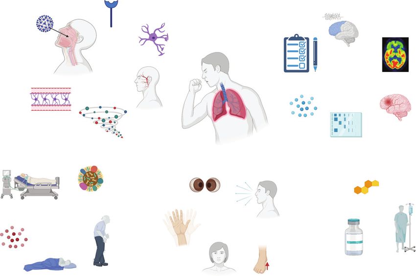
13]. In this review, focusing on the reciprocal link pocampal regions [19]. This does not seem to applyS. Toniolo et al. / Dementia and COVID-19 885 Fig. 1. Overview of the pathophysiological mechanisms, risk factors, symptoms, and biomarkers of cognitive impairment and therapy in COVID-19 patients. Made in ©BioRender - biorender.com. to SARS-CoV-2, as the relatively limited neuropatho- would ultimately lead to CNS damage [26]. As a logical data available show a primary involvement of result, markers of BBB disruption, such as serum medulla oblongata, basal ganglia, cerebellum, frontal S100B, and glial activation, as glial fibrillary acidic lobes and olfactory bulbs [6, 20]. No proven cases protein (GFAP), are increased in patients with of AD or frontotemporal dementia (FTD) have been COVID-19-related encephalopathy [27–29]. More- associated to SARS and MERS coronaviruses, but over, this cytokine storm is also associated with this evidence is limited by small numbers. Despite increased levels of ferritin, lactate dehydrogenase the growing amount of literature, clinical evidence for (LDH) and D-dimer, which in turn can lead to an viral presence of SARS-CoV-2 in the cerebrospinal hypercoagulable state and increased cerebrovascular fluid (CSF) is still limited, even if viral particles events [30]. Lastly, hypoxia secondary to respiratory can be found in the brain [20–22]. COVID-19 is insufficiency needs to be considered among the major often associated with a cytokine storm, with increased causative mechanisms leading to transient and possi- release of interleukin 6 (IL-6), tumor necrosis fac- bly permanent cognitive impairment [31] (Fig. 1). tor alpha (TNF-alpha), interleukin 1 beta (IL-1), and interleukin 8 (IL-8), which strongly resembles an immune effector cell-associated neuro-toxicity Dementia as a risk factor for COVID-19 syndrome (ICANS) [23]. Increased cytokine levels, especially IL-6, have been shown to be positively Dementia, and particularly AD, is associated with correlated with COVID-19 severity and mortality, higher rates of infection and hospitalization due to leading to multiorgan failure [24, 25]. This cytokine COVID-19 [9, 32]. A large UK biobank study showed storm induces a massive activation of macrophages that AD was the major risk factor for hospital admis- and monocytes and compromises the integrity of sion for COVID-19 (OR = 2.29) [9]. According to an the BBB, which in turn, can induce astrocyte and American study, dementia was associated with an OR glial cells to produce reactive oxygen species, which of 5.2 and AD of 7.7 of COVID-19 infection [32].
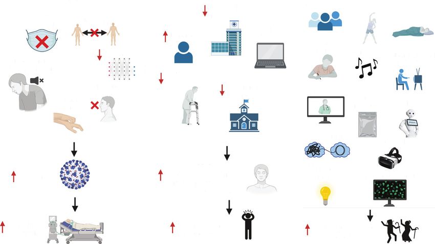
886                                           S. Toniolo et al. / Dementia and COVID-19

Fig. 2. Risk factors for increased infection rates and BPSD in patients with dementia, and possible interventions. WM, working memory;
BPSD, behavioral and psychological symptoms of dementia; CBT, Cognitive Behavioral Therapy; CST, Cognitive Stimulation Therapy;
MBI, Mindfulness-Based Intervention. Made in ©BioRender - biorender.com.

   Multiple factors could contribute to these increased               distancing. Therefore, it is not surprising that AD
rates of infection. Patients with dementia might                      patients, who have lower WM capacity and have
have difficulties in remembering safeguarding pro-                    poorer response inhibition, could have higher rates
cedures, such as wearing masks, or understanding                      of infection.
the public health information issued [33] (Fig. 2).                      However, not only is the incidence of COVID-19
A Japanese study on AD and FTD patients showed                        higher in patients with dementia, but these patients are
that both patient groups had troubles understand-                     also more likely to have more severe presentations,
ing the meaning of the requests, and AD patients                      and higher mortality rates due to COVID-19 infec-
often forgot the instructions [33], while FTD patients                tion [10, 38]. An epidemiological study among 185
struggled more to follow safeguarding procedures,                     countries found a significant correlation between bur-
especially washing hands, social distancing, and stay-                den of dementia and life expectancy, and COVID-19
ing at home, because of their socially inappropriate                  related deaths [39]. The highest numbers of con-
and stereotyped behaviors. In care homes, increased                   firmed cases and deaths were reported in high-income
intrusiveness and wandering undermine efforts to                      countries, where also the prevalence of dementia is
maintain isolation, thus increasing the risk of conta-                higher, and more patients live in long-term care facil-
gion [34, 35]. Even in healthy people, a UK Biobank                   ities, where COVID-19 had a higher toll [39]. A
study showed that over 431,051 healthy subjects an                    cross-country study involving UK, Spain, Ireland,
elevated risk of COVID-19 incidence was related to                    Italy, Australia, US, India, Kenya, and Brazil found
performance on two tests of cognitive function, i.e.,                 that 29%–75% of the deaths in patients with demen-
verbal and numerical reasoning and reaction speed                     tia in care homes were linked to COVID-19 [40].
on a ‘Go/No-go task’ [36]. Similarly, an American                     In a UK Biobank study, pre-existing dementia was
study on 850 healthy subjects found that participants’                the most important factor associated with COVID-19
social-distancing compliance could be predicted by                    severity (OR = 3.07) [10]. An Italian observational
individual differences in working memory (WM)                         study showed that severe dementia was an indepen-
capacity [37], with high WM capacity individu-                        dent risk factor for death [38]. In a Korean study,
als understanding better the true merits of social                    age, chronic lung disease, and AD were the only
S. Toniolo et al. / Dementia and COVID-19 887
significant parameters for predicting COVID-19 non- from COVID-19 patients with AD show that ACE-2
survival [41]. A Spanish study in 204 patients with levels are actually upregulated in AD brains [54], and
AD and FTD showed that AD patients had higher increased ACE-2 levels could therefore constitute an
mortality rates compared to patients with FTD, even intrinsic vulnus and an additional risk factor for viral
though the AD ones were older in this study, and that entry.
risk of death was much more common in patients APOE4 genotype, a known risk factor for late-
living in care homes (72.0%) compared to home- onset sporadic AD, has been acknowledged as a
dwelling patients (7.3%), regardless of the etiology possible additional predisposing factor for COVID-
[13]. 19 infection and severity [55] (Fig. 1). A UK-Biobank
Several explanations might account for the higher study showed that, after adjusting for pre-existing
rate of COVID-19 mortality in patients with demen- dementia, hypertension, coronary artery disease, and
tia. This has often been attributed exclusively to Type-2 diabetes, APOE4 homozygotes have a 2.2-
age, but comorbid diseases such as AD may be fold increased risk for COVID-19 infection, and more
more influential than aging [42, 43], and age and severe outcomes, specifically a 4.3-fold case-fatality
dementia are independently associated with a higher rate, compared to APOE3 homozygotes [10, 12, 56].
mortality [44]. Moreover, there might be local hos- This finding has been questioned with respect to
pital guidelines and ethical reasons accounting for the adjustment of p-value in genome-wide associa-
higher mortality in patients with dementia. In case of tion studies (GWAS) and possible selection bias [57,
shortage of ventilators, rationing of health care may 58]. Its replication could help to unravel a possible
be required, which could result in elderly patients detrimental effect of ApoE4 in inducing neuroinflam-
with dementia being denied intensive care or ven- matory microglia response, as in AD [59], and further
tilatory support, resulting in higher mortality [45, neurodegeneration in COVID-19 patients [51].
46]. Reports of ‘ageism’, i.e., discrimination against Another possible biological link between COVID-
elderly people, and especially patients with dementia, 19 and AD is represented by a pro-inflammatory
have been frequently described during the COVID-19 cytokine profile. IL-6 and IL-1 have been frequently
pandemic [40]. As an example, in the UK, people liv- associated with increased mortality in COVID-19
ing with dementia often reported being denied access patients [2], and currently IL-6 and IL-1 inhibitors,
to hospital or being told that they had to sign “do not such as tocilizumab and anakinra, are among the
attempt resuscitation (DNAR)” arrangements [40]. most used treatment options for COVID-19 [60, 61]
Alzheimer Europe has made a public statement that (Fig. 1). Neuroinflammation has received much atten-
a diagnosis of dementia should never be a reason to tion in recent years as a driver of cognitive impairment
refuse people access to treatment, care and support, as in AD [62]. Pre-COVID-19 data indicate that inflam-
would violate basic human rights [47]. Ethical con- matory markers in AD show different patterns across
siderations arise also when artificial intelligence is disease progression, with some cytokines peaking in
proposed to optimize triage [48]. the prodromal phase and then declining, while others,
including IL-1 and IL-6, increasing alongside dis-
Shared biological links between dementia and ease progression [63–65]. Preclinical data in animal
COVID-19 infection models of AD show that counteracting inflammation
leads to better cognitive performances [66–68], while
Several shared neurobiological substrates have evidence on the use of anti-inflammatory drugs in
been suggested to mediate the pathological synergy AD has been inconclusive, given their poor BBB per-
between AD and COVID-19 infection [49]. While meability [69]. SARS-CoV-2 open reading frame 3a
ACE-2 receptors overexpression could facilitate viral (ORF3a) protein stimulates NLRP3 inflammasome,
access into the CNS, their role in neurodegenera- which has also been found to cause pathological accu-
tive diseases is still debated, with some evidence mulation of A [70, 71]. Galectin-3, a key element
claiming that the physiological decline in ACE-2 in COVID-19 inflammation [72], is involved in A
expression could explain the increased severity of oligomerization and toxicity, and has been proposed
cognitive symptoms in elderly patients [50], while as shared anti-inflammatory target [73]. Neverthe-
other advocate a neuroprotective effect [51, 52]. Pre- less, these potential mechanisms remain speculative
COVID-19 data showed reduced ACE-2 levels in AD, at the moment, given the absence of reliable post-
which inversely correlated with A and tau burden mortem data on A and neurofibrillary tangles in
[51–53]. On the other hand, preliminary preprint data COVID-19 patients.888 S. Toniolo et al. / Dementia and COVID-19
Delirium as a clinical presentation of COVID-19 signs and myoclonus, has been frequently described
patients with dementia: Cognitive features and in hospitalized COVID-19 patients, whether in inten-
biomarkers sive care unit (ICU) on non-ICU wards [83–85]. At
neuropsychological testing, deficits in frontal lobe
Among patients diagnosed with dementia, one of functions have been reported, even in young COVID-
the most frequent symptoms of COVID-19 at onset 19 patients, and seem to be correlated with the
is delirium, especially in the hypoactive form [74, severity of respiratory symptoms and mechanical
44]. By contrast, fever, cough, and dyspnea are less ventilation, but not with gender, length of ICU stay or
common [38, 75, 76]. A French study in 353 elderly duration of mechanical ventilation [86–88]. In these
showed that patients with dementia compared to those cases, magnetic resonance imaging (MRI) findings
without, more often exhibited hypoactive (27.6% ver- can be either unremarkable, or show fronto-temporal
sus 11.4%) and hyperactive delirium (14.9% versus hypoperfusion at arterial spin labeling, or even atro-
5.5%), altered consciousness (17.2% versus 6.4%), phy in multiple regions, including the frontal grey
and lymphopenia (79.3 % versus 66.7%), and less matter [78, 83, 88]. Electroencephalogram (EEG) fre-
often hyperthermia (47.0% versus 61.6 %), cough quently shows frontal abnormalities, including slow
(46.3% versus 66.7%) and loss of taste (2.3% versus waves or epileptiform discharges, in up to 1/3 of the
10.1%) [77]. Prevalence of delirium increases with cases, and has recently been proposed as a biomarker
age and with more severe cognitive impairment, and for COVID-19 encephalopathy [89]. An inflamma-
can be associated with as high as 17 times greater tory profile in the CSF, such as increased IL-6 or
mortality rates [74]. oligoclonal bands has also been described [78, 83,
Pharmacological management has also been impli- 79, 80, 90]. Frontal hypometabolism on positron
cated in induction or exacerbation of delirium in emission tomography with F-18 fluorodeoxyglucose
COVID-19 patients [110, 111]. Benzodiazepines (FDG-PET) imaging has been reported in these
(BDZ) (OR = 1.59), opioids (OR = 1.39), antipsy- patients with acute frontal type behavioral changes
chotics (OR = 1.59) and vasopressor infusions [79, 80]. Moreover, frontal hypometabolism is found
(OR = 1.25) have been found to be associated with a in COVID-19 patients without evidence of acute
higher risk of delirium in these patients [110]. Phar- delirium or frontal signs, but with anosmia and
macodynamic interactions with drug-induced effects ageusia, and in old as well as young patients [90]. Pre-
on increased QTc prolongation. coagulation abnor- COVID-19 literature on 18F-FDG-PET in patients
malities, and immunosuppression also need to be with delirium is not vast, but the few available studies
considered in COVID-19 patients [112]. Haloperidol show a more diffuse pattern of hypometabolism [91,
has shown a favorable profile in terms of morbidity 92]. There are no pre-delirium scans in most COVID-
and mortality, while BDZ and anticholinergic drugs 19 studies, so these changes might also reflect in some
should be avoided for the high risk of respiratory studies premorbid patterns of cognitive impairment
depression and confusion [110, 112]. Whenever pos- as in AD or FTD, delirium potentially being only an
sible, non-pharmacological management of agitation epiphenomenon [93]. Nevertheless, even in patients
should be preferred, in order to avoid acute delirium with no previous history of cognitive impairment,
and a post-intensive care syndrome (PICS) [110]. there are reports of a fronto-parietal 18F-FDG-PET
Given the high frequency of delirium in patients hypometabolism coupled with relative striatal and
with COVID-19 infection, it is crucial to understand cerebellar hypermetabolism in the acute stage of
whether there are any specific cognitive, imag- COVID-19, which correlates with the degree of cog-
ing, electrophysiological, or laboratory predictors nitive impairment at testing [94, 95]. After 6 months,
(biomarkers) of CNS involvement. Moreover, it is one study showed a significant reduction of an ini-
important to ascertain whether these elderly COVID- tial frontoparietal hypometabolism, accompanied by
19 patients with delirium are more likely to develop a significant improvement in cognition, even if there
a neurodegenerative disorder in the future or are were still residual mild deficits at neuropsychological
already affected by preclinical disease. There is some testing and their 18F-FDG-PET did not completely
limited evidence that SARS-CoV-2 could preferen- revert back to normal [94].
tially target the frontal lobes [78–82] (Fig. 1). A What is the underlying cause of this frontal lobe
clinical phenotype characterized by acute frontal type dysfunction? The role of direct viral damage is at
behavior such as attention deficits, abulia, alogia, the moment limited, and even if viral particles can
absence of goal directed behavior, frontal release be found in the brain, the few neuropathologicalS. Toniolo et al. / Dementia and COVID-19 889
data available argue in favor of an inflammatory years, conducted 10–35 days after hospital discharge,
process rather than direct damage by SARS-CoV-2, found that patients who reported headache, anosmia,
involving primarily the brainstem, cerebellum, and dysgeusia, diarrhea, and those who required oxy-
olfactory bulbs, and to a lesser extent, the basal gen therapy had lower scores in several cognitive
ganglia and frontal lobes [6, 20]. Notably, in some domains, such as long-term episodic memory and
of these cases, there is evidence that counteract- WM, attention, executive function, processing speed
ing neuroinflammation could have a positive impact and naming, compared to asymptomatic patients [86].
on the patients’ outcomes, either through steroids Performance on digit span backwards was consis-
administration, intravenous immunoglobulin (IVIG), tently impaired, even if only anosmia and ageusia
or tocilizumab [24, 79, 80, 84, 85] (Fig. 1). The were present. Preliminary preprint data from a large
frontal lobes are particularly vulnerable to hypoxic UK online study on 84,285 people, including 9,201
damage, but given the response to immunosuppres- without and 3,466 with respiratory symptoms, and
sion, it is unlikely that this is the sole source of the 361 with laboratory-confirmed SARS-CoV-2 infec-
cognitive impairment [96] (Fig. 1). Lack of standard- tion, showed that global cognitive scores covaried
ized diagnostic protocols, as well as polymorphic with respiratory COVID-19 symptom severity after
nomenclatures such as COVID-19 related delirium correcting for multiple socio-economical cofounders,
[78], encephalopathy [79], encephalitis [97], akinetic including age, and people who required ventilation
mutism [85], and frontal lobe dysfunction [80], ham- performed significantly worse [87]. Notably, in this
per comparability across studies [98]. It is not known study only a small proportion (0.76%) of partici-
whether these patients are more likely to develop FTD pants reported having residual symptoms, although
or AD at the present time, and large-scale data on this was much higher in patients who required ven-
blood and CSF biomarkers of neurodegeneration is tilation, in which case 78% had residual cognitive
lacking, with reports of increased neurofilament light symptoms. Semantic problem solving, visual atten-
chain and total tau in severe COVID-19 cases [29]. tion, and visual WM were the most affected domains,
Moreover, COVID-19 is related to increased risk especially in hospitalized patients. The scores in
of vascular changes and stroke [99], which can also patients who required ventilation were equivalent
impact cognition acutely, and are important comor- to 10-year older subjects in this dataset. One small
bidities in subjects with cognitive impairment. American study in 57 COVID-19 patients, 88% of
which had documented hypoxemic respiratory fail-
Long-term effects on cognition and cognitive ure, showed deficits in working memory, set-shifting,
profile divided attention, and processing speed in 40–55%
of patients, which were not significantly associated
Only future longitudinal data will be able to assess with intubation length or the time from extubation
the long-term impact of COVID-19 on cognition. to assessment, psychiatric diagnosis, or preexisting
Currently, a few studies have described the cogni- cardiovascular/metabolic disease [102]. Preliminary
tive status of COVID-19 patients at discharge or preprint data on Long COVID-19 symptoms after 7
after a short follow-up period. In a French study months in a sample of 3,762 subjects support deficits
in COVID-19 patients hospitalized in an ICU, 33% in attention, short-term memory and executive func-
out of 45 patients had a dysexecutive syndrome tions, with a great impact on the ability to return to
at discharge [83] (Fig. 1). Another French study work [103].
reported that after a mean of 110.9 days, the most Is this dysexecutive/frontal syndrome character-
frequently reported persistent symptoms were fatigue istic of COVID-19 infection? Deficits in executive
(55%), dyspnea (41.7%), followed by loss of memory function are also prominent in patients with acute
(34.2%), attention deficits (26.7%), and sleep disor- respiratory distress syndrome (ARDS) [104–106],
ders (30.8%) [100]. A small Chinese study on 29 and a significant percentage of ARDS survivors suf-
patients who recovered from COVID-19 infection fer long-term cognitive impairment with prominent
and 29 controls found lower scores in COVID-19 dysexecutive traits [107], as part of PICS [106].
patients at the continuous performance test (CPT), The major risk factors for the development of PICS
which measures sustained and selective attention, and are age, prior cognitive deficits, delirium, sepsis,
the CPT score was positively correlated with their sleep deprivation, ARDS, prolonged mechanical ven-
C-reactive protein (CRP) levels [101]. One small tilation, and multiorgan failure [107–109], many
Spanish study in 35 COVID-19 patients aged 20–60 of which are experienced by COVID-19 patients890 S. Toniolo et al. / Dementia and COVID-19
(Fig. 1). Mechanical ventilation has been associ- COVID-19 patients had statistically significantly
ated with executive dysfunction during and after ICU higher grey matter volumes bilaterally in the olfactory
treatment, and is a risk factor for developing delirium cortex, hippocampus, insula, left rolandic operculum,
[105]. Nevertheless, not all COVID-19 patients who left Heschl’s gyrus and right cingulate gyrus, reduced
develop cognitive dysfunction in the aftermath have mean diffusivity, axial diffusivity, and radial diffu-
undergone mechanical ventilation, or have been in sivity, and increased fractional anisotropy, which are
ICU, so this might be a causal agent as well as a cofac- opposite findings compared to what usually observed
tor. A limitation of the available studies on COVID-19 in neurodegenerative diseases, and therefore more
is that many patients are not usually screened for base- data are needed to draw definite conclusions.
line cognitive status, nor for biomarkers of cognitive Lastly, there is general expectation that COVID-
impairment. 19 vaccination would bring current epidemy under
It is important to keep in mind that post-ICU control. Recent data has shown that influenza and
patients frequently experience a worsening of cog- pneumonia vaccination might be associated with
nition, due to depression, anxiety, and PTSD [113]. reduced risk of AD [124]. Bidirectional approach
PTSD has also been reported in MERS and SARS to AD and COVID-19 should include designing of
patients, even 18–30 months after illness [114, 115], proper studies to evaluate effect of various anti-
and by COVID-19 patients [116]. Subjective cog- COVID vaccination on incidence or course of AD.
nitive complaints are also frequently reported by
COVID-19 patients, in the acute and chronic phase, MANAGEMENT OF PEOPLE WITH
and in patients with or without a pre-existing psy- DEMENTIA DURING THE COVID-19
chiatric diagnosis [103, 117]. Subjective complaints OUTBREAK
often correlate with executive dysfunction, quality of
life and heave a substantial impact on patients’ ability Impact of COVID-19 on patients’ and
to return to work [103, 118]. Multiple centers already caregivers’ well-being
advise to monitor cognitive and psychological symp-
toms of PICS, and encourage the use of cognitive Most of the literature so far has focused on the
behavioral therapy, counseling sessions, and psycho- impact of COVID-19 on AD patients, described as
logical support [119, 120]. a “double burden”, as the pandemic both increases
To summarize, the etiology of cognitive impair- their vulnerability and disrupts their social network,
ment is probably multifactorial [117]. including their access to the healthcare system [39,
Discussing the cognitive long-term implications of 125]. Scheduled outpatient activities such as blood
vascular brain damage due to COVID-19 is beyond sampling or neuroimaging appointments might be
the scope of this review, but vascular ischemic and postponed or missed altogether, due to hospital man-
hemorrhagic events are frequently seen in COVID-19 agement decisions, but also to the reluctance of
patients [121, 122], and the point prevalence of long- vulnerable patients to go to the hospital, and a timely
term vascular dementia in these patients will need to diagnosis and initiation of treatment could be delayed
be ascertained. in patients referred for diagnostic evaluation [126]
Only few studies so far have examined voxel-wise (Fig. 2). The ‘Digital Divide’, i.e., little knowledge of
structural, non-lesional changes in patients recov- telecommunication and other digitally-based modes
ered from COVID-19 [88, 123]. A small study on 13 of communication, is frequent among elderly people,
COVID-19 patients admitted to ICU showed brain especially patients with dementia, and they depend
atrophy in regions including primarily, but not lim- primarily on in-person support, which might be lack-
ited to, frontal regions [88]. A subset of patients ing. Family members are often banned from visiting
had executive dysfunction and deficits in lexical flu- long-term care facilities, and patients with dementia
ency at testing within 2 weeks of ICU discharge, might not understand its reason, and feel abandoned
but brain atrophy was present regardless of the pres- [126]. Group activities in nursing homes may also be
ence of cognitive impairment [88]. Another study prohibited, leading to physical and social isolation.
enrolled 60 COVID-19 patients 3 months after ill- This might increase behavioral and psychological
ness, with negative SARS-CoV-2 PCR at the time symptoms of dementia (BPSD), such as anxiety,
of enrolment, and 39 age- and sex-matched controls agitation, irritability, sleep disturbances, apathy, and
that had not been exposed to SARS-CoV-2 [123]. depression, which might in turn also impact the indi-
Their study results are somehow unexpected, as viduals’ ability to report symptoms of the infectionS. Toniolo et al. / Dementia and COVID-19 891 [127, 128]. Social restrictions induce several lifestyle regarding a faster cognitive decline were reported by modifications eventually increasing psychological 44% of patients with objective cognitive decline and distress in healthy people and even more in patients 14% of SCIs [136]. 76% of the caregivers reported an with dementia, and the lack of social, physical and increase in BPSD, and two third reported discontinu- cognitive stimulation causes in these patients a partial ation of care, which correlated with a higher caregiver “deprivation syndrome” effect [129]. burden [136]. During quarantine, worsening of cognitive symp- Caregivers have also been highly affected by the toms, particularly of memory and orientation abilities pandemic, and higher burden has been associated and increased BPSD in patients with cognitive with longer confinement periods and with caring for impairment have often been reported, especially in more severe cases [131, 137–141] (Fig. 2). Caregivers patients with severe dementia [130]. An Argentinian might be the only point of contact between the patient study found that 48% patients with AD reported an and the outside world, and being cut off from their increased level of anxiety in the context of lockdown personal network, might feel isolated [129]. Across [131], and a French study showed higher depression studies, at least two-thirds reported increased distress and anxiety rates during the COVID-19 crisis in AD [136]. Nevertheless, is important to keep in mind that patients [132]. AD is not the only dementia who might most of these studies lack adequate controls and lon- be affected by COVID-19. One Italian multicen- gitudinal data, and that COVID-19 has had an impact tric study on 4,913 patients with different dementia on mental health even in healthy people [142]. types showed an increased burden of neuropsychi- atric symptoms in 59.6% of patients with dementia, Adaptive strategies and challenges for clinical either worsening of preexisting symptoms (51.9%) or assessment and non-pharmacological new onset (26%), which required drug modifications interventions in 27.6% of the cases [129]. Irritability, apathy, agi- tation, and anxiety were the most frequently reported The healthcare system should plan adequate strate- worsening symptoms, and sleep disorder and irri- gies to counteract these neuropsychological and tability the most reported new symptoms. Anxiety behavioral consequences of quarantine, also through and depression were correlated with a diagnosis of the use of communication technologies [143]. Indeed, AD, mild disease severity, and female gender. Hav- the COVID-19 pandemic has confronted us with new ing a diagnosis of dementia with Lewy bodies (DLB) diagnostic challenges, but also, many suggest, with an increased the risk of worsening hallucinations and opportunity to positively revolutionize our diagnostic sleep disorders, while FTD increased the risk of aber- system [35, 127, 144]. rant motor behavior and change of appetite. Performing neuropsychological assessments wear- Worsening of cognition and BPSD have been ing Level 2 personal protective equipment (PPE) reported also in patients with mild cognitive impair- with COVID-19 patients poses several logistical chal- ment (MCI), who have experienced higher levels lenges [145]. Paper and pencil tasks cannot be taken of apathy during quarantine, clearly increasing over into the ward during the assessment [145]. For timed time [133]. A Spanish study in patients with MCI or tasks, even where is possible to wear a wristwatch, mild dementia found that only 61% declared feeling it cannot be seen underneath the PPE, and speaking well, 29% felt sad, 22% worried, 11% afraid, 24% through a mask to patients with perceptual or cog- anxious, and 14% bored [134]. Those living alone nitive deficits makes bedside cognitive assessment reported greater negative psychological effects and difficult to perform [145]. Moreover, COVID-19 sleeping problems. One Italian study in patients at patients often experience debilitating fatigue, which risk for dementia, i.e., patients with subjective cogni- may adversely affect their cognitive performance, and tive impairment (SCI) and MCI, found that patients increased anxiety, which can influence the reliability led an increased sedentary lifestyle, switched to a of their cognitive scores [145]. more unhealthy diet and had lower engagement in Even in outpatient settings, is not only the Dig- active recreational activities, which can increase the ital Divide wide among elderly and patients with risk of developing dementia [135]. One Dutch study dementia, but in addition, they might have further in patients with SCI, MCI, and dementia due to AD difficulties in performing a phone or video consulta- or DLB showed that 46% of the patients with objec- tion, as microphone and speakers may be insufficient tive cognitive decline (MCI, dementia) and 38% of for some patients with dysarthric speech, hearing, and SCIs reported an increase in BPSD, and concerns ambient and background noises may exacerbate these
892                                     S. Toniolo et al. / Dementia and COVID-19

difficulties [146]. Additionally, it can be challeng-              Lastly, funding for dementia research has been
ing to ensure input from caregivers over the phone,             drastically reduced due to COVID-19 [158]. There-
adding to possible barriers for telecommunication as            fore, action needs to be taken to avoid a pauperization
a substitute for face-to-face outpatient consultations.         of dementia researchers, and more importantly, detri-
However, telemedicine is having an essential, positive          mental effects on AD patients [158].
role during this pandemic, with some studies claim-
ing non-inferiority of telemedicine compared with
                                                                CONCLUSION
face to face evaluations in terms of patient and care-
giver satisfaction, with benefits in increasing access
                                                                   As we still are in the peak of the pandemic, most
and reducing cost [147, 148]. Obvious limitations to
                                                                studies focus on acute cognitive symptoms, while
some parts of the neurological examination, such as
                                                                longitudinal data are scarce, and whether COVID-19
fundoscopy or reflexes, might also not be essential in
                                                                is the initial pathogenic trigger for further neurode-
all follow-ups of patients with cognitive impairment.
                                                                generation is yet to be determined. However, current
   The important role of non-pharmacological treat-
                                                                evidence shows a bidirectional relationship between
ments for patients with dementia and their caregivers
                                                                COVID-19 global pandemic and dementia. Indeed,
has been underscored by the COVID-19 crisis
                                                                data on cognitive dysfunction in young patients with
(Fig. 2). Having a support network that prevents
                                                                COVID-19, who do not fall into high-risk age groups
risks of exposure to COVID-19 and guarantees food
                                                                for dementia, seem to support a possible frontal
and medical supplies, a daily routine with healthy
                                                                type cognitive dysfunction in COVID-19 patients, at
sleeping habits and leisure activities, staying physi-
                                                                least in the acute state [86, 87]. COVID-19 specific
cally and mentally active with cognitive stimulation
                                                                biomarkers of cognitive impairment are lacking, but
exercises, and ensuring social contacts using tech-
                                                                some common features have increasingly been rec-
nology should be encouraged [149, 150]. Emphasis
                                                                ognized, as a pro-inflammatory profile in the CSF,
on communicating the situation in an understandable
                                                                MRI fronto-temporal hypoperfusion, EEG frontal
and calming way, including the necessity of PPE, is
                                                                slowing, and 18 F-FDG-PET frontal hypometabolism.
imperative [151]. Continuous cognitive, social and
                                                                On the other hand, patients who have already been
physical stimulation has previously been used to
                                                                diagnosed with dementia had to face multiple chal-
reduce cognitive decline and modulate BPSD [152,
                                                                lenges due to COVID-19, including reduced access
153] and remain an appropriate approach during
                                                                to care, isolation, but also increased rates of hospi-
the ongoing epidemic. Physical and mental activi-
                                                                talization and mortality. This is true across different
ties have been modified for remote implementation,
                                                                types of disease (AD, FTD, DLB, vascular demen-
such as aerobic exercises, yoga, Pilates, music ther-
                                                                tia) and also across all disease stages (SCI, MCI,
apy, and singing [40]. Combined patient-caregiver
                                                                dementia). Worsening of cognitive, behavioral, and
psychoeducational interventions have also been tri-
                                                                psychological symptoms of dementia have had an
aled in the context of COVID-19 to reduce BPSD,
                                                                impact on the patients, but also on their caregivers.
such as cognitive behavioral therapy, cognitive
                                                                Non-pharmacological interventions show a potential
stimulation therapy, multisensory stimulation, and
                                                                benefit in improving patients’ symptoms and care-
mindfulness-based intervention [154]. Depending on
                                                                givers’ burden.
the country, other strategies have also been adopted
such as robots [155], virtual cognitive stimulation
therapy [156], television-based assistive integrated            ACKNOWLEDGMENTS
technology [134], and virtual reality [157]. Some
countries, as South Korea, have distributed “memory                This research did not receive funding from any
aid-packages” to quarantined elderly patients with              agency in the public, commercial, or non-profit sec-
dementia, containing a variety of helpful items such            tors.
as guidelines to prevent COVID-19, face masks, exer-               All authors are members of the EAN Dementia and
cise tools such as stretching bands, plant raising kits,        Cognitive Disorders Scientific Panel. All members
and learning materials or tools for cognitive activi-           of the Management Group of the panel approved the
ties [138]. In Ireland, in response to the closure of           addition of the phrase “the Management Group of the
day care centers, the delivery of a ‘Golden Moments’            Dementia Panel of the EAN”.
pack has been arranged, providing different activities             Authors’ disclosures available online (https://
to support people with dementia at home [40].                   www.j-alz.com/manuscript-disclosures/21-0335r1).
S. Toniolo et al. / Dementia and COVID-19 893
REFERENCES Results from an observational cohort study. Brain Behav
Immun 91, 383-392.
[1] Ellul MA, Benjamin L, Singh B, Lant S, Michael BD, [10] Atkins JL, Masoli JAH, Delgado J, Pilling LC, Kuo C-L,
Easton A, Kneen R, Defres S, Sejvar J, Solomon T (2020) Kuchel GA, Melzer D (2020) Preexisting comorbidities
Neurological associations of COVID-19. Lancet Neurol predicting COVID-19 and mortality in the UK Biobank
19, 767-783. Community Cohort. J Gerontol A Biol Sci Med Sci 75,
[2] Varatharaj A, Thomas N, Ellul MA, Davies NWS, Pol- 2224-2230.
lak TA, Tenorio EL, Sultan M, Easton A, Breen G, [11] Yu Y, Travaglio M, Popovic R, Leal NS, Martins LM
Zandi M, Coles JP, Manji H, Al-Shahi Salman R, Menon (2021) Alzheimer’s and Parkinson’s diseases predict
DK, Nicholson TR, Benjamin LA, Carson A, Smith C, different COVID-19 outcomes: A UK biobank study. Geri-
Turner MR, Solomon T, Kneen R, Pett SL, Galea I, atrics (Basel) 6, 10.
Thomas RH, Michael BD, Allen C, Archibald N, Arkell [12] Kuo CL, Pilling LC, Atkins JL, Masoli JAH, Delgado J,
J, Arthur-Farraj P, Baker M, Ball H, Bradley-Barker V, Kuchel GA, Melzer D (2020) ApoE e4e4 genotype and
Brown Z, Bruno S, Carey L, Carswell C, Chakrabarti A, mortality with COVID-19 in UK Biobank. J Gerontol A
Choulerton J, Daher M, Davies R, Di Marco Barros R, Biol Sci Med Sci 75, 1801-1803.
Dima S, Dunley R, Dutta D, Ellis R, Everitt A, Fady [13] Matias-Guiu JA, Pytel V, Matı́as-Guiu J (2020) Death rate
J, Fearon P, Fisniku L, Gbinigie I, Gemski A, Gillies due to COVID-19 in Alzheimer’s disease and frontotem-
E, Gkrania-Klotsas E, Grigg J, Hamdalla H, Hubbett J, poral dementia. J Alzheimers Dis 78, 537-541.
Hunter N, Huys AC, Ihmoda I, Ispoglou S, Jha A, Joussi [14] Park MD (2020) Macrophages: A Trojan horse in COVID-
R, Kalladka D, Khalifeh H, Kooij S, Kumar G, Kyaw 19? Nat Rev Immunol 20, 351.
S, Li L, Littleton E, Macleod M, Macleod MJ, Madi- [15] Zubair AS, McAlpine LS, Gardin T, Farhadian S,
gan B, Mahadasa V, Manoharan M, Marigold R, Marks Kuruvilla DE, Spudich S (2020) Neuropathogenesis and
I, Matthews P, Mccormick M, Mcinnes C, Metastasio A, neurologic manifestations of the coronaviruses in the age
Milburn-McNulty P, Mitchell C, Mitchell D, Morgans C, of coronavirus disease 2019: A review. JAMA Neurol 77,
Morris H, Morrow J, Mubarak Mohamed A, Mulvenna P, 1018-1027.
Murphy L, Namushi R, Newman E, Phillips W, Pinto A, [16] Fenrich M, Mrdenovic S, Balog M, Tomic S, Zjalic
Price DA, Proschel H, Quinn T, Ramsey D, Roffe C, Ross M, Roncevic A, Mandic D, Debeljak Z, Heffer M
Russell A, Samarasekera N, Sawcer S, Sayed W, Sekaran (2020) SARS-CoV-2 dissemination through peripheral
L, Serra-Mestres J, Snowdon V, Strike G, Sun J, Tang nerves explains multiple organ injury. Front Cell Neurosci
C, Vrana M, Wade R, Wharton C, Wiblin L, Boubriak 14, 229.
I, Herman K, Plant G (2020) Neurological and neu- [17] Lukiw WJ, Pogue A, Hill JM (2020) SARS-CoV-2 infec-
ropsychiatric complications of COVID-19 in 153 patients: tivity and neurological targets in the brain. Cell Mol
A UK-wide surveillance study. Lancet Psychiatry 7, Neurobiol. doi: 10.1007/s10571-020-00947-7.
875-882. [18] Chen R, Wang K, Yu J, Chen Z, Wen C, Xu Z (2021) The
[3] Alonso-Lana S, Marquié M, Ruiz A, Boada M (2020) Cog- spatial and cell-type distribution of SARS-CoV-2 recep-
nitive and neuropsychiatric manifestations of COVID-19 tor ACE2 in human and mouse brain. Front Neurol 11,
and effects on elderly individuals with dementia. Front 573095.
Aging Neurosci 12, 588872. [19] Jacomy H, Fragoso G, Almazan G, Mushynski WE, Tal-
[4] Pinna P, Grewal P, Hall JP, Tavarez T, Dafer RM, Garg R, bot PJ (2006) Human coronavirus OC43 infection induces
Osteraas ND, Pellack DR, Asthana A, Fegan K, Patel V, chronic encephalitis leading to disabilities in BALB/C
Conners JJ, John S, Silva I Da (2020) Neurological mani- mice. Virology 349, 335-346.
festations and COVID-19: Experiences from a tertiary care [20] Paniz-Mondolfi A, Bryce C, Grimes Z, Gordon RE, Reidy
center at the Frontline. J Neurol Sci 415, 116969. J, Lednicky J, Sordillo EM, Fowkes M (2020) Central
[5] Pezzini A, Padovani A (2020) Lifting the mask on neuro- nervous system involvement by severe acute respiratory
logical manifestations of COVID-19. Nat Rev Neurol 16, syndrome coronavirus-2 (SARS-CoV-2). J Med Virol 92,
636-644. 699-702.
[6] Matschke J, Lütgehetmann M, Hagel C, Sperhake JP, [21] Moriguchi T, Harii N, Goto J, Harada D, Sugawara
Schröder AS, Edler C, Mushumba H, Fitzek A, All- H, Takamino J, Ueno M, Sakata H, Kondo K, Myose
weiss L, Dandri M, Dottermusch M, Heinemann A, N, Nakao A, Takeda M, Haro H, Inoue O, Suzuki-
Pfefferle S, Schwabenland M, Sumner Magruder D, Bonn Inoue K, Kubokawa K, Ogihara S, Sasaki T, Kinouchi
S, Prinz M, Gerloff C, Püschel K, Krasemann S, Aepfel- H, Kojin H, Ito M, Onishi H, Shimizu T, Sasaki Y,
bacher M, Glatzel M (2020) Neuropathology of patients Enomoto N, Ishihara H, Furuya S, Yamamoto T, Shi-
with COVID-19 in Germany: A post-mortem case series. mada S (2020) A first case of meningitis/encephalitis
Lancet Neurol 19, 919–929. associated with SARS-Coronavirus-2. Int J Infect Dis 94,
[7] Rogers JP, Chesney E, Oliver D, Pollak TA, McGuire 55-58.
P, Fusar-Poli P, Zandi MS, Lewis G, David AS (2020) [22] Huang YH, Jiang D, Huang JT (2020) SARS-CoV-2
Psychiatric and neuropsychiatric presentations associated detected in cerebrospinal fluid by PCR in a case of COVID-
with severe coronavirus infections: A systematic review 19 encephalitis. Brain Behav Immun 87, 149.
and meta-analysis with comparison to the COVID-19 pan- [23] Pensato U, Muccioli L, Cani I, Janigro D, Zinzani PL,
demic. Lancet Psychiatry 7, 611-627. Guarino M, Cortelli P, Bisulli F (2021) Brain dys-
[8] Herman C, Mayer K, Sarwal A (2020) Scoping review of function in COVID-19 and CAR-T therapy: Cytokine
prevalence of neurologic comorbidities in patients hospi- storm-associated encephalopathy. Ann Clin Transl Neurol
talized for COVID-19. Neurology 95, 77-84. 8, 968–979.
[9] Zhou J, Liu C, Sun Y, Huang W, Ye K (2020) Cognitive [24] Mehta P, McAuley DF, Brown M, Sanchez E, Tatter-
disorders associated with hospitalization of COVID-19: sall RS, Manson JJ (2020) COVID-19: Consider cytokine894 S. Toniolo et al. / Dementia and COVID-19
storm syndromes and immunosuppression. Lancet 395, [38] Covino M, De Matteis G, Santoro M, Sabia L, Simeoni B,
1033-1034. Candelli M, Ojetti V, Franceschi F (2020) Clinical char-
[25] Herold T, Jurinovic V, Arnreich C, Hellmuth JC, Bergwelt- acteristics and prognostic factors in COVID-19 patients
Baildon M, Klein M, Weinberger T (2020) Elevated levels aged≥80 years. Geriatr Gerontol Int 20, 704-708.
of IL-6 and CRP predict the need for mechanical ventila- [39] Azarpazhooh MR, Amiri A, Morovatdar N, Steinwender
tion in COVID-19. J Allergy Clin Immunol 146, 128–136. S, Rezaei Ardani A, Yassi N, Biller J, Stranges S, Tokaze-
[26] Meneses G, Cárdenas G, Espinosa A, Rassy D, Pérez- bani Belasi M, Neya SK, Khorram B, Sheikh Andalibi MS,
Osorio IN, Bárcena B, Fleury A, Besedovsky H, Fragoso Arsang-Jang S, Mokhber N, Di Napoli M (2020) Corre-
G, Sciutto E (2019) Sepsis: Developing new alternatives lations between COVID-19 and burden of dementia: An
to reduce neuroinflammation and attenuate brain injury. ecological study and review of literature. J Neurol Sci 416,
Ann N Y Acad Sci 1437, 43-56. 117013.
[27] Perrin P, Collongues N, Baloglu S, Bedo D, Bassand X, [40] Suárez-González A, Livingston G, Cahill S, Hennelly
Lavaux T, Gautier-Vargas G, Keller N, Kremer S, Fafi- N, Dawson WD, Weidner W, Ferri CP, Matias-Guiu
Kremer S, Moulin B, Benotmane I, Caillard S (2021) JA, Alladi S, Musyimi CW, Comas-Herrera A (2020)
Cytokine release syndrome-associated encephalopathy in Impact and mortality of COVID-19 on people living with
patients with COVID-19. Eur J Neurol 28, 248–258. dementia: Cross-country report. https://ltccovid.org/wp-
[28] Aceti A, Margarucci LM, Scaramucci E, Orsini M, Salerno content/uploads/2020/08/International-report-on-the-
G, Di Sante G, Gianfranceschi G, Di Liddo R, Valeriani F, impact-of-COVID-19-on-people-living-with-dementia-
Ria F, Simmaco M, Parnigotto PP, Vitali M, Romano Spica 19-August-2020.pdf.
V, Michetti F (2020) Serum S100B protein as a marker of [41] Hwang J moon, Kim JH, Park JS, Chang MC, Park D
severity in Covid-19 patients. Sci Rep 10, 18665. (2020) Neurological diseases as mortality predictive fac-
[29] Pilotto A, Masciocchi S, Volonghi I, De Giuli V, Capri- tors for patients with COVID-19: A retrospective cohort
oli F, Mariotto S, Ferrari S, Bozzetti S, Imarisio A, Risi study. Neurol Sci 41, 2317-2324.
B, Premi E, Benussi A, Focà E, Castelli F, Zanusso G, [42] Hashim MJ, Alsuwaidi AR, Khan G (2020) Population
Monaco S, Stefanelli P, Gasparotti R, Zekeridou A, McK- risk factors for COVID-19 mortality in 93 countries. J
eon A, Ashton NJ, Blennov K, Zetterberg H, Padovani Epidemiol Glob Health 10, 204-208.
A (2021) SARS-CoV-2 encephalitis is a cytokine release [43] Harrison SL, Fazio-Eynullayeva E, Lane DA, Underhill P,
syndrome: Evidences from cerebrospinal fluid analyses. Lip GYH (2020) Comorbidities associated with mortality
Clin Infect Dis. doi: 10.1093/cid/ciaa1933. in 31,461 adults with COVID-19 in the United States: A
[30] Jose RJ, Manuel A (2020) COVID-19 cytokine storm: The federated electronic medical record analysis. PLoS Med
interplay between inflammation and coagulation. Lancet 17, e1003321.
Respir Med 8, e46–e47. [44] Bianchetti A, Rozzini R, Guerini F, Boffelli S, Ranieri P,
[31] Guo YR, Cao QD, Hong ZS, Tan YY, Chen SD, Jin HJ, Minelli G, Bianchetti L, Trabucchi M (2020) Clinical pre-
Tan K Sen, Wang DY, Yan Y (2020) The origin, transmis- sentation of COVID19 in dementia patients. J Nutr Health
sion and clinical therapies on coronavirus disease 2019 Aging 24, 560-562.
(COVID-19) outbreak- An update on the status. Mil Med [45] Cesari M, Proietti M (2020) COVID-19 in Italy: Ageism
Res 7, 11. and decision making in a pandemic. J Am Med Dir Assoc
[32] Chang T, Ding Y, Freund M, Johnson R, Schwarz T, Yabu 21, 576-577.
J, Hazlett C, Chiang J, Wulf A, Geschwind D, Butte M, [46] Martı́n-Jiménez P, Muñoz-Garcı́a MI, Seoane D, Roca-
Pasaniuc B (2020) Prior diagnoses and medications as risk Rodrı́guez L, Garcı́a-Reyne A, Lalueza A, Maestro G,
factors for COVID-19 in a Los Angeles Health System. Folgueira D, Blanco-Palmero VA, Herrero-San Martı́n
medRxiv Prepr Serv Health Sci, doi: 10.1101/2020.07.03. A, Llamas-Velasco S, Pérez-Martı́nez DA, González-
20145581. Sánchez M, Villarejo-Galende A (2020) Cognitive impair-
[33] Suzuki M, Hotta M, Nagase A, Yamamoto Y, Hirakawa N, ment is a common comorbidity in deceased COVID-
Satake Y, Nagata Y, Suehiro T, Kanemoto H, Yoshiyama K, 19 patients: A hospital-based retrospective cohort study. J
Mori E, Hashimoto M, Ikeda M (2020) The behavioral pat- Alzheimers Dis 78, 1367-1372.
tern of patients with frontotemporal dementia during the [47] Alzheimer Europe (2020) Alzheimer Europe position
COVID-19 pandemic. Int Psychogeriatr 32, 1231-1234. regarding the allocation of scarce medical resources for
[34] Gerritsen DL, Oude Voshaar RC (2020) The effects of the intensive care services during the COVID-19 pandemic.
COVID-19 virus on mental healthcare for older people in https://www.alzheimer-europe.org/Policy/Our-opinion-
the Netherlands. Int Psychogeriatr 32, 1353-1356. on/Triage-decisions-during-COVID-19-pandemic.
[35] Simonetti A, Pais C, Jones M, Cipriani MC, Janiri D, [48] Jiang X, Coffee M, Bari A, Wang J, Jiang X, Huang J,
Monti L, Landi F, Bernabei R, Liperoti R, Sani G (2020) Shi J, Dai J, Cai J, Zhang T, Wu Z, He G, Huang Y (2020)
Neuropsychiatric symptoms in elderly with dementia Towards an artificial intelligence framework for data-
during COVID-19 pandemic: Definition, treatment, and driven prediction of coronavirus clinical severity. Comput
future directions. Front Psychiatry 11, 579842. Mater Contin 63, 537-551.
[36] Batty GD, Deary IJ, Luciano M, Altschul DM, Kivimäki [49] Rahman MA, Islam K, Rahman S, Alamin M (2020)
M, Gale CR (2020) Psychosocial factors and hospitalisa- Neurobiochemical cross-talk between COVID-19 and
tions for COVID-19: Prospective cohort study based on a Alzheimer’s disease. Mol Neurobiol 58, 1017-1023.
community sample. Brain Behav Immun 89, 569–578. [50] Hascup ER, Hascup KN (2020) Does SARS-CoV-2
[37] Xie W, Campbell S, Zhang W (2020) Working memory infection cause chronic neurological complications? Gero-
capacity predicts individual differences in social-distanc- Science 42, 1083-1087.
ing compliance during the COVID-19 pandemic in the [51] Abate G, Memo M, Uberti D (2020) Impact of COVID-
United States. Proc Natl Acad Sci U S A 117, 17667- 19 on Alzheimer’s disease risk: Viewpoint for research
17674. action. Healthcare 8, 286.S. Toniolo et al. / Dementia and COVID-19 895
[52] Li J, Long X, Huang H, Tang J, Zhu C, Hu S, Wu J, Li J, [64] Cojocaru IM, Cojocaru M, Miu G, Sapira V (2011) Study
Lin Z, Xiong N (2020) Resilience of Alzheimer’s disease of interleukin-6 production in Alzheimer’s disease. Rom J
to COVID-19. J Alzheimers Dis 77, 67-73. Intern Med 49, 55-8.
[53] Kehoe PG, Wong S, Al Mulhim N, Palmer LE, Miners [65] Brosseron F, Krauthausen M, Kummer M, Heneka MT
JS (2016) Angiotensin-converting enzyme 2 is reduced (2014) Body fluid cytokine levels in mild cognitive impair-
in Alzheimer’s disease in association with increasing ment and Alzheimer’s disease: A comparative overview.
amyloid- and tau pathology. Alzheimers Res Ther Mol Neurobiol 50, 534-44.
8, 50. [66] Bialuk I, Taranta A, Winnicka MM (2018) IL-6 deficiency
[54] Ding Q, Shults N V, Harris BT, Suzuki YJ (2020) alters spatial memory in 4- and 24-month-old mice. Neu-
Angiotensin-converting enzyme 2 (ACE2) is upregulated robiol Learn Mem 155, 21-29.
in Alzheimer’s disease brain. bioRxiv, doi: 10.1101/2020. [67] Rachal Pugh C, Fleshner M, Watkins LR, Maier SF, Rudy
10.08.331157. JW (2001) The immune system and memory consolida-
[55] Liu CC, Kanekiyo T, Xu H, Bu G (2013) Apolipoprotein tion: A role for the cytokine IL-1. Neurosci Biobehav
e and Alzheimer disease: Risk, mechanisms and therapy. Rev 25, 29-41.
Nat Rev Neurol 9, 106–118. [68] Sheng JG, Ito K, Skinner RD, Mrak RE, Rovnaghi CR,
[56] Kuo C-L, Pilling LC, Atkins JL, Masoli JAH, Delgado J, Van Eldik LJ, Griffin WST (1996) In vivo and in vitro
Kuchel GA, Melzer D (2020) APOE e4 genotype predicts evidence supporting a role for the inflammatory cytokine
severe COVID-19 in the UK Biobank Community Cohort. interleukin-1 as a driving force in Alzheimer pathogenesis.
J Gerontol A Biol Sci Med Sci 75, 2231-2232. Neurobiol Aging 17, 761-6.
[57] Nikogosov DA, Shevlyakov AD, Baranova A V (2020) [69] Jaturapatporn D, Isaac MGEKN, McCleery J, Tabet N
Comment on “ApoE e4e4 genotype and mortality with (2012) Aspirin, steroidal and non-steroidal anti-inflamma-
COVID-19 in UK Biobank” by Kuo et al. J Gerontol A tory drugs for the treatment of Alzheimer’s disease. Coch-
Biol Sci Med Sci 75, 2233–2234. rane Database Syst Rev, CD006378.
[58] Kuo C-L, Melzer D (2020) Response to Comment on [70] Tejera D, Mercan D, Sanchez-Caro JM, Hanan M, Green-
“ApoE e4e4 genotype and mortality with COVID-19 in berg D, Soreq H, Latz E, Golenbock D, Heneka MT (2019)
UK Biobank” by Kuo et al. J Gerontol A Biol Sci Med Sci Systemic inflammation impairs microglial A clearance
75, 2235-2236. through NLRP 3 inflammasome. EMBO J 38, e101064.
[59] Motta C, Finardi A, Toniolo S, Di Lorenzo F, Scari- [71] Heneka MT, Golenbock D, Latz E, Morgan D, Brown
camazza E, Loizzo S, Mercuri NB, Furlan R, Koch G, R (2020) Immediate and long-term consequences of
Martorana A (2020) Protective role of cerebrospinal fluid COVID-19 infections for the development of neurological
inflammatory cytokines in patients with amnestic mild disease. Alzheimers Res Ther 12, 69.
cognitive impairment and early Alzheimer’s disease car- [72] Garcia-Revilla J, Deierborg T, Venero JL, Boza-Serrano A
rying apolipoprotein E4 genotype. J Alzheimers Dis 76, (2020) Hyperinflammation and fibrosis in severe COVID-
681-689. 19 patients: Galectin-3, a target molecule to consider.
[60] Guaraldi G, Meschiari M, Cozzi-Lepri A, Milic J, Tonelli Front Immunol 11, 2069.
R, Menozzi M, Franceschini E, Cuomo G, Orlando G, [73] Tao CC, Cheng KM, Ma YL, Hsu WL, Chen YC, Fuh JL,
Borghi V, Santoro A, Di Gaetano M, Puzzolante C, Carli Lee WJ, Chao CC, Lee EHY (2020) Galectin-3 promotes
F, Bedini A, Corradi L, Fantini R, Castaniere I, Tabbı̀ L, A oligomerization and A toxicity in a mouse model of
Girardis M, Tedeschi S, Giannella M, Bartoletti M, Pascale Alzheimer’s disease. Cell Death Differ 27, 192-209.
R, Dolci G, Brugioni L, Pietrangelo A, Cossarizza A, Pea [74] Poloni TE, Carlos AF, Cairati M, Cutaia C, Medici V,
F, Clini E, Salvarani C, Massari M, Viale PL, Mussini C Marelli E, Ferrari D, Galli A, Bognetti P, Davin A, Cir-
(2020) Tocilizumab in patients with severe COVID-19: rincione A, Ceretti A, Cereda C, Ceroni M, Tronconi L,
A retrospective cohort study. Lancet Rheumatol 2, e474- Vitali S, Guaita A (2020) Prevalence and prognostic value
e484. of delirium as the initial presentation of COVID-19 in
[61] Huet T, Beaussier H, Voisin O, Jouveshomme S, Dauriat G, the elderly with dementia: An Italian retrospective study.
Lazareth I, Sacco E, Naccache JM, Bézie Y, Laplanche S, EClinicalMedicine 26, 100490.
Le Berre A, Le Pavec J, Salmeron S, Emmerich J, Mourad [75] Isaia G, Marinello R, Tibaldi V, Tamone C, Bo M (2020)
JJ, Chatellier G, Hayem G (2020) Anakinra for severe Atypical presentation of Covid-19 in an older adult with
forms of COVID-19: A cohort study. Lancet Rheumatol severe Alzheimer disease. Am J Geriatr Psychiatry 28,
2, e393-e400. 790–791.
[62] Heneka MT, Carson MJ, Khoury J El, Landreth GE, [76] Graham NSN, Junghans C, Downes R, Sendall C, Lai H,
Brosseron F, Feinstein DL, Jacobs AH, Wyss-Coray T, McKirdy A, Elliott P, Howard R, Wingfield D, Priestman
Vitorica J, Ransohoff RM, Herrup K, Frautschy SA, M, Ciechonska M, Cameron L, Storch M, Crone MA,
Finsen B, Brown GC, Verkhratsky A, Yamanaka K, Freemont PS, Randell P, McLaren R, Lang N, Ladhani
Koistinaho J, Latz E, Halle A, Petzold GC, Town T, S, Sanderson F, Sharp DJ (2020) SARS-CoV-2 infection,
Morgan D, Shinohara ML, Perry VH, Holmes C, Bazan clinical features and outcome of COVID-19 in United
NG, Brooks DJ, Hunot S, Joseph B, Deigendesch N, Kingdom nursing homes. J Infect 81, 411–419.
Garaschuk O, Boddeke E, Dinarello CA, Breitner JC, [77] Annweiler C, Sacco G, Salles N, Aquino J-P, Gautier J,
Cole GM, Golenbock DT, Kummer MP (2015) Neuroin- Berrut G, Guérin O, Gavazzi G (2020) National French
flammation in Alzheimer’s disease. Lancet Neurol 14, survey of COVID-19 symptoms in people aged 70 and
388-405. over. Clin Infect Dis 72, 490–494.
[63] Motta M, Imbesi R, Di Rosa M, Stivala F, Malaguarnera L [78] Helms J, Kremer S, Merdji H, Schenck M, Severac F,
(2007) Altered plasma cytokine levels in Alzheimer’s dis- Clere-Jehl R, Studer A, Radosavljevic M, Kummerlen C,
ease: Correlation with the disease progression. Immunol Monnier A, Boulay C, Fafi-Kremer S, Castelain V, Ohana
Lett 114, 46-51. M, Anheim M, Schneider F, Meziani F (2020) DeliriumYou can also read