Decreased microRNA-155 in Behcet's disease leads to defective control of autophagy thereby stimulating excessive proinflammatory cytokine production
←
→
Page content transcription
If your browser does not render page correctly, please read the page content below
Liang et al. Arthritis Research & Therapy  (2021) 23:135
https://doi.org/10.1186/s13075-021-02517-8

 RESEARCH ARTICLE                                                                                                                                    Open Access

Decreased microRNA-155 in Behcet’s
disease leads to defective control of
autophagy thereby stimulating excessive
proinflammatory cytokine production
Liang Liang*, Qingyun Zhou and Lujia Feng

  Abstract
  Background: Earlier, we reported that the microRNA (miR)-155 expression in dendritic cells (DCs) from Behcet’s
  disease (BD) patients was decreased and affected cytokine production of DCs. In this study, we investigated the
  mechanisms whereby miR-155 regulates cytokine production by DCs.
  Methods: The formation of autophagosomes in DCs was detected by transmission electron microscopy. Western
  blotting was used to detect the protein levels of LC3, Beclin-1, P62, p-mTOR, and p-Akt in DCs. TNF-α, IL-6, and IL-
  1β expression were investigated by ELISA. MiR-155 mimics were transfected to DCs to evaluate its effects on
  autophagy and cytokine production. RNA interference was used to downregulate the expression of TAB2.
  Results: The formation of autophagosomes was found in DCs of active BD patients. The expressions of LC3-II,
  Beclin-1, and P62 were significantly increased in DCs of active BD patients compared to that of inactive BD patients
  and healthy controls. The expressions of IL-6, IL-1β, and TNF-α were significantly increased in DCs of active BD
  patients compared to that of healthy controls. The autophagy promoter (3-MA) and inhibitor (rapamycin)
  significantly decreased or increased the expression of TNF-α, IL-6, and IL-1β by DCs. The expression of LC3-II and
  Beclin-1 was significantly increased, but the expression of P62 proteins was decreased in DCs transfected with miR-
  155 mimics or after TAB2 was downregulated. The expression of TNF-α, IL-6, and IL-1β was decreased in DCs after
  miR-155 was upregulated or TAB2 was downregulated. The ratios of p-Akt/Akt and p-mTOR/mTOR were decreased
  in DCs after miR-155 was upregulated.
  Conclusions: These results suggest that miR-155 affects the production of TNF-α, IL-6, and IL-1β by DCs through
  activation of the Akt/mTOR signaling pathway and by affecting the process of autophagy.
  Keywords: Behcet’s disease, Autophagy, MicroRNA-155

* Correspondence: llqx2006@126.com
The First Affiliated Hospital of Chongqing Medical University, Chongqing Key
Lab of Ophthalmology, Chongqing Eye Institute, Chongqing Branch of
National Clinical Research Center for Ocular Diseases, Chongqing, P. R. China

                                         © The Author(s). 2021 Open Access This article is licensed under a Creative Commons Attribution 4.0 International License,
                                         which permits use, sharing, adaptation, distribution and reproduction in any medium or format, as long as you give
                                         appropriate credit to the original author(s) and the source, provide a link to the Creative Commons licence, and indicate if
                                         changes were made. The images or other third party material in this article are included in the article's Creative Commons
                                         licence, unless indicated otherwise in a credit line to the material. If material is not included in the article's Creative Commons
                                         licence and your intended use is not permitted by statutory regulation or exceeds the permitted use, you will need to obtain
                                         permission directly from the copyright holder. To view a copy of this licence, visit http://creativecommons.org/licenses/by/4.0/.
                                         The Creative Commons Public Domain Dedication waiver (http://creativecommons.org/publicdomain/zero/1.0/) applies to the
                                         data made available in this article, unless otherwise stated in a credit line to the data.
Liang et al. Arthritis Research & Therapy    (2021) 23:135                                                         Page 2 of 11

Introduction                                                        between July 2015 and November 2020. The BD patients
Behcet’s disease (BD) is a major uveitis entity in China,           were diagnosed according to the criteria of the Inter-
which symptoms include skin lesions, genital ulcers, oral           national Study Group for Behcet’s Disease [20]. None of
aphthae, and recurrent uveitis [1–3]. BD is now consid-             the active BD patients enrolled in this study were taking
ered as an autoinflammatory disease. Many studies have              immunosuppressive agents when first visiting our hos-
demonstrated that dendritic cells (DCs) play a key role             pital. The inactive BD patients were enrolled after
in modulating the aberrant immune reaction during the               complete control of the intraocular inflammation and
development of BD [4–7]. Earlier research from our                  termination of all medications for at least 6 months. The
group showed that the expression of miR-155 in DCs of               healthy controls had no clinical history of systemic dis-
BD patients was decreased [8]. However, the exact                   eases or uveitis. Venous blood samples were taken from
mechanism of how miR-155 regulates the function of                  patients and controls. Written consent was collected
DCs in patients with BD is still not completely eluci-              from each healthy control and BD patient. This study
dated and was therefore the subject of the study re-                obeyed the tenets of the Declaration of Helsinki and was
ported here, whereby we focused on the possible role of             approved by our Clinical Ethical Research Committee.
autophagy.
  Autophagy is an evolutionarily catabolic pathway that             Cell culture
degrades abnormal proteins, damaged organelles, and re-             Peripheral blood mononuclear cells (PBMCs) and
cycles cellular components [9]. This process is initiated           CD14+ monocytes were separated from the peripheral
by a double-membraned autophagosome, in which the                   blood and purified according to the methods described
autophagosomes fuse with the lysosomes, resulting in                earlier [21]. Immature monocyte-derived DCs were ob-
their degradation by acidic hydrolases. Lysosomes subse-            tained as described earlier [22]. DCs were treated with
quently release the end-products of autophagic digestion            autophagy activator (rapamycin (100nM, Sigma-Aldric,
into the cytoplasm, which then participate in cellular              St Louis, MO, USA) or inhibitor (3-MA(10mM, Sigma-
metabolism [10]. Autophagy has been shown to play a                 Aldrich, St Louis, MO, USA) together with LPS (Sigma-
crucial role in maintaining normal intracellular homeo-             Aldrich, St Louis, MO, USA) for 24 h.
stasis of eukaryotic cells and the dysfunction of autoph-
agy may be involved in the pathogenesis of various                  Transmission electron microscopy
diseases, including cancer, inflammation, neurodegener-             The collected DCs were prepared according to the
ative disease, and autoimmune disease [11–15]. Our                  methods described earlier [23]. An H-7500 transmission
earlier studies showed that autophagy-related gene                  electron microscope (Hitachi, Japan) was used to identify
(ATGs) polymorphisms were associated with the devel-                autophagosome-like vesicles at 80 kV.
opment of BD [16]. MiR-155 can modulate the process
of autophagy and participates in the development of                 Western blotting
various diseases [17–19]. In this study, we show that au-           Western blotting was performed following methods de-
tophagy was activated in DCs from active BD patients,               scribed earlier [8]. All the band detection is within the
but that autophagic degradation was decreased. We fur-              linear range.
thermore show that autophagy was involved in the miR-
155-dependent regulation of cytokine release from DCs.              MiRNAs and siRNAs transfection
                                                                    The miR-155 mimics from GenePharma (Shanghai,
Materials and methods                                               China) were transfected into DCs according to methods
Subjects                                                            described previously [8]. The siTAB2-RNAi-LV and
Twenty-three active BD patients (13 males and 10 fe-                pGC-FU-RNAi-NC-LV as controls were from Gene-
males, with an average age of 36.5 years), 8 inactive BD            Chem (Shanghai, China) and transfected into DCs ac-
patients (5 males and 3 females, with an average age of             cording to the user’s manual.
37.1 years) and 28 healthy individuals (16 males and 12
females, with an average age of 34.4 years) were included           ELISA
in this study (some patients were included in two or                The concentrations of TNF-α, IL-6, and IL-1β in cell su-
three experiments) (Table 1). All subjects were enrolled            pernatants were measured by ELISA (Human DuoSet
Table 1 Clinical parameters
                                  Active BD patients (23)    Inactive BD patients (8)       Normal controls (28)       P value
Age                               36±2                       37±5                           34±3                       P>0.05
Gender (male/female)              13/10                      5/3                            16/12                      P>0.05
Values are expressed as mean±SD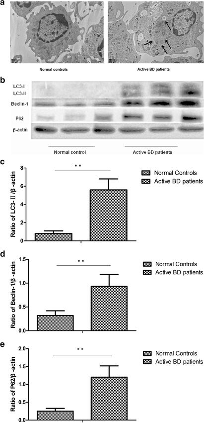
Liang et al. Arthritis Research & Therapy   (2021) 23:135                                                 Page 3 of 11

ELISA Development Kit; R&D Systems, Minneapolis,            expression levels of TNF-α, IL-6, and IL-1β by DCs
MN).                                                        treated with LPS (Fig. 2d–o). These data suggest that au-
                                                            tophagy plays a role as a negative regulator of inflamma-
Statistical analysis                                        tion by regulating the expression of inflammatory
One-way ANOVA and Student’s t test were carried out         cytokines.
by using SPSS17.0 software (SPSS Inc., Chicago, IL,
USA). P values < 0.05 were considered significant.          MiR-155 is involved in the cytokine production of DCs
                                                            induced by dysfunctional autophagy
Results                                                     Earlier, we showed that the expression of miR-155 was
Autophagy is activated, but the capability of autophagic    decreased in DCs from active BD patients [8]. Moreover,
degradation is decreased in DCs from active BD patients     miR-155 has been reported to be involved in autophagy
The most important manifestation of autophagy is the        regulation [17, 27–29]. A further experiment was carried
formation of autophagosomes and this was investigated       out to investigate if miR-155 was associated with cyto-
by TEM. Our results showed that autophagosomes were         kine production by DCs induced by dysfunctional au-
only found in DCs from active BD patients, but not in       tophagy. The results showed that in DCs overexpressing
DCs from inactive BD patients and healthy controls          miR-155, the expression of LC3-II and Beclin-1 was sig-
(Fig. 1a). To further identify whether autophagy was ac-    nificantly increased after stimulation with LPS. The ex-
tivated in patients with active BD, the expression of       pression of P62 was significantly decreased in DCs
Beclin-1 and LC3, which are two important markers of        overexpressing miR-155 after stimulation with LPS
autophagy, were investigated by Western blot. The re-       (Fig. 3a–d). As compared with DCs transfected with the
sults showed that compared with inactive BD patients        control sequence, TNF-α, IL-6, and IL-1β production
and healthy controls, and the protein levels of Beclin-1    were significantly decreased in DCs overexpressing miR-
and LC3-II were significantly increased in DCs from ac-     155. However, there was no difference concerning the
tive BD patients after stimulation with LPS (Fig. 1b–d).    expression of TNF-α, IL-6, and IL-1β in LPS-treated
There was no difference concerning the expression of        DCs overexpressing miR-155 after treatment with 3-MA
LC3 and Beclin-1 between the inactive BD patients and       compared to DCs transfected with the control sequence
healthy controls. Taken together, these results suggested   (Fig. 3e–g).
that autophagy was activated in DCs from active BD
patients.                                                   TAB2 is involved in the effects of MiR-155 on autophagy
   P62/SQSTM1 (hereafter referred to as P62), serving as    Transforming growth factor β-activated kinase 1-binding
anautophagic substrate, is degraded during autophagy        protein 2 (TAB2) is the direct target of miR-155. The
[24]. P62 accumulates in cells when degradation capacity    expression of TAB2 in DCs from active BD patients is
via autophagy is impaired. We next investigated the ex-     increased, and its expression can be suppressed by miR-
pression of P62 in DCs from active BD patients. The re-     155 [8]. Since the above findings showed that miR-155
sults showed that the protein expression level of P62       was involved in the cytokine production of DCs induced
was significantly higher in DCs of active BD patients       by autophagy, a further experiment was performed to in-
than that of inactive BD patients and healthy controls,     vestigate whether TAB2 was involved in the effects of
which indicates that autophagic degradation was de-         miR-155 on autophagy. The results showed that the ex-
creased in active BD patients, despite the presence of a    pression of LC3-II and Beclin-1 was significantly in-
large number of autophagosomes (Fig. 1b, e).                creased, but that the expression of P62 was decreased in
                                                            DCs after TAB2 was downregulated (Fig. 4a–d). The ex-
Autophagy is involved in cytokine production of DCs         pression of TNF-α, IL-6, and IL-1β by DCs treated with
In view of the dysfunctional autophagy in DCs of active     LPS was significantly decreased after TAB2 was down-
BD patients, further experiments were performed to          regulated (Fig. 4e–g).
examine whether autophagy had an effect on cytokine
production by DCs. The results demonstrated that the        MiR-155 regulates autophagy through Akt/mTOR
expression of TNF-α, IL-6, and IL-1β of LPS-treated         pathway
DCs from active BD patients was significantly higher        The aforementioned results showed that miR-155 could
than that of healthy controls (Fig. 2a–c). It has been      regulate cytokine production of DCs by controlling au-
proven that 3-MA could inhibit autophagy through PI3K       tophagy. The Akt/mTOR pathway is the most common
pathway, and rapamycin could activate autophagy by          signaling pathway involved in autophagy. We further ex-
mTOR pathway [25, 26]. The results showed that 3-MA         amined whether miR-155 exerted its effects on autoph-
and rapamycin, which are the inhibitor or promoter of       agy by activating the Akt/mTOR pathway. The results
autophagy, significantly increased or decreased the         showed that the ratios of p-Akt/Akt and p-mTOR/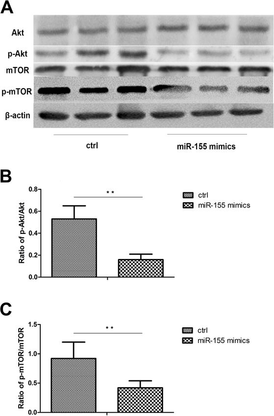
Liang et al. Arthritis Research & Therapy   (2021) 23:135   Page 4 of 11

 Fig. 1 (See legend on next page.)
Liang et al. Arthritis Research & Therapy     (2021) 23:135                                                                               Page 5 of 11

 (See figure on previous page.)
 Fig. 1 Autophagy is involved in the cytokine production of DCs. a Representative TEM photomicrographs of DCs from normal controls (n=3),
 inactive BD patients (n=3), and active BD patients (n=3). b Expression levels of LC3-I, LC3-II, Beclin-1, and P62 protein in DCs from normal controls
 (n=8), inactive BD patients (n=8), and active BD patients (n=8) were quantified by Western blot. c–e Quantification of expression levels of LC3-II,
 Beclin-1, and P62 proteins. *p
Liang et al. Arthritis Research & Therapy    (2021) 23:135                                                                             Page 6 of 11

 Fig. 3 MiR-155 is involved in the cytokine production of DCs induced by dysfunctional autophagy. DCs from normal controls (n=8) and active BD
 patients (n=8) were assessed. DCs were transfected with control mimics and miR-155 mimics at a final concentration of 100 nM. After 48 h, DCs
 were stimulated with 100ng/mL LPS for 24 h. a Expression levels of LC3-I, LC3-II, Beclin-1, and P62 protein in DCs from normal controls and active
 BD patients were quantified by Western blot. b–d Quantification of expression levels of LC3-II, Beclin-1, and P62 proteins. e–g TNF-α, IL-1β, and
 IL-6 were measured in the culture supernatants by ELISA. *p
Liang et al. Arthritis Research & Therapy   (2021) 23:135                                                                             Page 7 of 11

 Fig. 4 TAB2 is involved in the effects of miR-155 on autophagy. DCs from normal controls (n=8) and active BD patients (n=8) were assessed. DCs
 were transfected with siRNA or control siRNA and then stimulated with 100ng/mL LPS for 24 h. a Expression levels of LC3-I, LC3-II, Beclin-1, and
 P62 protein in DCs from the normal controls and active BD patients were quantified by Western blot. b–d Quantification of expression levels of
 LC3-II, Beclin-1, and P62 proteins. e–g TNF-α, IL-1β, and IL-6 were measured in the culture supernatants by ELISA. *p
Liang et al. Arthritis Research & Therapy   (2021) 23:135                                                                           Page 8 of 11

 Fig. 5 MiR-155 regulates autophagy through Akt/mTOR pathway. DCs from normal controls (n=8) and active BD patients (n=8) were assessed.
 After 48 h, DCs were stimulated with 100ng/mL LPS for 24 h. a Total levels of Akt and mTOR, together with phosphorylation levels of Akt and
 mTOR in DCs were determined by Western blotting. b–c Quantification of expression levels of p-Akt/Akt and p-mTOR/mTOR proteins.
 *p
Liang et al. Arthritis Research & Therapy   (2021) 23:135                                                                     Page 9 of 11

DCs. Our results showed that compared with healthy             The Akt/mTOR pathway is one of the most common
controls, the production of TNF-α, IL-6, and IL-1β by        signaling pathways involved in the control of autophagy
DCs from active BD patients stimulated with LPS were         [39, 40]. Rapamycin, as the inhibitor of mTOR, has been
significantly increased. We also showed that the autoph-     shown to alleviate inflammation of the retina [41]. It has
agy inhibitor (3-MA) or promoter (rapamycin) could up-       also been reported that the use of this mTOR inhibitor
regulate or downregulate the production of TNF-α, IL-6,      was safe and effective in treating non-infectious uveitis
and IL-1β. These results proved that the autophagy ac-       [42–44]. In this study, we found that miR-155 could
tivity level correlated with the production of cytokines     downregulate the phosphorylation level of mTOR and
by DCs. These results are consistent with previous stud-     Akt in DCs.
ies showing that IL-1β, IL-17, and IL-18 levels were           The limitation of our research is that we did not inves-
downregulated in murine DCs after autophagy was              tigate the function of autophagy in other types of uveitis
inhibited [32]. Others showed that the production of IL-     and the detailed mechanisms on how a decreased au-
6, IFN-β, and TNF-α by DCs were decreased after              tophagic degradation results in increased cytokine pro-
Beclin-1 was knocked out [33]. These results indicate        duction. Further experiments are needed to explore the
that a defective control of autophagy may trigger cyto-      effects of treatment on the function of autophagy in DCs
kine production by DCs.                                      and if the dysfunction of autophagy is a common
   Earlier, we reported that the expression of miR-155       phenomenon of uveitis.
was downregulated in DCs obtained from active BD pa-
tients and was involved in cytokine production by DCs
[8]. It has also been reported that the expression of miR-   Conclusions
155 was increased or decreased in various diseases and       Collectively, these results show that miR-155 can control
was involved in the development of these diseases by         the production of cytokines by DCs via the process of
regulating autophagy [17, 19, 34, 35]. In view of the        autophagy. Our study provides a greater depth of under-
aforementioned reports, we next investigated whether         standing about the mechanism on how miR-155 is in-
miR-155 exerted its effects on cytokine production by        volved in the development of BD and provides evidence
DCs via the process of autophagy. To answer this ques-       for the use of autophagy as a potential therapeutic target
tion, an experiment with miR-155 mimic transfection          of uveitis.
was performed. The results showed that an miR-155
mimic could promote the expression of LC3-II and             Abbreviations
                                                             miR: MicroRNA; BD: Behcet’s disease; DCs: Dendritic cells; ATGs: Autophagy-
Beclin-1, which meant that autophagy activity was in-        related gene; PBMCs: Peripheral blood mononuclear cells;
creased. More importantly, the protein level of P62 was      TAB2: Transforming growth factor β-activated kinase 1-binding protein 2
downregulated in DCs after miR-155 mimic was trans-
fected, showing that autophagic degradation was in-          Acknowledgements
creased. Additionally, miR-155 mimic transfection            We thank all the patients and medical staff who generously contributed to
reduced the production of TNF-α, IL-1β, and IL-6. The        this study.

autophagy inhibitor, 3-MA, was able to counteract the
effects of miR-155 on cytokine production. All these re-     Authors’ contributions
                                                             LL and Q-YZ conceived and designed the study, performed the experiments
sults suggested that autophagy was involved in the ef-
                                                             and statistical analysis, and drafted the manuscript. L-FQ participated in per-
fects of miR-155 on cytokine production by DCs.              forming the experiments and revised the manuscript. All authors critically re-
   TAB2 has been reported to be the target of miR-155,       vised the manuscript for important intellectual content and approved its
                                                             final version. The authors contributed to the final manuscript.
and its protein expression level was elevated in DCs de-
rived from active BD patients [8]. It also been reported
that TAB2 could bind to Beclin1 or ATG13 to regulate         Funding
                                                             This work was supported by the Basic Research program of Chongqing
autophagy [36–38]. A further experiment was performed        (cstc2020jcyj-msxmX0898), Basic Research program of Chongqing
to investigate whether miR-155 regulates the process of      (cstc2015jcyjA10112), Natural Science Foundation Major International
autophagy through TAB2. The results showed that              (Regional) Joint Research Project (grant no. 81720108009), Natural Science
                                                             Foundation Project of Chongqing (cstc2017shmsA130073), Chongqing Key
downregulation of TAB2 could promote the autophagic          Laboratory of Ophthalmology (grant no. CSTC, 2008CA5003), Chongqing
flux. The expression of TNF-α, IL-6, and IL-1β was de-       Science & Technology Platform and Base Construction Program (grant no.
creased after TAB2 was downregulated. Taken together,        cstc2014pt-sy10002), Science and Technology Project Foundation of Chong-
                                                             qing (cstc2016jcyjA0597), and National Natural Science Foundation Project
these data indicate that miR-155 regulates autophagy by      (grant no 81870673).
controlling the expression of TAB2. Our results are in
agreement with previous studies that showed that miR-
                                                             Availability of data and materials
155 promoted autophagy via a decrease in TAB2 expres-        The datasets used and/or analyzed during the current study are available
sion, thereby stimulating osteoclast formation [17].         from the corresponding author on reasonable request.
Liang et al. Arthritis Research & Therapy (2021) 23:135 Page 10 of 11
Declarations 18. Wang Y, Zheng ZJ, Jia YJ, Yang YL, Xue YM. Role of p53/miR-155-5p/sirt1
loop in renal tubular injury of diabetic kidney disease. J Transl Med. 2018;
Ethics approval and consent to participate 16(1):146.
This study obeyed the tenets of the Declaration of Helsinki and was 19. Yang Y, Zhang N, Wang S, Wen Y. MicroRNA-155 regulates inflammatory
approved by our Clinical Ethical Research Committee. Written consent was response in ischemic cerebral tissues through autophagy. Curr Neurovasc
collected from each healthy control and BD patient. Res. 2018;15(2):103–10. https://doi.org/10.2174/15672026156661806010814
09.
20. Criteria for diagnosis of Behcet’s disease. International Study Group for
Consent for publication Behcet’s Disease. Lancet. 1990, 335(8697):1078-1080.
All participants have approved to publish the data in this manuscript. 21. Liang L, Tan X, Zhou Q, Zhu Y, Tian Y, Yu H, et al. IL-1beta triggered by
peptidoglycan and lipopolysaccharide through TLR2/4 and ROS-NLRP3
inflammasome-dependent pathways is involved in ocular Behcet’s disease.
Competing interests Invest Ophthalmol Vis Sci. 2013;54(1):402–14. https://doi.org/10.1167/
The authors declare that they have no competing interests. iovs.12-11047.
22. Wang C, Tian Y, Lei B, Xiao X, Ye Z, Li F, et al. Decreased IL-27 expression in
Received: 27 June 2020 Accepted: 20 April 2021 association with an increased Th17 response in Vogt-Koyanagi-Harada
disease. Invest Ophthalmol Vis Sci. 2012;53(8):4668–75. https://doi.org/10.11
67/iovs.12-9863.
23. Lopes de Faria JM, Duarte DA, Montemurro C, Papadimitriou A, Consonni
References
SR, Lopes de Faria JB. Defective autophagy in diabetic retinopathy. Invest
1. Suzuki Kurokawa M, Suzuki N. Behcet’s disease. Clin Exp Med. 2004;4(1):10–
Ophthalmol Vis Sci. 2016;57(10):4356–66.
20. https://doi.org/10.1007/s10238-004-0033-4.
24. Katsuragi Y, Ichimura Y, Komatsu M. p62/SQSTM1 functions as a signaling
2. Bonfioli AA, Orefice F. Behcet’s disease. Semin Ophthalmol. 2005;20(3):199–
hub and an autophagy adaptor. FEBS J. 2015;282(24):4672–8. https://doi.
206. https://doi.org/10.1080/08820530500231953.
org/10.1111/febs.13540.
3. Deuter CM, Kotter I, Wallace GR, Murray PI, Stubiger N, Zierhut M. Behcet’s
25. Noda T. Regulation of autophagy through TORC1 and mTORC1.
disease: ocular effects and treatment. Progress Retin Eye Res. 2008;27(1):
Biomolecules. 2017;7(3):52. https://doi.org/10.3390/biom7030052.
111–36. https://doi.org/10.1016/j.preteyeres.2007.09.002.
4. Wang C, Ye Z, Kijlstra A, Zhou Y, Yang P. Activation of the aryl hydrocarbon 26. Wu Y, Wang X, Guo H, Zhang B, Zhang XB, Shi ZJ, et al. Synthesis and
receptor affects activation and function of human monocyte-derived screening of 3-MA derivatives for autophagy inhibitors. Autophagy. 2013;
dendritic cells. Clin Exp Immunol. 2014;177(2):521–30. https://doi.org/1 9(4):595–603. https://doi.org/10.4161/auto.23641.
0.1111/cei.12352. 27. Etna MP, Sinigaglia A, Grassi A. Mycobacterium tuberculosis-induced miR-
5. He Y, Wang C, Su G, Deng B, Ye Z, Huang Y, et al. Decreased expression of 155 subverts autophagy by targeting ATG3 in human dendritic cells. PLoS
A20 is associated with ocular Behcet’s disease (BD) but not with Vogt- Pathog. 2018;14(1):e1006790.
Koyanagi-Harada (VKH) disease. Br J Ophthalmol. 2018;102(8):1167–72. 28. Liu F, Nie C, Zhao N, Wang Y, Liu Y, Li Y, et al. MiR-155 alleviates septic lung
https://doi.org/10.1136/bjophthalmol-2017-311707. injury by inducing autophagy via inhibition of transforming growth factor-
6. Wan CK, He C. Cutting edge: IL-1 receptor signaling is critical for the beta-activated binding protein 2. Shock. 2017;48(1):61–8.
development of autoimmune uveitis. J Immunol. 2016;196(2):543–6. 29. Zarogoulidis P, Petanidis S, Domvri K, Kioseoglou E, Anestakis D, Freitag L,
7. Ture-Ozdemir F, Tulunay A, Elbasi MO, Tatli I, Maurer AM, Mumcu G, et al. et al. Autophagy inhibition upregulates CD4(+) tumor infiltrating
Pro-inflammatory cytokine and caspase-1 responses to pattern recognition lymphocyte expression via miR-155 regulation and TRAIL activation. Mol
receptor activation of neutrophils and dendritic cells in Behcet’s disease. Oncol. 2016;10(10):1516–31. https://doi.org/10.1016/j.molonc.2016.08.005.
Rheumatology (Oxford). 2013;52(5):800–5. 30. Santeford A, Wiley LA, Park S, Bamba S, Nakamura R, Gdoura A, et al. Impaired
8. Zhou Q, Xiao X, Wang C, Zhang X, Li F, Zhou Y, et al. Decreased microRNA- autophagy in macrophages promotes inflammatory eye disease. Autophagy.
155 expression in ocular Behcet’s disease but not in Vogt Koyanagi Harada 2016;12(10):1876–85. https://doi.org/10.1080/15548627.2016.1207857.
syndrome. Invest Ophthalmol Vis Sci. 2012;53(9):5665–74. https://doi.org/1 31. Jia X, Li J, Shi D, Zhao Y, Dong Y, Ju H, et al. Grouping annotations on the
0.1167/iovs.12-9832. subcellular layered interactome demonstrates enhanced autophagy activity
9. Levine B, Kroemer G. Biological functions of autophagy genes: a disease in a recurrent experimental autoimmune uveitis T cell line. Plos one. 2014;
perspective. Cell. 2019;176(1-2):11–42. https://doi.org/10.1016/j.cell.2018.09. 9(8):e104404. https://doi.org/10.1371/journal.pone.0104404.
048. 32. Mantegazza AR, Wynosky-Dolfi MA. Increased autophagic sequestration in
10. Yang Z, Klionsky DJ. Eaten alive: a history of macroautophagy. Nat Cell Biol. adaptor protein-3 deficient dendritic cells limits inflammasome activity and
2010;12(9):814–22. https://doi.org/10.1038/ncb0910-814. impairs antibacterial immunity. PLoS Pathog. 2017;13(12):e1006785.
11. Boutouja F, Stiehm CM, Platta HW. mTOR: a cellular regulator interface in 33. Zang F, Chen Y, Lin Z, Cai Z, Yu L, Xu F, et al. Autophagy is involved in regulating
health and disease. Cells.2019;8(1):18. https://doi.org/10.3390/cells8010018. the immune response of dendritic cells to influenza A (H1N1) pdm09 infection.
12. Choi AM, Ryter SW, Levine B. Autophagy in human health and disease. N Immunology. 2016;148(1):56–69. https://doi.org/10.1111/imm.12587.
Engl J Med. 2013;368(7):651–62. https://doi.org/10.1056/NEJMra1205406. 34. Wu K, Zhu C, Yao Y, Wang X, Song J, Zhai J. MicroRNA-155-enhanced
13. Lin DS, Huang YW, Ho CS, Hung PL, Hsu MH, Wang TJ, Wu TY, Lee TH, autophagy in human gastric epithelial cell in response to Helicobacter
Huang ZD, Chang PC, et al. Oxidative insults and mitochondrial DNA pylori. Saudi J Gastroenterol. 2016;22(1):30–6. https://doi.org/10.4103/1319-3
mutation promote enhanced autophagy and mitophagy compromising cell 767.173756.
viability in pluripotent cell model of mitochondrial disease. Cells. 2019;8(1): 35. Du F, Yu F, Wang Y, Hui Y, Carnevale K, Fu M, et al. MicroRNA-155
65. https://doi.org/10.3390/cells8010065. deficiency results in decreased macrophage inflammation and attenuated
14. Ma S, Attarwala I, Xie XS. SQSTM1/p62: a potential target for atherogenesis in apolipoprotein E-deficient mice. Arterioscler Thromb Vasc
neurodegenerative disease. ACS Chem Neurosci. 2019;10(5):2094–114. Biol. 2014;34(4):759–67. https://doi.org/10.1161/ATVBAHA.113.302701.
https://doi.org/10.1021/acschemneuro.8b00516. 36. Takaesu G, Kobayashi T, Yoshimura A. TGFβ-activated kinase 1 (TAK1)-
15. Lin TA, Wu VC. Autophagy in chronic kidney diseases. Cells. 2019;8(1):61. binding proteins (TAB) 2 and 3 negatively regulate autophagy. J Biochem.
https://doi.org/10.3390/cells8010061. 2012;151(2):157–66. https://doi.org/10.1093/jb/mvr123.
16. Zheng M, Yu H, Zhang L, Li H, Liu Y, Kijlstra A, et al. Association of ATG5 gene 37. Criollo A, Niso-Santano M, Malik SA, Michaud M, Morselli E, Mariño G, et al.
polymorphisms with Behcet’s disease and ATG10 gene polymorphisms with Inhibition of autophagy by TAB2 and TAB3. EMBO J. 2011;30(24):4908–20.
VKH syndrome in a Chinese Han population. Invest Ophthalmol Vis Sci. 2015; https://doi.org/10.1038/emboj.2011.413.
56(13):8280–7. https://doi.org/10.1167/iovs.15-18035. 38. Niso-Santano M, Criollo A, Malik SA, Michaud M, Morselli E, Mariño G, et al. Direct
17. Sul OJ, Sung YB, Rajasekaran M, Ke K, Yu R, Back SH, et al. MicroRNA-155 molecular interactions between Beclin 1 and the canonical NFκB activation
induces autophagy in osteoclasts by targeting transforming growth factor pathway. Autophagy. 2012;8(2):268–70. https://doi.org/10.4161/auto.8.2.18845.
beta-activated kinase 1-binding protein 2 upon lipopolysaccharide stimulation. 39. Paglin S, Lee NY, Nakar C, Fitzgerald M, Plotkin J, Deuel B, et al. Rapamycin-
Bone. 2018;116:279–89. https://doi.org/10.1016/j.bone.2018.08.014. sensitive pathway regulates mitochondrial membrane potential, autophagy,Liang et al. Arthritis Research & Therapy (2021) 23:135 Page 11 of 11
and survival in irradiated MCF-7 cells. Cancer Res. 2005;65(23):11061–70.
https://doi.org/10.1158/0008-5472.CAN-05-1083.
40. Takeuchi H, Kondo Y, Fujiwara K, Kanzawa T, Aoki H, Mills GB, et al. Synergistic
augmentation of rapamycin-induced autophagy in malignant glioma cells by
phosphatidylinositol 3-kinase/protein kinase B inhibitors. Cancer Res. 2005;65(8):
3336–46. https://doi.org/10.1158/0008-5472.CAN-04-3640.
41. Okamoto T, Ozawa Y, Kamoshita M, Osada H, Toda E, Kurihara T, et al. The
neuroprotective effect of rapamycin as a modulator of the mTOR-NF-
kappaB axis during retinal inflammation. Plos one. 2016;11(1):e0146517.
https://doi.org/10.1371/journal.pone.0146517.
42. Blair J, Barry R, Moore DJ, Denniston AK. A comprehensive review of mTOR-
inhibiting pharmacotherapy for the treatment of non-infectious uveitis. Curr
Pharm Des. 2017;23(20):3005–14. https://doi.org/10.2174/13816128236661
70111125550.
43. Blair J, Barry R, Murray PI, Moore DJ, Denniston AK: mTOR-inhibiting
pharmacotherapy for the treatment of non-infectious uveitis: a systematic
review protocol. Syst Rev. 2018;7(1):83.
44. Ibrahim MA, Sepah YJ, Watters A, Bittencourt M, Vigil EM, Do DV, et al. One-
year outcomes of the SAVE study: sirolimus as a therapeutic approach for
UVEitis. Transl Vis Sci Technol. 2015;4(2):4. https://doi.org/10.1167/tvst.4.2.4.
Publisher’s Note
Springer Nature remains neutral with regard to jurisdictional claims in
published maps and institutional affiliations.You can also read