Cryotherapy versus Phenol Chemical Peeling for Solar Lentigines: A Clinical, Histologic, Immunohistochemical and Ultrastructural Study
←
→
Page content transcription
If your browser does not render page correctly, please read the page content below
Original Article
Cryotherapy versus Phenol Chemical Peeling for Solar
Lentigines: A Clinical, Histologic, Immunohistochemical and
Ultrastructural Study
Enas A. S. Attia, M.D.*, May H. El Samahy, M.D.* and
Shereen A. Mahmoud, M.D.†
*Department of Dermatology and Venereology, Faculty of Medicine, Ain Shams University and
†El-Khazendarah Hospital, Cairo, Egypt
Background. Solar lentigines are the most common benign sun induced lesions. Their management includes chemical
peeling and cryotherapy. Objective. to compare cryotherapy to phenol chemical peeling in treatment of solar lentigines.
Patients and methods. Twenty patients with solar lentigines on the dorsa of hands underwent liquid nitrogen spray
cryotherapy of the right hand lesions for 3-5 seconds and focal Baker-Gordon phenol peeling of the left hand lesions.
Results. Cryotherapy to 181 solar lentigines resulted in complete disappearance of 26 lesions (14.35%), marked
improvement of 18 (9.95%), moderate improvement of 86 (47.5%), mild improvement of 44 (24.3 %), and bad response
in 7 (3.9 %), while phenol peeling to 158 lesions resulted in complete disappearance of 49 lesions (31.02 %), marked
improvement of 36 (22.78%) and moderate improvement of 73 (46.2%) (p value = 0.001). Higher patient`s satisfaction
was shown with phenol peeling but with more pain, prolonged healing time and more prolonged erythema. Histological
and immunohistochemical assessment revealed normalized epidermis with phenol peeling and only markedly reduced
melanocytes with cryotherapy. Electron microscopic examination revealed small dark melanin granules with some
aggregated complexes in melanocytes and keratinocytes with cryotherapy. However, with phenol peeling, small
dispersed melanin granules, mostly with light melanization were present. Conclusion. Our results suggested phenol
peeling to have better results due to acting on both melanocytes and keratinocytes, normalizing their proliferative and
melanization properties. However, by “cryo-peeling” comparable results could be achieved. (J Egypt Women Dermatol
Soc 2010; 7: - )
Keywords. Cryotherapy, phenol chemical peeling, solar lentigines
S
olar lentigines (actinic lentigo, senile lentigo, freezing with liquid nitrogen (melanocytes freeze at
sun spots, liver spots or age spots) are the -4 to -7°C), whereas squamous cells resist injury at
most common benign hyper-pigmented sun -20°C3.
induced lesions, that occur in sun exposed areas; Phenol peeling is an example of deep chemical
face, arms, dorsa of the hands and upper part of the peeling, in which there is necrosis followed by
trunk. Although these lesions are most common in regeneration of the epidermis and papillary dermis
individuals aged 30 - 50 years, they are now seen that extends to the reticular dermis. Phenol was
in younger individuals, because of their increased used long time ago in treatment of pigmented lesions
exposure to sun tanning, and the use of artificial such as melasma, freckles and solar lentigines,
sources of ultraviolet light (UV)1. Solar lentigines since it is known to be toxic to melanocytes4,5. The
possibly occur due to melanocytes mutation which aim of this work was to compare between the
results in proliferation and enhanced pigment effect of cryotherapy and chemical peeling with
production in response to UV radiation. Moreover, it phenol in treatment of solar lentigines, clinically,
is possible that there is a genetic susceptibility to the histologically, immunohistochemically and on basis
development of solar lentigo in response to acute or of ultrastructural changes.
chronic UV radiation and that melanocytes in these
circumscribed proliferations are permanently altered2. PATIENTS AND METHODS
Cryotherapy has been used to treat skin lesions for
approximately 100 years. Liquid nitrogen is currently The study included 20 patients attending the
the most widely used cryogen. It may be considered Dermatology Outpatient Clinic of Ain Shams
the first line therapy for treatment of solar lentigines, University Hospitals complaining of solar lentigines,
because of the susceptibility of melanocytes to 7 males (35%) and 13 females (65%), 47 - 70 years
Corresponding Author. Enas A. S. Attia, M.D., Lecturer of Conflict of interest. None declared.
Dermatology and Venereology, Faculty of Medicine, Ain Shams Copyright © 2010 Egyptian Women Dermatologic Society. All rights
University, Egypt. reserved.
E mail. annosah74@hotmail.com
56
57                                                 Enas A. S. Attia et al.

old with mean age 57 ± 6.92. The skin phototypes                  on the lesion until ice ball formation had spread from
according to Fitzpatrick skin phototyping6 were:                  the centre to include a margin of
Cryotherapy versus Phenol Chemical Peeling for Solar Lentigines: 58
A Clinical, Histologic, Immunohistochemical and Ultrastructural Study
by boiling in 10 mM citrate buffer solution (pH greater than that during cryotherapy and lasted
6) for 20 minutes followed by cooling at room for 4-6 hours after the session. However, with
temperature for 20 minutes. After blocking with cryotherapy the pain was less intense and lasted
normal serum (Lab Vision Corp., Fremont, CA), no longer than 15 minutes after the session.
the slides were incubated with MART-1/Melan-A Comparison between both modalities revealed
rabbit monoclonal antibodies (Lab Vision- cat. statistically matched results in all patients except
RB- 9054-R7-Ready to use), for 32 minutes in 6 patients; number 1, 6, 9, 10, 16 and 17, in whom
at room temperature, then with a secondary phenol chemical peeling showed statistically
biotinylated primary antibody, and finally with significant better response (p value 0.001, 0.001,
avidin-biotin-peroxidase complex. Hematoxylin 0.0016, 0.001, 0.006, and 0.001 respectively)
was used as counter-stain. Melan-A gave brown (Table 1). Collectively, cryotherapy was
cytoplasmic immunoreactivity in melanocytes. applied to 181 solar lentigines lesions and
resulted in bad response (G0) in 7 (3.9 %) of
Electron microscopy the lesions, mild improvement (G1) of 44 (24.3
Biopsy specimems were fixed in 3% buffered %), moderate improvement (G2) in 86 (47.5%),
glutaraldehyde for 2 hours at 4˚C and then marked improvement (G3) in 18 (9.95%) and
washed with the cacodylate buffer solution (pH complete disappearance (G4) in 26 (14.35%) of
7.3) for 15 minutes at 4˚C. This was followed by the lesions. On the other hand, phenol chemical
post-fixation in 1% buffered osmium tetraoxide, peeling was done to 158 lesions and resulted
dehydration in ascending grades of ethyl in moderate improvement (G2) in 73 (46.2%)
alcohol, and embedding in Epon 812. Sections of the lesions, marked improvement (G3) in 36
were prepared using Nova ultramicrotome and (22.78%) and complete disappearance (G4) in 49
stained with uranyl acetate and lead citrate, (31.02 %) of the lesions. Thus, phenol chemical
and examined under JEOL 100C-X (JEOL Ltd., peel showed an overall better clinical response
Tokyo, Japan) transmission electron microscope. as the results showed a statistically highly
Lesional basal keratinocytes contained increased significant difference (p value = 0.001) (Table
melanosomes and melanosome complexes 2). Figure 1 illustrates the clinical response of
(polymelanosomes), which formed massive caps both modalities in one patient.
on the nuclei. Even in the upper layers of the As regards patient’s satisfaction with
epidermis, including the horny layer, numerous cryotherapy: 2 patients (10%) reported
melanosomes are present, largely in a dispersed no satisfaction, 7 (35%) reported minimal
state rather than as complexes9. satisfaction, 8 (40%) expressed moderate
satisfaction and 3 (15%) expressed great
Statistical analysis satisfaction. With phenol chemical peel we
Statistical analysis of the results was done found that: no patients (0%) were not satisfied,
using Statistical Package for the Social Sciences 4 patients (20%) expressed minimal satisfaction,
(SPSS) version 12 program. Comparison 8 (40%) expressed moderate satisfaction and 8
between both modalities as regards clinical (40%) expressed great satisfaction. Comparison
response of patients was done using Mann- between both modalities revealed no statistically
Whitney test. Results in relation to gender were significant difference (p = 0.114).
analyzed using Student`s t test and results in Both modalities showed statistically matched
relation to Fitzpatrick skin type and Glogau`s response in both males and females using t test
photoaging classification were done using (p = 0.584 for cryotherapy and p = 0.357 for
ANOVA and Student`s t test respectively. phenol peeling). The response grades to either
Comparison between both modalities as regards modality in relation to Fitzpatrick skin photo-
patient׳s satisfaction was done using Chi-square type, and Glogau’s photoaging classification
test. Results were considered significant when showed no statistically significant difference (p
p value was ≤0.05, and highly significant when = 0.899 and 0.985 for cryotherapy, and 0.842
p value ≤ 0.001. and 0.387 for phenol peeling respectively).
With cryotherapy, we found that blistering
RESULTS was minimal or not at all. The post-healing time
in the lesions which developed blisters was
Clinical assessment within 1 week, while it took 3 weeks with phenol
Our study included 20 patients underwent chemical peel in all lesions. Only prolonged
treatment with cryotherapy on solar lentigine erythema (more than 2 months) occurred in
lesions of the right hand and phenol peeling on 3 patients at sites of phenol chemical peel (2
the lesions of the left hand. Pain was a common patients with type III and 1 patient with type
complaint in all patients. Pain during phenol ІV Fitzpatrick skin). No other complications
chemical peel was intense burning, and was occurred with either technique.
J Egypt Women Dermatol Soc. Vol. 7, No. 1, 2010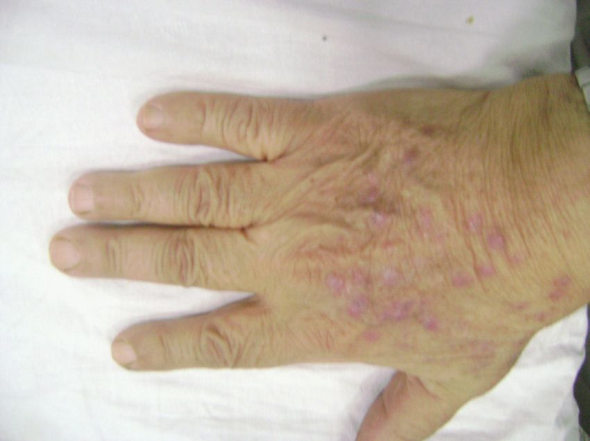
59 Enas A. S. Attia et al.
Table 1. Comparison between the results of both treatment modalities as regards clinical response of patients using Mann-Whitney Test.
No. of Response Median
Patients No Treatment Mean rank z p value
lesions range grade
cryo 13 G0 - G4 G1 8.615
P1 -3.969 0.001** HS
peeling 14 G4 - G4 G4 19.000
cryo 7 G2 - G4 G4 7.429
P2 -1.232 0.218 NS
peeling 5 G2 - G4 G2 5.200
cryo 14 G2 - G4 G2 12.964
P3 -1.457 0.145 NS
peeling 9 G2 - G2 G2 10.500
cryo 8 G2 - G4 G2 6.750
P4 -0.931 0.352 NS
peeling 6 G2 - G4 G3 8.500
cryo 8 G2 - G4 G4 6.125
P5 -0.264 0.792 NS
peeling 3 G2 - G4 G4 5.667
cryo 14 G1 - G1 G1 7.500
P6 -5.422 0.001** HS
peeling 22 G2 - G4 G4 25.500
cryo 5 G2 - G2 G2 5.000
P7 0.000 1.000 NS
peeling 4 G2 - G2 G2 5.000
cryo 6 G2 - G2 G2 5.000
P8 -1.225 0.221 NS
peeling 4 G2 - G4 G2 6.250
cryo 8 G1 - G2 G2 5.125
P9 -2.419 0.016* S
peeling 5 G2 - G4 G4 10.000
cryo 16 G1 - G1 G1 8.500
P10 -5.612 0.001** HS
peeling 20 G2 - G3 G3 26.500
cryo 6 G2 - G3 G3 7.167
P11 -1.476 0.140 NS
peeling 5 G2 - G3 G2 4.600
cryo 8 G2 - G4 G2 6.875
P12 -1.225 0.221 NS
peeling 7 G2 - G4 G4 9.286
cryo 5 G2 - G2 G2 6.000
P13 0.000 1.000 NS
peeling 6 G2 - G2 G2 6.000
cryo 15 G2 - G4 G2 14.333
P14 -1.746 0.081 NS
peeling 10 G2 - G2 G2 11.000
cryo 6 G2 - G4 G2 5.667
P15 -0.267 0.789 NS
peeling 4 G2 - G4 G2 5.250
cryo 4 G1 - G1 G1 2.500
P16 -2.739 0.006** HS
peeling 6 G2 - G3 G3 7.500
cryo 16 G0 - G3 G2 10.000
P17 -3.765 0.001** HS
peeling 12 G3 - G3 G3 20.500
cryo 6 G2 - G3 G3 6.000
P18 -0.707 0.480 NS
peeling 4 G2 - G4 G2 4.750
cryo 10 G2 - G3 G2 9.000
P19 -0.510 0.610 NS
peeling 8 G2 - G4 G2 10.125
cryo 6 G2 - G3 G2 4.667
P20 -1.225 0.221 NS
peeling 4 G2 - G3 G3 6.750
*p ≤ 0.05 is significant
**p ≤0.01 is highly significant
NS = Non significant
S = Significant
HS = Highly significant
J Egypt Women Dermatol Soc. Vol. 7, No. 1, 2010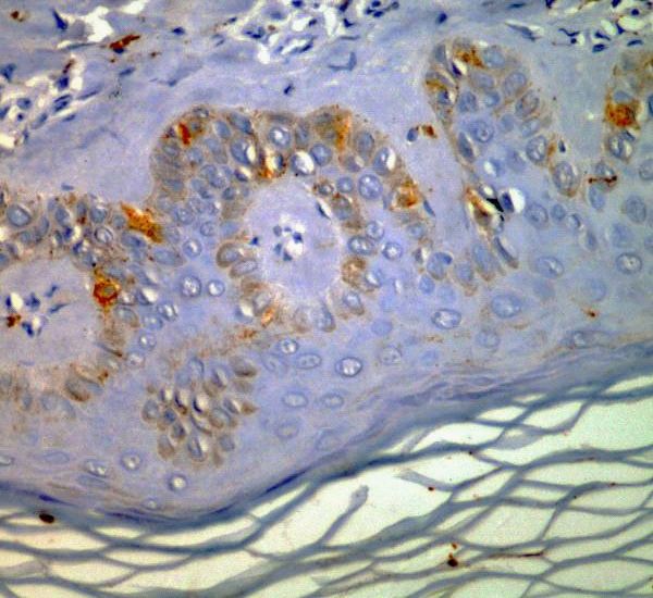
Cryotherapy versus Phenol Chemical Peeling for Solar Lentigines: 60
A Clinical, Histologic, Immunohistochemical and Ultrastructural Study
Table 2. Comparison between the total results of both modalities as regards clinical response of patients using Mann-Whitney Test.
Treatment No. of lesions Response range Median grade Mean rank Z p value
Cryo 181 G0 - G4 G2 136.57 0.001**
7.167
Peeling 158 G2 - G4 G3 208.30 (HS)
**p ≤0.01 is highly significant
HS = Highly significant
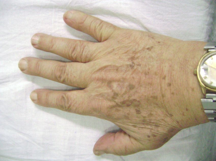
Figure 1A. Before phenol peeling: 22 solar lentigines on left hand. Figure 1B. After 6 weeks of phenol peeling: 16 lesions (G4) but with
occurrence of persistent erythema, and 6 lesions (G2) (arrows).
Figure 1C. Before cryotherapy: 14 solar lentigines on right hand. Figure 1D. After 6 weeks of cryotherapy: all lesions (G1).
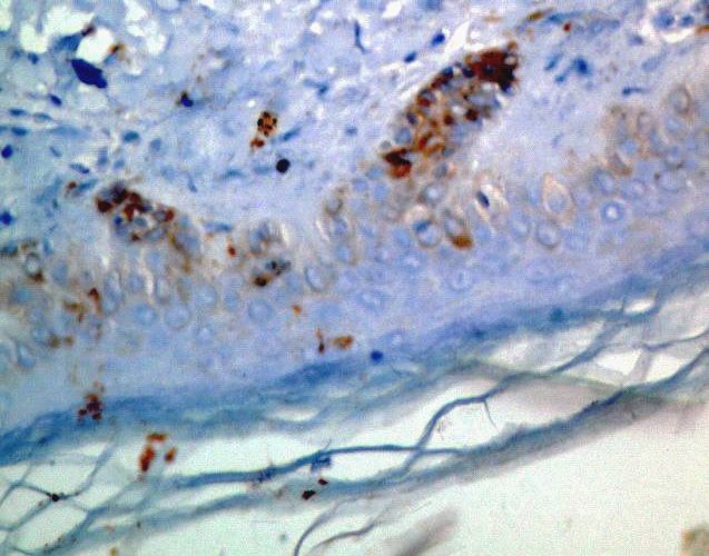
Histologic and immunohistochemical assessment 2E). Melan-A immunostaining revealed reduced but
Solar lentigine lesions showed club-shaped more melanocytes than with cryotherapy (figure 2F).
elongated rete ridges composed of deeply pigmented
basaloid cells intermingled with melanocytes (Figure Ultrastructural assessment
2A). Melan-A staining showed that melanocytes Following cryotherapy, melanocytes contained
were significantly increased in number (Figure 2B). small dark melanin granules, with few of them in
Following cryotherapy, the rete ridges were still aggregated complexes (Figure 3A, B). Keratinocytes
elongated with bud-like extensions, but pigmentation also showed more and darker melanin granules (figure
was markedly reduced (Figure 2C). Melanocytes were 3C), than with phenol peeling. After phenol peeling,
markedly reduced in number as evidenced by Melan-A melanocytes contained small dispersed melanin
immunostaining (Figure 2D). Phenol chemical peeling granules with different degrees of melanization; most
resulted in better arrangement of rete ridges than of them had light melanin granules (Figure 3D, E).
with cryotherapy. Although significantly reduced, Keratinocytes also showed different melanin granules;
pigmentation was more than with cryotherapy (figure most of them were light (Figure 3F).
J Egypt Women Dermatol Soc. Vol. 7, No. 1, 2010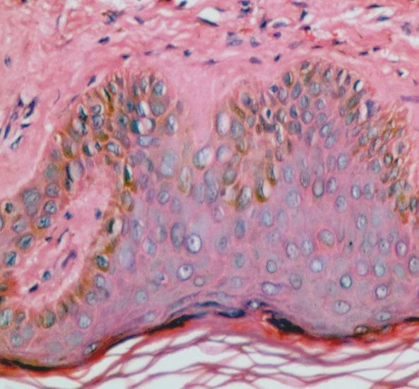
61                                                            Enas A. S. Attia et al.

  A                                                                       B

 C                                                                        D

 E                                                                            F

Figure 2. A: Solar lentigine before therapy (H&E x400). B: Solar lentigine before therapy (Anti Melan-A x400). C: After cryotherapy (H&E x400). D:
After cryotherapy (Anti Melan-A x400). E: After phenol peeling (H&E x400). F: After Phenol peeling (Anti Melan-A x400).

J Egypt Women Dermatol Soc. Vol. 7, No. 1, 2010
Cryotherapy versus Phenol Chemical Peeling for Solar Lentigines:                                       62
   A                                 A Clinical, Histologic, ImmunohistochemicalA
                                                                                Dand Ultrastructural Study                                           D

   ABA                                                                           DBD
                                                                                   E                                                                 E

A BCBD                                                                           DC
                                                                                  EEF                                                                F

  CC                                                                             FF
 B E                                                                             E
Figure 3. After cryotherapy; A: Melanocyte contains small dark melanin granules; few of them are seen in complex aggregates (marked by arrows)
(EM x6000). B: Another melanocyte showing small-sized dark aggregated melanin granules (EM x8000). C: Keratinocytes with dark melanin particles
(EM x1500). After Phenol peeling; D: Melanocyte contains small dispersed melanin granules with different sizes and degree of melanization; most of
them with light melanization (EM x8000). E: Another melanocyte with light melanin granules (EM x8000). F: Keratinocytes with melanin particles
variable in size and melanization (EM x1500).

                                                                                                 J Egypt Women Dermatol Soc. Vol. 7, No. 1, 2010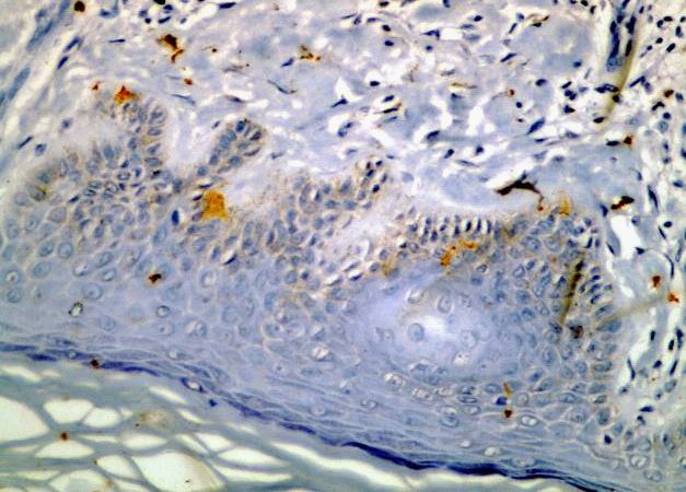
63                                                Enas A. S. Attia et al.

                     DISCUSSION                                  as regards cryotherapy, we showed similar response
                                                                 range, but with no reported complications.
    Although benign, solar lentigines are of                         In the present study, treatment of 158 lesions
great aesthetical concern for the patient1. Many                 with focal Baker-Gordon phenol chemical peeling
therapeutic modalities have been advocated in                    resulted in improvement of all lesions with complete
treatment of solar lentigines, including topical                 disappearance in 49 (31.02 %) of them. All patients
tretinoin, hydroquinone, adapalene, combination                  expressed some degree of satisfaction to the results,
of mequinol and tretinoin, cryotherapy, chemical                 but 3 patients showed prolonged erythema as a
peels, and laser surgery10. Because cryotherapy and              side effect after treatment. To our knowledge, no
chemical peeling are accessible and cost effective               previous studies have been reported in treatment
treatments and have shown good results in some                   of solar lentigines with phenol chemical peel.
reports, we decided to use these modalities in the               However, Hopking et al.15 and Piamphongsant16
treatment of solar lentigines over the back of the               reported good results by using phenol in treatment
hands.                                                           of other pigmented lesions (congenital pigmented
    Treatment of 181 lesions with 3-5 seconds                    nevi and melasma respectively).
of liquid nitrogen spray cryotherapy resulted in                     We found that phenol chemical peeling showed
improvement 148 (81.8 %) of the lesions and                      better clinical response than cryotherapy (G2-G4 in
complete disappearance in 26 (14.35%), with no                   100% of the lesions with phenol while G1-G4 in
reported complications. Eighteen patients (90%)                  96.13% of the lesions with cryotherapy) and higher
reported some degree of satisfaction towards the                 patient’s satisfaction. However, more pain, longer
results.                                                         healing time and more complications (prolonged
    Zouboulis et al.11 reported the use of a single              erythema) were reported with phenol chemical
FTC of 30 to 40 seconds nitrous oxide contact                    peel than cryotherapy. These results could not be
surgery in 6 patients with solitary large solar                  compared with others. Both modalities showed
lentigo lesions (3-16 cm in diameter) for one or two             statistically matched response in both males and
sessions. Full remission of all lesions was reported             females. In contrast, Fintisi and Landau17 reported
with excellent cosmetic results. The discrepancy                 that thick male skin is usually less responsive to
between these results and ours could be attributed to            deep peel. Further studies on larger numbers of
the different cryogen and our use of spray technique             patients are needed to establish such a conclusion.
in a single session-based therapy only for 3-5                   Again, the response grades to either modality in
seconds. Almond-Roesler and Zouboulis12 treated                  relation to Fitzpatrick skin phototype, showed no
20 patients with small solar lentigines with 5 or                statistically significant difference. Similarly, the
10 seconds of liquid nitrogen contact cryosurgery.               same authors17 reported that although the ideal
Substantial lightening was observed in 80% of                    patient for deep chemical peeling is a blond, blue-
patients treated for 5 seconds and 100% of patients              eyed with a fair complexion, previous experience
treated for 10 seconds. Minimal skin atrophy was                 showed that phenol-based peels could be performed
observed in 10% and 60% of patients treated for 5                on patients with olive and dark skin. We also found
and 10 seconds, respectively. In our study, using 3-5            statistically matched results in Glogau’s photoaging
seconds of spray cryotherapy resulted in substantial             type II and III patients with both modalities. Glogau
lightening of 96.13% of the lesions. Further studies             and matarasso18 stated that photoaging type I is not
using other techniques of cryotherapy for different              an indication for deep chemical peeling because
durations are recommended.                                       it may be more damaging than beneficial, while
    Compared to Trichloroacetic acid (TCA)                       photoaging type II, III and particularly type IV may
chemical peeling, Janer et al. 13 treated randomly               benefit from deep peeling.
21 patients with solar lentigo on the dorsa of the                   Both keratinocytes and melanocytes contribute
hands with either TCA 30% solution or 1-5 seconds                to the histopathological changes of solar lentigines.
of liquid nitrogen spray cryosurgery. Cryotherapy                The epidermis of solar lentigo shows significantly
showed better results than TCA, with no                          elongated rete ridges, composed, especially in their
complications for any treatment, except for a small              lower portion, of deeply pigmented basaloid cells
hypertrophic scar in one of the cryotherapy treated              intermingled with melanocytes. The melanocytes
patients. Moreover, Raziee et al. 14 treated randomly            appear significantly increased in number in some
25 women with solar lentigines of both hands, with               cases, but only slightly or not at all increased in others8.
either liquid nitrogen cryotherapy by pressing cotton            We demonstrated that both modalities reduced
tipped applicator for 3-5 seconds or TCA 33%                     the number of lesional melanocytes. However,
solution. Cryotherapy was more likely to produce                 the number was reduced more with cryotherapy,
substantial lightening of the solar lentigines than              compared to phenol peeling. Nevertheless,
TCA 33% solution particularly in lower Fitzpatrick               elongated tortuous rete ridges were less reduced
skin types (p = 0.025), but it was more painful and              by cryotherapy than with phenol chemical peeling.
took more time to heal. Compared to these studies,               This is attributed to the fact that pigmented epithelial

J Egypt Women Dermatol Soc. Vol. 7, No. 1, 2010
Cryotherapy versus Phenol Chemical Peeling for Solar Lentigines:                                           64
                             A Clinical, Histologic, Immunohistochemical and Ultrastructural Study

cells and melanocytes are more cryosensitive than                  Conclusion
other cell types. As early as one hour post-freezing,                  we realize that each of cryotherapy and phenol
the melanocytes show swelling of mitochondria                      chemical peeling is effective in treatment of solar
and nuclear damage. Tissue oedema is marked with                   lentigines. However, Baker-Gordon phenol chemical
pigment dispersed outside the cells, while adjacent                peel showed better clinical response and higher
keratinocytes are still unchanged19. Keratinocytes                 patient’s satisfaction, compared to 3-5 seconds of
need to be frozen to -50°C for optimum destruction.                liquid nitrogen spray cryotherapy. Phenol chemical
Melanocytes are more delicate and only require a                   peeling better response is due to acting on both
temperature of -5°C for destruction20. This fact is                melanocytes and keratinocytes, normalizing their
the reason for the resulting hypo-pigmentation                     proliferative and melanization properties. However,
following cryotherapy, with minimal changes in                     by “cryo-peeling” comparable results could be
keratinocytes. Yet, the response to freezing injury                achieved. Besides, more complications were
varies from inflammatory to destructive, depending                 reported with phenol peeling (greater pain intensity,
upon the severity of freezing. Minor freezing injury               prolonged healing time, and prolonged post treatment
(short cryogen spurts) produces only inflammatory                  erythema). Nevertheless, resulting in better response
responses, while severe freezing injury (long                      or less complications is not the only parameter to
cryogen spurts) destroys cells and tissues producing               choose a specific therapy for solar lentigines, as each
coagulation necrosis in the frozen tissue the days                 modality of treatment is not recommended in the
after thawing21. Thus, we recommend further studies                presence of certain medical condition. For example,
with longer cryogen application periods that likely                phenol chemical peel is contraindicated in significant
induce epidermal separation and re-epithelialization,              cardiac and renal diseases while cryotherapy is not
with better clinical response.                                     recommended in case of cold intolerance and cold
    In 1985, Kligman et al.22 conducted long-                      urticaria.
term histologic follow up of phenol face peels.
They demonstrated normal epidermal pattern                                                REFERENCES
in peeled skin, without cytologic irregularities,
lentigos, and microscopic actinic keratoses. The                   1.    Bastiaens M, Hoefnagel J, Westendorp R, Vermeer BJ, Bouwes
basal keratinocytes contained many fine pigment                          Bavinck JN. Solar lentigines are strongly related to sun exposure in
granules rather evenly dispersed, with abundance of                      contrast to ephelides. Pigment Cell Res 2004; 17: 225-9.
intermingled melanocytes. They concluded that the                  2.    Aoki H, Moro O, Tagami H, Kishimoto J. Gene expression profiling
bleaching effect of phenol is not due to destruction of                  analysis of solar lentigo in relation to immunohistochemical
melanocytes, but due to impaired melanin synthesis,                      characteristics. Br J Dermatol 2007; 156: 1214-23.
which is very long lasting (up to 20 years or even                 3.    Andrews MD. Cryosurgery for common skin conditions. Am Fam
more). The long-lasting effect could be explained                        Physician 2004; 69: 2365-72.
on basis of evidenced anti-proliferative activities                4.    Stuzin JM. Phenol peeling and the history of phenol peeling. Clin
of phenolic compounds against melanocytes23. On                          Plast Surg 1998; 25: 1-19.
basis of the reported mechanisms of interference                   5.    Hilinski JM. Skin resurfacing, chemical peels. www.emedicine.
with melanin synthesis and deposition, phenols                           com, 2008.
were categorized as peroxidase inhibitors, acting                  6.    Clark CP. Office-based skin care and superficial peels: The scientific
during active melanin synthesis24. The inhibition                        rationale. Plast Reconstr Surg 1999; 104: 854-64.
of peroxidase results in depigmentation or actually                7.    Ramos-e-Silva M, Da Silva Carneiro SC. Elderly skin and its
hypopigmentation by reduction of polymerization                          rejuvenation: Products and procedures for the aging skin. J Cosmet
of melanogenic intermediates25. Therefore, phenol                        Dermatol 2007; 6: 40-50.
peeling bleaching effect results from melanopenia,                 8.    Montagna W, Hu F, Carlisle K. A reinvestigation of solar lentigines.
melanocytopenia, and even dispersion of fine melanin                     Arch Dermatol 1980; 116: 1151-4.
in keratinocytes.                                                  9.    Noblesse E, Nizard C, Cario Andre M, Lepreux S, Pain C,
    Since Melan-A immunostaining identifies                              Schnebert S, et al. Skin ultrastructure in senile lentigo. Skin
melanocytes whether viable or not, electron                              Pharmacol Physiol 2006; 19: 95-100.
microscopic examination was performed. We noticed                  10.   Ortonne JP, Pandya AG, Lui H, Hexsel D. Treatment of solar
that the melanocytes which escaped cryotherapy                           lentigines. J Am Acad Dermatol 2006; 54: S262-71.
destructive effect contained small dark melanin                    11.   Zouboulis CC, Rosenberger AD, Adler Y, Orfanos CE. Treatment
granules with some aggregated complexes, and                             of solar lentigo with cryosurgery. Acta Derm Venereol 1999;
similarly did the keratinocytes. On the other hand,                      79: 489-90.
phenol-induced hypopigmentation was obviously                      12.   Almond-Roesler B, Zouboulis CC. Successful treatment of solar
due to impaired melanin synthesis, reflected as                          lentigines by brief gentle cryosurgery using a Kryomed device. Br
small dispersed melanin granules, mostly with                            J Dermatol 2000; 143: 216-8.
light melanization. Follow up is recommended                       13.   Janer AL, Somolinos AL, Sanchez JL. Comparison of trichloroacetic
to investigate the long-lasting effect of phenol on                      acid solution and cryosurgery in the treatment of solar lentigines.
melanin synthesis.                                                       Int J Dermatol 2003; 42: 829-31.

                                                                                         J Egypt Women Dermatol Soc. Vol. 7, No. 1, 2010
65                                                                   Enas A. S. Attia et al.

14.   Raziee M, Balighi K, Shabanzadeh Dehkordi H, Robati RM. Efficacy              21. Gage AA. Selective cryotherapy. Cell Preservation Technology
      and safety of cryotherapy vs. Trichloroacetic acid in the treatment of            2004; 2: 3-14.
      solar lentigo. J Eur Acad Dermatol Venereol 2008; 22: 316-9.                  22. Kligman AM, Baker TJ, Gordon HL. Long-term histologic
15.   Hopkins JD, Smith AW, Jackson IT. Adjunctive treatment of                         follow-up of phenol face peels. Plast Reconstr Surg 1985;
      congenital pigmented nevi with phenol chemical peel. Plast Reconstr               75: 652-9.
      Surg 2000; 105: 1-11.                                                         23. Yáñez J, Vicente V, Alcaraz M, Castillo J, Benavente García O,
16.   Piamphongsant T. Phenol-castor oil: Modified peel for dermal                      Canteras M, et al. Cytotoxicity and antiproliferative activities
      melasma. Dermatol Surg 2006; 32 :611-7.                                           of several phenolic compounds against three melanocytes cell
17.   Fintsi Y, Landau M. Exoderm: Phenol-based peeling in olive and                    lines: Relationship between structure and activity. Nutrition
      dark-skinned patients. Int J Cosm Surg Aesth Dermatol 2001;                       Cancer 2004; 49: 191-9.
      3: 173-8.                                                                     24. Briganti S, Camera E, Picardo M. Chemical and instrumental
18.   Glogau RG, Matarasso SL. Chemical peels. Trichloroacetic acid and                 approaches to treat hyperpigmentation. Pigment Cell Res 2003;
      phenol. Dermatol Clin 1995; 13: 263-76.                                           16: 101-10.
19.   Lindo SD, Daniels Jr F. Cryosurgery of junction nevi. Cutis 1975;             25. Kasraee B. Peroxidase-mediated mechanisms are involved in
      16: 492-6.                                                                        the melanocytotoxic and melanogenesis-inhibiting effects of
20.   Kuwahara RT. Cryotherapy. www.emedicine.com, 2007.                                chemical agents. Dermatology 2002; 205: 329-39.

J Egypt Women Dermatol Soc. Vol. 7, No. 1, 2010
You can also read