COVID-19 and the 1918 influenza pandemics: a concise overview and lessons from the past - De Gruyter
←
→
Page content transcription
If your browser does not render page correctly, please read the page content below
Open Health 2021; 2: 40-49

Review Article

Madiha Asghar, Misbahud Din*, Abdul Waris, Muhammad Talha Yasin, Tanzeel Zohra,
Muhammad Zia

COVID-19 and the 1918 influenza pandemics:
a concise overview and lessons from the past
https://doi.org/10.1515/openhe-2021-0003
                                                                       demics and various possibilities for the future course of
received February 3, 2021; accepted September 12, 2021
                                                                       COVID-19 are also highlighted.

Abstract: The coronavirus disease 2019 (COVID-19), which
                                                                       Keywords: pandemic, COVID-19, influenza pandemic,
is caused by severe acute respiratory syndrome coronavi-
                                                                       SARS-CoV-2
rus-2 (SARS-CoV-2), was first reported in December, 2019,
in Wuhan, China. Even the public health sector experts
could not anticipate that the virus would spread rapidly to
create the worst worldwide crisis in more than a century.
The World Health Organization (WHO) declared COVID-
19 a public health emergency on January 30, 2020, but it               1 Introduction
was not until March 11, 2020 that the WHO declared it a
global pandemic. The epidemiology of SARS-CoV-2 is dif-                In the 21st century, emerging viral infections are among
ferent from the SARS coronavirus outbreak in 2002 and                  the greatest challenges in the public health sector [1].
the Middle East Respiratory Syndrome (MERS) in 2012;                   Among these are zoonotic viruses, which jump to humans
therefore, neither SARS nor MERS could be used as a suit-              from other mammals. By crossing species barriers, coro-
able model for foreseeing the future of the current pan-               naviruses have infected the human population for the
demic. The influenza pandemic of 1918 could be referred                third time in the 21st century. In December, 2019, in China,
to in order to understand and control the COVID‐19 pan-                a novel coronavirus provisionally named 2019-nCoV was
demic. Although influenza and the SARS-CoV-2 are from                  identified in individuals linked to the seafood market
different families of viruses, they are similar in that both           in China. In January, 2020, the virus was identified as
silently attacked the world and the societal and political             SARS-CoV-2 [2]. On February 11, 2020, the World Health
responses to both pandemics have been very much alike.                 Organization (WHO) named the outbreak COVID-19. The
Previously, the 1918 influenza pandemic and unpredicta-                patient with confirmed COVID-19 displayed distinctive
bility of the second wave caused distress among people                 respiratory symptoms, such as dry cough, fever, myalgia
as the first wave of that outbreak (so-called Spanish flu)             (lung damage), and other signs including nasal conges-
proved to be relatively mild compared to a much worse                  tion, aches and diarrhea [3]. Like the SARS outbreak in
second wave, followed by smaller waves. As of April,                   2002 and MERS in 2012, SARS-CoV-2 is another coronavi-
2021, the second wave of COVID-19 has occurred around                  rus that emerged in the human population causing severe
the globe, and future waves may also be expected, if the               respiratory disease [4]. As of June, 2020, the WHO reported
total population of the world is not vaccinated. This article          millions of laboratory-confirmed cases with more than a
aims to highlight the key similarities and differences in              million deaths globally.
both pandemics. Similarly, lessons from the previous pan-                   Coronaviruses are RNA viruses that circulate among
                                                                       mammals, such as bats and humans, causing hepatic,
                                                                       enteric, neurologic and severe respiratory diseases [5,6].
                                                                       The six coronavirus species that infect humans are HKU1,
*Corresponding author: Misbahud Din, Department of Biotechnolo-        NL63, OC43, 2293, SARS-CoV and MERS-CoV [7]. The last
gy, Quaid-i-Azam University, Islamabad, Pakistan,                      two viruses (SARS-CoV and MERS-CoV) have a zoonotic
e-mail: misbah@bs.qau.edu.pk                                           origin and have resulted in severe outbreaks in 2002 in
Madiha Asghar, Abdul Waris, Muhammad Talha Yasin, Tanzeel
                                                                       China and 2012 in the Middle East [8–12]. SARS-CoV-2 is
Zohra, Muhammad Zia, Department of Biotechnology, Quaid-i-Azam
University, Islamabad, Pakistan                                        now considered to be the seventh species of coronavirus

  Open Access. © 2021 Madiha Asghar et al., published by De Gruyter.         This work is licensed under the Creative Commons Attribution 4.0
International License.
COVID-19 and the 1918 influenza pandemics 41
to infect humans. The genome of the novel coronavirus of these pandemics in Tables 1 and 2 and Figure 1. Sim-
has now been sequenced, which represents more than ilarly, the comparative mortality rate of both pandemics
75% genetic similarity to SARS-CoV [13]. Further explora- has been represented in Tables 3–7. As the second wave
tion of the epidemiological characteristics of COVID-19 is of COVID-19 has started globally and with more severity,
critical for developing and implementing effective control which also happened in the 1918 influenza pandemic,
strategies. lessons need to be learned from that pandemic. The lack
Both SARS and MERS infect the human population, of strong health care facilities, malnutrition, improper
but in different ways. COVID‐19 shows distinct epidemi- hygiene, and less responsible attitudes toward proper care
ological, pathogenetic, and clinical features compared to and management (e.g., not following standard operating
SARS and MERS, so it is difficult to predict the COVID‐19 procedures) of disease in various areas of the world might
situation from the data available. SARS-CoV-2 has a fatality result in greater severity of the pandemic [16]. Since more
rate of 2.3%, which is much lower than that of SARS (9.5%) waves of COVID-19 are expected based on the previous
and even lower than that of MERS (34.4%). However, it has influenza pandemic, we are highlighting a few aspects of
higher transmission rate than SARS and MERS. both pandemics.
On the other hand, the influenza pandemic can be a
good comparative model and could be used to prepare for
the future course of COVID-19. In the 20th century, there
were eight influenza pandemics, and 3 of them occurred
2 Origin of influenza in 1918 and
after 1918 (1957, 1968 and 2009) [14,15]. By highlighting COVID-19
the key differences and similarities in the epidemiology
of the influenza and coronavirus pandemics, we can envi- The 1918–1919 flu was the most disastrous pandemic
sion various possibilities for the future course of COVID- in human history, with the projected number of deaths
19. We have summarized some differences and similarities ranging from 20 to 150 million [17,18]. There is still disa-
Table 1: Common features between the 1918-19 influenza and COVID-19
Common features 1918-19 influenza and COVID-19
Causative agent RNA virus
Mode of transmission Aerosol or droplet infection, touching infected surfaces
Symptoms Fever, mild illness, tiredness, shortness of breath, aches and pains
Preventive measures Social distancing, wearing masks, avoid touching people and surfaces, staying home
Epidemic or pandemic Pandemic
Disease type Zoonotic diseases
Table 2: Differences between the 1918-19 influenza and COVID-19
Differences 1918-19 influenza COVID-19
Origin Many theories but mostly say USA China
Strain of virus H1N1 SARS-CoV-2
Mostly effected age group 20–40 years Over 60
Incubation period Average 1 to 2 days Average 5–6 days, can be up to 14 days
Total deaths 50 million 2,901,60042 Madiha Asghar et al.
greement regarding the name given to that pandemic. origin [19,20]. After 90 years, virologists and epidemi-
Since the beginning, because of the misinformation of ologists globally agreed that the virus did not originate
the virus origin, the pandemic has been called “Spanish in Spain. Some theories indicate that the virus probably
flu”. Despite the name, little information about the con- started in British Army camps in mainland Europe in
sequences and course are available regarding a Spanish 1916–1917. However, others consistently date its appear-
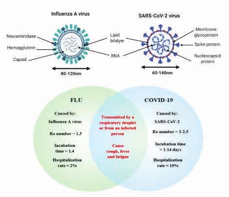
Figure 1: A comparison of influenza A virus and SARS-CoV-2 along with the diseases they cause i.e. flu and COVID-19.
Table 3: Comparative mortality rate of the 1918-19 influenza and COVID-19 (up to April 7, 2021): Africa
Country Total population Published death rate Total population 2021 Published death rate from
1918–19 from 1918 flu (per 1,000) COVID-19 (per 1,000)
Gambia 211,000 ~50 2,468,006 0.067
Ghana 2,298,000 ~40 31,564,990 0.024
Kenya 2,596,000 40 54,669,365 0.041
Nigeria 18,631,000 30 210,008,723 0.010
Sierra Leone 1,541,000 30 8,100,074 0.010
South Africa 6,769,000 43.97 59,877,996 0.885
The source of the data was World Health Organization, an article published by Johnson and Mueller [17] and Google (Worldometer 2020).COVID-19 and the 1918 influenza pandemics 43
Table 4: Comparative mortality rate of the 1918-19 influenza and COVID-19 (up to April 7, 2021): the Americas
Country Total population Published death rate Total population 2021 Published death rate from
1918–19 from 1918 flu (per 1,000) COVID-19 (per 1,000)
Argentina 8,517,000 1.20 45,511,967 1.24
Brazil 26,277,000 6.00 213,712,338 1.57
Canada 8,148,000 6.25 37,994,490 0.60
Mexico 14,556,000 23.00 129,964,977 1.57
Uruguay 1,439,000 1.40 3,482,929 0.34
The source of the data was World Health Organization, an article published by Johnson and Mueller [17] and Google (Worldometer 2020).
Table 5: Comparative mortality rate of the 1918-19 influenza and COVID-19 (up to April 7, 2021): Europe
Country Total population Published death rate Total population 2021 Published death rate from
1918–19 from 1918 flu (per 1,000) COVID-19 (per 1,000)
Austria 6,131,445 3.00 9,045,631 1.055
Denmark 3,010,000 3.50 5,807,787 0.418
Ireland 4,280,000 4.04 4,979,804 0.949
England & Wales 34,020,000 ~4.90 68,158,309 1.861 (UK)
Finland 3,120,000 5.80 5,547,325 0.155
France 32,830,000 3.90 65,384,318 1.494
Germany 58,450,345 3.70 83,989,395 0.930
Italy 36,280,000 11.00 60,393,531 1.860
Norway 2,580,000 5.70 5,453,693 0.125
Portugal 6,010,000 9.70 10,173,859 1.660
Russia 184,000,000 5.00 145,982,499 0.695
Spain 20,880,000 12.00 46,768,680 1.625
Sweden 5,810,000 5.41 10,147,504 1.338
Switzerland 3,880,000 6.00 8,703,085 1.198
The source of the data was World Health Organization, an article published by Johnson and Mueller [17] and Google (Worldometer 2020).
ance in the United States Army training camps to about form of transmission still needs to be identified. Similarity
1918. Even more recent data shows that the disease could of the SARS-CoV-2 to bat coronaviruses indicates that the
have appeared in New York City [21]. primary reservoirs of the virus are bats. Previously, SARS-
Coronaviruses, on the other hand, have infected the CoV and MERS-CoV were transmitted to humans from
human population by crossing the species barrier for the exotic animals (through the seafood market) and camels,
third time in the 21st century. In December, 2019, in China, respectively. However, the primary hosts were bats in both
a novel coronavirus, provisionally named as 2019-nCoV, cases. The spread of SARS-CoV-2 from bats directly or via
was identified in individuals and linked to the seafood other routes should be studied to explain the pattern of
market of China [3]. Much information about the origin and zoonotic transmission.44 Madiha Asghar et al.
Table 6: Comparative mortality rate of the 1918-19 influenza and COVID-19 (up to April 7, 2021): Oceania
Country Total population Published death rate Total population 2021 Published death rate from
1918–19 from 1918 flu (per 1,000) COVID-19 (per 1,000)
Australia 5,304,000 2.8 25,726,041 0.035
Fiji 164,000 52.0 901,419 0.002
New Zealand 1,158,000COVID-19 and the 1918 influenza pandemics 45
4 Demographical comparison of the phase. Despite the low viral load, individuals can still be
1918 influenza and COVID-19 infected. The viral load can be predicted by using real-
time reverse transcription-polymerase chain reaction (RT-
PCR).
There was no vaccine for the 1918 influenza virus; however,
several vaccines approved by the Food and Drug Admin-
istration (FDA) are available for COVID-19. The unavaila-
5.2 Phase 2: Upper and conducting airways
bility of antibiotics to treat secondary bacterial infections
basically made the 1918 influenza more lethal. Therefore, response
the only solution was isolation, quarantine and use of dis-
In this phase, the virus further spreads and moves down
infectants. The 1918 flu pandemic ended in the summer
the respiratory tract, and a stronger response is triggered.
of 1919, which could be linked to fewer deaths and higher
The SARS-CoV-2 can be clinically apparent in this phase.
immunity levels [16]. Since the world was at war in 1918,
The C-X-C motif chemokine 10 (CXCL10) level can be predic-
soldiers were considered to spread the virus globally [32].
tive of the clinical course [38]. The infected epithelial cells
Up to November, 2020, more than 52 million people had
become the major source of beta and lambda interferon
confirmed COVID-19 infections worldwide, with more 1.2
[39]. In about 80% of the cases, the disease is restricted
million deaths reported. The total world population was
to the upper conducting airways, and these patients can
about 1.8 billion in 1918. The estimate of 50 million deaths
be monitored with conventional symptomatic treatment.
indicates that the 1918 influenza killed 2.7% of the world
population.
The current world population is about 8 billion, with
5.3 Phase 3: Hypoxia and progression to
an overall lower death rate from COVID-19. The lower rate
is likely to be related to greater awareness about viruses, acute respiratory distress syndrome (ARDS)
improved health care facilities, and COVID-19 being less
More than 20% of the infected individuals can progress
lethal than the 1918 influenza. The health care facilities
to this phase. The overall mortality rate is approximately
were impacted in 1918 not only by the pandemic but also
2%, but this varies with age. At this phase, the virus enters
by mass casualties and war injuries; many medical staff
the lungs and infects alveolar type 2 cells. The infected
were stationed with troops, and physicians themselves
alveolar cells tend to be peripheral and subpleural [40,41].
were infected with the influenza virus.
The virus propagates inside type 2 cells, and as a result, a
significant number of viral particles are released. Conse-
quently, the cell undergoes apoptosis. The released viral
5 Comparison of the 1918 particles infect type 2 cells of the adjacent units. The pre-
influenza and COVID-19 based on cursors of type 1 cells are type 2 cells, and this series of
events is shown in the murine model of influenza pneu-
pathogenesis monia [41,42]. Elderly people are at high risk because
of their weak immune response and impaired ability to
On the basis of the cells being infected, COVID-19 could be repair the damaged epithelium. This basically allows
divided into three phases representing different disease the propagation of viruses to the gas exchange unit [43].
stages [32]. Despite rapid progress in the understanding of COVID-19
pathogenesis, it is not yet fully explained. However, viral
entry is the same for both SARS-CoV and SARS-CoV-2.
5.1 Phase 1: The symptomatic state (initial
1–2 days of infection)
5.4 Pathogenesis of the 1918 influenza in
The SARS-CoV-2 targets and replicates in the epithelial reconstructed 1918 virus
cells of the nasal cavity. Angiotensin-converting enzyme-2
(ACE2) is the major receptor for SARS-CoV-2 [33–35]. It has To gain insight into the pathogenesis of the 1918 flu,
been reported that the primary infected cells in the con- the reconstructed 1918 virus was injected in mouse and
ducting airways are the ciliated cells [36,37]. There is a non-human primate models [44–46]. In the infected
confined spread of the virus with limited innate immune animal models, virus propagated at a high rate and
response. Virus can be detected by nasal swabs at this migrated throughout the respiratory tract. Severe lung46 Madiha Asghar et al.
damage, such as extensive edema and hemorrhagic exu- patients showed that out of 27 patients who were asymp-
dates, was shown in the virus-infected models, which tomatic during initial testing, symptoms appeared in
eventually led to acute respiratory distress and death [45]. 24 individuals after 4 days, supporting the potential for
A high titer of 1918 virus was observed in upper and lower several days of pre-symptomatic infection [52].
respiratory tracts. The molecular mechanism of the 1918 With the 1918 virus, research revealed that infection
virus was also studied in non-human primates, which peaked 1 to 2 days after the onset of symptoms, showing
showed a high expression of the genes involved in innate less pre-symptomatic shedding for the influenza pan-
immune response [45]. Moreover, the 1918 virus triggered demic as compared to that of SARS-CoV-2 [53]. All these
fewer type 1 interferon genes, which led to more viral rep- factors lead to virus transmission. One method to quantify
lication [45]. This uncontrolled innate immune response viral transmission could be finding the basic reproduc-
has also been shown in mouse models [46]. This data tive number (R0) for that virus. The R0 is considered the
suggest that vigorous innate response is a hallmark of average number of new viral infections resulting from one
high influenza viral infection. infected individual in a total vulnerable population [54].
The R0 varies by a factor that influences the contact
rate among individuals, such as the number of lockdowns
6 Epidemiologic similarities and proposed to drive the R0 below 1. An R0 below 1 indicates
the outbreak is shrinking rather than expanding, because
differences between the 1918 flu each infected individual is infecting less than 1 person.
and COVID-19 R0 could not be affected by herd immunity, no matter
whether produced by immunization or natural infection
[54]. Immunity of the population can influence an out-
Even though influenza and coronaviruses are different,
break if the R0 is below 1 [55]. The R0 during the initial
both pandemics share several significant similarities.
pandemic course was estimated to be 2.0 to 2.5 for SARS-
First, they both were novel pathogens to which the world
CoV-2 [56]. However, one study revealed that the R0 value
had little or no immunity, making people susceptible to
for SARS-CoV-2 could have been higher in several regions;
infection. Second, their mode of transmission was the res-
the R0 was problematic because of the difficulties in
piratory route by droplets and smaller aerosols. Asympto-
detecting and testing infected individuals [57].
matic transmission is a contributing factor in both viruses.
The R0 for SARS-CoV-2 is not the same for each person;
Both the viruses infected millions of individuals world-
it is different with the natural inconsistency in viral infec-
wide. There are a few differences, such as the average
tion by infected individuals. The average R0 value is not a
incubation period for influenza is 1–4 days, whereas it
purely biological quantity; it relies on behavior and con-
is 2–15 days for COVID-19 [47]. This longer incubation
tacts. For example, R0 for SARS-CoV-2 could be greater in
period allows the virus to spread silently without being
densely populated areas, such as large cities. Moreover,
detected, and this results in an initial complexity before
some studies indicate that a few individuals are super-
an individual becomes aware of possible infection [48,49].
spreaders, as shown in cases of MERS-CoV and SARS-CoV
The asymptomatic fraction could be another important
[58,59]. The countries in the studies were able to keep their
element in both infections. The asymptomatic fraction
R0 for SARS-CoV-2 less than 1 with mitigation measures,
for coronavirus may not yet be completely explained, but
but as these mitigation measures were increased, the R0
health care professionals have stated that 25% cases could
in a particular region could increase to 1 or above, eventu-
be asymptomatic [49].
ally leading to infection reappearance over time. The R0 of
A number of studies have described a mutual mean
influenza pandemics has varied, but showed consistency
for an asymptomatic fraction of 16% (45 to 28%) [50]. Both
at approximately 2 or less, which suggests that the influ-
viruses could lead to asymptomatic infection; however,
enza viruses of past pandemics were more transmissible
the asymptomatic infection rate could be higher for
than SARS-CoV-2 [60].
COVID-19 than for influenza. Another significant concern
is the time period for pre-symptomatic viral shedding in
infected people. A recent study suggested that the viral
load is elevated at the symptom onset, indicating that 7 Conclusion
infection could be at peak before the occurrence of symp-
toms and, hence, lead to substantial pre-symptomatic Exploring important differences and similarities in the
transmission [51]. A study of SARS-CoV-2 in hospitalized epidemiology of the 1918 influenza and COVID-19 pan-COVID-19 and the 1918 influenza pandemics 47
[10] Ksiazek TG, Erdman D, Goldsmith CS, Zaki SR, Peret T, Emery
demics could help provide numerous possible scenarios
S, et al. A novel coronavirus associated with severe acute
for the future course of the current pandemic. We have respiratory syndrome. N Engl J Med. 2003;348(20):1953–66.
highlighted the key resemblances and differences in [11] Drosten C, Günther S, Preiser W, Van Der Werf S, Brodt
several aspects of both pandemics. COVID-19 could be a H-R, Becker S, et al. Identification of a novel coronavirus in
part of the future; however, social distancing (physical patients with severe acute respiratory syndrome. N Engl J
Med. 2003;348(20):1967–76.
distance of 1.5 meter), use of masks and gloves, extensive
[12] Zaki AM, Van Boheemen S, Bestebroer TM, Osterhaus
testing, hospital preparation and vaccine development are AD, Fouchier RA. Isolation of a novel coronavirus from
needed for the timely management of the disease [61,62]. a man with pneumonia in Saudi Arabia. N Engl J Med.
The most urgent need is to vastly increase the information 2012;367(19):1814–20.
about the genomic sequencing of coronavirus, so that [13] Zhou P, Yang X-L, Wang X-G, Hu B, Zhang L, Zhang W, et al.
mutations can be tracked efficiently and vaccines can be Discovery of a novel coronavirus associated with the recent
pneumonia outbreak in humans and its potential bat origin.
updated accordingly [63]. Similarly, lessons need to be
bioRxiv. 2020. doi: 10.1101/2020.01.22.914952.
learned from past pandemics. [14] Kilbourne ED. Influenza pandemics of the 20th century.
Emerg Infect Dis. 2006;12(1):9–14.
Funding information: The authors state no funding [15] Girard MP, Tam JS, Assossou OM, Kieny MP. The 2009
involved. A (H1N1) influenza virus pandemic: A review. Vaccine.
2010;28(31):4895–902.
[16] Murray CJ, Lopez AD, Chin B, Feehan D, Hill KH. Estimation of
Conflict of interest: The authors state no conflict of inter- potential global pandemic influenza mortality on the basis of
est. vital registry data from the 1918–20 pandemic: a quantitative
analysis. Lancet. 2006;368(9554):2211–8.
Data availability statement: Data sharing is not appli- [17] Johnson NP, Mueller J. Updating the accounts: global
cable to this article as no datasets were generated or ana- mortality of the 1918-1920 “Spanish” influenza pandemic.
Bull Hist Med. 2002;76(1):105–15.
lyzed during the current study.
[18] Taubenberger JK, Morens DM. 1918 Influenza: the mother of
all pandemics. Rev Biomed. 2006;17(1):69–79.
[19] Oxford JS, Sefton A, Jackson R, Innes W, Daniels RS,
References Johnson NP. World War I may have allowed the emergence of
“Spanish” influenza. Lancet Infect Dis. 2002;2(2):111–4.
[20] Reid AH, Taubenberger JK, Fanning TG. The 1918 Spanish
[1] Gao GF. From “A” IV to “Z” IKV: attacks from emerging and influenza: integrating history and biology. Microbes Infect.
re-emerging pathogens. Cell. 2018;172(6):1157–9. 2001;3(1):81–7.
[2] Burki TK. Coronavirus in China. Lancet Respir Med. [21] Olson DR, Simonsen L, Edelson PJ, Morse SS. Epidemiological
2020;8(3):238. evidence of an early wave of the 1918 influenza pandemic in
[3] Huang C, Wang Y, Li X, Ren L, Zhao J, Hu Y, et al. Clinical New York City. Proc Natl Acad Sci. 2005;102(31):11059–63.
features of patients infected with 2019 novel coronavirus in [22] Holmes EC. Error thresholds and the constraints to RNA virus
Wuhan, China. Lancet. 2020;395(10223):497–506. evolution. Trends Microbiol. 2003;11(12):543–6.
[4] Sajjad H, Majeed M, Imtiaz S, Siddiqah M, Sajjad A, Din [23] Hay A, Zambon M, Wolstenholme A, Skehel J, Smith M.
M, et al. Origin, Pathogenesis, Diagnosis and Treatment Molecular basis of resistance of influenza A viruses to
Options for SARS-CoV-2: A Review. Biologia (Bratisl). amantadine. J Antimicrob Chemother. 1986;18(Suppl
2021;76:2655–73. B):19–29.
[5] Weiss SR, Leibowitz JL. Coronavirus pathogenesis. Adv Virus [24] Taubenberger JK, Reid AH, Krafft AE, Bijwaard KE, Fanning
Res. 2011;81:86–164. TG. Initial genetic characterization of the 1918 “Spanish”
[6] Schmaljohn C, Nichol S. Bunyaviridae. In: Knipe DM, Howley influenza virus. Science. 1997;275(5307):1793–6.
PM, editors. Fields Virology. Philadelphia, PA: Lippincott [25] Steinhauer DA, Holland J. Rapid evolution of RNA viruses.
Williams and Wilkins; 2007. Annu Rev Microbiol. 1987;41(1):409–33.
[7] Su S, Wong G, Shi W, Liu J, Lai AC, Zhou J, et al. Epidemiology, [26] Blyth C, Kelso A, McPhie K, Ratnamohan V, Catton M, Druce
genetic recombination, and pathogenesis of coronaviruses. J, et al. The impact of the pandemic influenza A (H1N1)
Trends Microbiol. 2016;24(6):490–502. 2009 virus on seasonal influenza A viruses in the southern
[8] Cui J, Li F, Shi Z-L. Origin and evolution of pathogenic corona- hemisphere, 2009. Euro Surveill. 2010;15(31):19631.
viruses. Nat Rev Microbiol. 2019;17(3):181–92. [27] Tang X, Wu C, Li X, Song Y, Yao X, Wu X, et al. On the origin
[9] Zhong N, Zheng B, Li Y, Poon L, Xie Z, Chan K, et al. and continuing evolution of SARS-CoV-2. Natl Sci Rev.
Epidemiology and cause of severe acute respiratory 2020;7(6):1012–23.
syndrome (SARS) in Guangdong, People’s Republic of China, [28] Korber B, Fischer W, Gnanakaran SG, Yoon H, Theiler J,
in February, 2003. Lancet. 2003;362(9393):1353–8. Abfalterer W, et al. Spike mutation pipeline reveals the
emergence of a more transmissible form of SARS-CoV-2.
bioRxiv. 2020. doi: 10.1101/2020.04.29.069054.48 Madiha Asghar et al.
[29] Forster P, Forster L, Renfrew C, Forster M. Phylogenetic [43] Ho JC, Chan KN, Hu WH, Lam WK, Zheng L, Tipoe GL, et al.
network analysis of SARS-CoV-2 genomes. Proc Natl Acad Sci. The effect of aging on nasal mucociliary clearance, beat
2020;117(17):9241–3. frequency, and ultrastructure of respiratory cilia. Am J Respir
[30] Harcourt J, Tamin A, Lu X, Kamili S, Sakthivel SK, Murray J, Crit Care Med. 2001;163(4):983–8.
et al. Severe acute respiratory syndrome coronavirus 2 from [44] Tumpey TM, Basler CF, Aguilar PV, Zeng H, Solórzano A,
patient with coronavirus disease, United States. Emerg Infect Swayne DE, et al. Characterization of the reconstructed
Dis. 2020;26(6):1266–73. 1918 Spanish influenza pandemic virus. Science.
[31] Steinhauer DA, Holland JJ. Direct method for quantitation of 2005;310(5745):77–80.
extreme polymerase error frequencies at selected single base [45] Kobasa D, Jones SM, Shinya K, Kash JC, Copps J, Ebihara
sites in viral RNA. J Virol. 1986;57(1):219–28. H, et al. Aberrant innate immune response in lethal
[32] Wever PC, Van Bergen L. Death from 1918 pandemic influenza infection of macaques with the 1918 influenza virus. Nature.
during the First World War: a perspective from personal 2007;445(7125):319–23.
and anecdotal evidence. Influenza Other Respir Viruses. [46] Kash JC, Tumpey TM, Proll SC, Carter V, Perwitasari O, Thomas
2014;8(5):538–46. MJ, et al. Genomic analysis of increased host immune and
[33] Wu Z, McGoogan JM. Characteristics of and important lessons cell death responses induced by 1918 influenza virus. Nature.
from the coronavirus disease 2019 (COVID-19) outbreak 2006;443(7111):578–81.
in China: summary of a report of 72 314 cases from the [47] Lauer SA, Grantz KH, Bi Q, Jones FK, Zheng Q, Meredith
Chinese Center for Disease Control and Prevention. JAMA. HR, et al. The incubation period of coronavirus disease
2020;323(13):1239–42. 2019 (COVID-19) from publicly reported confirmed
[34] Wan Y, Shang J, Graham R, Baric RS, Li F. Receptor recognition cases: estimation and application. Ann Intern Med.
by the novel coronavirus from Wuhan: an analysis based on 2020;172(9):577–82.
decade-long structural studies of SARS coronavirus. J Virol. [48] Kahn R, Peak CM, Fernández-Gracia J, Hill A, Jambai A, Ganda
2020;94(7):e00127-20. L, et al. Incubation periods impact the spatial predictability of
[35] Hoffmann M, Kleine-Weber H, Schroeder S, Krüger N, Herrler cholera and Ebola outbreaks in Sierra Leone. Proc Natl Acad
T, Erichsen S, et al. SARS-CoV-2 cell entry depends on ACE2 Sci. 2020;117(9):5067–73.
and TMPRSS2 and is blocked by a clinically proven protease [49] Li R, Pei S, Chen B, Song Y, Zhang T, Yang W, et al.
inhibitor. Cell. 2020;181(2):271–80.e8. Substantial undocumented infection facilitates the rapid
[36] Sims AC, Baric RS, Yount B, Burkett SE, Collins PL, Pickles dissemination of novel coronavirus (SARS-CoV-2). Science.
RJ. Severe acute respiratory syndrome coronavirus infection 2020;368(6490):489–93.
of human ciliated airway epithelia: role of ciliated cells in [50] Leung NH, Xu C, Ip DK, Cowling BJ. The fraction of influenza
viral spread in the conducting airways of the lungs. J Virol. virus infections that are asymptomatic: a systematic review
2005;79(24):15511–24. and meta-analysis. Epidemiology. 2015;26(6):862–72.
[37] Reyfman PA, Walter JM, Joshi N, Anekalla KR, McQuat- [51] He X, Lau EH, Wu P, Deng X, Wang J, Hao X, et al. Temporal
tie-Pimentel AC, Chiu S, et al. Single-cell transcriptomic dynamics in viral shedding and transmissibility of COVID-19.
analysis of human lung provides insights into the Nat Med. 2020;26(5):672–5.
pathobiology of pulmonary fibrosis. Am J Respir Crit Care [52] Arons MM, Hatfield KM, Reddy SC, Kimball A, James A,
Med. 2019;199(12):1517–36. Jacobs JR, et al. Presymptomatic SARS-CoV-2 infections
[38] Tang NL-S, Chan PK-S, Wong C-K, To K-F, Wu AK-L, Sung Y-M, and transmission in a skilled nursing facility. N Engl J Med.
et al. Early enhanced expression of interferon-inducible 2020;382(22):2081–90.
protein-10 (CXCL-10) and other chemokines predicts adverse [53] Ip DK, Lau LL, Chan K-H, Fang VJ, Leung GM, Peiris MJ, et
outcome in severe acute respiratory syndrome. Clin Chem. al. The dynamic relationship between clinical symptom-
2005;51(12):2333–40. atology and viral shedding in naturally acquired seasonal
[39] Hancock AS, Stairiker CJ, Boesteanu AC, Monzón-Casanova and pandemic influenza virus infections. Clin Infect Dis.
E, Lukasiak S, Mueller YM, et al. Transcriptome analysis 2016;62(4):431–7.
of infected and bystander type 2 alveolar epithelial cells [54] Delamater PL, Street EJ, Leslie TF, Yang YT, Jacobsen KH.
during influenza A virus infection reveals in vivo Wnt pathway Complexity of the basic reproduction number (R0). Emerg
downregulation. J Virol. 2018;92(21):e01325-18. Infect Dis. 2019;25(1):1–4.
[40] Wu J, Wu X, Zeng W, Guo D, Fang Z, Chen L, et al. Chest [55] Fine P, Eames K, Heymann DL. “Herd immunity”: a rough
CT findings in patients with coronavirus disease 2019 guide. Clin Infect Dis. 2011;52(7):911–6.
and its relationship with clinical features. Invest Radiol. [56] Anderson RM, Heesterbeek H, Klinkenberg D, Hollingsworth
2020;55(5):257–61. TD. How will country-based mitigation measures
[41] Zhang S, Li H, Huang S, You W, Sun H. High-resolution influence the course of the COVID-19 epidemic? Lancet.
computed tomography features of 17 cases of coronavirus 2020;395(10228):931–4.
disease 2019 in Sichuan province, China. Eur Respir J. [57] Sanche S, Lin YT, Xu C, Romero-Severson E, Hengartner
2020;55(4):2000334. N, Ke R. High Contagiousness and Rapid Spread of Severe
[42] Yee M, Domm W, Gelein R, Bentley KLdM, Kottmann RM, Sime Acute Respiratory Syndrome Coronavirus 2. Emerg Infect Dis.
PJ, et al. Alternative progenitor lineages regenerate the adult 2020;26(7):1470–7.
lung depleted of alveolar epithelial type 2 cells. Am J Respir [58] Frieden TR, Lee CT. Identifying and interrupting
Cell Mol Biol. 2017;56(4):453–64. superspreading events—implications for control of severeCOVID-19 and the 1918 influenza pandemics 49
acute respiratory syndrome coronavirus 2. Emerg Infect Dis. [61] Franchini AF, Auxilia F, Galimberti PM, Piga MA, Castaldi S,
2020;26(6):1059–66. Porro A. COVID 19 and Spanish flu pandemics: All it changes,
[59] Wong G, Liu W, Liu Y, Zhou B, Bi Y, Gao GF. MERS, SARS, and nothing changes. Acta Biomed. 2020;91(2):245–50.
Ebola: the role of super-spreaders in infectious disease. Cell [62] Din M, Asghar M, Ali M. Delays in polio vaccination programs
Host Microbe. 2015;18(4):398–401. due to COVID-19 in Pakistan: a major threat to Pakistan’s long
[60] Biggerstaff M, Cauchemez S, Reed C, Gambhir M, Finelli war against polio virus. Public Health. 2020;189:1–2.
L. Estimates of the reproduction number for seasonal, [63] Hafeez S, Din M, Zia F, Ali M, Shinwari ZK. Emerging concerns
pandemic, and zoonotic influenza: a systematic review of the regarding COVID‐19; second wave and new variant. J Med
literature. BMC Infect Dis. 2014;14(1):480. Virol. 2021;93(7):4108–10.You can also read