Convenient Auto-Processing Vector Based on Bamboo Mosaic Virus for Presentation of Antigens Through Enzymatic Coupling
←
→
Page content transcription
If your browser does not render page correctly, please read the page content below
ORIGINAL RESEARCH
                                                                                                                                             published: 14 October 2021
                                                                                                                                       doi: 10.3389/fimmu.2021.739837

                                          Convenient Auto-Processing Vector
                                          Based on Bamboo Mosaic Virus for
                                          Presentation of Antigens Through
                                          Enzymatic Coupling
                                          Ming-Hao Yang 1, Chung-Chi Hu 1,2, Chi-Hzeng Wong 1, Jian-Jong Liang 3, Hui-Ying Ko 3,
                                          Meng-Hsun He 4, Yi-Ling Lin 3,5, Na-Sheng Lin 4 and Yau-Heiu Hsu 1,2*
                                          1 Graduate Institute of Biotechnology, National Chung Hsing University, Taichung, Taiwan, 2 Advanced Plant Biotechnology

                                          Center, National Chung Hsing University, Taichung, Taiwan, 3 Institute of Biomedical Sciences, Academia Sinica, Taipei, Taiwan,
                                          4 Institute of Plant and Microbial Biology, Academia Sinica, Taipei, Taiwan, 5 Biomedical Translation Research Center, Academia

                                          Sinica, Taipei, Taiwan

                                          We have developed a new binary epitope-presenting CVP platform based on bamboo
                                          mosaic virus (BaMV) by using the sortase A (SrtA)-mediated ligation technology. The
                                          reconstructed BaMV genome harbors two modifications: 1) a coat protein (CP) with N-
                                          terminal extension of the tobacco etch virus (TEV) protease recognition site plus 4 extra
                                          glycine (G) residues as the SrtA acceptor; and 2) a TEV protease coding region replacing
                         Edited by:       that of the triple-gene-block proteins. Inoculation of such construct, pKB5G, on Nicotiana
                Sandip D. Kamath,
     James Cook University, Australia
                                          benthamiana resulted in the efficient production of filamentous CVPs ready for SrtA-
                      Reviewed by:
                                          mediated ligation with desired proteins. The second part of the binary platform includes an
                     Edward Rybicki,      expression vector for the bacterial production of donor proteins. We demonstrated the
University of Cape Town, South Africa
                                          applicability of the platform by using the recombinant envelope protein domain III (rEDIII) of
             Morten Agertoug Nielsen,
 University of Copenhagen, Denmark        Japanese encephalitis virus (JEV) as the antigen. Up to 40% of the BaMV CP subunits in
                  *Correspondence:        each CVP were loaded with rEDIII proteins in 1 min. The rEDIII-presenting BaMV CVPs
                       Yau-Heiu Hsu       (BJLPET5G) could be purified using affinity chromatography. Immunization assays
                  yhhsu@nchu.edu.tw
                                          confirmed that BJLPET5G could induce the production of neutralizing antibodies
                  Specialty section:      against JEV infections. The binary platform could be adapted as a useful alternative for
        This article was submitted to     the development and mass production of vaccine candidates.
Vaccines and Molecular Therapeutics,
              a section of the journal    Keywords: Bamboo mosaic virus (BaMV), chimeric virus particle (CVP), nanoparticle, virus-like particle (VLP),
             Frontiers in Immunology      antigen presentation platform, Tobacco etch virus protease (TEVp), sortase A, Japanese encephalitis virus (JEV)

           Received: 14 July 2021
     Accepted: 28 September 2021          Abbreviations: AlMV, alfalfa mosaic virus; BaMV, bamboo mosaic virus; B5G CP, BaMV coat protein with 5 protruding G
       Published: 14 October 2021         residues; B5G CVP, chimeric virus particle made of B5G coat protein; BJLPET5G, rEDIII-presenting BaMV chimeric virus
                                          particles; BVP1LPET5G, VP1 peptide-presenting BaMV chimeric virus particles; CaM, calmodulin; CBS, Coomassie Blue
                              Citation:
                                          Staining; cGMP, Current Good Manufacturing Practice; CP, coat protein; CVPs, chimeric virus particles; DAPI, 4’, 6-
      Yang M-H, Hu C-C, Wong C-H,
                                          diamidino-2-phenylindole; DCs, dendritic cells; DPI, days post-inoculation; EGTA, ethylene glycol tetraacetic acid; ELISA,
           Liang J-J, Ko H-Y, He M-H,
                                          enzyme-linked immunosorbent assay; FMDV, foot-and-mouth disease virus; G, glycine; GraphPad, GraphPad Prism version
         Lin Y-L, Lin N-S and Hsu Y-H     6.0; HRP, horseradish peroxidase; IBDV, infectious bursal disease virus; JEV, Japanese encephalitis virus; JEV-eGFP, JEV (RP-
  (2021) Convenient Auto-Processing       9) expressed enhanced green fluorescent protein; N. benthamiana, Nicotiana benthamiana; NBT/BCIP, Nitro-blue
    Vector Based on Bamboo Mosaic         tetrazolium/5-bromo-4-chloro-3’-indolyphosphate; PapMV, papaya mosaic virus; PCR, Polymerase chain reaction; PEG,
     Virus for Presentation of Antigens   polyethylene glycol; PVX, potato virus X; rEDIII, recombinant envelope protein domain III; rEDIII-mCP, rEDIII-LPET-5G-
         Through Enzymatic Coupling.      mCP; RVNT, reporter virus neutralization test; SDS-PAGE, polyacrylamide gel containing 1% sodium dodecyl sulfate; SrtA,
           Front. Immunol. 12:739837.     sortase A; TEV, tobacco etch virus; TGBps, triple-gene-block proteins; TM, TEV protease cleavage motif (ENLYFQG); VLPs,
    doi: 10.3389/fimmu.2021.739837        virus-like particles; 4G, tetra-glycine; 6×His, 6×His-tagged; 7×His-TEV protease, histidine-tagged TEV protease.

Frontiers in Immunology | www.frontiersin.org                                      1                                        October 2021 | Volume 12 | Article 739837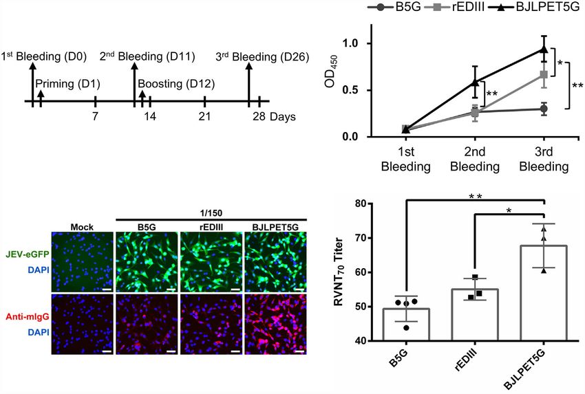
Yang et al.                                                                                       Auto-Processing BaMV Scaffold for Protein-Presenting

INTRODUCTION                                                                our previous design for the production of BaMV-based vaccine
                                                                            candidate against JEV (14). Another approach is to attach the
Virus-like particles (VLPs) or chimeric virus particles (CVPs) have         antigens after the assembly of the VLPs or CVPs, through
been utilized extensively as effective scaffolds for the presentation       chemical or enzymatic conjugation, such as sortase A (SrtA)-
of epitopes or antigens in the development of vaccine candidates            mediated ligation (16–18).
(1–4). The antigens displayed on the surfaces of VLPs and CVPs                  SrtA, an enzyme with both peptidase and ligase activities, was
can stimulate strong and long-lasting immune responses, since the           originally identified in Staphylococcus aureus and found to
VLPs and CVPs are assembled from hundreds or thousands of                   mediate the ligation of proteins on the surface of bacterial
highly ordered coat proteins (CPs) which present the antigens               cells. It has been shown that different SrtA may recognize and
repeatedly for the immune system. Furthermore, the size, shape,             cleave at specific amino acid sequences in the donor protein and
and rigidity of most viruses and the derived VLPs and CVPs are              mediate the ligation of the acceptor protein at the exposed N-
suitable to enter the lymphatic system for their uptake by antigen          terminus (19, 20). The coupling of target proteins on the surfaces
presenting cells of the immune system (5). Together with the                of VLPs or CVPs by SrtA-mediated ligation has been successfully
relative ease in manipulating the surface properties (6, 7), these          applied in different systems, including bacterial phage M13 (21)
features make VLPs and CVPs ideal alternative platforms for                 and papaya mosaic virus (PapMV) (17, 18, 22). However, these
vaccine development.                                                        platforms could be further improved with regard to the
    VLPs and CVPs derived from plant viruses exhibit additional             convenience in the mass-production of VLP or CVP scaffolds,
advantages as scaffolds for antigen presentation, compared to               which is another major concern in the development of VLP or
systems based on non-plant viruses. Plant viruses are not                   CVP antigen-presentation platforms.
pathogens to animals and humans, thus pose less biosafety                       To address these issues, we have devised a binary system in
threats. In addition, most plant viruses encode only a single CP            this study. The first part contains a BaMV-based vector which
with structural flexibility, which facilitates the ease in genetic or        facilitates the efficient in planta production of CVPs self-
chemical modifications of the VLPs or CVPs (1, 8). Many plant                assembled from CPs with 5 protruding glycine (G) residues as
virus-derived VLPs or CVPs have been developed as vaccine                   the acceptor for SrtA-mediated ligation (hereafter referred to as
candidates, such as the edible vaccine against rabies by                    SrtA-ready CVPs). The second part includes vectors for the
incorporation of a recombinant peptide from the G and N                     expression of target antigen, with calmodulin (CaM) fusion tag
proteins of rabies virus on the N-terminus of the alfalfa mosaic            to enhance solubility, and SrtA protein, respectively, in bacterial
virus (AlMV) (4). Some of plant virus-derived vaccine candidates,           cells. The applicability of this new system was demonstrated by
including malaria (9) and influenza (10), have been tested in                using the recombinant envelope protein domain III (rEDIII) of
human clinical trials, demonstrating the safety and efficacy of such         JEV (111 amino acids) as the target epitope, and the efficacy of
vaccines. In our previous studies, we have developed an efficient            the vaccine candidate in the induction of functionalized
epitope presentation CVP system based on Bamboo mosaic virus                antibodies was validated in BALB/c mice. The current BaMV-
(BaMV), a member of the genus Potexvirus, and validated its                 based system provides an efficient alternative for the rapid
applicability in stimulating protective immunity against foot-and-          development of bioactive vaccine candidates.
mouth disease virus (FMDV) (11, 12), infectious bursal disease
virus (IBDV) (13), or Japanese encephalitis virus (JEV) (14) in
different animal models. However, these previously developed                METHODS
BaMV-based epitope presenting systems, as well as other VLP
or CVP systems, still face several challenges that require                  Construction of Expression Vectors
further improvement.                                                        The constructs used for the production of BaMV CVPs were
    One of the major limitations of the epitope-presenting VLP or           developed based on the infectious clone of BaMV, pBS-d35CP
CVP systems is that the fusion of target proteins on the viral coat         (11). To enhance viral vector expression, the original pBS-d35CP
proteins may impair the self-assembly efficiency of the VLPs or              backbone was replaced by the binary plasmid pKn (23) to generate
CVPs, which are required for highly ordered presentation of                 pKBNd35 (Figure 1A), which could be introduced into plant cells
repetitive epitopes to stimulate strong immune responses. In                through Agrobacterium-mediated infiltration. The coding sequence
addition, the fusion with the viral CP may interfere with the               for TEV protease cleavage motif (ENLYFQG, abbreviated as TM)
proper folding of the antigen proteins, leading to inactive                 and tetra-glycine (4G) were amplified by polymerase chain reaction
conformation. Thus, the sizes of the target antigens to be                  (PCR) using the primer pair, TM-4G-F and TM-4G-R, and inserted
displayed on VLPs or CVPs are usually limited to short                      into pBS-d35CP at the AgeI/NotI sites, which was further digested
peptide fragments. However, some epitopes are structurally-                 with ApaLI and SacI and inserted into the corresponding restriction
dependent, which require the formation of specific                           sites of pKBNd35 to generate pKBTM4G. The coding region for
conformation from different domains of an antigen, not just                 BaMV triple-gene-block proteins (TGBps) was replaced by that of
linear amino acid sequences. The peptide epitopes might not                 Histidine-tagged TEV protease (7×His-TEV protease), which was
stimulate proper immune responses. Several strategies have been             amplified by PCR using the primer pair, 7×His-TEV protease-F and
developed to circumvent the above challenges. For example, we               TEV protease-stop-R, followed by digestion with DraIII and cloned
have adopted the FMDV 2A peptide sequence, which induces co-                into the corresponding site of pKBTM4G, generating pKB5G
translational “cleavage” (15) between target epitope and CP, in             (Figure 1A). The pKB-based constructs were transformed into

Frontiers in Immunology | www.frontiersin.org                           2                                  October 2021 | Volume 12 | Article 739837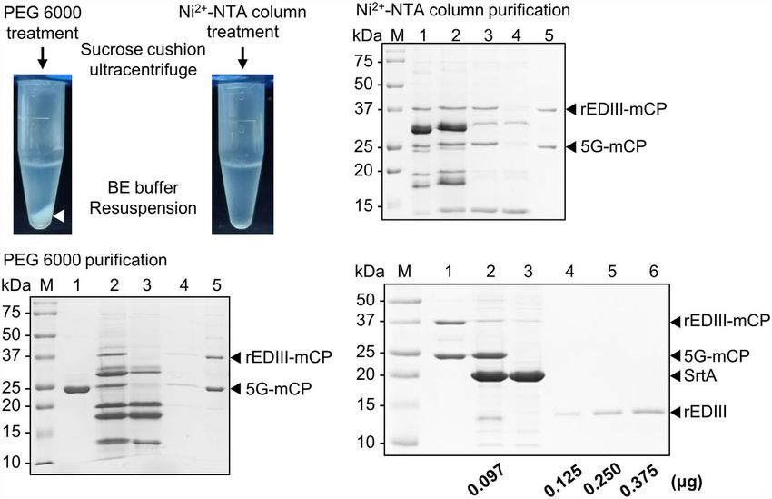
Yang et al.                                                                                                   Auto-Processing BaMV Scaffold for Protein-Presenting

                                                                                         the main CaM coding sequence with NdeI and EcoRI sites. The
                                                                                         amplified fragments of dual-6×His-tagged (6×His) CaM were
  A
                                                                                         digested with NdeI and EcoRI and ligated into the cognate sites of
                                                                                         pET29a (Invitrogen, Waltham, MA, USA) to generate pET29a-
                                                                                         6×His-CaM-6×His, abbreviated as pET29a-CaM. The coding
                                                                                         sequence of JEV rEDIII was amplified from pBJ2A (14) with the
                                                                                         primer pair, rEDIII-F and rEDIII-GSS-LPETG-GS-R, to add the
                                                                                         SrtA recognition sequence, LPETG-GS, to the C-terminus of
                                                                                         rEDIII. The amplified fragment was cloned into the NdeI site of
                                                                                         pET29a-CaM to generate pET29a-rEDIII-CaM (Figure 2A), which
                                                                                         harbors the coding sequence of rEDIII fused with the SrtA
                                                                                         recognition site, LPETG-GS, followed by the coding region of
                                                                                         CaM flanked by two His-tag (6×His) at both termini. We also
                                                                                         attempted to use the common thioredoxin (TrxA) fusion tag, which
                                                                                         was amplified from pET32a by using TGSS-TrxA-6×His-F and
                                                                                         TrxA-8×His-R primers to replace CaM with TrxA on pET29a-CaM
                                                                                         with AgeI and EcoRI site to generate pET29a-6×His-TrxA-8×His,
                                                                                         abbreviated as pET29a-TrxA. The coding sequence of VP1 peptide
  B                                       C                                              was amplified from pBVP1 (11) with the primer pair, VP1-F and
                                                                                         VP1-GSS-LPETG-R, to add the SrtA recognition sequence,
                                                                                         LPETG-GS, to the C-terminus of TrxA. The amplified fragment
                                                                                         was cloned into the NdeI and AgeI site of pET29a-TrxA to generate
                                                                                         pET29a-VP1 peptide-TrxA (Supplementary Figure 8). Primer
                                                                                         sequences for rEDIII-CaM and VP1 peptide-TrxA constructions
                                                                                         are listed in Supplementary Table 1. For production of SrtA, the
  D                                       E                                              plasmid pET29-eSrtA was purchased from Addgene (Watertown,
                                                                                         MA, USA). The pET29-based expression constructs were
                                                                                         transformed into the E. coli BL21 Star (DE3).

                                                                                         Protein Expression and Purification
                                                                                         Following cultivation of E. coli BL21 Star (DE3) cells harboring
                                                                                         the desired constructs, the induction and purification of target
                                                                                         proteins were performed as described previously (14). The His-
                                                                                         tagged target proteins were subjected to purification through a
 FIGURE 1 | Production of SrtA-ready CVPs of BaMV in Nicotiana benthamiana.
 (A) Schematic representation of the construction of infectious clones for the           Ni2+-NTA column (GE Healthcare, Chicago, IL, USA). The
 production of SrtA-ready CVPs in N. benthamiana for SrtA-mediated ligation of           fractions containing rEDIII-CaM and VP1 peptide-TrxA
 antigens. (B) Analysis of total proteins extracted from N. benthamiana leaves           proteins were confirmed by 12% polyacrylamide gel containing
 infiltrated with Agrobacterium tumefaciens harboring individual constructs by            1% sodium dodecyl sulfate (12% SDS-PAGE). The rEDIII
 electrophoresis through a 12% acrylamide gel containing 1% sodium dodecyl
 sulfate (12% SDS-PAGE), followed by staining with CBS. The positions of BaMV
                                                                                         protein, which was expressed from pET21d-rEDIII (14), was
 CP with various modifications were indicated by the bracket on the right. The            produced and purified as described previously (14, 25).
 proteins were then electro-blotted to PVDF membranes, and probed with
 antiserum specific to BaMV coat protein (C), TEV protease recognition site (D), or       BaMV CVP Production
 HC-Pro (E). Total proteins extracted from N. benthamiana leaves infiltrated with         The pKB-based clones and pBIN61-HC-Pro (26) were introduced
 buffer only (lane 1), or A. tumefaciens harboring pKBNd35 (lane 2), pKBTM4G
 (lane 3), or pKB5G (lane 4), or co-infiltrated with A. tumefaciens harboring pKB5G
                                                                                         into N. benthamiana plants through Agrobacterium-mediated
 and pBIN-HC-Pro (lane 5); lane M, molecular weight markers. The positions of            infiltration (27) with the concentrations of A. tumefaciens cells
 CP fused TEV site and HC-Pro are indicated by the solid and blank arrowheads            harboring pKBNd35, pKBTM4G, pKB5G, or pBIN61-HC-Pro
 on the right, respectively.                                                             adjusted to an OD600 of 0.5. For co-infiltration, equal volumes of
                                                                                         A. tumefaciens cells harboring pKB5G or pBIN61-HC-Pro were
                                                                                         mixed and adjusted to make a final concentration of OD600 = 0.5 for
Agrobacterium tumefaciens GV3850 for subsequent CVP expression                           each. The infiltrated leaves were harvested 7 days post-inoculation
following confirmation of the sequence. Primer sequences for CVPs                         (DPI), and BaMV CVPs were purified using the standard protocol
construction are listed in Supplementary Table 1.                                        described previously (28).
    To improve the solubility of the recombinant proteins, we have
employed CaM fusion tag, which has been used for proteins highly                         Production of Epitopes-Coupled CVPs
expressed in E. coli (24). The gene encoding CaM was amplified                            The CP concentration of BaMV CVP was adjusted to 1 mg/mL
from N. benthamiana by using 6×His-CaM-F and CaM-GSS-                                    and subject to SrtA-mediated ligation reaction. The ratio
6×His-R primers that incorporate 6×His tags at both termini of                           between the volumes of BaMV CVP and the total reaction

Frontiers in Immunology | www.frontiersin.org                                        3                                 October 2021 | Volume 12 | Article 739837
Yang et al. Auto-Processing BaMV Scaffold for Protein-Presenting
A B
C D
FIGURE 2 | Production and purification of JEV rEDIII and SrtA in Escherichia coli. (A) Schematic of the construct, pET29a-rEDIII-CaM, for the expression of rEDIII
fused with calmodulin (CaM) (rEDIII-CaM). The blank arrow and box represent the T7 promoter (T7) and T7 terminator (T7 ter), respectively. (B–D) SDS-PAGE
analysis of bacterially expressed rEDIII, rEDIII-CaM and SrtA proteins. E. coli BL21 cells were transformed with pET29a-rEDIII-CaM (B), pET21d-rEDIII (C), or pET29-
eSrtA (D). Protein samples taken from the following steps or fractions were analyzed by 15% SDS-PAGE and visualized by CBS. Lanes 1, and 2, before and after
IPTG induction, respectively; lanes 3 and 4, supernatant and pellet fraction, respectively, following sonication and centrifugation; lanes 5, 6, and 7, the flow-through,
washing, and elution fractions, respectively, following purification by Ni2+-NTA affinity column; lane M, molecular weight markers. The positions of rEDIII (≈12 kDa),
rEDIII-CaM (≈32 kDa), and SrtA (≈18 kDa) are indicated by the arrowheads on the right.
mixture was adjusted to 1:10. To determine the optimal reaction Borate–EDTA buffer (50 mM boric acid, pH 8.0, and 1 mM EDTA)
condition of the rEDIII coupling to BaMV CVPs, the ratio of and stored at 4°C. The BVP1LPET5G CVPs were inhibited SrtA
B5G CP: rEDIII-CaM: SrtA was varied as indicated in a 1.5-ml activity as described above and purified using the PEG 6000
Eppendorf tube containing 1× SrtA reaction buffer (50 mM Tris- precipitation protocol as described previously (28).
HCl, pH 8.0, 150 mM NaCl, 10 mM CaCl2, and 4 mM beta-
mercaptoethanol), and the reaction mixture was incubated at
28°C for different time period as indicated. SrtA activity was
Protein Analysis and Antibodies
Total proteins were extracted from infected N. benthamiana
terminated with the addition of 0.25 reaction volume of 5×
leaves and/or E. coli cultures, and subjected to analysis by SDS-
sample buffer (250 mM Tris-HCl, pH 6.8, 10% SDS, 40% (v/v)
PAGE and western blot (29) using specific antibodies against
glycerol, 2 mM beta-mercaptoethanol, 0.1% (w/v) bromophenol
BaMV CP (5000× dilution), HC-Pro (5000× dilution), JEV
blue). The protein products were analyzed by electrophoresis
rEDIII (5000× dilution), FMDV VP1 (5000× dilution), or
through 12% SDS-PAGE, followed by Coomassie Blue Staining
commercial polyclonal antibodies against TEV protease
(CBS) or western blotting with specific antibodies as described
recognition site (Abcam, Cambridge, United Kingdom; 2500×
below. Large-scale coupling reactions were performed in a 100-
dilution), or mouse monoclonal antibody against His-tag (Bio-
ml beaker, and the mixture was incubated for 15 min at 28°C
Rad, Hercules, CA, USA; 2000× dilution), followed by
with gentle stirring. Another epitope candidate, the VP1 peptide-
visualization using a nitro-blue tetrazolium/5-bromo-4-chloro-
presenting BaMV CVPs (designated BVP1LPET5G) were also
3’-indolyphosphate (NBT/BCIP) color development substrate
produced as described above.
(Thermo Scientific, Waltham, MA, USA) or horseradish
peroxidase (HRP) reagent (Merck Millipore, Darmstadt,
Purification of Epitopes-Coupled
Germany). The Precision Plus Protein Dual Color standard
BaMV CVPs marker (Bio-Rad, Hercules, CA, USA) was used as the
The rEDIII-presenting BaMV CVPs (designated BJLPET5G)
molecular weight standard.
were purified as described above with the following
modifications: (1) the chelator (10 mM EGTA) and disodium
phosphonate (100 mM K2HPO3) were gently added to inhibit Quantification of CVPs and Proteins
SrtA activity; (2) the calcium complex was removed by The amount of CVPs was determined spectrophotometrically as
centrifugation at 7,600 ×g at 4°C for 10 min; (3) the described previously (28). The concentrations of rEDIII-CaM,
supernatant was purified with the Ni2+-NTA column to remove SrtA, and rEDIII proteins were determined through the Bradford
the unreacted His-tagged proteins; (4) the flow-through fraction assay (Sigma-Aldrich, St. Louis, MO, USA). To quantify the
was collected and subjected to ultracentrifugation through a 20% concentration of rEDIII proteins on the CVPs, SrtA was used to
sucrose cushion; and (5) the virion pellet was resuspended with release the rEDIII coupled on CVPs and the digested products
Frontiers in Immunology | www.frontiersin.org 4 October 2021 | Volume 12 | Article 739837Yang et al. Auto-Processing BaMV Scaffold for Protein-Presenting
were subjected to analysis by 12% SDS-PAGE followed by vaccinated mice with 2.5-fold serial dilutions (starting at 1:40)
staining with CB and quantification. Known amounts of in Opti-MEM medium (Gibco, Grand Island, NY, USA) at 37°C
rEDIII were used to create a standard for quantification using for 1 h. The mixtures were individually incubated with a
ImageJ software (30). monolayer of BHK-21 cells in the 96-well plates at 37°C for
2 h. After incubation, the inoculum in the plates was replaced
Immunoelectron Microscopy with fresh medium containing 2.5% FBS for incubation at 37°C
Negative staining and immunoelectron microscopy with gold- for 20 h. After fixation and permeabilization, the nuclei were
labeled antibodies specific to the BaMV virion or JEV rEDIII stained with 4′, 6-diamidino-2-phenylindole (DAPI; Invitrogen,
were performed as described previously (31). CVP conformation Waltham, MA, USA). Finally, the plates were scanned under a
and immunolabeling were examined through transmission fluorescence microscope to generate the panels, and the cells
electron microscopy (FEI Tecnai G2 Spirit; FEI Company, were scored (percentage of infectivity = fluorescent cells/total
Hillsboro, OR, USA) at 80 kV. The control grids were loaded cells) using MetaXpress software (Molecular Devices, San Jose,
with BNd35 and B5G CVPs. CA, USA) for high content screening. The 70%-neutralization
titer in RVNT (RVNT70) data for each antibody were used to
Animals and Vaccination calculate relative reduction in fluorescence of JEV-eGFP,
Three groups of five 3-week-old female BALB/c mice were determined as follows: % reduction = 100 × [1 − (average
obtained from the National Laboratory Animal Center, number of fluorescent cells for each dilution/average number
Taiwan. Freund’s complete (priming) or incomplete adjuvants of fluorescent cells in B5G control group)]. The RVNT70 is
(boosting) (Sigma-Aldrich, St. Louis, MO, USA) were mixed evaluated by the concentration that gave 70% fluorescent cells
with B5G (BaMV virion scaffold), rEDIII (5 mg), and BJLPET5G reduction, determined by nonlinear, dose-response regression
(≈5 mg of rEDIII-coupled BaMV virion scaffold) for priming on analysis with GraphPad Prism version 6.0 (GraphPad, San
day 1 and boosting on day 12, respectively, for intraperitoneal Diego, CA, USA).
inoculation. Whole blood from the orbital sinus was collected on
days 0, 11, and 26; the blood cells were removed through Streamlined Production of Epitope-
centrifugation (13,000 ×g, 10 min) twice, and the sera were Presenting CVPs
collected. The titers of sera were tested through indirect enzyme- His-tagged rEDIII-CaM and SrtA proteins were purified from
linked immunosorbent assay (ELISA). bacterial cell lysates (following sonication) with Ni2+-NTA resin
in batch mode, and bound on resin for the coupling reaction with
Mice Immunization Assay SrtA-ready B5G CVPs. Mass-produced SrtA-ready B5G CVPs
C6/36 cells were infected with JEV (RP-9) (32) at a multiplicity of were added to the resin with rEDIII-CaM and SrtA, incubated
infection (MOI) of 0.1 and cultured for 4 days in a 96-well plate. for 15 min in the SrtA reaction buffer containing 300 mM
The infected cells were fixed and examined by indirect ELISA imidazole. The SrtA-mediated ligation reaction was terminated
with diluted serum samples collected from immunized mice with the addition of EGTA (10 mM), and the imidazole was
overnight at 4°C followed by detection with goat anti-mouse removed by using spin column (Amicon Ultra 10K, Merck
IgG-HRP conjugate (Jackson ImmunoResearch Laboratories, Millipore). Supernatant was incubated with Ni2+-NTA resin
West Grove, PA, USA) as described previously (33). The again to remove his-tagged unreacted proteins. The BJLPET5G
OD450 was measured by using an ELISA reader (Spectramax CVPs were then harvested from the supernatant after low-
M2; Molecular Devices, Sunnyvale, CA, USA). speed centrifugation.
Immunofluorescence Assay
BHK-21 cells were infected with the reporter virus, JEV (RP-9)
Statistical Analysis
Statistical analysis and graphical visualizations were performed
expressed enhanced green fluorescent protein (JEV-eGFP) (34), at
in Microsoft Excel 2010 (Microsoft, Redmond, WA, USA) and
an MOI of 2 for 15 h. Following fixation and permeabilization, the
GraphPad Prism version 6.0. A two-tailed Student’s t test was
cells were incubated overnight at 4°C with a serial dilution of
used to compare between the means of two groups; p < 0.05
different sera from mice vaccinated with B5G, rEDIII, or
indicates statistical significance.
BJLPET5G followed by detection with goat anti-mouse IgG-Alexa
568 conjugate (Invitrogen, Waltham, MA, USA), and examined
under a fluorescence microscope. Fluorescent signal images were
captured at 200× magnification with an Olympus IX71 inverted RESULTS
fluorescence microscope (Olympus, Tokyo, Japan).
Production of SrtA-Ready Chimeric BaMV
Reporter Virus Neutralization Test Particle In Planta
The titers of the neutralizing antibodies in the sera from mice In this study, we have developed a CVP scaffold based on BaMV
vaccinated with various CVPs were determined using the that is ready for the presentation of target protein through SrtA-
neutralization assay with BHK-21 cells and the reporter virus mediated ligation, as shown in Figure 1A. This design ensured
JEV-eGFP (at an MOI of 0.5) in triplicates following the standard the production BaMV CP and TEV protease within the same
protocol (35). The JEV-eGFP was incubated with sera from plant cell. The TEV protease could thus recognize the TM at the
Frontiers in Immunology | www.frontiersin.org 5 October 2021 | Volume 12 | Article 739837Yang et al. Auto-Processing BaMV Scaffold for Protein-Presenting
N-terminus of the modified BaMV CP and cleave in between Q induced by the addition of IPTG. As shown in Figure 2B, the
and G, generating BaMV CP with 5 protruding G residues which expected fusion protein rEDIII-CaM (lane 2) was successfully
could then self-assemble into BaMV CVPs, designated as B5G, expressed and detected by the previously prepared JEV-specific
susceptible for SrtA-mediated ligation of target proteins. The antibodies (Supplementary Figures 1A, B, lane 4). In
assembled BaMV CVPs are then equipped with thousands of comparison with the rEDIII expression system without CaM-
protruding 4G extensions ready for the SrtA-mediated ligation of fusion, the level of soluble rEDIII protein in E. coli cells
the antigens. The above constructs were then used to inoculate expressing CaM-fused rEDIII was increased in the supernatant,
Nicotiana benthamiana via Agrobacterium-mediated infiltration. resulting in the highest amount of total rEDIII protein extracted
Total proteins were extracted from the inoculated leaves 7 DPI after sonication (Figures 2B, C, lanes 3 and 4).
and analyzed by electrophoresis through 12% SDS-PAGE, To provide SrtA proteins for ligation reactions, the construct
followed by CBS (Figure 1B) and western blot analysis using pET29-eSrtA purchased from Addgene (19), which harbors the
specific antiserum (Figures 1C–E). As shown in Figures 1B, C, coding sequence of SrtA with 6×His tag, was transformed into E.
BaMV CP could be detected in total protein extracts from N. coli. Following IPTG induction, the SrtA proteins could be
benthamiana leaves inoculated with A. tumefaciens harboring readily detected in the total protein extracts by 15% SDS-
pKBNd35 and pKBTM4G (lanes 2 and 3, respectively), but not PAGE through CBS (Figure 2D, lane 2). Both the
pKB5G (lane 4). We hypothesized that the low expression level 6×His-tagged rEDIII-CaM and SrtA could be easily purified by
might be due to the replacement of the original BaMV RNA Ni2+-NTA resin (Figures 2B, D, lanes 5–7), and the protein
silencing suppressor, TGBp1, by the TEV protease in pKB5G samples were stored in 50% glycerol at −20°C for subsequent
construct, leading to the silencing of the expressions of B5G CP. SrtA-mediated ligation reactions.
To address this problem, we resorted to the usage of the well-
known potyvirus silencing suppressor, HC-Pro (36). A. SrtA-Mediated Surface Modification on
tumefaciens cells harboring pKB5G were co-infiltrated with B5G CVPs
those harboring pBIN61-HC-Pro (26) to complement the To evaluate the feasibility of SrtA-mediated coupling on the B5G
silencing suppressor function of BaMV TGBp1. As expected, viral surface, small-scale reactions were performed using equal
the co-expression of potyviral HC-Pro facilitated the efficient amounts of B5G CP, rEDIII-CaM, and SrtA. Time-course assays
accumulation of B5G proteins in the infiltrated leaves were then performed to test the optimal reaction time to
(Figures 1B, C, lane 5). To determine whether the TM on maximize the yield of epitope-presenting BaMV CVPs. The
BaMV CPs is functional in plants, the total proteins extracted LPETG-GS motifs on rEDIII-CaM were cleaved and ligated on
from infiltrated tissues were probed using antibodies specific to B5G CPs by SrtA, resulting in the generation of rEDIII-LPET-
TM (Figure 1D). The result showed that only the BaMV CP 5G-mCP (abbreviated as rEDIII-mCP) with a size of
from leaves inoculated by pKBTM4G could be detected approximately 35 kDa, as observed by SDS-PAGE analysis
(Figure 1D, lane 3), indicating that the TM could indeed be (Figure 3A). The result revealed that the SrtA-mediated
recognized and digested by the TEV protease expressed in the coupling of rEDIII on BaMV CVPs could be achieved in 1 min
same plant cell infiltrated with pKB5G, rendering it undetectable (Figure 3A), generating rEDIII-presenting BaMV CVPs
by the TM-specific antibodies (Figure 1D, lane 5). The higher (designated BJLPET5G). The identities of the coupled proteins
mobility of B5G (Figures 1B, C, lane 5) compared to that of the and rEDIII proteins were further confirmed by western blot
CP from leaves infiltrated with pKBTM4G (Figures 1B, C, lane analysis with specific antibodies against BaMV CP or rEDIII
3) provide further evidence indicating that the TM on the B5G (Supplementary Figure 2). To evaluate the relative coupling
was digested to a similar size of BNd35 CP (Figures 1B, C, lane efficiency, we calculated the ratio between the band intensities of
2) by TEV protease. The presence of HC-Pro in the leaves co- the rEDIII-coupled CPs and the total CPs in SDS-PAGE
infiltrated with pKB5G and pBIN61-HC-Pro was confirmed by (Figures 3A–C) using the software ImageJ (National Institutes
HC-Pro-specific antibodies (Figure 1E, lane 5). of Health, Bethesda, MD, USA) (Figures 3D–F). It was found
that up to 35% of the B5G CPs were labeled with rEDIII by SrtA
Protein Expression and Purification in in 1–10 min, whereas only 3% to 18% of B5G CPs retained the
Escherichia coli rEDIII after 15 min (Figures 3A, D). Longer reaction time (more
To prepare the donor protein, rEDIII, to be presented on the than 15 min) resulted in significantly decreased yields of
surface of B5G CVPs, we have developed a vector, designated BJLPET5G, presumably caused by the removal of rEDIII from
pET29a-rEDIII-CaM (Figure 2A), for the expression of target BaMV CVPs by SrtA, since the ligation of the rEDIII on the B5G
proteins in E. coli with the C-terminal fusion of CaM. The CVPs would create another SrtA-recognition site which could be
protein product expected to be translated from such construct cleaved again in a prolonged reaction (37, 38). With this
would be rEDIII-LPETG-GS-6×His-CaM-6×His, abbreviated as knowledge of the SrtA reaction time, the reaction condition
rEDIII-CaM, in which the rEDIII-LPET fragment could be was optimized by increasing the amount of rEDIII-CaM from 2
attached to the G residue of the acceptor protein by SrtA- to 10 folds relative to that of B5G CP to increase the amount of
mediated ligation and the released G-GS-6×His-CaM-6×His rEDIII protein labeled on the B5G CVPs. The result showed that
fragment could be removed by Ni2+-NTA affinity column. To the amount of conjugated rEDIII on the CVPs reached a plateau
test the functionality, the construct pET29a-rEDIII-CaM was when the ratio between rEDIII-CaM and B5G CP was increased
transformed into E. coli, and the target protein expression was to 3:1 or more (Figures 3B, E). To avoid the removal of rEDIII
Frontiers in Immunology | www.frontiersin.org 6 October 2021 | Volume 12 | Article 739837Yang et al. Auto-Processing BaMV Scaffold for Protein-Presenting
A D
B E
C F
FIGURE 3 | Condition optimization of SrtA-mediated ligation of rEDIII on the B5G CVP surface. (A) Effect of reaction time. Equal amounts of B5G CP, rEDIII-CaM,
and SrtA were incubated for different reaction times as indicated on top of each lane. (B) Effect of different ratios of reactants. The amount of rEDIII-CaM was
increased by 2 to 10 folds, as indicated on top of each lane, relative to that of B5G CP, with a reaction time of 1 min. (C) Effect of reaction time with reduced SrtA
concentration. The amount of SrtA was reduced to 0.2 mg and the reaction time was varied as indicated on top of each lane. The components in each reaction are
shown on top of each lane. Protein samples were analyzed by 12% SDS-PAGE, and the protein bands were visualized by CBS. The expected sizes of different
forms of proteins were indicated by the arrowheads and brackets on the right. The relative coupling efficiencies under different reaction conditions were shown in the
respective bar charts (D–F). Statistical significance was analyzed using two-tailed Student’s t tests; *p < 0.05; **p < 0.01; ***p < 0.001. Error bars represent the
standard deviation of three independent experiments.
following SrtA-mediated ligation, the ratio between B5G CP and the corresponding concentrations of approximately 4.38, 9.48, and
SrtA was decreased to 1:0.2 in the subsequent time-course 1.12 mM, respectively.
experiment. It was found that similar coupling efficiency could To demonstrate the applicability of the B5G CVP platform
be observed with longer reaction times (10-15 min), whereas the with SrtA-mediated ligation for other antigens, we used the VP1
labeled rEDIII proteins were re-cleaved after 30 min peptide derived from FMDV (14) as the target protein. The result
(Figures 3C, F). Accordingly, the reaction time was extended showed that the VP1 peptide could also be coupled to B5G CP at
in the following experiments to increase coupling efficiency and a ratio of 1:2:0.2 for B5G CP, VP1-TrxA, and SrtA, with an
facilitate subsequent purification. The coupled proteins on the efficiency up to 42% (Supplementary Figure 9).
CVPs were then quantified following SDS-PAGE with the
software ImageJ. It was found that the coupling efficiency with Optimization of Purification Process and
the rEDIII protein was approximately 22.5-43.0% (Figures 3C, F). Quantification of SrtA-Coupled CVPs
Based on these observations, the optimal ratio for coupling was Following the addition of 10 mM ethylene glycol tetraacetic acid
determined to be 1:3:0.2 for B5G CP, rEDIII-CaM, and SrtA, with (EGTA) to terminate SrtA reaction, the virions were extracted from
Frontiers in Immunology | www.frontiersin.org 7 October 2021 | Volume 12 | Article 739837Yang et al. Auto-Processing BaMV Scaffold for Protein-Presenting
the mixture as described (28). Unexpectedly, the polyethylene lanes 1 and 5, respectively). To evaluate the amount of coupled
glycol (PEG)-mediated CVPs precipitation resulted in the rEDIII on the CVP CP, SrtA-mediated cyclic cleavage was
generation of insoluble white precipitates (Figure 4A, left panel). performed to release the coupled rEDIII, and the products were
Analysis by SDS-PAGE revealed that most of the free CPs or rEDIII- analyzed by SDS-PAGE (Figure 4D). The result of quantification
coupled CPs were detected in the pellet fractions (either before or using software ImageJ revealed that approximately 97 ng rEDIII
after sucrose cushion purification), indicating that PEG treatment were coupled on 1 mg of B5G CP. As indicated by the structural
severely reduced the solubility of CVPs (Figure 4B, compare lanes model of the BaMV virion, each CP unit interacts with estimated 5.2
4 and 5). To enhance the solubility of the final product, we employed bases (39). Based on this calculation and the observed coupling
affinity column purification using Ni2+-NTA resin. It was found efficiency, the estimated numbers of BJLPET5G CPs per CVP are
that the affinity purification method prevented the precipitation approximately 345 out of 1150 copies.
problem (Figure 4A, right panel). The result of SDS-PAGE analysis
revealed that most bands corresponding to CP with or without the Identification of rEDIII on the
rEDIII labels were harvested in the flow-through fraction BJLPET5G Surface Through
(Figure 4C; lane 3, indicated by arrowheads). In addition, most Immunoelectron Microscopy
CPs could be easily separated from free SrtA and rEDIII-CaM by To verify whether the CVPs of B5G and BJLPET5G were
ultracentrifugation through a 20% sucrose cushion, suggesting properly assembled, the purified samples were examined by
that CPs were assembled into BJLPET5G CVPs (Figure 4C, lane electron microscopy, with the BNd35 virion as a control. The
5). The recovery rate of CVP CP was approximately 82-98%, as result showed that the CVPs of B5G and BJLPET5G exhibited
determined from the band intensities in SDS-PAGE (Figure 4C, similar flexible filamentous virion conformations as those of
A C
B D
FIGURE 4 | Purification and quantification of B5G CVPs presenting rEDIII. (A) Comparison of purification by polyethylene glycol (PEG) 6000 precipitation (left) or Ni2+-NTA
affinity column (right). The BaMV CVPs presenting rEDIII following SrtA-mediated ligation were separated from the unreacted rEDIII and SrtA by an initial purification of
PEG 6000 precipitation or Ni2+-NTA affinity column, followed by ultracentrifugation through a 20% (w/v) sucrose cushion. For the initial separation using PEG 6000,
white precipitate was observed (white arrowhead). (B) SDS-PAGE analysis of BJLPET5G CVPs purified by PEG precipitation. Lanes 1, B5G alone; 2, unpurified SrtA
reaction mixture; 3, supernatant collected after PEG precipitation. The pellet fraction obtained following PEG precipitation was resuspended and subjected to
ultracentrifugation, and the resulting supernatant (lane 4) and final pellet (lane 5) fractions were analyzed. (C) SDS-PAGE analysis of BJLPET5G CVPs purified
through Ni2+-NTA affinity column. Lanes 1, unpurified SrtA reaction mixture; 2, reaction mixture containing EGTA and K2HPO4 for inhibiting SrtA activity; 3, flow-
through sample from Ni2+-NTA column; 4 and 5, the supernatant and pellet fraction, respectively, obtained following ultracentrifugation through a 20% sucrose
cushion. (D) SDS-PAGE analysis of the amount of the rEDIII proteins coupled on the CVP surface. Purified BJLPET5G CP (2 mg) were cyclically digested by SrtA
(4 mg) at 28°C overnight to release the rEDIII. The digestion product was analyzed by 12% SDS-PAGE and visualized by CBS. Lanes M, molecular weight standard;
1, undigested BJLPET5G CVPs; 2, digested BJLPET5G CVPs; 3, SrtA protein control; 4-6, increasing amounts (0.125–0.375 mg) of purified rEDIII from E. coli as the
quantitative standard. The relative amount of coupled rEDIII on the CVPs was quantified by analyzing the band intensities using ImageJ, as indicated at the bottom.
The positions of various proteins were indicated by arrowheads on the right.
Frontiers in Immunology | www.frontiersin.org 8 October 2021 | Volume 12 | Article 739837Yang et al. Auto-Processing BaMV Scaffold for Protein-Presenting
BNd35 (Figures 5A–C, respectively). Immunoelectron (5 µg), or BJLPET5G (corresponding to 5 µg of rEDIII
microscopy was performed to confirm the presence of rEDIII on conjugated on 52.25 µg of B5G CP) following the scheme
the surface of BJLPET5G virions. As expected, BNd35, B5G, and illustrated in Figure 6A. It was found that the antibody titers
BJLPET5G virions could be specifically decorated with gold against JEV in serum samples collected from mice injected with
particles following the reaction with antibodies against BaMV BJLPET5G were increased more rapidly following the priming
virions and gold-labeled secondary antibodies (Figures 5D–F, injection, as compared with the other groups (Figure 6B). After
respectively). The presence of rEDIII on the surface of BJLPET5G boosting, the JEV-specific antibody titers in the sera of mice in
virion scaffolds was verified by antibodies specific to rEDIII and the BJLPET5G group have increased significantly as compared
gold-conjugated secondary antibodies, whereas little or no gold with those in the B5G and rEDIII groups (Figure 6B and
particles were observed on the BNd35 or B5G virions following the Supplementary Figure 4). To compare the sera titers of
same treatment (Figures 5G–I). Quantification of the gold particles different treatment groups, the titration curves for different
decorating virion surface revealed that each BJLPET5G virion was time-points (i.e., pre-immune, priming, boosting) were plotted
labeled by approximately 14 gold particles (an average of 50 as shown in Supplementary Figure 5. In the analysis, the half-
virions, Figure 5 and Supplementary Figures 3A–C). Compared maximal effective concentration (EC50) values could not be
to our previous BaMV-based vector with FMDV 2A peptide-fusion accurately determined since the reaction curves had not
[BJ2A CVPs, (14)], it was found that BJLPET5G particles exhibited reached a plateau at 1/100 dilution in our experiments. The
a 7.5-fold increase of rEDIII on the CVP surface (Supplementary observation indicated that immune responses elicited by the
Figures 3C, D). The result confirmed that rEDIII was successfully treatments were not especially strong. However, the data
presented on the BJLPET5G virion surface. indicated that BJLPET5G stimulated higher or comparable
titers than rEDIII did at the priming and boosting stages,
respectively. In addition, the activities of antibodies were
BJLPET5G Elicited Specific Antibody evaluated by using the JEV-eGFP reporter system (34), in
Responses and Virus-Neutralization which the cultured BHK-21 cells infected with JEV-eGFP were
in Mice fixed and reacted with antisera (150-fold dilution) collected from
To assess the immunogenicity of BJLPET5G CVPs, we injected mice in the aforementioned groups, followed by the detection of
BALB/c mice (5 per group) with B5G (52.25 µg), free rEDIII the bound antibodies by Alexa Fluor 568-conjugated secondary
FIGURE 5 | Examination of BaMV CVPs through transmission electron microscopy. Purified CVPs of BNd35 (A, D, G), B5G (B, E, H), and BJLPET5G (C, F, I)
were observed by transmission electron microscopy using uranyl acetate (UA) negative staining (A–C), or immuno-gold staining using antisera specific to BaMV virion
(D–F) or JEV rEDIII (G–I) followed by labeling with the gold-conjugated goat anti-rabbit IgG antibodies. Average numbers of gold particles on each CVP surface (n =
50) treated with antisera-specific rEDIII (G–I) are indicated below each panel. Scale bars, 100 nm.
Frontiers in Immunology | www.frontiersin.org 9 October 2021 | Volume 12 | Article 739837Yang et al. Auto-Processing BaMV Scaffold for Protein-Presenting
A B
C D
FIGURE 6 | Immunization and virus-neutralization analysis. (A) Vaccination and bleeding scheme. The bleeding and injection date are indicated by the arrows.
(B) The serum collected at each stage was diluted 500-fold, incubated with JEV-infected (RP-9) C6/36 cells coated on plates, and analyzed for antibody titers in an
enzyme-linked immunosorbent assay (ELISA). The line chart represents the mean values of optical density at 450 nm (OD450) obtained in the ELISA with sera from
individual mouse. (C) In the immunofluorescence assay, BHK-21 cells infected with JEV-eGFP (green) were fixed and stained with pooled serum diluted 150-fold
from the immunized groups of mice. The antibodies were labeled with Alexa Fluor 568-conjugated secondary antibody against mice IgG (anti-mIgG, red), and the cell
nuclei were stained with DAPI (blue). The samples were examined for antibody labeling by using an inverted fluorescence microscope. Scale bars, 50 mm. (D) The
70%-neutralizing antibody titrations of reporter virus neutralization test (RVNT70) against JEV-eGFP were preformed using serum samples collected in third bleeding
from B5G, rEDIII, and BJLPET5G-vaccinated mice (n = 5). Individual samples were serially diluted from 1:40 to 1:625 and RVNT70 were performed in 96-well plates
as described in material and methods. Sera from mice immunized with B5G were used as negative controls. rEDIII was used as the positive control. Approximately 5
mg of rEDIII was used for immunization in both the rEDIII and BJLPET5G groups. The relative mean and standard deviation of titers were shown in the bar chart. The
standard deviations are indicated by the error bars. Statistical significance was analyzed using two-tailed Student’s t tests; *p < 0.05; **p < 0.01.
antibodies against mice IgG. The numbers of green fluorescence- (Supplementary Figure 6). The result indicated that sera from
positive cells were used as an indicator of infection, while the mice in the BJLPET5G group had a significantly superior ability
intensity of the red fluorescence represented the titer of the in suppressing JEV infection compared to those from mice in the
rEDIII-specific antibodies present in the serum samples. The rEDIII group.
result of fluorescence microscopy examination revealed that To further quantitate the JEV-inhibition efficiency, the pooled
while the BHK-21 cells were infected with relatively equal sera were serially diluted, and the inhibition rate was determined
amounts of JEV-eGFP (Figure 6C, upper panel), the sera from by the RVNT70 (14, 35). The result showed that the JEV-specific
mice in the BJLPET5G group exhibited the highest titer against neutralizing antibody titers of the BJLPET5G group were
JEV, giving the highest intensities of red fluorescence as significantly higher (Figure 6D), demonstrating the
compared to those from mice in the B5G or rEDIII groups immunogenicity of BJLPET5G against JEV.
(Figure 6C, lower panel). This observation demonstrated that
injection of BJLPET5G in mice could stimulate effective immune Streamlined Production of Epitope-
response, leading to the production of antibodies against JEV. To Presenting CVPs
further verify whether the injection of BJLPET5G could induce The workflow of the binary system is illustrated in Figure 7A. To
the generation of neutralizing antibodies against JEV, we demonstrate the applicability of the system in the efficient
performed a RVNT, which is correlated with immune development of new vaccine candidates, we have tested the
protection (34, 35). The result showed that JEV-eGFP pre- feasibility of a simplified protocol as follows (Figure 7B). The
incubated with sera from mice in the BJLPET5G group E. coli cells expressing the epitope (rEDIII-CaM in this example)
resulted in fewer JEV-positive cells, as compared with those and SrtA proteins were lysed by ultrasonication, and the lysates
treated with sera from mice in the B5G or rEDIII groups were directly mixed with Ni2+-NTA resin in the Eppendorf tube
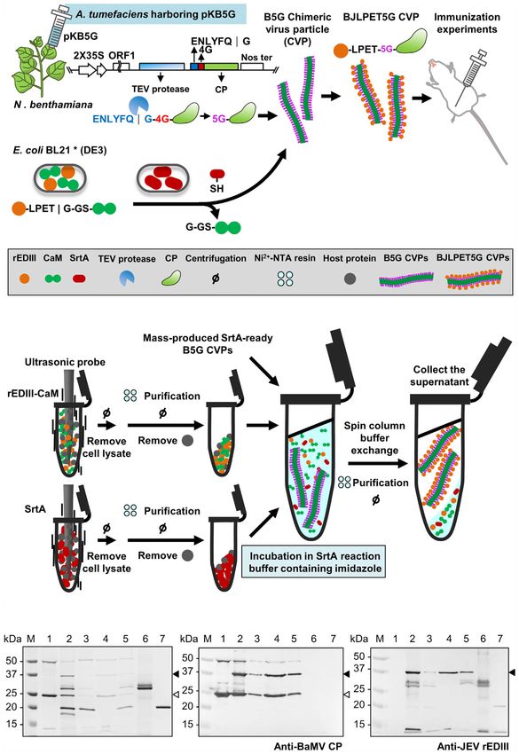
Frontiers in Immunology | www.frontiersin.org 10 October 2021 | Volume 12 | Article 739837Yang et al. Auto-Processing BaMV Scaffold for Protein-Presenting
followed by the addition of previously prepared SrtA-ready B5G indistinguishable from that of BNd35. Similarly, the payload of
CVPs. The supernatant containing BJLPET5G CVPs was then the current system is higher (meaning that B5G could be applied
recovered. However, the result of SDS-PAGE analysis of the for the presentation of larger epitopes), since the epitopes were
products (Figure 7C) showed that the activity of SrtA-mediated ligated after the formation of virions. Thus, the interference of
ligation reaction was inhibited when SrtA proteins bound on the large sizes of epitopes on CVPs assembly was eliminated. Our
Ni2+-NTA resin. Thus, a modification of the above protocol was new system is more cost-effective, since the TEV protease is
made in which the imidazole was added to release SrtA proteins directly produced by the chimeric virus in plants, and the
(Figure 7B). Analysis of the final products by SDS-PAGE and processing of CP and auto-assembly of the CVPs were
western blot revealed that BJLPET5G CVPs could be readily completed within the plants. In addition, the unreacted rEDIII-
obtained through this simplified protocol within 6 hrs CaM and SrtA proteins could be recovered from the Ni2+-NTA
(Figures 7C–E, lane 4). The result suggested that the system column and reused in another ligation reaction, reducing the cost
developed in this study could be applied in the rapid for the production of SrtA-ready B5G and target epitopes. In the
development of new vaccine candidates. current binary system, the production of B5G CVPs in plants is
separated from that of the target antigens in bacteria. The B5G
CVPs could be mass-produced in the greenhouse or fields
following the regulation of Current Good Manufacturing
DISCUSSION Practice (cGMP), tested for quality control, and stored or
Development of a Binary System for the applied immediately by SrtA-mediated ligation with desired
antigens for the development of other vaccine candidates. In
Production of SrtA-Ready CVP Scaffolds
contrast, in the previous CP-epitope fusion approaches, such as
and Antigens (14), a new chimeric virus construct has to be generated for every
The design of B5G CVP system ensured the production of TEV
new epitope, some of which may severely interfere with the
protease and BaMV CP within the same cell, and the processed
infectivity or assembly of the chimeric virus. Therefore, the
BaMV CP could auto-assemble into the SrtA-ready virions in
current binary system requires less time and efforts in the
planta. In addition, the yield of B5G virions, reached about 1.7
development cycles of new vaccine candidates. Furthermore,
mg per 100 g of fresh N. benthamiana leaves, with the co-
the results of immunization assays revealed that BJLPET5G
expression of HC-Pro, which is comparable to the yields of wild
stimulated the production of neutralizing antibodies with
type BaMV virions or CVPs in inoculated leaves or transgenic
higher titers against JEV infection.
plant cell‐suspension system (11, 40). For the target antigen
production, the fusion of 6×His-CaM-6×His tag at the C-
terminus following the SrtA recognition site of the target Possible Means for Further Optimization
antigen increased the solubility (Figures 2B, C) and allowed of the SrtA-Mediated Ligation
for easy purification of the target protein. By separating the It has been reported that the coupling density of M2e peptide
production processes of CVPs and target antigens, this approach onto the PapMV-N nanoparticles (made of PapMV CP with N-
exhibits the potential to facilitated the auto-assembly of terminal acceptor site) could reach up to 83% by increasing the
filamentous CVP scaffolds and the presentation of large incubation time and the concentration of M2e peptide (22).
peptides on the surface of such CVP scaffolds through SrtA- However, in our system, the efficiency of SrtA-mediated ligation
mediated ligation. We have showed the successful coupling of was only around 20% to 40% (Figures 3C, F and 4D). One of the
two different antigens, rEDIII and VP1 peptides, with B5G CVPs major reasons might be the reversibility of SrtA-mediated
(Figure 4 and Supplementary Figure 9), demonstrating the ligation, in which the ligation of donor and acceptor restored
applicability of the current system for antigen presentation. the original SrtA recognition sequence LPETG-GS, which could
Compared to our previous design (14), the system developed be re-cleaved by SrtA. The evidence for our reasoning is that the
in this study exhibited the following potential advantages: (1) longer incubation time for SrtA reaction resulted in less yield of
better biocontainment of the chimeric virus; (2) better antigen-coupled B5G CPs (Figures 3A, D), suggesting that the
conformation of virions; (3) increased payload on the CVPs; ligated rEDIII proteins were re-cleaved by SrtA. Several methods
(4) higher cost-effectiveness in the production process; and (5) have been developed to minimize the reversibility of the SrtA-
faster development of new vaccine candidates. The reason for mediated ligation [as comprehensively reviewed by Antos et al.,
better containment of the chimeric virus is that the coding region (41)]. The first type of approaches is to increase the supply of
for TGBps, involved in virus movement, on the chimeric BaMV either the donor or acceptor. The second is the separation of
genome was replaced by that of the TEV protease, rendering the desired ligation product from the by-products by selective
chimeric BaMV unable to move out of the inoculated regions in removal of the cleaved amino-glycine peptide fragment by
plants. Therefore, the biosafety concern for the chimeric virus dialysis, centrifugal filtration, or affinity immobilization (42–
could be reduced. For better virion conformation, the reason is 47). As for our system, the B5G CVPs provide a highly
that the CP of the chimeric virus is essentially the same as that of ordered arrangement of a large amount of CPs as receptors
the original BNd35, except for 5 extra glycine residues at the N- [estimated to be about 1150 copies per virion, based on structure
terminus following the cleavage by TEV protease. As shown in of BaMV (39)], which should have driven the reaction towards
Figure 5, the morphology of B5G and BJLPET5G virions was the formation of BJLPET5G. However, the coupling efficiency
Frontiers in Immunology | www.frontiersin.org 11 October 2021 | Volume 12 | Article 739837Yang et al. Auto-Processing BaMV Scaffold for Protein-Presenting
A
B
C D E
FIGURE 7 | Illustration of the processes for the production of antigen-presenting CVPs. (A) Schematic representation of BaMV B5G-based SrtA-mediated CVP
platform for JEV vaccine. The identities of the icons are indicated in the box at bottom. (B) Schematic representation of the simplified production process of epitope-
presenting CVPs as new vaccine candidates. (C–E) SDS-PAGE and western blot analysis of BJLPET5G CVPs purified through Ni2+-NTA batch purification. The
proteins were separated by 12% SDS-PAGE, visualized by CBS (C), or electro-blotted to PVDF membranes and probed with antiserum specific to BaMV coat
protein (D) or JEV rEDIII protein (E). Lanes 1, unpurified SrtA reaction mixture; lanes 2 and 3, the supernatant and pellet fraction, respectively, obtained through a
low-speed centrifuge after EGTA treatment; lanes 4 and 5, the supernatant and pellet fraction, respectively, obtained following buffer exchange, Ni2+-NTA resin
incubation, and a low-speed centrifugation. Purified rEDIII-CaM (lane 6) and SrtA (lane 7) proteins were used as size markers. The positions of rEDIII-mCP and 5G-
mCP are indicated by the solid and blank arrowheads on the right, respectively.
Frontiers in Immunology | www.frontiersin.org 12 October 2021 | Volume 12 | Article 739837Yang et al. Auto-Processing BaMV Scaffold for Protein-Presenting
was not as high as expected, suggesting that the increase of The platform presented in current study would produce
acceptor supply was not sufficient to increase the coupling CVPs with sizes in the range of 488-504 nm, which possibly
efficiency. In addition, the ratio among B5G, rEDIII, and SrtA affect the efficiency of lymph node drainage as they required the
also plays a crucial role in the coupling reaction (Figure 3). Thus, assistance of dendritic cells (DCs) to enter the lymphatic vessels
the increase of the amount of one reactant might have resulted in (5). However, in a previous study, PVX particles, with similar
the decrease in coupling efficiency (Supplementary Figures 7A, B). sizes as BaMV virions, were used as the scaffold to be conjugated
Another type of approach is the de-activation of the ligation product with the antigen of interest, and the results of immunization
or the by-product through modification of the donor and acceptor assays showed promising levels of both B-cell and T-cell
sequences to form unreactive b-hairpin around the SrtA recognition stimulatory effect (58). In addition, we have previously
site or to release of unreactive by-products following the desired developed BaMV-based epitope presentation platform to
ligation (48–54). These de-activation strategies could be applied in produce vaccine candidates against FMDV and IBDV, and
further studies to enhance the coupling efficiency of the current demonstrated the efficient stimulation of both humoral and
system. On the other hand, it has been shown that particles of cellular immune responses against target antigens in swine
certain plant viruses may serve as the adjuvant in vaccination (55). and chickens (11, 13). The vaccinated chickens showed similar
Potato virus X (PVX), the type species of the genus Potexvirus, is one protective effect as commercially available D78 vaccine in the
of the viruses with adjuvant potential (10). BaMV is also a member IBDV challenges (13). It is also worth noting that the larger
of the genus Potexvirus, with similar virion morphology, suggesting particles are advantageous in presenting more antigens, and with
that even if some BaMV virions were not labeled with rEDIII, these the assistance of proper adjuvants could form a local depot at the
virions may serve as adjuvants in the vaccine preparations. injection site, resulting in prolonged stimulation of immune
system to build up the similar protective effect (5).
Unique Features of the Current
Binary System
In contrast to the previous designs, our current system exhibits
several unique features. Firstly, the BaMV CVP scaffolds are CONCLUSIONS
produced in plants, as opposed to those produced in bacteria or
animal cells (16–18, 21, 22, 56, 57). The plant production system An alternative approach for the production of BaMV-based
reduced the risk of contamination from prokaryotic or animal CVPs was provided, facilitating the presentation of antigens
sources, and allows for easy scale-up. The requirement for the (rEDIII and VP1 peptide) through SrtA-mediated coupling.
inoculation process could be eliminated by using transgenic Our approach consists of two subsystems: one for the
approaches. Previously, we have developed transgenic N. production of SrtA-ready B5G CVPs in N. benthamiana, the
benthamiana for the production of BaMV-based vaccine other for the generation of target antigens and SrtA in E. coli.
candidates (40). With simple modifications, transgenic N. Upon mixing the B5G CVPs with the antigen proteins and SrtA,
benthamiana expressing B5G CVPs could be generated, which antigen-presenting CVPs (BJLPET5G) could be generated in 1
would also compensate for the deletion of TGBps (viral min and readily purified by removing the unreacted antigen
movement proteins) in the B5G construct and ensure the proteins and SrtA through Ni2+-NTA column. Injection of mice
production of B5G virions in each cell of the transgenic plants. with the BJLPET5G CVPs resulted in the stimulation of immune
Secondly, our design facilitated auto-processing of CP and self- responses against JEV, demonstrating the prospective
assembly CVP scaffold. In comparison, the scheme for PapMV- applicability of our approach. This study thus provides an
based platform recently published (22) requires pH and efficient system for the development of vaccine candidates,
temperature shift with buffer exchange and an incubation time adding to the arsenal against the ever-emerging pandemics.
of 16-24 hrs for intein removal following the harvesting of
PapMV CP from the bacterial cells. Thirdly, our system
provides a vector for the expression of target antigens with
improved solubility by fusing with the 6×His-CaM-6×His tag,
DATA AVAILABILITY STATEMENT
which could be removed upon ligation with the B5G CP by SrtA. The datasets presented in this study can be found in online
In our previous study (14), the solubility of rEDIII expressed in repositories. The names of the repository/repositories and
bacterial cells was only moderate, leading to significant loss in the a c c e s s i o n n u m b e r ( s ) ca n b e f o u n d i n t h e a r t i c l e /
purification process and the requirement for re-solubilization. Supplementary Material.
Our current design enhanced the solubility of target proteins,
and facilitated the separation of target protein-labeled B5G CVPs
from the cleaved 6×His-CaM-6×His tag and the unreacted target
proteins. Lastly, our system allowed for highly efficient SrtA- ETHICS STATEMENT
mediated ligation reaction, which could be completed in 1 min
(Figures 3C, F), as opposed to the previous systems which This animal protocol was approved by the Academia Sinica
require hours of reaction time (16–18, 21, 22, 56, 57). All these Institutional Animal Care and Use Committee (Protocol no. 17-
features contributed to the applicability of the current system. 11-1123) and was performed in accordance with the guidelines.
Frontiers in Immunology | www.frontiersin.org 13 October 2021 | Volume 12 | Article 739837You can also read