Combined DLL3- targeted bispecific antibody with PD-1 inhibition is efficient to suppress small cell lung cancer growth
←
→
Page content transcription
If your browser does not render page correctly, please read the page content below
Open access Original research
Combined DLL3-targeted bispecific
J Immunother Cancer: first published as 10.1136/jitc-2020-000785 on 17 June 2020. Downloaded from http://jitc.bmj.com/ on June 30, 2022 by guest. Protected by copyright.
antibody with PD-1 inhibition is
efficient to suppress small cell lung
cancer growth
Xin Chen,1 Norhan Amar ,1 Yuankui Zhu,1 Chunguang Wang,2 Chunjiao Xia,1
Xiaoqing Yang,3 Dongde Wu,4 Mingqian Feng 1
To cite: Chen X, Amar N, Zhu Y, Abstract which is frequently accompanied with distant
et al. Combined DLL3-targeted Background Small cell lung cancer (SCLC) accounts for metastasis.1 2
bispecific antibody with PD-1 15% of lung cancers, and the primary treatment of this
inhibition is efficient to suppress
By far, systemic chemotherapy and localized
malignancy is chemotherapy and radiotherapy. Delta-like radiotherapy still represent the main treat-
small cell lung cancer growth.
Journal for ImmunoTherapy 3 (DLL3) is an attractive target for SCLC immunotherapy ment modalities for SCLC. According to the
of Cancer 2020;8:e000785. since its expression is highly restricted to SCLC with a
international standard of care, patients with
doi:10.1136/jitc-2020-000785 neglectable appearance on normal adult tissues. In the
SCLC with extensive stage and widespread
current study, we aimed to explore the efficacy of DLL3-
targeted SCLC immunotherapy via the engagement of T
metastasis are typically treated with chemo-
Accepted 11 May 2020
cell. therapy cisplatin or carboplatin together with
Methods As a proof of concept, we constructed DLL3- etoposide.5 For early stage of SCLC, local
targeted bispecific antibody and chimeric antigen radiotherapy combined with chemotherapy
receptor (CAR)-modified T cells. In vitro and in vivo is recommended. Although SCLC is sensitive
tumor-suppression activity of these treatments alone or to the initial chemotherapy and radiotherapy,
in combination with a Program Death-1 (PD-1) inhibitory it has a very high rate of relapse and drug
antibody was evaluated. tolerance.2
Results In vitro studies showed that both DLL3 bispecific In recent years, immunotherapy has
antibody and CAR-T efficiently killed DLL3-positive cancer achieved great clinical success in non-SCLC
cells, including the native SCLC cell lines H446, H196,
and other cancer types. However, the prog-
H82, and the artificial A431 cells that were forcefully
overexpressing DLL3. In vivo studies in xenograft mouse
ress on SCLC immunotherapy is far from
models demonstrated that both bispecific antibody and satisfaction, and the mainstay of clinical
CAR-T suppressed the tumor growth, and combination trials in SCLC immunotherapy is restricted
© Author(s) (or their
employer(s)) 2020. Re-use therapy with PD-1 inhibitory antibody dramatically to immune checkpoint inhibitors (ICIs).6
permitted under CC BY-NC. No improved the efficacy of the DLL3 bispecific antibody, but CheckMate032 was a phase I/II clinical trial to
commercial re-use. See rights not the CAR-T cells. evaluate the efficacy and safety of nivolumab
and permissions. Published by Conclusions Our results demonstrated that DLL3- (anti-PD-1) plus ipilimumab (anti- CTLA4)
BMJ.
1
targeted bispecific antibody plus PD-1 inhibition was on extensive-stage SCLC.7 In this trial, a total
College of Life Science effective in controlling SCLC growth.
and Technology, Huazhong of 216 patients were enrolled and treated.
Agricultural University, Wuhan, An objective response rate was 10%–33%
Hubei, China depending on the drug combinations and
2
Department of Thoracic Background dosage. However, high rate of grade 3 or 4
Surgery, Jilin University Second
Small cell lung cancer (SCLC) is a subtype of treatment-related adverse events (13%–30%,
Hospital, Changchun, Jilin, China
3
Hospital of Huazhong lung cancer, which accounts for about 15% depending on the dosage) was not satis-
Agricultural University, Huazhong of all lung cancer cases.1–3 SCLC is notorious factory. Other clinical trials of ICIs include
Agricultural University, Wuhan, for its high degree of malignant attributes, KeyNote-158 (ClinicalTrials.gov identifier:
Hubei, China including fast growth, early tendency to wide- NCT02628067, a phase II trial investigating
4
Department of Hepatobiliary
and Pancreatic Surgery, Hubei
spread metastasis, and poor prognosis.1–3 pembrolizumab in relapsed or refractory
Cancer Hospital, Wuhan, Hubei, According to the staging system of the SCLC)1 and IMpower133 that combined
China Veterans Administration Lung Study Group, atezolizumab (anti- PD-
L1) with carboplatin
SCLC can be classified as the limited disease and etoposide.8 Overall, the clinical data on
Correspondence to
Dr Mingqian Feng;
(early stage) and extensive disease (late or the efficacy of ICIs monotherapy in SCLC are
fengmingqian@mail.h zau. advanced stage).4 Approximately 70% of not very promising.2 Therefore, seeking other
edu.cn patients are diagnosed at extensive stage, forms of immunotherapy with more potency
Chen X, et al. J Immunother Cancer 2020;8:e000785. doi:10.1136/jitc-2020-000785 1
Open access
is desperately needed, for example, bispecific antibody Scientific (Rockford, Illinois). Fifty micrograms of total
J Immunother Cancer: first published as 10.1136/jitc-2020-000785 on 17 June 2020. Downloaded from http://jitc.bmj.com/ on June 30, 2022 by guest. Protected by copyright.
and chimeric antigen receptor (CAR)-modified T cells. protein were run on reducing sodium dodecyl sulfate-
Delta-like 3 (DLL3) is a single-pass type I transmem- polyacrylamide gel electrophoresis (SDS- PAGE) for
brane protein. The extracellular domain of DLL3 inter- western blot analysis.
acts with Notch receptor.9 A host of studies repeatedly
and consistently indicated that DLL3 is an attractive Construction of DLL3 bispecific antibody
target for cancer immunotherapy, particularly in SCLC The DLL3-targeted SC16.15 scFv (sequence from patent
and to a less extent in other cancer types.10–14 DLL3 is US9173959B1) was fused with hFc knob mutant (N297A/
selectively expressing on the vast majority of SCLC (~70% S354C/T366W). By using an EcoRV/BspEI double diges-
in prevalence) with minimal or absent expression in tion cloning method, the scFv coding sequence was cloned
normal adult tissues, even though the expression level into the knob expression plasmid, placed downstream of
might be magnitude lower than a typical immunotherapy a mouse IgG secretion signal peptide. The amino acid
target.10 11 15 DLL3- targeted antibody–drug conjugate sequence of the knob cassette reads as: MEWSWVFLF-
(ADC) rovalpituzumab tesirine (Rova-T, SC16LD6.5) has FLSVTTGVHSDIQVQLQQSGAELAKPGASVKMSCKASGYT
shown durable tumor regression in vivo across multiple FTRYWIHWIKQRPGQGLEWIGYINPTTVYTEFNQNFKDKA
Patient-Derived Xenograft (PDX) models.16 Phase I clin- TLTADKSSTTASMQLSSLTSEDSAVYYCARGGSNFFDYWG
ical trial of Rova-T on SCLC presented some promising QGTTLTVSSGGGGSGGGGSGGGGSDIQMTQSPASLAASVG
results in terms of efficacy, with 18% (11 out of 60) of ETVAITCRASENIYYNLAWYQQKQGKSPQLLIYTANSLED
treated patients having shown objective response, and GVPSRFSGSGSGTQYSLKINSMQPEDSATYFCKQAYDVPP
68% (41 out of 60) disease stabilization.17 However, TFGG G TKLEIKS G D KTH T CPP C PAP E LLG G PSV F LFP
the outcomes of the other following clinical trials were PKPK D TLM I SRT P EVT C VVV D VSH E DPE V KFN W YVD
questionable or somewhat dampening.9 Nevertheless, GVEVHNAKTKPREEQYASTYRVVSVLTVLHQDWLNGKE
the specificity of this target in SCLC encouraged us to YKCKVSNKALPAPIEKTISKAKGQPREPQVYTLPPCREEM
seek the efficacy of DLL3-targeted T cell-based immuno- TKNQVSLWCLVKGFYPSDIAVEWESNGQPENNYKTT
therapy. As a proof of concept, we made a T cell-engaging PPVL D SDG S FFL Y SKL T VDK S RWQ Q GNV F SCS V MHE
bispecific antibody and CAR based on the mAb SC16.15. ALHNHYTQKSLSLSPGK (EcoRV/BspEI sites under-
The cytotoxicity of DLL3-targeted bispecific antibody and lined, scFv in italic, and N297A/S354C/T366W mutations
CAR-T was tested in vitro and in xenograft mice. highlighted). The CD3- targeted OKT3 scFv was fused
with hFc hole mutant (N297A/Y349C/T366S/L368A/
Y407V). The OKT3 scFv coding sequence was cloned into
Methods the hole expression plasmid in a similar way. The amino
Cell lines acid sequence of the hole cassette reads as: M EWSWV-
Three SCLC cell lines (H446, H196, H82) and DLL3- FLFFLSVTTGVHSDIDIKLQQSGAELARPGASVKMSCKT
negative A431 cell line were maintained as adherent SGYTFTRYTMHWVKQRPGQGLEWIGYINPSRGYTNYNQKF
monolayer cultures in Dulbecco's Modified Eagle KDKATLTTDKSSSTAYMQLSSLTSEDSAVYYCARYYDDHY
Medium (DMEM) medium (Invitrogen, Carlsbad, Cali- CLDYWGQGTTLTVSSVEGGSGGSGGSGGSGGVDDIQLTQS
fornia) supplemented with 10% fetal bovine serum PAIMSASPGEKVTMTCRASSSVSYMNWYQQKSGTSPKRWI
(HyClone, Logan, Utah), 1% L-glutamine, and 1% peni- YDTSKVASGVPYRFSGSGSGTSYSLTISSMEAEDAATYYC
cillin–streptomycin (Invitrogen) and incubated in 5% QQWSSNPLTFGAGTKLELKSGDKTHTCPPCPAPELLG
CO2 with a balance of air at 37°C. Cells were harvested GPSVFLFPPKPKDTLMISRTPEVTCVVVDVSHEDPEVKFN
and the media were refreshed two times a week. DLL3- WYVDGVEVHNAKTKPREEQYASTYRVVSVLTVLHQD
negative A431 cells (human epithelial carcinoma cell WLNG K EYK C KVS N KAL PAPI E KTI S KAK G QPR E PQV
line) were engineered to overexpress DLL3 by transfec- CTLP P SRE E MTK N QVS L SCA V KGF Y PSD I AVE W ESN
tion with a plasmid encoding the full-length DLL3 (Gene- GQPENNYKTTPPVLDSDGSFFLVSKLTVDKSRWQQG
Bank accession number NP_058637.1). Both A431 and NVFSCSVMHEALHNHYTQKSLSLSPGK (EcoRV/BspEI
the stably transfected A431 (DLL3) cells were maintained sites underlined, scFv in italic, and N297A/Y349C/T366S/
in DMEM medium. L368A/Y407V mutations highlighted). The knob and hole
plasmids were mixed at 1:1 ratio and introduced into 293 F
Antibodies and western blot cells by polyethylenimine (PEI) transfection. The heterod-
Antibody used in western blot for DLL3 was from imerized bispecific antibody was secreted into the medium
Proteintech Group (Wuhan, China). Cells were cultured and purified via protein A chromatography.
in T-25 or T-75 flasks for 3–5 days at a final confluence
of 70%. Cells were collected and lysed with lysis buffer Production of CAR-T cells
(50 mM Tris–HCl, pH 7.5, 50 mM NaCl, 5 mM EDTA, 1% The CAR expression cassette adopted the third gener-
Triton X-100, mixed with cocktail proteinase inhibitors ation format and was constructed by de novo gene
from Roche Applied Science (Indianapolis, Indiana)). synthesis, which consisted of the CD8a leader sequence
Protein concentration of the cell lysate was measured by (NP_001139345, a.a. 1–21), followed by anti- DLL3
Coomassie Plus (Bradford) Protein Assay from Thermo scFv (SC16.15, sequence from patent US9173959B1)
2 Chen X, et al. J Immunother Cancer 2020;8:e000785. doi:10.1136/jitc-2020-000785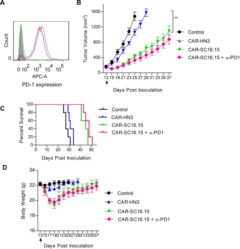
Open access
or isotype control HN3 (a human antibody that targets analysis, a PD-1 inhibitory antibody (in-house made as
J Immunother Cancer: first published as 10.1136/jitc-2020-000785 on 17 June 2020. Downloaded from http://jitc.bmj.com/ on June 30, 2022 by guest. Protected by copyright.
glypican-3, sequence from patent US20140044714), the scFv-hFc format, using nivolumab VH and VL sequences)
CD8α hinge (NP_001139345, a.a. 138–182), CD8α trans- was incubated with CAR-T cells and the antibody binding
membrane region (NP_001139345, a.a. 183–210), CD28 was detected by APC- conjugated goat-anti-
human IgG
intracellular domain (NP_006130.1, a.a. 180–220), 4-1BB (Jackson ImmunoResearch, West Grove, Pennsylvania).
intracellular domain (NP_001552.2, a.a. 214–255), CD3ζ
intracellular domain (NP_932170, a.a. 52–164), internal In vitro cytotoxicity assay
ribosome entry site (from encephalomyocarditis virus), In vitro cell killing activity of the bispecific antibody and
and a red fluorescent protein mScarlet- I. The amino CAR- T cells was assessed by firefly luciferase reporter
acid sequence of the CAR-HN3 cassette reads as: M ALP assay. Cancer cells were stably transduced with a lentiviral
VTALLLPLALLLHAARP DIQVQLVQSGGGLVQPGGSLRL vector that carries a luciferase reporter gene (ffLuc2).
SCAASYFDFDSYEMSWVRQAPGKGLEWIGSIYHSGSTYYN Two hundred microliters of stably transduced cells were
PSLKSRVTISRDNSKNTLYLQMNTLRAEDTATYYCAR VNM seeded on a 96-well plate (104 cells/well), with the addi-
DRFD Y WGQ G TLVTVSSS G T TTP A PRP P TPA P TIA S QPL tion of PBMC and bispecific antibody at the indicated
SLRP E ACR PAAG G AVH T RGL D FAC D IYI WAPL A GTC concentrations, or addition of CAR-T cells at the indi-
GVLL L SLV I TLY C NHR N RSK R SRL L HSD Y MNM T PRR cated amount. Untreated cells were used as control.
PGPT R KHY Q PYA P PRD FAAY R SKR G RKK L LYI F KQP The cell mixtures were incubated at 37°C for 48 hours
FMRP V QTT Q EED G CSC R FPE E EEG G CEL RVKF S RSA and then collected by spinning down. Collected cells
DAPAYQQGQNQLYNELNLGRREEYDVLDKRRGRDPE were then lysed by two rounds of freezing–thawing. The
MGGKPQRRKNPQEGLYNELQKDKMAEAYSEIGMKGE released luciferase activity was measured to represent the
RRRG K GHD G LYQ G LST ATKD T YDA L HMQ A LPPR cell viability. The in vitro cytotoxicity assay was repeated
(restriction enzyme sites underlined, HN3 in italic). CAR- three times and PBMC from a total of three donors was
SC16.15 had the same sequence except that HN3 part was tested. Reported was the representative result.
replaced by SC16.15 coding sequence. Human periph-
eral blood mononuclear cells (PBMC) were isolated In vivo efficacy study
from the whole blood of healthy donors (Wuhan Blood Five million H446 and A431 (DLL3) cells were subcuta-
Center) by a Ficoll separation method (Stem Cell Tech- neously injected into NOD/SCID/IL2Rg(null) (NSG)
nologies, Vancouver, British Columbia, Canada). PBMC mice. After the tumor formed and reached the size of
were cultured in RPMI 1640 medium supplemented with 100–200 mm3, the treatment was started by splenocyte
200 IU/mL human recombinant interleukin (IL)-2 and depletion with 200 mg/kg cyclophosphamide (Sigma-
activated by Dynabeads CD3/CD28 human T- activator Aldrich, St. Louis, Missouri). One day later, mice were
(Cat. 11 131D, ThermoFisher, Waltham, Massachusetts) intraperitoneally injected with 10 millions of PBMC cells
for 3 days according to the manufacturer’s instruction. or 5 millions of CAR-T cells, and intravenously injected
The activated PBMC were transduced with lentivirus with bispecific antibody and PD-1 antibody. The bispe-
carrying the CAR cassette for 24 hours. After trans- cific antibody was given two times at the indicated time
duction, refreshed medium containing 200 IU/mL of point, and PD-1 antibody was given once every week.
IL-2 was used to culture the CAR-T cells for additional Tumor dimensions were determined every 2 or 3 days
3 days before the functional studies. The expression of with a caliper. Tumor volume (mm3) was calculated by
CAR genes was indicated by the expression of mScarlet-I the formula: (a) × (b2) ×0.5, where a is tumor length and
protein as detected by flow cytometry. b is tumor width in millimeters. Five mice per group were
assigned. The in vivo study was repeated two times with
Flow cytometry method two different donors as the source of PBMC.
Cells were harvested by detaching with trypsin–EDTA
(ThermoFisher, Waltham, Massachusetts), washed by Statistical analysis
centrifugation, and resuspended in ice-cold phosphate All statistical analyses were conducted using GraphPad
buffer saline (PBS) containing 5% bovine serum albumin Prism5 (GraphPad Software, La Jolla, California)
(BSA). One million of cells per mL were incubated with and expressed as the mean±SEM. Comparison of two
10 µg/mL of DLL3 bispecific antibody. The antibody groups was performed using paired Student’s t-test (two
binding was detected by phycoerythrin-conjugated goat tailed). Comparisons among three or more groups were
antihuman IgG (Jackson ImmunoResearch, West Grove, performed using one-way analysis of variance. P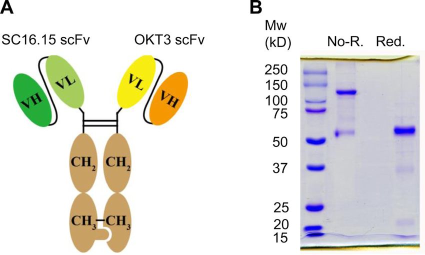
Open access
J Immunother Cancer: first published as 10.1136/jitc-2020-000785 on 17 June 2020. Downloaded from http://jitc.bmj.com/ on June 30, 2022 by guest. Protected by copyright.
Figure 1 Preparation of delta-like 3 (DLL3) bispecific
antibody. (A) Schematic diagram of the primary structure of
the DLL3 bispecific antibody. The anti-DLL3 scFv (SC16.15)
was fused with hFc knob, and the anti-CD3 scFv (OKT3) was
fused with hFc hole. (B) SDS-PAGE analysis of the purified
bispecific antibody. Two micrograms of protein were loaded
for each lane. Non-R., non-reduced condition, showing the
dimerized bispecific antibody; Red., 2-mercaptoethanol
reduced condition, showing the reduced monomer of the Figure 2 Binding properties of the delta-like 3 (DLL3)
bispecific antibody. bispecific antibody. (A) Flow cytometry analysis of the
bispecific antibody binding to different cancer cell lines. Ten
micrograms of the bispecific antibody were coincubated
OKT3 was fused with Fc hole (figure 1A). Both the knob with one million of cells. Antibody binding was detected
and hole plasmid were coexpressed in 293F cells. The by phycoerythrin-conjugated goat antihuman IgG. Shaded
heterodimerized bispecific antibody was purified via area, secondary antibody staining; dashed lines, isotype
protein A affinity chromatography and the purity was control (pooled human IgG) staining; red solid line, bispecific
accessed by SDS-PAGE (figure 1B). As expected, the non- antibody staining. (B) T cell binding analysis of the bispecific
reduced heterodimer migrated mainly as 120 kD and the antibody. Same experimental settings were used as above
reduced monomers of both knob and hole migrated as mentioned, except that the T cell line Jurkat and peripheral
blood mononuclear cells were tested. (C) Western blot
about 60 kD.
analysis of the DLL3 expression in different cancer cell lines.
Fifty micrograms of total protein from each cell lysate were
Cell binding specificity of the bispecific antibody
run on reduced SDS-PAGE, followed by anti-DLL3 antibody
Cell binding was checked on both DLL3-negative and
staining. The β-actin was used loading control. A431 (DLL3)
DLL3- positive cancer cell lines, T lymphoma cell line was an artificial cell line that was forcefully overexpressing
Jurkat, and primary human T cells (PBMC; figure 2). DLL3. The band intensity from each lane was quantified by
Since DLL3 are generally expressed at a very low level using ImageJ software, and presented as mean±SEM.
on SCLC,10 as expected, the bispecific antibody margin-
ally bound to SCLC cell line H446, H196, and H82
(figure 2A). To confirm the cell binding activity, we made antibody. As shown in figure 3, the bispecific antibody
an artificial A431 (DLL3) cell line by overexpressing efficiently killed the DLL3-positive H446 and H196 cells
DLL3 on A431 cells via lentiviral transduction and cell in a dose-dependent manner in the presence of unstimu-
sorting. A little surprised, it was difficult to get DLL3 super lated PBMC, while had little influence on the A431 cells.
high expressers (figure 2A, A431 (DLL3)), which prob- The artificial A431 (DLL3) and H82 cells were relatively
ably explained why DLL3 is generally low expressing in resistant to the bispecific antibody-mediated T cell killing.
SCLC cell lines and tissues. The binding of the bispecific
antibody to T cells was apparent (figure 2B), as shown in Tumor-suppression activity of DLL3 bispecific antibody in
both the Jurkat cell line and PBMC. To further confirm xenograft mice
the DLL3 expression in the tested cell lines, we also ran The efficient in vitro killing result of the bispecific anti-
western blot (figure 2C), which was consistent with the body encouraged us to investigate its in vivo efficacy.
cell binding data. Because the classical PD-L1/PD-1 immune checkpoint
plays significant roles in limiting the efficacy of T cell
Cytotoxicity of DLL3 bispecific antibody immunotherapy in many cancer types, and SCLC cells
Cytotoxicity of the DLL3 bispecific antibody was tested have generally low-level expression of PD-L1,19 we decided
in DLL3-negative A431 cells and DLL3-positive SCLC cell to include PD-1 blockade as a combination therapy with
lines. All the tested cell lines were stably transduced with a the DLL3 bispecific antibody for the in vivo study. First,
lentiviral vector to express the firefly luciferase gene that we confirmed the PD-L1 expression on H446 and A431
was utilized to measure the cytotoxicity of the bispecific (DLL3) cells by flow cytometry (figure 4A), and the PD-1
4 Chen X, et al. J Immunother Cancer 2020;8:e000785. doi:10.1136/jitc-2020-000785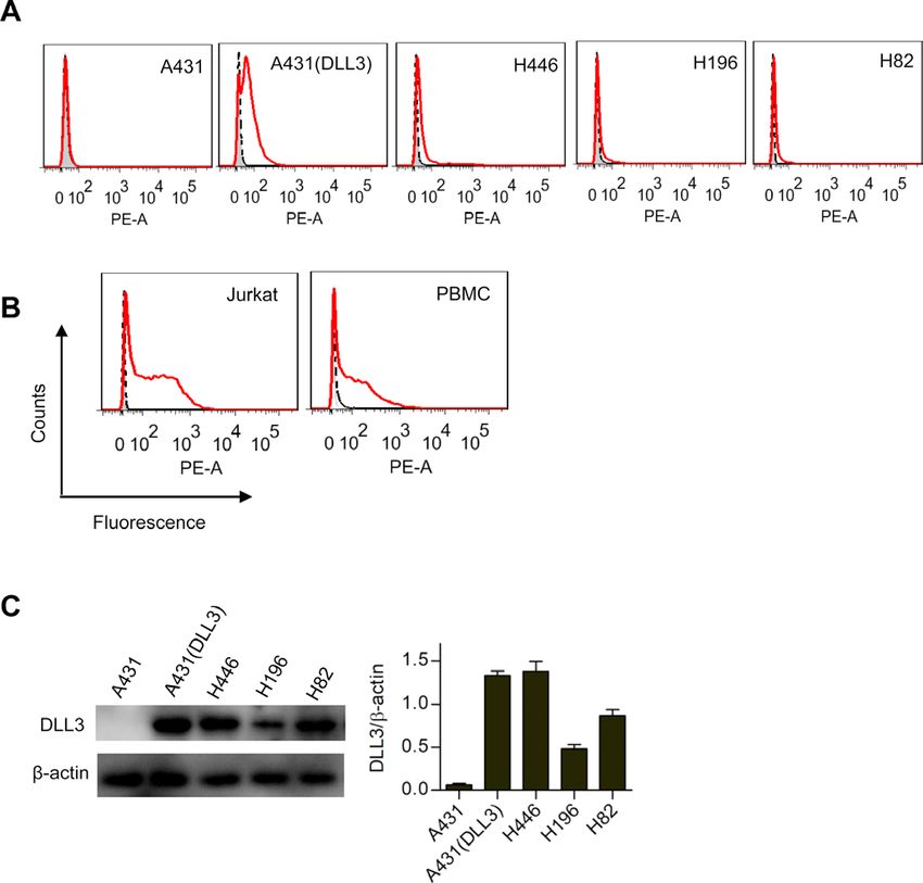
Open access
biomarker that is mainly expressing on hepatocellular
J Immunother Cancer: first published as 10.1136/jitc-2020-000785 on 17 June 2020. Downloaded from http://jitc.bmj.com/ on June 30, 2022 by guest. Protected by copyright.
carcinoma.20 The in vitro killing of DLL3 CAR-T cells was
apparent, especially on native SCLC cells H446 and H196
(figure 5C). The maximal killing was seen at 10:1 ratio
(CAR-T: tumor cells) for A431 (DLL3) and H446, and 5:1
for H196. No maximal killing was achieved for H82 at up
to 15:1 ratio.
In vivo efficacy of DLL3 CAR-T cells
The in vivo efficacy was tested on H446 model. Based on
the same rationale as in the bispecific antibody combina-
Figure 3 In vitro cell killing assay of the delta-like 3 (DLL3)
bispecific antibody. All the tested cell lines were stably
tion treatment, we also included PD-1 blockade as a combi-
transduced to constitutively express a fire fly luciferase nation in the CAR-T treatment. As shown in figure 6A,
reporter gene (ffLuc2). Ten thousand cancer cells were both the DLL3 CAR-T and the HN3 control CAR-T cells
coincubated with two hundred thousand unstimulated expressed PD-1 as examined by flow cytometry. The in
peripheral blood mononuclear cells (PBMC) cells for 48 hours vivo study showed that the DLL3 CAR- T significantly
in the presence of variable concentrations of the bispecific slowed down the tumor growth, but surprisingly, addition
antibody as indicated. The cell viability was quantified by of PD-1 inhibitory antibody did not enhance the tumor
measuring the intracellular luciferase activity. DLL3-negative growth suppression (figure 6B) or prolong the survival
A431 was served as a control of the non-specific killing by
of the mice (figure 6C), which was not the case for the
the bispecific antibody. Statistical comparisons between the
A431 control and other cell lines at each bispecific antibody
bispecific antibody combination treatment (figure 4C).
concentration was calculated and labeled on the top of the The loss of body weight in the CAR-T treatment group
bar. Data represent mean±SEM. was also apparent (figure 6D). Taken together, these
data suggested that combination of DLL3-targeted bispe-
expression on the DLL3 bispecific antibody activated cific antibody and PD-1 blockade was superior to CAR-T
PBMC (figure 4B). Then, NSG mice were subcutaneously combination.
inoculated with H446 and A431 (DLL3) cells. When the
tumor reached the size of 100–200 mm3, treatment was
started by intraperitoneal injection of 10 million PBMC Discussion
and tail vein injection of 1.0 or 0.5 mg/kg bispecific anti- SCLC is a highly aggressive disease that novel curative
body. As shown in figure 4, the bispecific antibody signifi- therapies are imperative, given that the overall prog-
cantly suppressed tumor growth in both H446 and A431 nosis remains poor. Conventional chemotherapy plus
(DLL3) cells (figure 4C), with better efficacy in A431 radiotherapy are not very effective due to the rapid onset
(DLL3) than H446, which was in contrast with the in vitro of relapse and drug resistance, and the survival rate of
killing data that the bispecific antibody had better killing patient with SCLC has not been improved over the past
on H446 than A431 (DLL3). Addition of the PD-1 inhib- three decades.2 21 In this disappointing scenario, there is a
itory antibody (in-house made as scFv-hFc format, using strong rationale to test immunotherapy that has changed
nivolumab VH and VL sequences) dramatically enhanced the paradigm of treatment of some cancer types, espe-
the antitumor activity of the bispecific antibody and cially circulating tumors. So far, ICI antibodies and DLL3-
prolonged the survival of both H446 and A431 (DLL3; targeted ADC are the two front-running forms of SCLC
figure 4E), indicating that combination therapy of DLL3 immunotherapy.6 15 22 In the current study, we aimed to
bispecific antibody with PD-1 inhibition may potentially explore the therapeutic potential of the third form of
maximize the benefit of immunotherapy in SCLC. The immunotherapy, bispecific antibody and CAR-T, both of
loss of the body weight in the treated mice was apparent which are based on the mobilization/reactivation of T
and associated with the dosage of the bispecific antibody cells.
(figure 4G). We used the classical knob-into-hole structure to make
the bispecific antibody in the heterodimerized scFv-hFc
Cytotoxicity of DLL3-targeted CAR-T cells format. DLL3-specific and CD3-specific scFv was utilized
As we have already seen the cytotoxicity and in vivo effi- as the targeting moieties. Cell binding and cytotoxicity
cacy of the bispecific antibody, it would be interesting assays showed that the bispecific antibody was able to
to see the potency of the DLL3-targeted CAR-T cells. As kill DLL3-positive cancer cells efficiently in the working
shown in figure 5, the CAR was constructed by de novo concentration of 1–1000 ng/mL (figure 3), even though
gene synthesis (figure 5A), packed into a lentiviral vector, the cell binding of the bispecific antibody was very weak
and transduced into PBMC. The transduction efficiency (figure 2A). Previous studies demonstrated that DLL3
was indicated by the parentage of the mScarlet-I posi- is remarkably low-abundance protein on the surface of
tive population (figure 5B), which was about 50% in tumor cells, in the order of only 10 000 molecules per cell,
both DLL3 and HN3 control CAR-T group. HN3 was a and H82 (the same cell line used in the current study) is
VH-domain antibody that targets glypican-3, a cell surface representative of median DLL3 expression, with 14 000
Chen X, et al. J Immunother Cancer 2020;8:e000785. doi:10.1136/jitc-2020-000785 5
Open access
J Immunother Cancer: first published as 10.1136/jitc-2020-000785 on 17 June 2020. Downloaded from http://jitc.bmj.com/ on June 30, 2022 by guest. Protected by copyright.
Figure 4 In vivo efficacy testing of the delta-like 3 (DLL3) bispecific antibody. (A) PD-L1 expression on H446 and A431 (DLL3)
cells. One million of cells was stained with PD-L1 rabbit monoclonal antibody, followed by detection with APC-conjugated goat-
anti-rabbit IgG. Shaded area, secondary antibody staining; blue curve, isotype control (pooled rabbit IgG) staining; red curve,
PD-L1 antibody staining. (B) PD-1 expression on the DLL3 bispecific antibody-stimulated peripheral blood mononuclear cells
(PBMC) cells. H446 (red curve) and A431(DLL3) cells (blue curve) were incubated with PBMC for 48 hours in the presence of
100 ng/mL DLL3 bispecific antibody, followed by flow cytometry to analyze PD-1 expression. Shaded area, unstimulated PBMC
cell staining; green curve, isotype control (pooled human IgG) staining of H446-stimulated PBMC. (C) Tumor growth curve of
native small cell lung cancer cell line NCI-H446. Five million cells were subcutaneously inoculated in each NSG mouse. After
the tumor formed and reached a size of 100–200 mm3, treatment was started. Bispecific antibody alone (0.5 mg/kg or 1.0 mg/
kg body weight), or in combination of anti-PD-1 antibody (in-house made scFv-hFc format, 5.0 mg/kg) were tested. Ten million
unstimulated human PBMC were intraperitoneally given immediately before the first intravenous delivery of the antibody. Arrows
indicated the injection time point of the bispecific antibody. The PD-1 antibody was intravenously given once every week since
the start of the treatment. Both tumor volume and body weight were measured every two or 3 days. **pOpen access
J Immunother Cancer: first published as 10.1136/jitc-2020-000785 on 17 June 2020. Downloaded from http://jitc.bmj.com/ on June 30, 2022 by guest. Protected by copyright.
Figure 5 In vitro cell killing assay of the delta-like 3 (DLL3)-targeted chimeric antigen receptor (CAR)-T. (A) Schematic
diagram of the CAR structure. Anti-DLL3 scFv (SC16.15) or isotype control (HN3, a human antibody that targets glypican-3,
sequence from patent US20140044714) was fused with the following domains, the CD8α hinge, CD8α transmembrane region,
CD28 intracellular domain, 4-1BB intracellular domain, CD3ζ intracellular domain, internal ribosome entry site (IRES, from
encephalomyocarditis virus), and a red fluorescent protein mScarlet-I. (B) Transduction efficiency analysis of the CAR-T cells,
as indicated by the expression of mScarlet-I. Untransduced PBMC was used as negative control. (C) Cell killing measurement
of the CAR-T cells. All the cancer cell lines were stably transduced to constitutively express a fire fly luciferase reporter gene
(ffLuc2). Ten thousand cancer cells were coincubated with different amount of CAR-T cells for 48 hours. The cell viability was
quantified by measuring the intracellular luciferase activity. HN3 was a negative control CAR that targets glypican-3. Data
represent mean±SEM. Statistical comparisons between the DLL3 CAR-T (SC16.15) and the control (HN3) at each CAR-T:
tumor cell ratio was calculated and labeled on the top of the bar. Comparisons between different DLL3 CAR-T ratios were also
calculated. ns, not significant, *p<0.05, ** p<0.01, ***pOpen access
J Immunother Cancer: first published as 10.1136/jitc-2020-000785 on 17 June 2020. Downloaded from http://jitc.bmj.com/ on June 30, 2022 by guest. Protected by copyright.
Figure 6 In vivo efficacy testing of the delta-like 3 (DLL3)-targeted chimeric antigen receptor (CAR)-T cells. (A) PD-1
expression on CAR-T cells. DLL3 CAR-T cells (red curve) and HN3 control CAR-T cells (blue curve) were stained with the
PD-1 inhibitory antibody (in-house made scFv-hFc format), followed by APC-conjugated goat-anti-human IgG. Shaded area,
peripheral blood mononuclear cells cell staining; green curve, isotype control (pooled human IgG) staining of the DLL3 CAR-T
cells. (B) Tumor growth curve of NCI-H446. Five million cells were subcutaneously inoculated in NSG mice. After the tumor
formed and reached a size of 100–200 mm3, treatment was started by intraperitoneal delivery of five million CAR-T cells. Arrow
indicated the time point of the CAR-T treatment. The anti-PD-1 antibody (in-house made scFv-hFc format, 5.0 mg/kg) was
intravenously given once every week since the start of the treatment. Both tumor volume and body weight were measured every
2 or 3 days. **pOpen access
in the T cells, which delivers the cross-membrane activa- designed the project and finalized the manuscript. All authors read and approved
J Immunother Cancer: first published as 10.1136/jitc-2020-000785 on 17 June 2020. Downloaded from http://jitc.bmj.com/ on June 30, 2022 by guest. Protected by copyright.
tion signals in T cells. In the current study, it was found the final manuscript.
that the potency of the DLL3-targeted CAR-T cells was Funding This work was supported by the National Natural Science Foundation of
China (31670943), the Fundamental Research Funds for the Central Universities
similar as the bispecific antibody. However, the CAR-T
(2662016PY113, 2662017PY111, and 2662019YJ013), the Applied Basic Research
cells could not be potentiated by the inclusion of PD-1 Program of Wuhan Science and Technology Bureau (2017060201010195),
inhibition, which was in contrast with that of the bispe- Bethune Medical Program of Jilin Provincial Science and Technology Department
cific antibody combination, suggesting that the fate of T (20160101125JC).
cells activated by bispecific antibody differs substantially Competing interests None declared.
from that by CAR. A similar observation was also found Patient consent for publication Not required.
that the GD2-targeted bispecific antibody had superior Ethics approval All the procedures used in the animal studies were approved by
activity than CAR- T in a melanoma xenograft mouse the Animal Care and Use Committee of Huazhong Agricultural University.
model, because the bispecific antibody did not induce Provenance and peer review Not commissioned; externally peer reviewed.
T-cell death mediated by CAR.23 In the same study, the Data availability statement Data sharing not applicable as no datasets generated
authors found that majority of high CAR density T cells and/or analyzed for this study. Data sharing not applicable as no datasets
were depleted on exposure to target cells while the bispe- generated and/or analyzed for this study.
cific antibody redirected T cells survived, and blockade of Open access This is an open access article distributed in accordance with the
PD-1 did not prevent CAR-T cell depletion. Another inter- Creative Commons Attribution Non Commercial (CC BY-NC 4.0) license, which
esting study showed that long-lasting PD-1 blockade in permits others to distribute, remix, adapt, build upon this work non-commercially,
and license their derivative works on different terms, provided the original work is
CAR-T cells by shRNA actually impaired T cell antitumor properly cited, appropriate credit is given, any changes made indicated, and the use
function, mainly because long- lasting PD-1 blockade is non-commercial. See http://creativecommons.org/licenses/by-nc/4.0/.
inhibited T cell proliferation and prevented effector T
ORCID iDs
cells from differentiating into effect memory T cells.24 Norhan Amar http://orcid.org/0000-0002-9921-824X
Clinical experience employing the combination of CAR-T Mingqian Feng http://orcid.org/0000-0002-0654-8224
and immune checkpoint blockade is in its early stages,
and there are both encouraging preliminary results and
some unsatisfactory outcomes.25 A small phase I clinical References
trial in a GD2-CAR for relapsed or refractory neuroblas- 1 Wang S, Zimmermann S, Parikh K, et al. Current diagnosis
and management of small-cell lung cancer. Mayo Clin Proc
toma did not find significant benefit of combining PD-1 2019;94:1599–622.
blockade.26 2 Pavan A, Attili I, Pasello G, et al. Immunotherapy in small-cell lung
cancer: from molecular promises to clinical challenges. J Immunother
It should be noted that the epitopes of the bispecific Cancer 2019;7:205.
antibody may have pivotal impacts on the activity.18 27 3 Povsic M, Enstone A, Wyn R, et al. Real-world effectiveness and
Bispecific antibodies that target epitopes closer to the cell tolerability of small-cell lung cancer (SCLC) treatments: a systematic
literature review (SLR). PLoS One 2019;14:e0219622.
membrane are generally advantageous as this may facili- 4 Micke P, Faldum A, Metz T, et al. Staging small cell lung cancer:
tate to form a stronger immune synapse between T cells Veterans Administration Lung Study Group versus International
Association for the Study of Lung Cancer--what limits limited
and cancer cells.27 It is unclear whether the epitope of disease? Lung Cancer 2002;37:271–6.
SC16.15 is the best fit for DLL3-targeted bispecific anti- 5 Waqar SN, Morgensztern D. Treatment advances in small cell lung
body. Therefore, following-up studies are needed to test cancer (SCLC). Pharmacol Ther 2017;180:16–23.
6 Regzedmaa O, Zhang H, Liu H, et al. Immune checkpoint inhibitors
more bispecifics that target different epitopes of DLL3 to for small cell lung cancer: opportunities and challenges. Onco
achieve the optimal therapeutic benefit of DLL3-targeted Targets Ther 2019;12:4605–20.
7 Antonia SJ, López-Martin JA, Bendell J, et al. Nivolumab alone
immunotherapy. and nivolumab plus ipilimumab in recurrent small-cell lung cancer
(CheckMate 032): a multicentre, open-label, phase 1/2 trial. Lancet
Oncol 2016;17:883–95.
8 Horn L, Mansfield AS, Szczęsna A, et al. First-Line Atezolizumab plus
Conclusion chemotherapy in extensive-stage small-cell lung cancer. N Engl J
The current study demonstrated that both DLL3-targeted Med 2018;379:2220–9.
9 Leonetti A, Facchinetti F, Minari R, et al. Notch pathway in small-cell
bispecific antibody and CAR-T cells were able to selec- lung cancer: from preclinical evidence to therapeutic challenges. Cell
tively kill DLL3-positive cancer cells in vitro and suppress Oncol 2019;42:261–73.
tumor growth in vivo. Combination of PD-1 blockade was 10 Sharma SK, Pourat J, Abdel-Atti D, et al. Noninvasive interrogation
of DLL3 expression in metastatic small cell lung cancer. Cancer Res
capable of enhancing the activity of bispecific antibody 2017;77:3931–41.
but not the CAR-T cells, suggesting its superiority over 11 Tanaka K, Isse K, Fujihira T, et al. Prevalence of delta-like protein
3 expression in patients with small cell lung cancer. Lung Cancer
CAR-T in terms of efficacy. Further studies to optimize 2018;115:116–20.
the DLL3 bispecifics are worthwhile to fully take the 12 Koshkin VS, Garcia JA, Reynolds J, et al. Transcriptomic and protein
advantage of DLL3-targeted immunotherapy. analysis of small-cell bladder cancer (SCBC) identifies prognostic
biomarkers and DLL3 as a relevant therapeutic target. Clin Cancer
Res 2019;25:210–21.
Acknowledgements The authors would like to thank Dr. Shaozhong Wei, 13 Puca L, Gavyert K, Sailer V, et al. Delta-like protein 3 expression and
president of Hubei Cancer Hospital, for discussing the project and for reviewing the therapeutic targeting in neuroendocrine prostate cancer. Sci Transl
manuscript. Med 2019;11. doi:10.1126/scitranslmed.aav0891. [Epub ahead of
print: 20 Mar 2019].
Contributors XC performed the experiments, analyzed the data, prepared the 14 Spino M, Kurz SC, Chiriboga L, et al. Cell surface Notch ligand DLL3
figures, and drafted the manuscript. NA, YZ, and CX carried out the flow cytometry. is a therapeutic target in isocitrate Dehydrogenase-mutant glioma.
CW, XY, and DW provided assistance with experiments and data analysis. MF Clin Cancer Res 2019;25:1261–71.
Chen X, et al. J Immunother Cancer 2020;8:e000785. doi:10.1136/jitc-2020-000785 9Open access
15 Saito M, Saito K, Shiraishi K, et al. Identification of candidate 21 Van Den Borg R, Leonetti A, Tiseo M, et al. Novel targeted strategies
J Immunother Cancer: first published as 10.1136/jitc-2020-000785 on 17 June 2020. Downloaded from http://jitc.bmj.com/ on June 30, 2022 by guest. Protected by copyright.
responders for anti-PD-L1/PD-1 immunotherapy, Rova-T therapy, to overcome resistance in small-cell lung cancer: focus on PARP
or EZH2 inhibitory therapy in small-cell lung cancer. Mol Clin Oncol inhibitors and rovalpituzumab tesirine. Expert Rev Anticancer Ther
2018;8:310–4. 2019;19:461–71.
16 Saunders LR, Bankovich AJ, Anderson WC, et al. A DLL3-targeted 22 Lashari BH, Vallatharasu Y, Kolandra L, et al. Rovalpituzumab
antibody-drug conjugate eradicates high-grade pulmonary Tesirine: a novel DLL3-Targeting antibody-drug conjugate. Drugs R D
neuroendocrine tumor-initiating cells in vivo. Sci Transl Med 2018;18:255–8.
2015;7:302ra136. 23 Hoseini SS, Dobrenkov K, Pankov D, et al. Bispecific antibody does
17 Rudin CM, Pietanza MC, Bauer TM, et al. Rovalpituzumab tesirine, a not induce T-cell death mediated by chimeric antigen receptor
DLL3-targeted antibody-drug conjugate, in recurrent small-cell lung against disialoganglioside GD2. Oncoimmunology 2017;6:e1320625.
cancer: a first-in-human, first-in-class, open-label, phase 1 study. 24 Wei J, Luo C, Wang Y, et al. PD-1 silencing impairs the anti-tumor
Lancet Oncol 2017;18:42–51. function of chimeric antigen receptor modified T cells by inhibiting
18 Qi J, Li X, Peng H, et al. Potent and selective antitumor activity of a proliferation activity. J Immunother Cancer 2019;7:209.
T cell-engaging bispecific antibody targeting a membrane-proximal 25 Grosser R, Cherkassky L, Chintala N, et al. Combination
epitope of ROR1. Proc Natl Acad Sci U S A 2018;115:E5467–76. immunotherapy with CAR T cells and checkpoint blockade for the
19 Carbotti G, Nikpoor AR, Vacca P, et al. IL-27 mediates HLA class treatment of solid tumors. Cancer Cell 2019;36:471–82.
I up-regulation, which can be inhibited by the IL-6 pathway, in 26 Heczey A, Louis CU, Savoldo B, et al. CAR T cells administered in
HLA-deficient small cell lung cancer cells. J Exp Clin Cancer Res combination with Lymphodepletion and PD-1 inhibition to patients
2017;36:140. with neuroblastoma. Mol Ther 2017;25:2214–24.
20 Feng M, Gao W, Wang R, et al. Therapeutically targeting 27 Li J, Stagg NJ, Johnston J, et al. Membrane-Proximal epitope
glypican-3 via a conformation-specific single-domain antibody facilitates efficient T cell synapse formation by Anti-FcRH5/
in hepatocellular carcinoma. Proc Natl Acad Sci U S A CD3 and is a requirement for myeloma cell killing. Cancer Cell
2013;110:E1083–91. 2017;31:383–95.
10 Chen X, et al. J Immunother Cancer 2020;8:e000785. doi:10.1136/jitc-2020-000785You can also read