Collagen IV 345 dysfunction in glomerular basement membrane diseases. I. Discovery of a COL4A3 variant in familial Goodpasture's and Alport diseases
←
→
Page content transcription
If your browser does not render page correctly, please read the page content below
Zurich Open Repository and
Archive
University of Zurich
Main Library
Strickhofstrasse 39
CH-8057 Zurich
www.zora.uzh.ch
Year: 2021
Collagen IV 345 dysfunction in glomerular basement membrane diseases. I.
Discovery of a COL4A3 variant in familial Goodpasture’s and Alport diseases
Pokidysheva, Elena N ; Seeger, Harald ; Pedchenko, Vadim ; Chetyrkin, Sergei ; Bergmann, Carsten ; et
al ; Gaspert, Ariana ; Kistler, Andreas D
Abstract: Diseases of the glomerular basement membrane (GBM), such as Goodpasture’s disease (GP)
and Alport syndrome (AS), are a major cause of chronic kidney failure and an unmet medical need. Col-
lagen IV345 is an important architectural element of the GBM that was discovered in previous research
on GP and AS. How this collagen enables GBM to function as a permselective filter and how structural
defects cause renal failure remain an enigma. We found a distinctive genetic variant of collagen IV345 in
both a familial GP case and four AS kindreds that provided insights into these mechanisms. The variant
is an 8-residue appendage at the C-terminus of the 3 subunit of the 345 hexamer. A knock-in mouse
harboring the variant displayed GBM abnormalities and proteinuria. This pathology phenocopied AS,
which pinpointed the 345 hexamer as a focal point in GBM function and dysfunction. Crystallography
and assembly studies revealed underlying hexamer mechanisms, as described in Companion Papers II
and III. Bioactive sites on the hexamer surface were identified where pathogenic pathways of GP and AS
converge, and, potentially, that of diabetic nephropathy (DN). We conclude that the hexamer functions
include signaling and organizing macromolecular complexes, which enable GBM assembly and function.
Therapeutic modulation or replacement of 345 hexamer could therefore be a potential treatment for
GBM diseases, and this knock-in mouse model is suitable for developing gene therapies.
DOI: https://doi.org/10.1016/j.jbc.2021.100590
Posted at the Zurich Open Repository and Archive, University of Zurich
ZORA URL: https://doi.org/10.5167/uzh-203375
Journal Article
Published Version
The following work is licensed under a Creative Commons: Attribution 4.0 International (CC BY 4.0)
License.
Originally published at:
Pokidysheva, Elena N; Seeger, Harald; Pedchenko, Vadim; Chetyrkin, Sergei; Bergmann, Carsten; et
al; Gaspert, Ariana; Kistler, Andreas D (2021). Collagen IV 345 dysfunction in glomerular basement
membrane diseases. I. Discovery of a COL4A3 variant in familial Goodpasture’s and Alport diseases.
Journal of Biological Chemistry, 296:100590.
DOI: https://doi.org/10.1016/j.jbc.2021.100590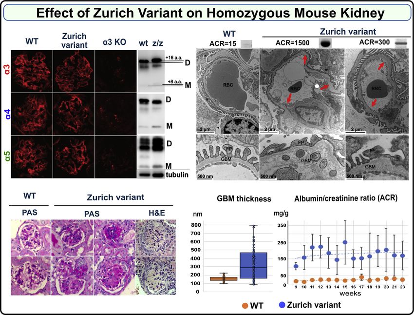
RESEARCH ARTICLE                                                                                          EDITORS’ PICK

Collagen IVα345 dysfunction in glomerular basement
membrane diseases. I. Discovery of a COL4A3 variant in
familial Goodpasture’s and Alport diseases
Received for publication, December 29, 2020, and in revised form, March 11, 2021 Published, Papers in Press, March 26, 2021,
https://doi.org/10.1016/j.jbc.2021.100590
Elena N. Pokidysheva1,2,‡, Harald Seeger3,‡, Vadim Pedchenko1,2,‡, Sergei Chetyrkin1,2,‡, Carsten Bergmann4,5,‡,
Dale Abrahamson6, Zhao Wei Cui7, Eric Delpire8, Fernando C. Fervenza9 , Aaron L. Fidler1,2,10 , Agnes B. Fogo1,11,
Ariana Gaspert12, Maik Grohmann13, Oliver Gross14 , George Haddad3, Raymond C. Harris1, Clifford Kashtan15,
A. Richard Kitching16 , Johan M. Lorenzen3, Stephen McAdoo17, Charles D. Pusey17, Marten Segelmark18 ,
Alicia Simmons1,2,10, Paul A. Voziyan1,2, Timo Wagner13, Rudolf P. Wüthrich3, Ming-Hui Zhao7,
Sergei P. Boudko1,2,18,§ , Andreas D. Kistler19,§, and Billy G. Hudson1,2,10,11,18,20,21,22, *,§
From the 1Division of Nephrology and Hypertension, Department of Medicine, 2Center for Matrix Biology, Vanderbilt University
Medical Center, Nashville, Tennessee, USA; 3Nephrology Division, University Hospital Zurich, Zurich, Switzerland; 4Department of
Medicine and Nephrology, University Hospital Freiburg, Freiburg, Germany; 5Medizinische Genetik Mainz, Limbach Genetics,
Mainz, Germany; 6Department of Anatomy and Cell Biology, University of Kansas Medical Center, Kansas City, Kansas, USA;
7
 Renal Division, Peking University First Hospital, Beijing, PR China; 8Department of Anesthesiology, Vanderbilt University School of
Medicine, Nashville, Tennessee, USA; 9Division of Nephrology and Hypertension, Mayo Clinic, Rochester, Minnesota, USA;
10
   Aspirnaut Program, 11Department of Pathology, Microbiology and Immunology, Vanderbilt University Medical Center,
Nashville, Tennessee, USA; 12Department of Pathology and Molecular Pathology, University Hospital Zurich, Zurich, Switzerland;
13
   Medizinische Genetik Mainz, Limbach Genetics, Mainz, Germany; 14Clinic of Nephrology and Rheumatology, University Medical
Center Goettingen, University of Goettingen, Goettingen, Germany; 15Division of Pediatric Nephrology, University of Minnesota
Medical School and Masonic Children’s Hospital, Minneapolis, Minnesota, USA; 16Centre for Inflammatory Diseases, Monash
University Department Medicine, Nephrology, Monash Health, Clayton, VIC, Australia; 17Centre for Inflammatory Disease,
Imperial College London, London, UK; 18Department of Biochemistry, Vanderbilt University, Nashville, Tennessee, USA;
19
   Department of Internal Medicine, Kantonsspital Frauenfeld, Frauenfeld, Switzerland; 20Center for Structural Biology,
21
   Department of Cell and Developmental Biology, 22Vanderbilt-Ingram Cancer Center, Vanderbilt University, Nashville, Tennessee,
USA
Edited by Gerald Hart

   Diseases of the glomerular basement membrane (GBM),                           conclude that the hexamer functions include signaling and
such as Goodpasture’s disease (GP) and Alport syndrome (AS),                     organizing macromolecular complexes, which enable GBM
are a major cause of chronic kidney failure and an unmet                         assembly and function. Therapeutic modulation or replace-
medical need. Collagen IVα345 is an important architectural                      ment of α345 hexamer could therefore be a potential treatment
element of the GBM that was discovered in previous research                      for GBM diseases, and this knock-in mouse model is suitable
on GP and AS. How this collagen enables GBM to function as a                     for developing gene therapies.
permselective filter and how structural defects cause renal
failure remain an enigma. We found a distinctive genetic
variant of collagen IVα345 in both a familial GP case and four                      Diseases of the glomerular basement membrane (GBM)
AS kindreds that provided insights into these mechanisms. The                    are a major cause of chronic kidney disease, a health
variant is an 8-residue appendage at the C-terminus of the α3                    problem affecting about 10% of the global population and
subunit of the α345 hexamer. A knock-in mouse harboring the                      an unmet medical need (1, 2). Prominent diseases are
variant displayed GBM abnormalities and proteinuria. This                        diabetic nephropathy (DN), Alport syndrome (AS), and
pathology phenocopied AS, which pinpointed the α345 hex-                         Goodpasture’s disease (GP) that are characterized by
amer as a focal point in GBM function and dysfunction.                           morphological abnormalities in GBM, ranging from
Crystallography and assembly studies revealed underlying                         thickening in DN to multilamellations in AS and ruptures
hexamer mechanisms, as described in Boudko et al. and Ped-                       due to specific GBM attack by antibodies in GP (3–5).
chenko et al. Bioactive sites on the hexamer surface were                        These abnormalities involve structural alterations in
identified where pathogenic pathways of GP and AS converge                        collagen IV, the major GBM component. How collagen IV
and, potentially, that of diabetic nephropathy (DN). We                          enables GBM to function as a permselective filter and how
                                                                                 its structural alterations cause GBM abnormalities and
                                                                                 dysfunction remain an enigma. New insights into these
‡
§
  Equal contribution authors.                                                    mysteries were revealed in the present study of a
  Co-senior authors.
* For correspondence: Billy G. Hudson, billy.hudson@vumc.org.
                                                                                 distinctive genetic variant of collagen IV, uniquely

                                                                                                                       J. Biol. Chem. (2021) 296 100590      1
© 2021 THE AUTHORS. Published by Elsevier Inc on behalf of American Society for Biochemistry and Molecular Biology. This is an open access article under the CC
BY license (http://creativecommons.org/licenses/by/4.0/).
EDITORS’ PICK: Pathobiology of collagen IV α345

associated with both AS and GP diseases, coupled with            gene that was found in both a familial GP case and four
crystallography and animal studies.                              kindreds of AS provided a unique opportunity to elucidate
   Collagen IV is a family of six homologous α-chains,           pathogenic mechanisms of these two GBM diseases.
which are distributed within basement membranes (BMs)
that underlie the epithelial architecture of the nephron. In     Results
pioneering studies of the GBM over 50 years ago, collagen
IV was identified as a novel collagen and shown to be             A distinctive variant of α3(IV) collagen (Zurich (Z)-variant) was
structurally altered in DN (6–12). It was first characterized     found in familial GP and Alport patients
as a supramolecular network of triple helical protomers
                                                                   Clinical histories of two patients with familial GP disease
composed of α1 and α2 chains (13, 14). In subsequent
                                                                 harboring the Z-variant
studies of the GBM in GP and AS, four additional chains
were discovered (α3, α4, α5, and α6) (4, 15–21). The six α-         A 24-year-old man (T.A.) presented with pulmonary-renal
chains coassemble into protomers with three distinct mo-         syndrome. His serum was positive for anti-GBM antibodies
lecular compositions: α121, α345, and α565, which in turn        at 151 U/ml (reference range:
EDITORS’ PICK: Pathobiology of collagen IV α345

Figure 1. Immunochemical and genetic analyses of index patient with Zurich variant. A, binding of T.A. circulating autoantibodies is restricted to the
α3NC1 domain of human collagen IV, while pooled serum from eight sporadic GP cases (GPc) also reacted to α5NC1. Normal human serum does not react
to any of NC1 domains as depicted by the dashed line (mean plus 3 × SD). B, index patient autoantibodies bind immunodominant epitopes EA and EB of
α3NC1, but not the parental α1NC1 domain used to create α1/α3 chimeric proteins bearing EA and EB epitopes. C, serum from the index patient reacts with
denatured (D), but not native (N) NC1 hexamers purified from human (hGBM) and bovine (bGBM) glomerular basement membrane (left panel). This pattern
is identical to the reactivity of pooled serum of eight sporadic GP patients (right panel). NC1 hexamer from bovine placenta (bPBM), which is composed of
GP nonreactive α1 and α2NC1 subunits served as negative control. D, the Zurich variant does not directly affect antigenicity of the α3NC1 monomer. Serum
from the index patient and from three sporadic GP patients (GP-1, GP-2, GP-3) displayed similar reactivity to the α3NC1 domain and α3NC1 domain variant
(α3-Z), but does not react with either α1NC1 domain or α1NC1 chimera (α1-Z) bearing the eight residues extension from the α3NC1 variant of the index
patient. These results suggest that Zurich appendage does not represent a neoepitope for patient autoantibodies or affect the presentation of immu-
nodominant EA and EB epitopes in the α3NC1 monomer. All bar graphs with T bars (A–D) indicate the means and standard errors. E, a heterozygous COL4A3
variant (rs765655100: c.5010_*del(p.His1670_*167delinsGln*9)) was discovered by next-generation sequencing in two patients with familial Goodpasture’s

                                                                                                                  J. Biol. Chem. (2021) 296 100590     3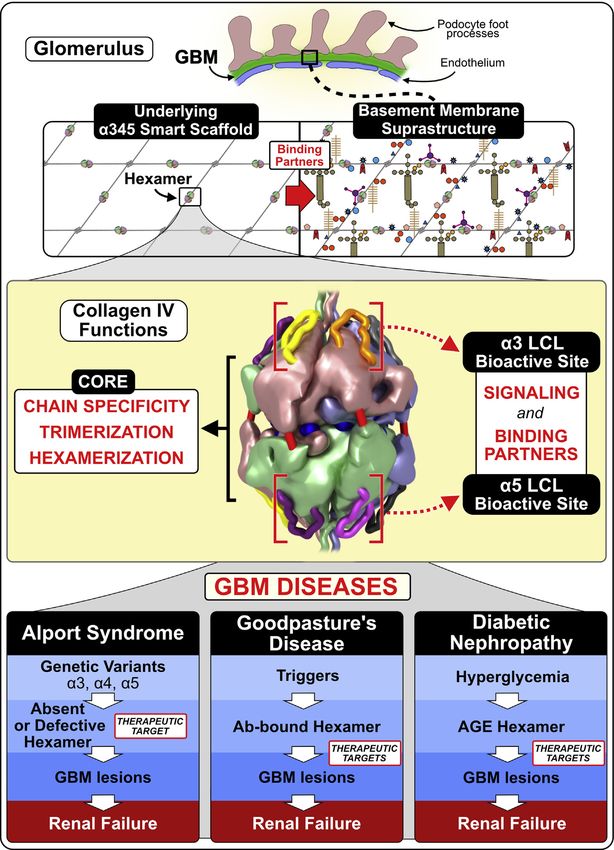
EDITORS’ PICK: Pathobiology of collagen IV α345

261 cases from five cohorts, which revealed the absence of the                  hearing loss had been previously attributed to job-related
Zurich variant in these patients (Fig. 2A and Supplementary                    noise exposure.
Section 3.1). Also, the variant was absent in three patients                      In family H (Arabic origin), the Alport genetic panel was
from a previously reported kindred of familial GP (43)                         ordered due to microhematuria (50 erythrocytes/μl) in a 10-
(Supplementary Section 3.2; Fig. S2, Table S1). However, the                   year-old girl with normal renal function (creatinine 42 μmol/
Zurich variant was found in a general population database                      l) and no proteinuria. The girl’s mother also has asymptomatic
(gnomAD; 16 out of ca. 124,000 individuals) of individuals                     microhematuria but no proteinuria and normal renal function.
with unknown health profile and in a clinical database (NCBI                    Her father suffers from recurrent urolithiasis, but has normal
ClinVar; 32 cases out of ca. 100,000 individuals)                              renal function, no proteinuria, and no microhematuria except
(Supplementary Section 3.3). Extrapolation of the gnomAD                       during symptomatic kidney stone episodes. The rs765655100
database resulted in an estimate of about one million people                   variant was found in the daughter and her father (who has no
worldwide harboring the Zurich variant (Fig. 2A).                              hematuria) but not in the mother (who has microhematuria),
                                                                               while an additional COL4A4 variant (c.1321_1369+3del, pre-
   Clinical histories of patients with Alport syndrome harboring the           viously associated with familial hematuria/AS) was identified
Z-variant                                                                      in the daughter and her mother, but not in the father. Neither
   The Z-variant was also identified in six non-GP individuals                  the index patient nor her parents reported hearing problems.
with variable renal phenotypes. These individuals were iden-                      In family U (mother Caucasian, father Turkish), a 7-year-old
tified in a cohort of patients subjected to targeted Next-                      girl with normal renal function (creatinine 0.37 mg/dl) and
Generation Sequencing of COL4A3, COL4A4, and COL4A5                            microhematuria (150 erythrocytes/μl) but no proteinuria
genes as part of commercial genetic screening panels                           (urinary protein 86 mg/g creatinine; urinary albumin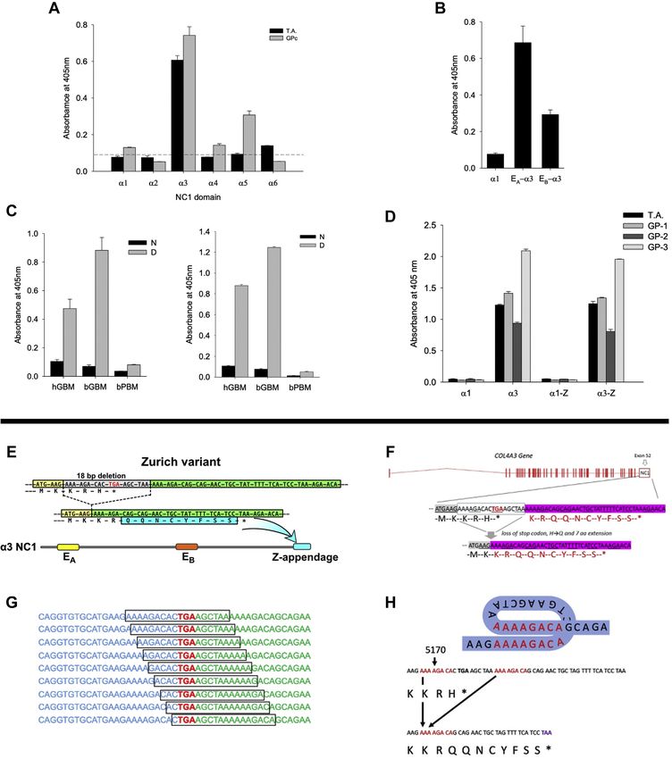
EDITORS’ PICK: Pathobiology of collagen IV α345

Figure 2. A genetic variant of α3(IV) collagen (Zurich variant) associated with familial Goodpasture’s disease and Alport syndrome is distinct
among the 1700 known Alport variants. A, Zurich variant is associated with both familial Goodpasture’s disease (GP) and Alport syndrome. This variant
was a risk factor in developing familial Goodpasture’s disease in two Zurich patients (red pie chart wedge and red circle, n = 2). In several cohorts of GP
patients from different countries, the presence of Zurich variant was not detected either by Sanger sequencing of exons 48 to 52 (Switzerland, n = 11;
Sweden, n = 15; China, n = 171; and UK, n = 29) or whole exome/genome sequencing (USA, n = 36) as shown in the gray pie chart. However, the Zurich
variant was detected in patients diagnosed with familial hematuria or suspected Alport syndrome (tan circle, n = 6) and in the general population with
unknown phenotype (dark blue circle, n = 48). The extrapolation of the data set to the world population resulted in estimated one million people carrying
the Zurich variant, and thus classified as Alport Syndrome. Therefore, the Zurich variant places them at risk of progressing to renal failure and developing
Goodpasture’s disease (light blue circle). B, the number and location of 1700+ genetic Alport-associated variants (indicated by yellow dots, zoom in to see
individual dots) in the α3, α4, and α5 chains of collagen IV (left). Pathogenic variants cause either loss of α345 protomers from the GBM or assembly of
defective α345 protomers that can incorporate into the GBM, causing a broad spectrum of GBM phenotypes. Significant numbers of Alport-associated
variants occur within α3, α4, and α5 NC1 domains (right). Zurich variant α3NC1 monomer with an 8-amino acid Z-appendage is shown at top right.
Analogous to Zurich variant, a novel variant α5NC1 monomer with a C-terminal 74-amino acid appendage (59) is shown at bottom right. Because these two
variants resulted in a C-terminal extension of the protein polypeptide chain, they stood out among over 1700 known variants in the COL4A3, COL4A4, and
COL4A5 genes associated with Alport syndrome. The Z-appendage incorporated into the collagen IV scaffold of the GBM and served as a pathogenic
reporter group that identified a new therapeutic target (vide infra).

                                                                                                                    J. Biol. Chem. (2021) 296 100590     5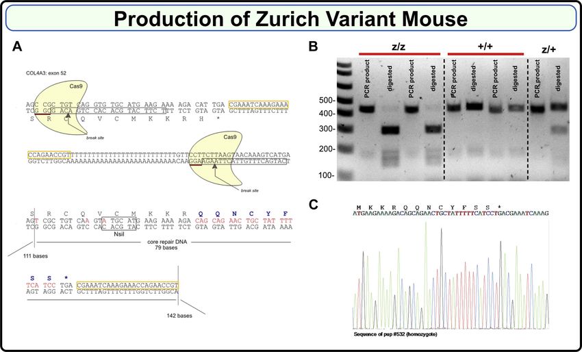
EDITORS’ PICK: Pathobiology of collagen IV α345

variant encodes a 74-residue on the α5NC1 domain (Fig. 2B).                  RNAs located within exon 52 of the COL4A3 gene were used.
Importantly, the Z-appendage is distinct among the hundreds                  Each guide RNA consisted of a 20-base DNA stretch
of variants for several reasons: 1) it occurred in patients with             (TCTTCATGCACACCTGACAG and CATGACTTTGTT
AS and in patients with familial GP disease and therefore,                   ACTTAAGA, boxed in Fig. 3A) directly preceding an NGG as
could provide insights into the pathogenesis of both diseases.               a proto-spacer adjacent motif. A single-strand oligonucleotide
2) Its location on the surface of the α345 hexamer places it in              repair DNA consisting of a 79 base repair fragment core and
juxtaposition with the GP epitopes and T-cell receptor epitope               flanking 111 and 142 base homology arms was designed
(27, 44–46), based on a predicted α345 hexamer model (22).                   (Fig. 3A). As shown in Figure 3A, mutations were introduced
The convergence of these pathogenic pathways suggests that a                 to substitute His1169 with eight new amino acids:
narrow site on the surface of the α345 hexamer is critical in                QQNCYFSS; disrupt the PAM sequences; introduce a unique
pathogenesis of GP and AS; 3) the Z-appendage is a non-                      NsiI restriction site for genotyping (Fig. 3, B and C); and
truncating variant, thus can incorporate into the GBM, and 4)                eliminate the polyT stretch. Two separate lines were estab-
its impact on hexamer function may serve as a vanguard for                   lished and backcrossed to C57BL6/J mice for four generations
understanding the unknown mechanisms of numerous other                       to eliminate possible off target events. Homozygote animals on
variants nearby on the hexamer surface. Collectively, these                  C57BL/6 background were then generated and line one was
features prompted us to assess the pathogenicity of the Z-                   expanded and studied. Mice were viable, fertile, and born in
appendage in a mouse model and to elucidate the underpin-                    the expected Mendelian ratios. For additional details on mouse
ning biology and chemistry by solving the crystal structure and              genotyping, see Supplementary Section 5 and Figure S5.
assembly mechanism of the α345 hexamer.
                                                                             Zurich variant phenocopies features of Alport syndrome in a
Generation of the Zurich variant knock-in mouse                              knock-in mouse (Col4a3z/z), revealing the α345 hexamer as a
  To assess pathogenicity, we developed a mouse harboring                    focal point in GBM function and dysfunction
the Zurich variant using CRISPR/Cas9 editing technology. To                    Homozygous knock-in mice (Col4a3z/z) on a C57BL6
increase the DNA break efficiency, two overlapping guide                      background were viable, fertile, and born in the expected

Figure 3. Production of the Zurich variant mouse. A, Zurich variant mouse CRISPR/Cas9 design scheme. Guide RNAs consisting of 20 base pairs each are
boxed; break sites are indicated. Lower part shows repair DNA, which introduces new eight amino acids. B, PCR products of 460 bp in wild type (+/+) and
446 bp in the mutant (z/z) were generated using genotyping primers (refer to the Experimental procedures section for the sequences). Mutant PCR product
had NsiI restriction site, while wild type did not. PCR products were then digested with NsiI enzyme resulting in appearance of 300 bp band for the
mutant. Heterozygous (z/+) mice had both undigested and digested bands. C, representative Sanger sequencing of gDNA isolated from homozygous F2
Zurich variant mouse.

6 J. Biol. Chem. (2021) 296 100590
EDITORS’ PICK: Pathobiology of collagen IV α345
Figure 4. Zurich variant knock-in homozygous mouse (Col4a3z/z mouse) phenocopies features of Alport syndrome in GBM. Immunofluorescence
staining of renal sections demonstrates deposition of genetically modified α3 chain along with native α4 and α5 chains within collagen IV α345 scaffold in a
Col4a3z/z mouse (top left). This scaffold did not form in the kidney of Col4a3 KO mouse lacking α3 chain (top left). The Zurich variant also did not interfere
with hexamer assembly and formation of sulfilimine cross-links as shown by western blot using collagen IV chain-specific antibodies (top left, D indicates
dimer and M indicates monomer). The shift in the position of mutant α3 bands relative to the control is due to the difference in sequence length. Samples
were normalized on kidney weight and tubulin (bottom of the western blot). Histological analysis of kidney sections revealed varying degree of glomerular
sclerosis in 20% to 70% of the glomeruli in the Col4a3z/z mice (periodic acid–Schiff (PAS)-stained representative glomeruli are shown, magnification 600×)
and occasional formation of crescents (bottom left, H&E staining). Representative transmission electron microscopy (TEM) images of the glomerular capillary
loops from two different Col4a3z/z mice and a wild-type control mouse are shown at the top right. In Col4a3z/z mice exhibiting high albuminuria (see
corresponding urine albumin-to-creatinine ratio (ACR) values and albumin SDS-PAGE bands above the TEM images), the glomerular basement membrane
(GBM) was irregularly thinned and thickened, lamellated, and occasionally split with foot process effacement. Animals with moderate albuminuria
demonstrated mainly irregular thickening of the GBM (arrows and higher magnification for GBM structure). GBM thickness (measured from two glomeruli
with five images each, from each mouse) was increased in WT (n = 2) versus Col4a3z/z (n = 3) mice (bottom middle) (p < 0.001, Student’s t test). Col4a3z/z
mice (n = 6) had significantly elevated ACR compared with WT mice (n = 5) (bottom right). Both experimental groups included male and female mice.
Mendelian ratios. The deposition of collagen IVα345 scaffold to variable GBM thickening (Fig. 4). The majority of Zurich mice
the GBM assessed by the immunofluorescence was unaffected developed moderate albuminuria from 9 to 23 weeks (Fig. 4
in the Col4a3z/z mice. Hexamer formation and cross-linking and Fig. S7), while two mice displayed a rapid tenfold in-
was also unaffected. Direct indication that the Z-appendage crease in urine albumin-to-creatinine ratios at 14 to 16 weeks
incorporated into the scaffold was obtained from the band (Fig. S8), demonstrating variability in the appendage effects.
shift of the α3 NC1 domain derived from Col4a3z/z mouse No circulating α3NC1-specific antibodies were detected
kidney. (Fig. 4 and Supplementary Section 6; Fig. S6). (Fig. S9). Aged heterozygous Col4a3+/z mice also displayed
Histological analysis of kidney sections revealed glomer- irregular thickened GBM and mild but significant albuminuria
ulosclerosis with rare crescents in homozygous Col4a3z/z mice (Fig. 5). This heterozygous phenotype is distinct from the
(Fig. 4). The GBM was irregular in thickness, lamellated with heterozygous knock-out model, which lacks the phenotype
occasional splitting and with podocyte foot processes efface- (47, 48). The difference highlights the toxicity of the Z-
ment while morphometry revealed substantial and widely appendage in the function of the collagen IVα345 scaffold.
J. Biol. Chem. (2021) 296 100590 7EDITORS’ PICK: Pathobiology of collagen IV α345 Figure 5. Zurich variant knock-in heterozygous mouse (Col4a3z/+ mouse) shows pathologic phenotype in kidneys at 1 year of age. Histological analysis of periodic acid–Schiff (PAS)-stained kidney sections (×400) revealed varying degree of glomerular sclerosis (top left). Representative transmission electron microscopy (TEM) images of the glomerular capillary loops from two different Col4a3z/+ mice and a wild-type control mouse are shown at the bottom. The glomerular basement membrane (GBM) was irregularly thickened in Col4a3z/+ mice (red arrows; TEM images scale bars are 1 μm). One-year-old Col4a3z/+ mice (n = 12) exhibited mild albuminuria compared with the age-matched WT mice (n = 9). Albumin-to-creatinine ratios are represented in graph (top right). Both experimental groups included male and female mice. The p value was 0.04. Collectively, these findings revealed that the Z-appendage Discussion rendered the α345 hexamer defective causing a wide range of In the present study of two rare diseases, familial GP and ultrastructural abnormalities in the GBM, proteinuria, and AS, a genetic collagen IVα345 variant encoding a short 8- glomerulosclerosis. Animals with high level of albuminuria residue appendage, common to both diseases, was found to displayed GBM changes characteristic for AS while moder- serve as a beacon into the inner workings of the GBM. The ately affected mice had GBM thickening. The mild phenotype appendage is pathogenic in Zurich mice, causing ultrastruc- observed in the heterozygous mice recapitulates the dominant tural abnormalities in the GBM, albuminuria, and glomer- trait in humans and suggests that defective α345 hexamer ulosclerosis. These features phenocopied the wide spectrum of “signals” the pathological changes. In contrast to knock-out glomerular phenotypes in human AS, which supports the mouse models that eliminate the collagen IVα345 scaffold recent nomenclature of COL4A3, COL4A4, and COL4A5 from basement membranes, the Zurich mouse provides variants as AS (37). Moreover, the prevalence of the Z-variant trackability to discover functions of the α345 scaffold at a is estimated to be approximately one million people world- specific site and to discover pathogenic mechanisms. The wide, placing them at risk of progressing to renal failure and absence of α3NC1 autoantibodies indicates that the developingGP (Fig. 2A). appendage is not causal, but a risk factor in GP disease. The The crystal structure and assembly mechanisms of the Z-appendage pathogenicity, in both homozygous and het- α345 hexamer, as presented in Boudko et al. (49) and Ped- erozygous mice, pinpointed the α345 hexamer as a critical chenko et al. (50), provided a framework to interpret how the structure, enabling the GBM to function as a permselective Z-appendage, a representative AS variant, played a role in AS filter. 8 J. Biol. Chem. (2021) 296 100590
EDITORS’ PICK: Pathobiology of collagen IV α345

Figure 6. Z-appendage can participate in multiple chemical interactions and thus can contribute to AS and GP pathogenesis. A, the Z-appendage is
an 8-residue extension of the α3 chain replacing the C-terminal histidine residue of the native structure. It is located at the apex of α3NC1 monomer, in
juxtaposition with EA and EB hypoepitope loops (left). The appendage is flexible and can assume multiple conformations as predicted by molecular dy-
namics (MD) simulations (left). Z-appendage features a cysteine residue with free sulfhydryl group (middle). This group can participate in multiple reactions
resulting in posttranslational modifications (PTMs), complexes with metal ions or small molecule drugs, and disulfide bonds with proteins (right) (49, 50). B,
the appendage has been predicted to assume multiple configurations (left); not shown is the Z-appendage of the opposing α345 trimer. Because of its
relatively short length, the expected impact of Z-appendage is on the structure and/or function of a specific area within the α345 hexamer including the EA

                                                                                                                     J. Biol. Chem. (2021) 296 100590      9
EDITORS’ PICK: Pathobiology of collagen IV α345
and a possible structural risk factor for GP (49, 50). Z- Whole-genome libraries were prepared from genomic DNA
appendage features a cysteine residue with free sulfhydryl with the NEBNext Ultra II DNA Library Prep Kit for Illumina
group. This group can participate in multiple reactions per manufacturer’s instructions. The DNA was fragmented on
resulting in posttranslational modifications (PTMs), com- the Covaris LE220 targeting an average insert size of 400 to
plexes with metal ions or small molecule drugs, and disulfide 500 bp. The blunt ends of each sample were end repaired,
bonds with proteins (Fig. 6A and Boudko et al. 49) (51, 52). adenylated for adaptor ligation, ligated to standard Illumina
The appendage is juxtaposed with the GP hypoepitopes adapters, and PCR amplified.
forming common “hotspots” of dysfunction, called the LCL Whole-exome libraries were generated from genomic DNA
site (49). This convergence of pathogenic pathways indicates extracted from FFPE kidney biopsies using the Exome
that the LCL site is bioactive, having functions that include captured with IDT xGen Exome Research Panel library (IDT).
signaling and organizing macromolecular complexes (49,
50). Since the LCL bioactive site is conformationally plastic Whole-exome and whole-genome data analysis
(50), its functions can be perturbed by the Z-appendage and
Both whole exome sequence and whole genome sequence
other genetic variants that occur in the hexamer in AS
were subjected to DNA sequencing on the NovaSeq Illumina
(Fig. 6B), endogenous and exogenous triggers in GP
platform, and genotype calls were made at targeted bases as
(Fig. 6C), and hyperglycemia in DN (Fig. 7 and Boudko et al.
described below (Zurich section). All targeted bases of
49). Therefore, we conclude that the α345 hexamer is a focal
COL4A3 exon 52 in WES experiments show ≥20 independent
point with bioactivity, enabling GBM morphology and its
reads in all samples and were subject for de novo mutation
function as a permselective filter, which can be altered in
detection using human reference genome hg19. Average
GBM diseases.
sequencing depth for all bases in targeted region (COL4A3
Collectively, these findings demonstrate that the α345 hex-
exon 52) in WGS experiments was at least ×50.
amer is a promising target for therapeutic intervention.
Attractive therapeutic strategies for AS include protein
replacement and pharmacological chaperones, because the Library sequencing
GBM is directly accessible to protein delivery via the blood- Captured libraries were sequenced using the Illumina
stream. Recent advances in delivery of full-length recombinant NovaSeq 6000 system (Illumina) with paired end reads of
laminin molecules to the GBM (53) set up a possibility that a 151 bp according to the manufacturer’s protocols. Raw reads
full-length or mini-α345 protomer can be delivered thera- in FASTQ format from WES or WGS were aligned to the hg19
peutically to the glomerulus, where it can oligomerize forming reference genome using the Burrows–Wheeler Aligner (BWA;
the collagen IVα345 scaffold in the GBM (50). Moreover, http://bio-bwa.sourceforge.net/). Duplicates were removed
because a significant number of hypomorph variants occurs with Picard (http://picard.Sourceforget.net). WES and WGS
within the hexamer (49), the multiple pores, crevices, and data were analyzed using GATK INDEL calling algorithm
cavities of the α345 hexamer can be potential targets for (GATK (http://www.broadinstitute.org/gatk/) following the
pharmacological chaperone interventions to correct misfold- guidelines provided in the user manuals.
ing or stabilize the protein and reduce degradation. In addi-
tion, the knock-in mouse is a promising model for the SNPs filtration and annotation
development of new drugs and gene editing therapies for AS.
INDELs and the variants were filtered using GATK and
annotated using the ANNOVAR program (http://www.
Experimental procedures openbioinformatics.org/annovar/).
Human subjects
All human studies were done in accordance with the Hel- Production of reagents for immunoassays of GP patient
sinki Principles. Informed consent was obtained from all pa- autoantibodies
tients included in the studies, and the Institutional Review Recombinant human α1-α6NC1 monomers and α3/α1
Board of the Vanderbilt University Medical Center and the monomeric chimeras bearing EA and EB regions of α3NC1 were
Cantonal Ethics Committee of Zurich approved this study. purified from the culture medium of stably transfected HEK
293 cells using anti-FLAG agarose (31, 54). For the construction
Genomic DNA extraction and library preparation of mutated α3NC1 or α1NC1 domains containing an 8-residue
Genomic DNA from FFPE samples was extracted with extension on carboxy termini, we used PCR mutagenesis with
either Covaris of QIAGEN FFPE DNA extraction kits. DNA corresponding pRcX expression vectors as template (55).
from blood cells was extracted with Promega’s Wizard Introduction of the target mutations was verified by automated
Genomic DNA kit (cat# A1120). sequencing. Native collagen IV NC1 hexamers were isolated
and EB hypoepitopes and the crevice between them called the loop-crevice-loop (LCL) site. In the pathogenesis of Alport syndrome, Z-appendage can block
the RGD integrin binding site (60) or phosphorylation site (61) located in the adjacent triple helical domain, interfere with the EA and EB hypoepitope loops,
and attract toxic small molecules and metals to the crevice (inset). C, in Goodpasture’s (GP) disease, Z-appendage can sensitized α345 hexamer, a GP
autoantigen, to different second-hit triggers of GP autoantibody production such as environmental toxins or endogenous pathogenic factors, e.g., in-
flammatory response to bacterial infections, glycoxidative stress in diabetes, etc.
10 J. Biol. Chem. (2021) 296 100590EDITORS’ PICK: Pathobiology of collagen IV α345
Figure 7. The α345 hexamer is a focal point in GBM function and dysfunction with LCL sites as potential therapeutic targets in the treatment of
GBM kidney diseases. The collagen IVα345 scaffold is a major structure underlying glomerular (GBM) basement membrane. In the GBM, it is deposited by
podocytes (62) in the form of protomers that self-assemble in the presence of extracellular levels of chloride ions (63). The scaffold has been defined as a
“smart” scaffold (64) due to the presence of multiple binding sites and surfaces that allow participation in quaternary and quinary interactions (65) within
the crowded macromolecular environment of the insoluble basement membrane. The α345 hexamer is a key connection module within the collagen IVα345
scaffold. The noncollagenous (NC1) domains of individual α-chains forming the hexamer encode its specific composition and assembly via intracellular
trimerization followed by extracellular hexamerization. Quaternary and quinary interactions involving the hexamer surface may include binding partners
within the basement membrane and cell surface receptors inducing signaling. There are functional loop-crevice-loop (LCL) bioactive sites at the apices of
the hexameric structure (indicated by red square brackets) where pathogenic mechanisms for Alport syndrome, Goodpasture’s disease, and potentially
diabetic nephropathy converge. This convergence of pathogenic pathways indicates that the LCL site harbors bioactive functions, including signaling and
organizing macromolecular complexes, which underlie the GBM biology. The LCL sites are targets for genetic variants and toxic triggers causing basement
membrane abnormalities and leading to renal, pulmonary, otic, and eye disorders. Triggers are envisioned as both environmental and endogenous, e.g.,
hyperglycemia in diabetes. The LCL sites and downstream pathways are potential targets for rational design of protein replacement and small-molecule
therapies. For additional details, see Boudko et al. (49), Fig. 6.
from human and bovine GBM after collagenase digestion (55). NC1 domain isolation from mouse tissues and western
In some experiments, the GBM NC1 hexamers were denatured blotting
by treatment with 6 mol/l guanidine-HCl for 30 min at 60o C The kidneys or lungs from wild-type control and homozy-
prior to coating on ELISA plates. gous Zurich mutant mice were homogenized using metal bug
J. Biol. Chem. (2021) 296 100590 11EDITORS’ PICK: Pathobiology of collagen IV α345
beads homogenizer system in TBS buffer containing 0.1% sodium cacodylate buffer, pH 7.5 overnight. Lungs were
Tween 20 (TBST). Beads were centrifuged and precipitated inflated with 2 ml of 2% PFA plus 2% glutaraldehyde and left
material was washed with TBST four times. At the final step immersed in the same fixative solution overnight. Small pieces
precipitate was dissolved in 250 μl of collagenase digestion were cut next day. Tissues were postfixed in 1% osmium te-
buffer (55) containing 100 μg/ml of collagenase and incubated troxide, followed by dehydration through a grade series of
overnight at 37 C. Soluble portion was loaded and separated ethanol to 100%. Samples were further dehydrated in propyl-
by SDS-PAGE using 4 to 20% gradient gel, transferred to ene oxide and infiltrated and embedded in Spurr’s epoxy. 70-
nitrocellulose membranes for probing with H31, H43, and nm ultrathin sections were collected on 300 mesh copper
Mab5 antibodies. grids and stained with 2% uranyl acetate followed by Reynold’s
lead citrate. Stained sections were examined using a T-12
Mouse tissue immunofluorescence and light microscopy electron microscope (Philips/FEI) operated at 100 kV and
analysis photographed using a 2K camera (AMT).
Kidneys were cut and dropped into near-freezing pentane
(Fisher Scientific). Lungs were first inflated with 1:1 solution of Expression and purification of α3, α4, and α5 NC1 monomers
optimal cutting temperature compound (Tissue-Tek) and PBS. Recombinant human NC1 domains were amplified by PCR
For cryosectioning, tissue was embedded into the mold and from human kidney cDNA library, cloned in derivative of pRc-
4 μm sections were cut with cryostat and placed on slides. CMV mammalian expression vector that includes BM-40
Slides were pretreated with 6 M urea in 0.1 M glycine-HCl signal peptide and N-terminal FLAG tag, and transfected
buffer, pH 3.0, for 10 min (24), followed by several washes into HEK-293 cells using HEPES-calcium phosphate (Pro-
with PBS and PBS/0.2% Tween. Slides were preincubated with Fection, Promega). Stable clones were selected using neomycin
10% normal goat serum (Invitrogen) for 1 h at RT to block (0.4 mg/ml) and clones with highest levels of NC1 expression
nonspecific binding of antibodies. The following primary an- after testing by western blotting were expanded into T225
tibodies were used for antigen detections: rat anti-collagen IV culture flasks. Conditioned medium was collected from
α3 NC1 (1:250 dilution, H31), rat anti-collagen IV α4 NC1 confluent cultures two times a week and recombinant proteins
(1:250 dilution, H43), and mouse anti-collagen IV α5 NC1 were purified by passing through anti-FLAG M2-agarose
(1:250, Mab5). The H31 and H43 antibodies were from Y. Sado (Sigma) columns with subsequent elution with FLAG peptide
(Shigei Medical Research Institute). The secondary antibodies (100 μg/ml, Sigma) and concentration on ultrafiltration con-
used for immunofluorescence detection were Alexa555 goat centrators (Amicon 10MWCO, Millipore) to 2 to 4 mg/ml.
anti-rat (1:1000 dilution; Abcam) and Alexa568 goat anti- Proteins were further purified by SEC on Superdex 200 col-
mouse (1:1000 dilution; Abcam). All antibodies were diluted umn in TBS buffer (55).
in PBS/0.1% Tween and 5% normal goat serum. Slides were
incubated with primary antibodies overnight at 4 C in a hu-
Data availability
midified chamber and then washed three times in PBS/0.2%
Tween before incubating with secondary antibodies for 1 h at All data described in this article is available in the main text
RT. Negative control slide was processed similarly to experi- or supporting information.
mental slide, but without primary antibodies.
For light microscopic examination, 5-μm paraffin sections
of the lung tissue (n = 4 for each genotype) were stained with Supporting information—This article contains supporting
Periodic acid–Schiff and hematoxylin and eosin. Slides were information.
reviewed by a pathologist in a blinded fashion and the Acknowledgments—We thank Neonila Danylevych, Mohamed Rafi,
morphometric data were acquired and subjectively analyzed. and Sergey Ivanov for their technical assistance, Dr Julie Hudson for
Morphometric analysis was performed as described previously editing. We are grateful to Valeryie Luyckx for valuable clinical
(56). input concerning the management of the index patient.
The atomic coordinates and structure factors (code 6wku) have
Mouse urine samples collection and ACR measurements been deposited in the Protein Data Bank (http://wwpdb.org/). This
research used samples provided, in part, by the Imperial College
Urine samples were collected according to animal protocol
Healthcare Tissue Bank (Application R18056); we acknowledge
(M1900063-00) approved by VUMC IACUC. Albumin con-
support from the Imperial NIHR Biomedical Research Centre (UK).
centrations were measured with Albuwell M kit (Exocell, Inc)
and creatinine concentrations were determined with Creati- Author contributions—E. N. P.: Acquisition analysis and interpre-
nine Companion kit (Exocell, Inc) according to the protocols tation of data, drafted the work; V. P.: Design of the work, acqui-
provided by the manufacturer. Electrophoresis SDS-PAGE sition analysis, and interpretation of data, drafted the work; H. S.:
analysis of 3 μl urine was also performed on most samples. Conception of the work, acquisition analysis, and interpretation of
data; S. C.: Acquisition analysis and interpretation of data, drafted
the work; D. A.: Interpretation of data; Z. W. C.: Acquisition anal-
Transmission electron microscopy of mouse tissues
ysis and interpretation of data; E. D.: Design of the work, acquisition
Freshly extracted kidneys were cut in small pieces (2 × 2 analysis, and interpretation of data; F. C. F.: Acquisition and analysis
mm) and fixed in 2.5% glutaraldehyde buffered in 0.1 M of data; A. L. F.: Interpretation of data, substantively revised the
12 J. Biol. Chem. (2021) 296 100590EDITORS’ PICK: Pathobiology of collagen IV α345
work; A. B. F.: Acquisition analysis and interpretation of data; A. G.: 6. Kefalides, N. A. (1968) Isolation and characterization of the collagen from
Acquisition of data; M. G.: Acquisition of data; O. G.: Interpretation glomerular basement membrane. Biochemistry 7, 3103–3112
of data; G. H.: Acquisition of data; R. C. H.: Interpretation of data; 7. Kefalides, N. A. (1973) Structure and biosynthesis of basement mem-
SI: Acquisition analysis and interpretation of data; C. K.: Interpre- branes. Int. Rev. Connect. Tissue Res. 6, 63–104
8. Spiro, R. G. (1967) Studies on the renal glomerular basement membrane.
tation of data; A. R. K.: Interpretation of data; J. M. L.: Acquisition of
Preparation and chemical composition. J. Biol. Chem. 242, 1915–1922
data; S. M.: Acquisition and analysis of data; C. D. P.: Acquisition
9. Beisswenger, P. G., and Spiro, R. G. (1970) Human glomerular basement
and analysis of data; JS: Acquisition analysis and interpretation of membrane: Chemical alteration in diabetes mellitus. Science 168, 596–
data; M. S.: Acquisition and analysis of data; A. S.: Acquisition of 598
data; P. A. V.: Interpretation of data, substantively revised the work; 10. Hudson, B. G., and Spiro, R. G. (1972) Studies on the native and reduced
T. W.: Acquisition of data; R. P. W.: Acquisition of data; M.-H. Z.: alkylated renal glomerular basement membrane. Solubility, subunit size,
Acquisition analysis and interpretation of data; C. B.: Acquisition and reaction with cyanogen bromide. J. Biol. Chem. 247, 4229–4238
analysis and interpretation of data; A. K.: Conception of the work, 11. Spiro, R. G. (1973) Biochemistry of the renal glomerular basement
acquisition analysis, and interpretation of data; S. P. B.: Design of membrane and its alterations in diabetes mellitus. N. Engl. J. Med. 288,
the work, acquisition analysis, and interpretation of data, drafted the 1337–1342
12. Mott, J. D., Khalifah, R. G., Nagase, H., Shield, C. F., 3rd, Hudson, J. K.,
work; B. G. H.: Conception of the work, design of the work, inter-
and Hudson, B. G. (1997) Nonenzymatic glycation of type IV collagen
pretation of data, drafted the work, substantively revised the work.
and matrix metalloproteinase susceptibility. Kidney Int. 52, 1302–1312
13. Timpl, R., Wiedemann, H., van Delden, V., Furthmayr, H., and Kuhn, K.
Funding and additional information—Supported by grants
(1981) A network model for the organization of type IV collagen mole-
R01DK18381-50 and a supplement R01DK018381 (to Dr Billy G cules in basement membranes. Eur. J. Biochem. 120, 203–211
Hudson), R24DK103067 (to Dr Raymond Harris and Dr Billy G 14. Yurchenco, P. D., and Ruben, G. C. (1987) Basement membrane structure
Hudson), R01DK065138 (to Dr Billy G Hudson and Dr Paul in situ: Evidence for lateral associations in the type IV collagen network.
Voziyan), R01DK93501 (to Dr Eric Delpire), and P30DK114809 (to J. Cell Biol. 105, 2559–2568
Dr Raymond Harris), and RO1DK56942 (to Dr Agnes Fogo) from 15. McCoy, R. C., Johnson, H. K., Stone, W. J., and Wilson, C. B. (1982)
the National Institute of Diabetes and Digestive and Kidney Dis- Absence of nephritogenic GBM antigen(s) in some patients with hered-
eases. Additional support was provided by the Aspirnaut program itary nephritis. Kidney Int. 21, 642–652
from the Center for Matrix Biology at Vanderbilt University Med- 16. Butkowski, R. J., Langeveld, J. P., Wieslander, J., Hamilton, J., and Hud-
son, B. G. (1987) Localization of the Goodpasture epitope to a novel chain
ical Center. Dr Sergei Boudko was supported, in part, by start-up
of basement membrane collagen. J. Biol. Chem. 262, 7874–7877
funding from Department of Medicine, Division of Nephrology,
17. Saus, J., Wieslander, J., Langeveld, J. P., Quinones, S., and Hudson, B. G.
Vanderbilt University Medical Center. Dr Oliver Gross is supported (1988) Identification of the Goodpasture antigen as the alpha 3(IV) chain
by the German Research Foundation DFG (GR 1852/6-1). Dr of collagen IV. J. Biol. Chem. 263, 13374–13380
Carsten Bergmann is supported by the Deutsche For- 18. Barker, D. F., Hostikka, S. L., Zhou, J., Chow, L. T., Oliphant, A. R.,
schungsgemeinschaft (DFG) (BE 3910/8-1, BE 3910/9-1, and Proj- Gerken, S. C., Gregory, M. C., Skolnick, M. H., Atkin, C. L., and
ect-ID 431984000 – SFB 1453) and the Federal Ministry of Tryggvason, K. (1990) Identification of mutations in the COL4A5
Education and Research (BMBF, 01GM1903I and 01GM1903G). collagen gene in Alport syndrome. Science 248, 1224–1227
19. Gunwar, S., Saus, J., Noelken, M. E., and Hudson, B. G. (1990) Glomer-
Conflict of interest—The authors declare that there is no conflict of ular basement membrane. Identification of a fourth chain, alpha 4, of type
interest. IV collagen. J. Biol. Chem. 265, 5466–5469
20. Hostikka, S. L., Eddy, R. L., Byers, M. G., Hoyhtya, M., Shows, T. B., and
Abbreviations—The abbreviations used are: AS, Alport syndrome; Tryggvason, K. (1990) Identification of a distinct type IV collagen alpha
BM, basement membrane; DN, diabetic nephropathy; GBM, chain with restricted kidney distribution and assignment of its gene to the
locus of X chromosome-linked Alport syndrome. Proc. Natl. Acad. Sci.
glomerular basement membrane; GP, Goodpasture’s disease; PTM,
U. S. A. 87, 1606–1610
posttranslational modification.
21. Myers, J. C., Jones, T. A., Pohjolainen, E. R., Kadri, A. S., Goddard, A. D.,
Sheer, D., Solomon, E., and Pihlajaniemi, T. (1990) Molecular cloning of
alpha 5(IV) collagen and assignment of the gene to the region of the X
References chromosome containing the Alport syndrome locus. Am. J. Hum. Genet.
46, 1024–1033
1. Levin, A., Tonelli, M., Bonventre, J., Coresh, J., Donner, J. A., Fogo, A. B., 22. Khoshnoodi, J., Cartailler, J. P., Alvares, K., Veis, A., and Hudson, B. G.
Fox, C. S., Gansevoort, R. T., Heerspink, H. J. L., Jardine, M., Kasiske, B., (2006) Molecular recognition in the assembly of collagens: Terminal
Kottgen, A., Kretzler, M., Levey, A. S., Luyckx, V. A., et al. (2017) Global noncollagenous domains are key recognition modules in the formation of
kidney health 2017 and beyond: A roadmap for closing gaps in care, triple helical protomers. J. Biol. Chem. 281, 38117–38121
research, and policy. Lancet 390, 1888–1917 23. Khoshnoodi, J., Pedchenko, V., and Hudson, B. G. (2008) Mammalian
2. Bikbov, B., Perico, N., Remuzzi, G., and on behalf of the GBD Genito- collagen IV. Microsc. Res. Tech. 71, 357–370
urinary Diseases Expert Group. (2018) Disparities in chronic kidney 24. Ninomiya, Y., Kagawa, M., Iyama, K., Naito, I., Kishiro, Y., Seyer, J. M.,
disease prevalence among males and females in 195 countries: Analysis of Sugimoto, M., Oohashi, T., and Sado, Y. (1995) Differential expression of
the global burden of disease 2016 study. Nephron 139, 313–318 two basement membrane collagen genes, COL4A6 and COL4A5,
3. Hudson, B. G., Reeders, S. T., and Tryggvason, K. (1993) Type IV demonstrated by immunofluorescence staining using peptide-specific
collagen: Structure, gene organization, and role in human diseases. Mo- monoclonal antibodies. J. Cell Biol. 130, 1219–1229
lecular basis of goodpasture and Alport syndromes and diffuse leiomyo- 25. Hudson, B. G. (2004) The molecular basis of Goodpasture and Alport
matosis. J. Biol. Chem. 268, 26033–26036 syndromes: Beacons for the discovery of the collagen IV family. J. Am.
4. Hudson, B. G., Tryggvason, K., Sundaramoorthy, M., and Neilson, E. G. Soc. Nephrol. 15, 2514–2527
(2003) Alport’s syndrome, Goodpasture’s syndrome, and type IV collagen. 26. Fidler, A. L., Darris, C. E., Chetyrkin, S. V., Pedchenko, V. K., Boudko, S.
N. Engl. J. Med. 348, 2543–2556 P., Brown, K. L., Gray Jerome, W., Hudson, J. K., Rokas, A., and Hudson,
5. Naylor, R. W., Morais, M., and Lennon, R. (2021) Complexities of the B. G. (2017) Collagen IV and basement membrane at the evolutionary
glomerular basement membrane. Nat. Rev. Nephrol. 17, 112–127 dawn of metazoan tissues. Elife 6, e24176
J. Biol. Chem. (2021) 296 100590 13EDITORS’ PICK: Pathobiology of collagen IV α345
27. Pedchenko, V., Bondar, O., Fogo, A. B., Vanacore, R., Voziyan, P., Kitch- 44. Ooi, J. D., Petersen, J., Tan, Y. H., Huynh, M., Willett, Z. J., Ramar-
ing, A. R., Wieslander, J., Kashtan, C., Borza, D. B., Neilson, E. G., Wilson, athinam, S. H., Eggenhuizen, P. J., Loh, K. L., Watson, K. A., Gan, P. Y.,
C. B., and Hudson, B. G. (2010) Molecular architecture of the Goodpasture Alikhan, M. A., Dudek, N. L., Handel, A., Hudson, B. G., Fugger, L., et al.
autoantigen in anti-GBM nephritis. N. Engl. J. Med. 363, 343–354 (2017) Dominant protection from HLA-linked autoimmunity by antigen-
28. Gunwar, S., Bejarano, P. A., Kalluri, R., Langeveld, J. P., Wisdom, B. J., Jr., specific regulatory T cells. Nature 545, 243–247
Noelken, M. E., and Hudson, B. G. (1991) Alveolar basement membrane: 45. Phelps, R. G., and Rees, A. J. (1999) The HLA complex in Goodpasture’s
Molecular properties of the noncollagenous domain (hexamer) of disease: A model for analyzing susceptibility to autoimmunity. Kidney Int.
collagen IV and its reactivity with Goodpasture autoantibodies. Am. J. 56, 1638–1653
Respir. Cell Mol. Biol. 5, 107–112 46. Xie, L. J., Cui, Z., Chen, F. J., Pei, Z. Y., Hu, S. Y., Gu, Q. H., Jia, X. Y.,
29. Turner, N., Mason, P. J., Brown, R., Fox, M., Povey, S., Rees, A., and Pusey, C. Zhu, L., Zhou, X. J., Zhang, H., Liao, Y. H., Lai, L. H., Hudson, B. G., and
D. (1992) Molecular cloning of the human Goodpasture antigen demonstrates Zhao, M. H. (2017) The susceptible HLA class II alleles and their pre-
it to be the alpha 3 chain of type IV collagen. J. Clin. Invest. 89, 592–601 senting epitope(s) in Goodpasture’s disease. Immunology 151, 395–404
30. Morrison, K. E., Mariyama, M., Yang-Feng, T. L., and Reeders, S. T. 47. Cosgrove, D., Meehan, D. T., Grunkemeyer, J. A., Kornak, J. M., Sayers, R.,
(1991) Sequence and localization of a partial cDNA encoding the human Hunter, W. J., and Samuelson, G. C. (1996) Collagen COL4A3 knockout: A
alpha 3 chain of type IV collagen. Am. J. Hum. Genet. 49, 545–554 mouse model for autosomal Alport syndrome. Genes Dev. 10, 2981–2992
31. Netzer, K. O., Leinonen, A., Boutaud, A., Borza, D. B., Todd, P., Gunwar, 48. Miner, J. H., and Sanes, J. R. (1996) Molecular and functional defects in
S., Langeveld, J. P., and Hudson, B. G. (1999) The Goodpasture auto- kidneys of mice lacking collagen alpha 3(IV): Implications for Alport
antigen. Mapping the major conformational epitope(s) of alpha3(IV) syndrome. J. Cell Biol. 135, 1403–1413
collagen to residues 17-31 and 127-141 of the NC1 domain. J. Biol. Chem. 49. Boudko, S. P., Bauer, R., Chetyrkin, S. V., Ivanov, S., Smith, J., Voziyan, P.
274, 11267–11274 A., and Hudson, B. G. (2021) Collagen IVα345 dysfunction in glomerular
32. Hellmark, T., Burkhardt, H., and Wieslander, J. (1999) Goodpasture basement membrane diseases. II. Crystal structure of the α345 hexamer. J.
disease. Characterization of a single conformational epitope as the target Biol. Chem. 296, 100591
of pathogenic autoantibodies. J. Biol. Chem. 274, 25862–25868 50. Pedchenko, V., Boudko, S. P., Barber, M., Mikhailova, T., Saus, J., Har-
33. Cui, Z., Zhao, M. H., Jia, X. Y., Wang, M., Hu, S. Y., Wang, S. X., Yu, F., mange, J.-C., and Hudson, B. G. (2021) Collagen IVα345 dysfunction in
Brown, K. L., Hudson, B. G., and Pedchenko, V. (2016) Antibodies to glomerular basement membrane diseases. III. A functional framework for
alpha5 chain of collagen IV are pathogenic in Goodpasture’s disease. J. α345 hexamer assembly. J. Biol. Chem. 296, 100592
Autoimmun. 70, 1–11 51. Backus, K. M. (2019) Applications of reactive cysteine profiling. Curr.
34. Pedchenko, V., Kitching, A. R., and Hudson, B. G. (2018) Goodpasture’s Top. Microbiol. Immunol. 420, 375–417
autoimmune disease - a collagen IV disorder. Matrix Biol. 71-72, 240–249 52. Paulsen, C. E., and Carroll, K. S. (2013) Cysteine-mediated redox
35. McAdoo, S. P., and Pusey, C. D. (2017) Anti-glomerular basement signaling: Chemistry, biology, and tools for discovery. Chem. Rev. 113,
membrane disease. Clin. J. Am. Soc. Nephrol. 12, 1162–1172 4633–4679
36. Groopman, E. E., Marasa, M., Cameron-Christie, S., Petrovski, S., 53. Lin, M. H., Miller, J. B., Kikkawa, Y., Suleiman, H. Y., Tryggvason, K.,
Aggarwal, V. S., Milo-Rasouly, H., Li, Y., Zhang, J., Nestor, J., Krithivasan, Hodges, B. L., and Miner, J. H. (2018) Laminin-521 protein therapy for
P., Lam, W. Y., Mitrotti, A., Piva, S., Kil, B. H., Chatterjee, D., et al. (2019) glomerular basement membrane and podocyte abnormalities in a model
Diagnostic utility of exome sequencing for kidney disease. N. Engl. J. Med. of Pierson syndrome. J. Am. Soc. Nephrol. 29, 1426–1436
380, 142–151 54. Sado, Y., Boutaud, A., Kagawa, M., Naito, I., Ninomiya, Y., and Hudson,
37. Kashtan, C. E., Ding, J., Garosi, G., Heidet, L., Massella, L., Nakanishi, K., B. G. (1998) Induction of anti-GBM nepritis in rats by recombinant
Nozu, K., Renieri, A., Rheault, M., Wang, F., and Gross, O. (2018) Alport α3(IV)NC1 and α4(IV)NC1 of type IV collagen. Kidney Int. 53, 664–671
syndrome: A unified classification of genetic disorders of collagen IV 55. Boudko, S. P., Danylevych, N., Hudson, B. G., and Pedchenko, V. K.
alpha345: A position paper of the Alport Syndrome Classification (2018) Basement membrane collagen IV: Isolation of functional domains.
Working Group. Kidney Int. 93, 1045–1051 Methods Cell Biol. 143, 171–185
38. Savige, J., Gregory, M., Gross, O., Kashtan, C., Ding, J., and Flinter, F. 56. Polosukhin, V. V., Stathopoulos, G. T., Lawson, W. E., and Blackwell, T. S.
(2013) Expert guidelines for the management of Alport syndrome and (2005) Variability of interalveolar septal remodeling after bleomycin
thin basement membrane nephropathy. J. Am. Soc. Nephrol. 24, 364–375 treatment in mice. Ultrastruct. Pathol. 29, 53–64
39. Savige, J., Storey, H., Il Cheong, H., Gyung Kang, H., Park, E., Hil- 57. Longo, I., Porcedda, P., Mari, F., Giachino, D., Meloni, I., Deplano, C.,
bert, P., Persikov, A., Torres-Fernandez, C., Ars, E., Torra, R., Hertz, Brusco, A., Bosio, M., Massella, L., Lavoratti, G., Roccatello, D., Frasca, G.,
J. M., Thomassen, M., Shagam, L., Wang, D., Wang, Y., et al. (2016) Mazzucco, G., Muda, A. O., Conti, M., et al. (2002) COL4A3/COL4A4
X-linked and autosomal recessive Alport syndrome: Pathogenic mutations: From familial hematuria to autosomal-dominant or recessive
variant features and further genotype-phenotype correlations. PLoS Alport syndrome. Kidney Int. 61, 1947–1956
One 11, e0161802 58. Fan, H., and Chu, J. Y. (2007) A brief review of short tandem repeat
40. Pirson, Y. (1999) Making the diagnosis of Alport’s syndrome. Kidney Int. mutation. Genomics Proteomics Bioinformatics 5, 7–14
56, 760–775 59. Yamamura, T., Nozu, K., Fu, X. J., Nozu, Y., Ye, M. J., Shono, A.,
41. Gross, O., Tonshoff, B., Weber, L. T., Pape, L., Latta, K., Fehrenbach, H., Yamanouchi, S., Minamikawa, S., Morisada, N., Nakanishi, K., Shima, Y.,
Lange-Sperandio, B., Zappel, H., Hoyer, P., Staude, H., Konig, S., John, U., Yoshikawa, N., Ninchoji, T., Morioka, I., Kaito, H., et al. (2017) Natural
Gellermann, J., Hoppe, B., Galiano, M., et al. (2020) A multicenter, history and genotype-phenotype correlation in female X-linked Alport
randomized, placebo-controlled, double-blind phase 3 trial with open- syndrome. Kidney Int. Rep. 2, 850–855
arm comparison indicates safety and efficacy of nephroprotective ther- 60. Borza, C. M., Borza, D. B., Pedchenko, V., Saleem, M. A., Mathieson,
apy with ramipril in children with Alport’s syndrome. Kidney Int. 97, P. W., Sado, Y., Hudson, H. M., Pozzi, A., Saus, J., Abrahamson, D. R.,
1275–1286 Zent, R., and Hudson, B. G. (2008) Human podocytes adhere to the
42. Seeger, H. P., V, Budko, S., Gaspert, A., Bergmann, C., Lorenzen, J., KRGDS motif of the alpha3alpha4alpha5 collagen IV network. J. Am.
Wuthrich, R., Hudson, B. G., and Kistler, A. D. (2018) Familial Good- Soc. Nephrol. 19, 677–684
pasture’s disease associated with a deletion in COL4A3: A potential clue 61. Raya, A., Revert, F., Navarro, S., and Saus, J. (1999) Characterization of a
to etiology. In Journal of the American Society of Nephrology: JASN. novel type of serine/threonine kinase that specifically phosphorylates the
American Society of Nephrology, San Diego, CA, 692 human goodpasture antigen. J. Biol. Chem. 274, 12642–12649
43. Angioi, A., Cheungpasitporn, W., Sethi, S., De Vriese, A. S., Lepori, N., 62. Abrahamson, D. R. (2012) Role of the podocyte (and glomerular endo-
Schwab, T. R., and Fervenza, F. C. (2017) Familial antiglomerular base- thelium) in building the GBM. Semin. Nephrol. 32, 342–349
ment membrane disease in zero human leukocyte antigen mismatch 63. Cummings, C. F., Pedchenko, V., Brown, K. L., Colon, S., Rafi, M.,
siblings. Clin. Nephrol. 88, 277–283 Jones-Paris, C., Pokydeshava, E., Liu, M., Pastor-Pareja, J. C., Stothers,
14 J. Biol. Chem. (2021) 296 100590You can also read