Clinical Study to Assess the Efficacy of a Novel Hair Loss Treatment
←
→
Page content transcription
If your browser does not render page correctly, please read the page content below
Volume 9, No 1/2022                                                                                                           ISSN 2313-0008 (Print); ISSN 2313-0016 (Online)

Case Report
Malays. j. med. biol. res.

Clinical Study to Assess the Efficacy of a Novel Hair Loss
Treatment
Jamie Thomas Parker
Department of Biological Sciences, Fordham University, 441 E Fordham Rd., Bronx, NY 10458, USA

*
Email for Correspondence: Jparker39@fordham.edu

ABSTRACT
Background: Hair loss affects men and women around the world.
Objective: This study analyzed the efficacy of a hair formula named Lotus 39 by Valentino De Salva
(Lotus 39) on hair loss.
Methods: Men and women between ages 42 and 72 applied Lotus 39 to their scalp every day for 8 weeks.
For ethical reasons, a nonrandomized controlled study protocol was selected
Results: According to the data, Lotus 39 stops hair loss, causes an increase in vellus hair, and an increase
total hair count.
Limitations: No control was assigned due to funding, baseline pictures were taken at the onset of the
study, during, and at the end of the study to draw a depiction of hair improvement over time.
Conclusion: This study suggests that Lotus 39 improves the health of the scalp and hair.

Keywords: hair, hair loss, vellus hair, scalp health, human hair regrowth, human hair

IRB Approval Status: Reviewed and approved by IntegReview IRB; approval #CRLNJ2019-0198.

    Manuscript Received: 11 January 2022                         -          Revised: 12 February 2022                     -          Accepted: 20 February 2022
    This article is is licensed under a Creative Commons Attribution-NonCommercial 4.0 International License.
    Attribution-NonCommercial (CC BY-NC) license lets others remix, tweak, and build upon work non-commercially, and although the new works must also
    acknowledge and be non-commercial.

INTRODUCTION
Hair loss and thinning are typical disorders in clinical dermatology (Gordon, 2011). Some potential reasons for hair
loss include androgenetic alopecia, alopecia areata, telogen effluvium, and chemotherapy-induced alopecia (Otberg et
al., 2007; Ghanaat, 2010; Wasserman et al., 2007; Trueb, 2009). Many communities place a large importance on hair.
Males and females alike are under constant societal pressure to maintain their physical appearance as it relates to body
image, hair, cosmetics and apparel (Helfert and Waschburger, 2013; Dohnt and Tiggemann, 2006; Chen and Jackson,
2012; Andersson, 1979; Jones et al., 2004). Hair loss studies have shown the importance of hair during social and sexual
interactions (Randall, 2001), as well as it being a contributing factor to the outlook of the human body (Randall, 2007;
Shorter et al., 2008).
Numerous studies conducted internationally have been directed toward determining the best route to regrow hair on
humans. Some researchers have attempted to use mammals like rats, hamsters, rabbits and sheep to understand the
hair regrowth process in laboratory conditions (Hamilton, 1958; Hamilton, 1951; Ludwig, 1977; Chase et al., 1951).
Other researchers have studied human hair and the effect of transplanting that patient’s hair from one location of their

Copyright © CC-BY-NC, i-Proclaim | MJMBR                                                                                                                            Page 7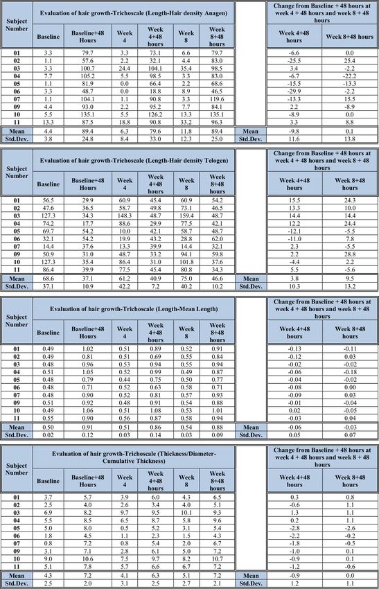
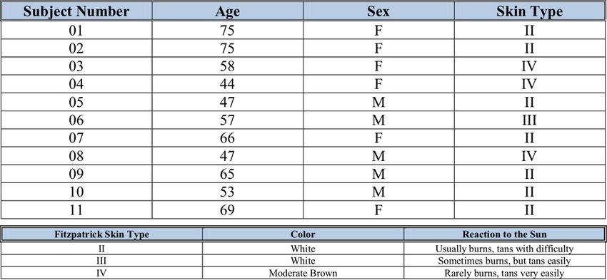
Parker: Clinical Study to Assess the Efficacy of a Novel Hair Loss Treatment (7-20) scalp to another, unfortunately, this option is limited by cost and patient supply of donor hair (Rogers and Avram, 2008). Although everyone is not eligible for hair transplantation, Abaci is studying ways to regrow human hair in a petri dish first, to open hair restoration surgery for more individuals (Abaci et al., 2018). Another method that has shown some success for human hair regrowth is light therapy (Metelitsa and Green, 2011; Avci et al., 2014). Currently, there are only two Food and Drug Administration (FDA) approved hair loss drugs available for medical management of hair loss: minoxidil and finasteride (Tsuboi et al., 2007; Price, 1999). Unfortunately the efficacy of Minoxidil ranges from 20-40%, causing discontinuity of treatment in the majority of patients (Tosti, 2009). Studies have shown Finasteride to have an adverse reaction with some individuals causing sexual dysfunction in 4% of cases (Mc Phee et al., 2007; Kondo et al., 1990). According to Orasan et al. (2016a) “finasteride and minoxidil (2% or 5%) have temporary effects and unpredictable efficacy, better pharmacological options are necessary for managing hair loss” (Gordon, 2011). Orasan et al. (2016b) went on to review hair loss techniques and stated “further studies are required not only to compare the efficiency of different therapies, but more importantly to establish their long term safety” (p.327). There is little data on topical formulations that can naturally stimulate new hair follicles to grow, improve skin health and increase hair count. This research will discuss the effects of a clinical study conducted by Eurofin Scientific whereby participants used Lotus 39 to address their hair loss. STUDY DESIGN A total of 11 people (6 men and 5 women) took part in this clinical trial to see how effective a hair treatment regimen was (Supplemental Data). Individuals ranging from white to brown based on the Fitzpatrick skin type participated in this study. Study evaluations included corneometer measurements, transepidermal water loss, ocular acuity evaluations, and hair counting. Two Materials were used in this study: Lotus 39 Enhancer (Enhancer) and Lotus 39 Elixir (Elixir). A study schedule appears in the Supplemental Data of this clinical study. Subject Termination and Withdrawal: All subjects were free to withdraw from participation at any time and without prejudice. Randomization: No randomization was required for this study. Blinding: Subjects were blinded to the name of the test materials STUDY EVALUATIONS Test Site Tattooing One professional micro tattoo was placed in the center of the 0.5 in.2 test site by a professional tattoo artist. The micro tattoo assisted in referencing the exact test site at subsequent study visits. Evaluation of Hair Growth The FotoFinder Medicam 1000 using the D-Scope IV 20x lens captured participant images at Baseline, 48 Hours Post- Application, Week 4, Week 4 + 48 Hours, Week 8, and Week 8 + 48 Hours. The images were captured in the Trichogram mode. The Trichoscale program generated the data for this study. The following parameters were collected for analysis. Relevant Hair: Hair Count, Hair Density Length: Hair Count Anagen, Hair Count Telogen, Hair density Anagen, Hair density Telogen, Mean Length Thickness/Diameter: Cumulative Thickness Corneometer The general appearance of a soft, smooth skin depends on the presence of an adequate amount of water in the stratum corneum. The Corneometer is an instrument designed to measure changes in the capacitance of the skin resulting from changes in the degree of hydration. It is sensitive to low hydration levels. Triplicate Corneometer measurements were obtained at Baseline and Week 8. Transepidermal Water Loss (TEWL) The VapoMeter is the only fully portable instrument available for the measurement of TEWL values and evaporation rates. TEWL is an indicator of the skin's barrier function. One TEWL measurement was obtained at Baseline and Week 8. Page 8 Malaysian Journal of Medical and Biological Research ● Volume 9, No 1/2022
Volume 9, No 1/2022                                                                         ISSN 2313-0008 (Print); ISSN 2313-0016 (Online)
Ocular Acuity
All subjects were tested for visual acuity using a Snellen Eye Chart. Ocular acuity evaluations were performed at
Baseline and Week 8.
Hair Count after Combing
Each subject’s hair was divided into four quadrants; front left, front right, back left, and back right. If hair length was
not sufficient enough to be parted in order to be divided, the hair was combed from the four quadrants without being
divided. The subjects combed the hair onto a white paper, applying two strokes to each quadrant, for a total of eight
strokes. Hairs that fell onto the white surface were counted. The paper was folded and collected by the technician. Hair
count was performed at Baseline, Week 4, and Week 8.

TEST METHOD
Screening: Participants reported to the testing facility with clean hair, having styled as normal without using any hair
products (every subsequent visit). Informed consent was obtained. A trained technician conducted an initial
examination of the scalp for signs of hair loss as it was part of the inclusion/exclusion. Participants who met all the
study requirements were enrolled into the study.
A patch test of the test materials was performed on the right volar forearm of each subject. A patch test was performed
for the hair dye (Just for MEN) and the test products. A trained technician selected a test area on the scalp, located
closest to the greatest amount of hair loss. A 0.5in.2 template was placed over the test area, and the exposed hair was
snipped (Hairliner HL 1 – Wella Professionals) for the tattoo.
One professional micro tattoo (Ink - Company: SOLIDINK; Color-Red and Needle: White Rose tight 3) was placed in
the center of the 0.5 in.2 test site. The micro tattoo assisted in referencing the exact test site at subsequent study visits.
A patch test was dispersed to participants to test for any irritation
Baseline: Participants acclimated to ambient laboratory conditions for approximately 15 minutes (and every
subsequent visit). Hair clips were used to keep the template in place. Using an 8” pin tail comb, the hair was pulled
through the holes of the template. First the hair was shortened with a pair of scissors to make the shaving easier. Using
the comb, the hair was cut close to the scalp. The shorter the hair, the easier it was to shave with the electric razor to
0.2-0.7mm in length (enough to leave short hair shafts visible). After shaving, the template was removed to enable
subsequent and even shortening of marginal hair. The results were checked with images. The shaved hair stubbles
were removed completely with paper towels and isopropyl alcohol (clipping process was completed the same way
each time).
The clipped hair was no longer than 0.5mm after clipping/shaving. Corneometer and TEWL measurements were
obtained from the shaved site. Ocular acuity was assessed. Hair count after combing was performed. Subjects applied
the test material for the first time in the testing facility under the supervision of study staff.
48 Hours Post-clipping: Participants returned to the testing facility, the 0.5in.2 test site was dyed and images were
captured.
Week 4: A trained technician located and identified the test area utilizing the micro tattoos as a reference. The hair was
clipped. Hair count after combing was performed. Images were captured.
Week 4 + 48 Hours: Participants returned to the testing facility, the 0.5in.2 test site was dyed and images were captured.
Week 8: The hair was clipped and a trained technician made sure the hair was even using images. Corneometer and
TEWL measurements were obtained. Ocular acuity was assessed. Hair count after combing was performed.
Week 8 + 48 Hours: Participants returned to the testing facility, the 0.5in.2 test site was dyed and images were captured.

DATA TABULATION
Ocular acuity was listed for each subject at each time interval.
Analysis of variance followed by Dunnett’s test was applied to determine the differences between baseline and each
post-treatment interval for the following parameters: evaluation of hair growth and hair counts.
A paired Student t-test or Wilcoxon signed ranks test depending on the normality of the data (checked by Shapiro-
Wilk test at 0.01% significance level) was performed to determine the difference between baseline and week 8 for
Corneometer and Vapometer measurements.

Copyright © CC-BY-NC, i-Proclaim | MJMBR                                                                                          Page 9
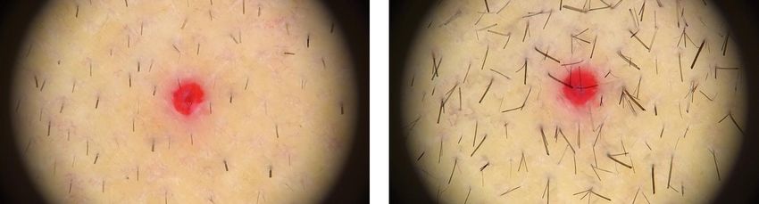
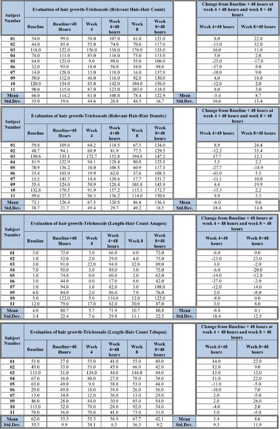
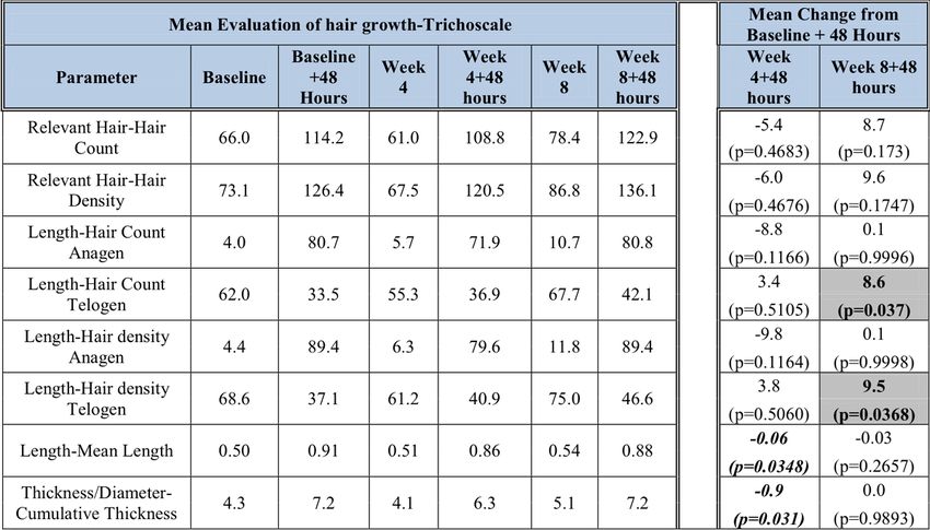
Parker: Clinical Study to Assess the Efficacy of a Novel Hair Loss Treatment                                                                        (7-20)
Change from baseline and % of subjects improved was calculated at each post-treatment interval for the above
mentioned parameter.

RESULTS
Completed and Discontinued Participants
A total of 10 participants completed the study ranging in age from 44 to 75. One subject (#08) was lost to follow up
(Supplemental Data).
Evaluation of Hair Growth: Fotofinder Medicam 1000 and D-Scope IV – 20x
Statistical summary tables for hair growth evaluations appear below along with a baseline photo and Week 8 + 48
hours photo of subject 2(randomly selected). Individual hair growth evaluations and % of subjects showing
improvement can be found in Appendix Table 1 of the supplemental data.
Table 1a: Mean Evaluation of Hair Growth

Bold/shaded indicates a statistically significant improvement
Bold/Italic indicates a statistically significant change but not improvement
Table 1b: Mean Evaluation of Hair Growth

Bold/shaded indicates a statistically significant improvement

Page 10                                                                        Malaysian Journal of Medical and Biological Research ● Volume 9, No 1/2022
Volume 9, No 1/2022                                                                   ISSN 2313-0008 (Print); ISSN 2313-0016 (Online)
Table 1c: Red tattoo was used as a marker for hair count and photo consistency

                         Subject 2 Baseline                                Subject 2 Week 8 + 48hrs
Corneometer
A statistical summary table for Corneometer measurements appears below. Individual Corneometer measurements
and % of subjects showing improvement appear in Table 2 of the supplemental data.
Table 2: Mean Corneometer measuremen

Bold/shaded indicates a statistically significant improvement
Transepidermal Water Loss
Statistical summary tables for VapoMeter measurements appear below. Individual VapoMeter measurements and %
of subjects showing improvement from baseline appear in Table 3 of the supplemental data.
Table 3: VapoMeter measurements

Ocular Acuity
Individual ophthalmic evaluations appear in Table 4 (supplemental data). There were no increases in subjective eye
irritation, including stinging, burning, itching, dryness, and/or foreign body sensation. There was no difference in
visual acuity at the Week 8 visit, compared to Baseline.
Hair Count after Combing
Statistical summary tables for hair counts after combing appear below. Individual hair counts after combing and % of
subjects showing improvement appear in Table 5 of the supplemental data.
Table 4: Bold/shaded indicates a statistically significant improvement

DISCUSSION
Society is shifting toward natural alternatives to everyday problems, hair health is one of those areas. Some not only
want their hair healthier, they want their hair regrown as quickly and naturally as possible. Hair loss in humans is

Copyright © CC-BY-NC, i-Proclaim | MJMBR                                                                                    Page 11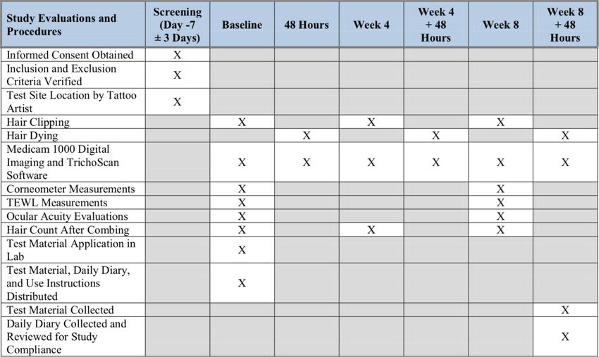
Parker: Clinical Study to Assess the Efficacy of a Novel Hair Loss Treatment (7-20)
characterized by a progressive alteration in the terminal hair population, typified by an increase in vellus hair and a
decline in total hair density (Rushton et al., 1983). Hair follicle restoration has been attributed to the reversal of this
process (Whiting et al., 1999, Mirmirani et al., 2015).
A hair follicle is capable of producing three different types of hair as follows: lanugo, vellus and terminal hairs. The
type of hair originated by an individual follicle can change with age or under the influence of hormones. The initial
hair produced is the lanugo hair. Lanugo hair is non pigmented hair which covers the body of newborns (Alves and
Grimalt, 2015), sheds within the womb, but can remain 3 to 4 months after birth. The lanugo hairs are replaced by
vellus hairs (Alves and Grimalt, 2016).
Vellus hair is short, light-colored, barely noticeable, and covers almost the whole body. Puberty can cause the vellus
hair to be converted to terminal hairs due to the increase in androgens which lead to secondary sex characteristics
(Moreno-Romero and Grimalt, 2014; Harrison and Sinclair, 2003).
Terminal hair is larger and thicker usually. This type of hair is strongly pigmented and is found on the scalp, eyebrows,
axillary and pubic areas, chest and face (Moreno-Romero and Grimalt, 2014). Someone who is losing their hair will
begin to have more terminal hair follicles convert to vellus hairs. Uno and Kurata (Uno and Kurata, 1993) studied hair
growth promoters, minoxidil, diazoxide, and Cooper peptides, on fuzzy rats. Their results with minoxidil
demonstrated a conversion of short vellus hairs to prolonged terminal hairs.
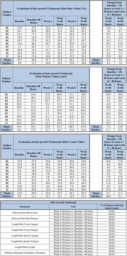
According to the results, individuals who used Lotus 39 for 8 weeks + 48hrs had a statistically significant increase in
vellus hair from the baseline. The Hair Rate Vellus (%) increased by 7.2 units (p =0.0063), The Hair Count Vellus
increased 11.7 units (p = 0.0087), and hair density vellus increased 13.0 units (p=0.0087). This data suggests new hair
follicles were formed that were not previously there. Not only did the amount of vellus hairs increase, 90% of the
subjects improved both their Relevant Hair Density and Hair Count. This is an indication that the amount of hair on
the subjects scalp increased over time. The amount of hair is important for hair to appear healthier, what often gets
overlooked is the thickness of each individual hair (Cohen, 2008). 60% of the subjects’ follicles hair grew in
Thickness/Diameter. Which means that the individual hairs on subjects grew bigger which can give the appearance
of healthier hair, decreases in the diameter of hair can be a sign of hair loss ( Lacharrière et al., 2001). We believe the
key to hair health is scalp health. Lotus 39 contains nutrients that protect the energy of the hypoderm of the skin so
they do no degenerate. Corneometer and Transepidermal water loss tests are ideal for examining skin health.
Corneometer measurements indicated a significant change from baseline of 6.5 units (p=0.0197) with 80% of the
subjects having improvement in skin health. Transepidermal water loss measurements indicated 60% of the subjects
had improvement in the hydration of their skin. Since most subjects improved the health of their scalp and most
subjects improved their hair, a healthy scalp can assist in the health of an individual’s hair (Trüeb et al., 2018).
Not only did the data suggest Lotus 39 can increase the amount of hair on someones scalp, the data also suggests that
individuals reduced or stopped losing their hair. From baseline to 8 weeks +48hrs 100% of subjects showed a
statistically significant difference in the amount of hair they lost. Stopping hair shedding is another way to make an
individual’s hair fuller and thicker. There was no change in ocular acuity and hair length. The ocular acuity of the
subjects was not altered because Lotus 39 is not expected to improve or harm the eyes. Hair length did not change
because the area of the hair that was used for measuring was shaved prior to taking the length of the hair.
CONCLUSION
Under the conditions of this study, the test material Lotus 39 can stop hair loss, increase the amount of hair on the
scalp, significantly improve scalp hydration all while causing no ocular irritation or adverse effects. Future studies
include increasing the amount of time subjects use Lotus 39, observing for changes in the restoration of hair color,
increasing the age range of the subjects, and incorporating a placebo.
REFERENCES
Abaci, H. E., Coffman, A., Doucet, Y., Chen, J., et al. (2018). Tissue engineering of human hair follicles using a
biomimetic developmental approach. Nat Commun, 9, 5301 https://doi.org/10.1038/s41467-018-07579-y
Alves, R. and Grimalt, R. (2015). Hair loss in children. Curr Probl Dermatol. 47, 55-
66. https://doi.org/10.1159/000369405
Alves, R. and Grimalt, R. (2016). Androgenetic Alopecia in Adolescents. In: Oranje AP, Al-Mutairi N, Shwayder T (Eds.).
Practical Pediatric Dermatology. Controversies in Diagnosis and Treatment. Switzerland: Springer. 187-196.
Andersson, B. (1979). Development trends in reaction to social pressure from adults versus peers. Int J Behav Dev, 2,
269-286.
Page 12 Malaysian Journal of Medical and Biological Research ● Volume 9, No 1/2022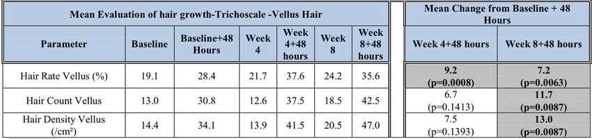
Volume 9, No 1/2022                                                                       ISSN 2313-0008 (Print); ISSN 2313-0016 (Online)
Avci, P., Gupta, G. K., Clark, J., Wikonkal, N., & Hamblin, M. R. (2014). Low-level laser (light) therapy (LLLT) for
       treatment of hair loss. Lasers in surgery and medicine, 46(2), 144-151. https://doi.org/10.1002/lsm.22170
Chase, H. B., Rauch, R. and Smith, V. W. (1951). Critical stages of hair development and pigmentation in the mouse.
      Physiol Zool, 24, 1–8.
Chen, J. and Jackson, T. (2012). Gender and age group differences in mass media and interpersonal influcence on body
      dissatisfaction among Chinese adolescents. Sex Roles, 66, 3-20.
Cohen, B. (2008). The cross‐section trichometer: a new device for measuring hair quantity, hair loss, and hair growth.
     Dermatol Surg, 34, 900‐911.
Dohnt, N. K. and Tiggemann, M. (2006). Body image concerns in young girls: the role of peers and media prior to
      adolescences. J Youth Adolesc. 35(2), 141-151.
Ghanaat, M. (2010). Types of hair loss and treatment options, including the novel low-level light therapy and its
     proposed mechanism. South Med J., 103(9), 917–921.
Gordon, K. A. (2011). Tosti A. Alopecia: evaluation and treatment. Clin Cosmet Investig Dermatol, 4, 101-106.
Hamilton, J. B. (1951). Patterned loss of hair in man; types and incidence. Ann N Y Acad Sci, 53, 708–728.
Hamilton, J. B. (1958). Age, sex and genetic factors in the regulation of hair growth in man: A comparison of Caucasian
      and Japanese populations. In: Montagna W, Ellis RA (eds). The Biology of Hair Growth. New York: Academic
      Press; 399–433.
Harrison, S., and Sinclair, R. (2003). Optimal management of hair loss (alopecia) in children. Am J Clin Dermatol, 4(11),
      757-770.
Helfert, S. and Waschburger, P. (2013). The face of appearance-related social pressure: gender, age and body mass
       varaintions in peer and parental pressure during adolescence. Child Adolesc Psychiatry Ment Health, 7.
Jones, D. C., Vigfusdottir, T. and Lee, Y. (2004). Body image and the appearance culture among adolescent girls and
       boys: An examination of friend conversations, peer criticism, appearance magazines and the internalization of
       appearance ideals. J Adolesc Res., 19, 323- 339.
Kondo, S., Hozumi, Y., Aso, K. (1990). Organ culture of human scalp hair follicles: effect of testosterone and oestrogen
     on hair growth. Arch Dermatol Res., 282, 442–445.
Lacharrière, O. d., Deloche, C., Misciali, C., Piraccini, B. M., Vincenzi, C., Bastien, P., Tardy, I., Bernard, B A., Tosti,
      A. (2001). Hair Diameter Diversity: A Clinical Sign Reflecting the Follicle Miniaturization. JAMA Dermatology,
         137(5), 641–646.
Ludwig, E. (1977). Classification of the types of androgenetic alopecia (common baldness) occurring in the female sex.
     Br J Dermatol, 97, 247–254.
Mc Phee, S. J., Papadakis, M. A. and Tierney, L. M. (2007). Current medical diagnosis and treatment. 46th ed. McGraw-
     Hill Medicale.
Metelitsa, A. I. and Green, J. B. (2011). Home-use laser and light devices for the skin: An update. Semin Cutan Med Surg,
       30(3), 144–147.
Mirmirani, P., Consolo, M., Oyetakin-White, P., Baron, E., Leahy, P., Karnik, P. (2015). Similar response patterns to
     topical minoxidil foam 5% in frontal and vertex scalp of men with androgenetic alopecia: a microarray analysis.
     Br J Dermatol, 172(6), 1555-1561. https://doi.org/10.1111/bjd.13399
Moreno-Romero, J. A. and Grimalt, R. (2014). Hair loss in infancy. G Ital Dermatol Venereol, 149(1), 55-78.
Orasan, M. S., Roman, I., Coneac, A., Muresan A., & Orasan, R. I. (2016b). Hair loss and regeneration performed on
      animal models. Clujul medical. 2016; (1957), 89(3), 327-334. https://doi.org/10.15386/cjmed-583
Orasan, M., & Bolfă, P., & Coneac, A., & Muresan, A., & Mihu, C. (2016a). Topical Products for Human Hair
      Regeneration: A Comparative Study on an Animal Model. Annals of Dermatology, 28(1), 65-73.
      https://doi.org/10.5021/ad.2016.28.1.65
Otberg, N., Finner, A. M., Shapiro, J. (2007). Androgenetic alopecia. Endocrinol Metab Clin North Am., 36(2), 379–398.
Price, V. H. (1999). Treatment of hair loss. N Engl J Med, 341, 964-973.

Copyright © CC-BY-NC, i-Proclaim | MJMBR                                                                                        Page 13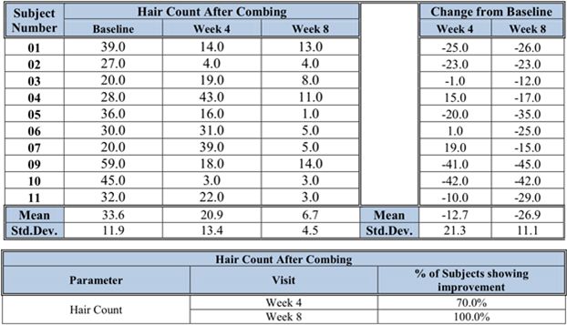
Parker: Clinical Study to Assess the Efficacy of a Novel Hair Loss Treatment                                                                           (7-20)
Randall V. A. (2001). Physiology and pathophysiology of androgenetic alopecia. In: De Groot LJ, Jameson JL (eds).
     Endocrinology. 4th edn. Philadelphia: WB Sanders, 2257-2268.
Randall, V. A. (2007). Hormonal regulation of hair follicles exhibits a biological paradox. Semin Cell Dev Biol, 18, 274-
     285.
Rogers, N. E. and Avram, M. R. (2008). Medical treatments for male and female pattern hair loss. J Am Acad Dermatol,
      59(4), 547–566.
Rushton, H., James, K., and Mortimer, C. (1983). The unit area trichogram in the assessment of androgen‐dependent
      alopecia. British Journal of Dermatology, 109, 429-437. https://doi.org/10.1111/j.1365-2133.1983.tb04617.x
Shorter, K., Farjo, N. P., Picksley, S. M., Randall, V. A. (2008). Human hair follicles contain two forms of ATP-sensitive
      potassium channels, only one of which is sensitive to minoxidil. FASEB J. 22(6), 1725-1736.
Tosti, A. (2009). Duque-Estrada B. Treatment strategies for alopecia. Expert Opin Pharmacother, 10, 1017-1026.
Trueb, R. M. (2009). Chemotherapy-induced alopecia. Semin Cutan Med Surg, 28(1), 11–14.
Trüeb, R. M., Henry, J. P., Davis, M. G., & Schwartz, J. R. (2018). Scalp Condition Impacts Hair Growth and Retention
      via Oxidative Stress. International journal of trichology, 10(6), 262–270. https://doi.org/10.4103/ijt.ijt_57_18
Tsuboi, R., Tanaka, T., Nishikawa, T., Ueki, R., Yamada, H., Katsuoka, K., et al. (2007). A randomized, placebo-
      controlled trial of 1% topical minoxidil solution in the treatment of and- rogenetic alopecia in Japanese women.
      Eur J Dermatol, 17, 37-44.
Uno, H. and Kurata, S. (1993). Chemical agents and peptides affect hair growth. J Invest Dermatol, 101(1 Suppl), 143S-
      147S.
Wasserman, D., Guzman-Sanchez, D. A., Scott, K., McMichael, A. (2007). Alopecia areata. Int J Dermatol, 46(2), 121–
     131.
Whiting, D. A., Waldstreicher, J., Sanchez, M., Kaufman, K. D. (1999). Measuring reversal of hair miniaturization in
      androgenetic alopecia by follicular counts in horizontal sections of serial scalp biopsies: results of finasteride 1
      mg treatment of men and postmenopausal women. J Investig Dermatol Symp Proc, 4(3), 282-284.
      https://doi.org/10.1038/sj.jidsp.5640230

                                                                               --0--

Page 14                                                                          Malaysian Journal of Medical and Biological Research ● Volume 9, No 1/2022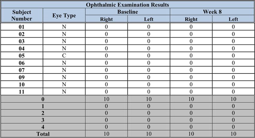
Volume 9, No 1/2022                                     ISSN 2313-0008 (Print); ISSN 2313-0016 (Online)
APPENDICES (SUPPLEMENTAL DATA)
Appendix Table 1: Trichoscale Hair Growth Evaluations

Copyright © CC-BY-NC, i-Proclaim | MJMBR                                                      Page 15
Parker: Clinical Study to Assess the Efficacy of a Novel Hair Loss Treatment                                                                        (7-20)
Appendix Table 1: Trichoscale Hair Growth Evaluations (Continued)

Page 16                                                                        Malaysian Journal of Medical and Biological Research ● Volume 9, No 1/2022
Volume 9, No 1/2022                                                 ISSN 2313-0008 (Print); ISSN 2313-0016 (Online)
Appendix Table 1: Trichoscale Hair Growth Evaluations (Continued)

Copyright © CC-BY-NC, i-Proclaim | MJMBR                                                                  Page 17
Parker: Clinical Study to Assess the Efficacy of a Novel Hair Loss Treatment                                                                        (7-20)
Appendix Table 1: Trichoscale Hair Growth Evaluations (Continued)

Appendix Table 2: Trichoscale Hair Growth Evaluations

Appendix Table 3: VapoMeter Measurements

Page 18                                                                        Malaysian Journal of Medical and Biological Research ● Volume 9, No 1/2022
Volume 9, No 1/2022                           ISSN 2313-0008 (Print); ISSN 2313-0016 (Online)
Appendix Table 4: Ophthalmic Evaluations

N = Non-Contact Lens Wearer
C = Contact Lens Wearer
Appendix Table 5: Hair Counts after Combing

Appendix Table: Study Schedule

Copyright © CC-BY-NC, i-Proclaim | MJMBR                                            Page 19
Parker: Clinical Study to Assess the Efficacy of a Novel Hair Loss Treatment                                                                           (7-20)
Appendix Table: Balding Scales
Scores of 3 to 7 on the Norwood Scale Required for Male Subject Enrollment

Image Source: American Hair Loss Association
Scores of 1 to 3 or Higher on Ludwig Scale Required for Female Subjects

Image Source: American Hair Loss Association
Appendix Table: Participant Demographics

                                                                               --0--

Page 20                                                                           Malaysian Journal of Medical and Biological Research ● Volume 9, No 1/2022
You can also read


