Clinical practice - Wounds ...
←
→
Page content transcription
If your browser does not render page correctly, please read the page content below
Clinical practice

                                         Osteomyelitis in diabetic foot ulcers:
                                         the Malaysian experience

                                         Osteomyelitis is defined as an inflammation of the bone marrow.
                                         Approximately 20% of patients with diabetes will develop osteomyelitis
                                         and it is linked to high rates of mortality, morbidity and amputation.
                                         Diagnosing osteomyelitis associated with a diabetic foot can be challenging
                                         as it is difficult to identify the infection in its initial phase and there is often
Authors:
                                         symptom and clinical manifestation variability. As there are no standardised
Harikrishna KR Nair,
Sylvia SY Chong                          tests or criteria for diagnosing osteomyelitis, it may be helpful to obtain a
                                         patient’s complete history of symptoms, including physiological state (risk
                                         factors) with clinical manifestation, laboratory tests, imaging and blood or
                                         bone cultures to come to a final diagnosis. This article looks at some of the
                                         tests that can be used in the diagnosis process.

                                         T
                                               he importance of wound irrigation and             As seen below, Lew and Waldvogel (Figure 1)
                                               cleansing solutions is often ignored,          with Cierny and Mader are two major clinical
                                               WAs Malhotra et al (2014) have shown,          classifications for osteomyelitis. According to
                                         osteomyelitis is defined as an inflammation          Lew and Waldvogel in 1970, osteomyelitis is
                                         of the bone marrow. A bacterial infection can        classified based (Table 1) on the length of
                                         cause inflammation of the bone tissue, which         evolution and pathophysiology.
                                         can result in inflammatory destruction, necrosis,       Cierny and Mader (1984) attempted to
                                         bone neoformation, and it can progress into a        address some aspects that were not covered
                                         chronic or persistent stage (Smith et al, 2006).     by Lew and Walvogel’s classification . They
                                         Staphylococcus aureus is the most common             classified osteomyelitis by anatomical stages
                                         pathogenic organism isolated in osteomyelitis,       according to bone infection and the type of
                                         although a variety of organisms can cause this       host health status, depending on the patient’s
                                         disease, as outlined by Lew and Woldvogel (2015).    clinical conditions (Table 2).

Harikrishna KR Nair is Head of
Wound Care Unit, Department
of Internal Medicine, Hospital
Kuala Lumpur; Sylvia SY
Chong is a Research Assistant,
Wound Care Unit, Department              Figure 1. Lew and Waldvogel with Cierny and Mader are two major clinical classifications
of Internal                              for osteomyelitis

Wounds Asia 2021 | Vol 4 Issue 1 | ©Wounds Asia 2021 | www.woundsasia.com                                                                  19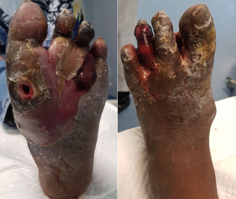
Clinical practice

                      Table 1. The Lew and Waldvogel osteomyelitis classification system
                      Duration of infection                      Description
                      Acute                                      Initial episodes of osteomyelitis with the presence
                                                                 of oedema, pus formation, vascular congestion and
                                                                 thrombosis of the small vessels
                      Chronic                                    Recurrence of acute cases with large areas of ischemia,
                                                                 necrosis and bone sequestra

                     Mechanism of bone infection                 Description
                     Hematogenous                                Commonly seen in children and occurs through
                                                                 secondary infection when bacterial is transported
                                                                 through the blood
                     Contiguous                                  Bacterial inoculation from an adjacent focus, e.g.
                                                                 post-traumatic osteomyelitis, or infections related to
                                                                 prosthetic devices
                     Associated with vascular insufficiency      Infections affecting the feet in patients with diabetes,
                                                                 hanseniasis or peripheral vascular insufficiency

                      Table 2. The Cierny and Mader osteomyelitis classification system
                      Anatomical stage        Description
                      1       Medullary       Infection restricted to the bone marrow
                      2       Superficial     Infection restricted to cortical bone
                      3       Localised       Infection with clearly defined edges and bone stability preserved
                      4       Diffuse         Infection spread to the entire bone circumference, with stability before or
                                              after debridement
                      Host health status      Description
                      A       Host healthy    Patients without comorbidities
                      Bl      Local           Smoking, chronic lymphedema, venous stasis, arthritis, large scars, fibrosis
                              compromise      by radiotherapy
                      Bs      Systemic        Diabetes mellitus, malnutrition, renal or hepatic failure, chronic hypoxia,
                              compromise      neoplasms, extremes of age
                      C       Poor clinical   Surgical treatment will have a higher risk than the osteomyelitis itself
                              conditions

                       Hematogenous osteomyelitis is most                   are Staphylococcus aureus (Asmar, 1992). As
                     commonly seen in infants and children, and             reported by Ramsey et al (1999) and Lavery et
                     usually involves the metaphysis of long bones,         al (2009), about 20% of osteomyelitis cases are
                     particularly the femur and tibia. Metaphyseal          acute hematogenous osteomyelitis. Of these,
                     spongiosa contains abundant blood vessels              children under the age of 5 account for 50%.
                     with leaky endothelium and a sluggish flow             This accumulates up to 85% for cases involving
                     that ends in capillary loops and provides a            children under the age of 17, as stated by
                     suitable environment for bacteria growth,              Cierny and Mader (1984).
                     as reported by Whyte and Bielski (2016). In               The most common infection site is the
                     children under the age of one, there have              vertebrae, but it can also occur in the pelvis,
                     been cases of osteomyelitis affecting the              clavicle and long bones, and, as reported by
                     epiphysis due to the connection of blood               Arciola et al (2005), only 2–7% of adults have
                     vessels passing through the metaphysis to              this condition. As Rao et al (2011) have shown,
                     the epiphysis. As shown by McPherson (2002),           hematogenous osteomyelitis is usually an acute
                     Liao et al (2005), and Qadir et al (2010), in          disorder and primarily treated conservatively.
                     new-borns the most common pathogens are                On the other hand, as stated by Lew and
                     Streptococcus agalactiae, Escherichia coli and         Waldvogel in 1970, chronic osteomyelitis is
                     Klebsiella pneumonia, while in children the            characterised by progressive reoccurrence or
                     common pathogens across all age groups                 multiple episodes of acute osteomyelitis at the

20                                                            Wounds Asia 2021 | Vol 4 Issue 1 | ©Wounds Asia 2021 | www.woundsasia.com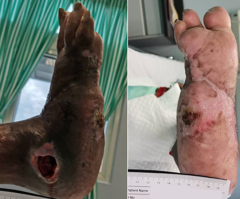
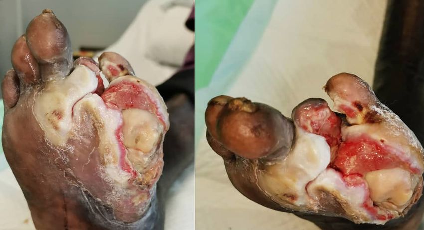
Figure 2. X Rays with bony erosions, subluxations, soft tissue swellings etc and pictures of the
                                           wounds with bone exposed or probe test positive with osteomyelitis are shown in a–c
                                           a. Patients with osteomyelitis and chronic wounds currently undergoing treatment under the
                                           Wound Care Unit, Hospital Kuala Lumpur

                                           b. Patients with osteomyelitis and chronic wounds currently undergoing treatment under the
                                           Wound Care Unit, Hospital Kuala Lumpur

                                           c. Patients with osteomyelitis and chronic wounds currently undergoing treatment under the
                                           Wound Care Unit, Hospital Kuala Lumpur

                                         same site, which can lead to bone necrosis (Lew        in patients with diabetes, as stated by Sia and
                                         and Waldvogel, 2004).                                  Berbari (2006). In elective trauma surgery,
                                            Other entry routes of infection are secondary       close fractures and first- to third-degree open
                                         to the direct inoculation of bacteria into             fractures had 1–5% and 3–50% of contagious
                                         the bone tissue. The can occur in acute                infection respectively, as reported by Gustilo
                                         trauma (an open fracture) and surgery (with            et al (1990). As Parvazi et al (2008) have shown,
                                         or without implantation), as well as poor              early infections are expected in 0.5% to 2%
                                         peripheral vascular supply with infection of           of primary hip and knee replacement cases,
                                         the surrounding tissues. This is especially seen       and more than 20% of septic revisions and

Wounds Asia 2021 | Vol 4 Issue 1 | ©Wounds Asia 2021 | www.woundsasia.com                                                                       21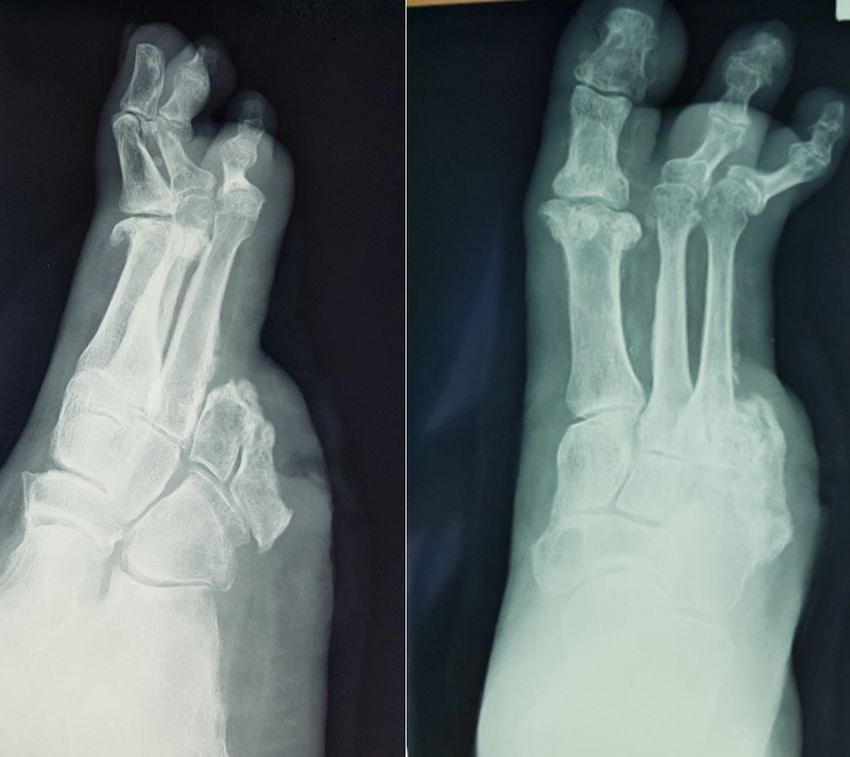
Case reports
 Clinical practice

                     5% of aseptic revisions have deep infections.           clinical manifestation variability. As Arias et
                     Generally, infectious complications occur in            al (2019) have shown, because there are no
                     5% of traumatic and orthopaedic implants                standardised tests or criteria for diagnosing
                     during the lifetime of the implant (Trampuz and         osteomyelitis, it may be helpful to see a patient’s
                     Zimmerli, 2006).                                        complete history of symptoms, including
                        Contiguous spread of pathogens from                  physiological state (risk factors) with clinical
                     infected diabetic foot ulcers (DFU) to the              manifestation, laboratory tests, imaging, and
                     bone is the pathogenesis of osteomyelitis               blood or bone cultures to come to a final
                     in a diabetic foot. Bacteria induce an acute            diagnosis. Furthermore, patients with diabetes
                     inflammatory reaction during infection of the           and peripheral neuropathy are also prone to
                     bone and the bacteria and inflammation affect           developing Charcot neuro-osteoarthropathy,
                     the periosteum and spread in the bone, causing          which closely resembles and may co-exist with
                     bone necrosis. Lifting of the periosteum further        diabetic foot-associated osteomyelitis (Berendt
                     impairs the blood supply to the affected bone,          et al, 2008).
                     causing segmental bone necrosis known as a                  Clinical suspicion is very important when
                     sequestrum. In the chronic stage, numerous              commencing a medical investigation for
                     inflammatory cells and their release of cytokines       osteomyelitis. A thorough assessment of the
                     stimulate osteoclastic bone resorption,                 foot or lower extremity should be performed,
                     ingrowth of fibrous tissue, and the deposition          including examination of the ulcer, presence of
                     of reactive new bone in the periphery. When             peripheral neuropathy (present in 88% of DFUs),
                     the newly deposited bone forms a sleeve of              peripheral vascular disease (present in 45–65%
                     living tissue around the segment of devitalised         of DFUs), and the extent of any underlying
                     infected bone, it is known as an involucrum.            infection (Nair, 2017). Infected DFUs usually
                     As Rosenberg (2010) has shown, a rupture of             have purulent secretions or at least two signs of
                     a subperiosteal abscess may lead to a soft-             inflammation, as stated by Giurato et al (2017)
                     tissue abscess and the eventual formation of a          and Jeffcoate and Lipsky (2004), yet diabetic
                     draining sinus.                                         foot-associated osteomyelitis can occur without
                        As reported in several publications Lipsky           any local signs of infection. Systemic symptoms
                     (2014), Lázaro-Martínez (2019) Optimal                  are rare due to the presence of diabetic-
                     management of diabetic foot osteomyelitis:              immunopathy, which impairs the patient’s
                     challenges and solutions. Diabetes Metab Syndr          response to inflammation and infection.
                     Obes12:947–59. https://doi.org/10.2147/dmso.                As Nair (2017) has shown, a simple clinical test
                     s181198 et al (2019) and Bond et al (2019),             commonly used for osteomyelitis is a probe-to-
                     approximately 20% of patients with diabetes             bone (PTB) test. This test evaluates the ability
                     will develop osteomyelitis, and it is linked to a       to contact a bone in the depth of an ulcer and
                     high burden of mortality and morbidity - and            it is performed by probing the ulcer area with
                     especially high rates of amputation. Of these           a sterile blunt metal probe. It is considered
                     10–20% account for moderate infection and               positive if the probe reaches the bone surface. A
                     50–60% of the remaining account for severe              study by Lavery et al (2007) showed a sensitivity
                     infections, as shown by Giurato et al (2017),           87% and specificity 91%, positive predictive
                     Berendt et al (2008) and Thomas-Ramoutar et             value of only 57%, and a negative predictive
                     al (2010). Diabetic foot osteomyelitis usually          value 98% for the PTB test, which concluded
                     develops by contiguous spread of bacteria from          that it is a better tool to use to exclude
                     overlying soft-tissue, infiltrating the cortex and      osteomyelitis. In contrast, Morales et al (2010)
                     eventually the bone marrow. In osteomyelitis,           and Aragon-Sanchez et al (2011), support it as
                     the pathogens found are more frequently poly-           a reliable test for osteomyelitis, with sensitivity
                     microbial. Staphylococcus aureus is the most            98%, specificity 79%, and sensitivity 95%,
                     commonly detected (up to 50%), followed by              specificity 93% respectively. Clinical signs that
                     Enterobacteriaceae (up to 40%), Streptococci            predict osteomyelitis are: an ulcer larger than
                     (~30%), and Staphylococcus epidermidis (~25%).          2cm2; ulcer depth >3mm; visible exposed bone;
                     This is stated by several publications (Jeffcoate       positive PTB test; presence of ‘sausage; toe; an
                     and Lipsky, 2004) Hartemann-Heurtier and                ulcer that fails to heal or is located over the bony
                     Sennivel, 2008; Nair, 2017).                            prominence, and the presence of soft-tissue
                        Diagnosing osteomyelitis associated with             sinus with purulent discharge. Leucocytosis and
                     a diabetic foot can be challenging, as it is            high CRP levels are a poor indicator for diabetic
                     difficult to identify the infection in its initial      foot-associated osteomyelitis, as they can be
                     phase, and there is often symptom and                   negative in osteomyelitis, but ESR>70mm/h

22                                                            Wounds Asia 2021 | Vol 4 Issue 1 | ©Wounds Asia 2021 | www.woundsasia.com
is highly suggestive of osteomyelitis, with a        attain limb salvage wherever possible. Current
                                         positive predictive value of 100%.                   management of diabetic foot osteomyelitis
                                               Laboratory tests such as whit blood cell       is evaluated by case basis with guidelines
                                         (WBC), erythrocyte sedimentation rate (ESR),         to advocate the specific conditions for
                                         and C-reactive proteins detect the presence of       surgical or medical approaches combined
                                         inflammation and are generally raised in acute       with conservative surgery. Antibiotic therapy
                                         hematogenous osteomyelitis, but they lack            alone can work for selected cases, particularly
                                         specificity as other sources of inflammation         uncomplicated forefoot osteomyelitis with no
                                         might also raise the readings. However, as           other indications for surgery, as stated by the
                                         stated by Saavedra-Lozano et al (2008), these        International Working Group on the Diabetic
                                         inflammatory markers are better used as a            Foot (2019). The antibiotic regimens are based
                                         monitor during treatment.                            on the likely causative pathogens and guided by
                                            Radiological investigations are useful to         culture results preferably from the bone, with a
                                         confirm a suspected case of osteomyelitis, to        treatment duration of 6 weeks or shorter if the
                                         detect bone involvement and to distinguish           infected bone is resected. As shown by Berendt
                                         a diabetic foot-associated osteomyelitis from        et al (2008) and Mutluoglu and Lipsky (2016),
                                         soft-tissue infection (Giurato et al, 2017). Plain   the patient’s clinical symptoms, abnormal lab
                                         radiography, although not as effective in the        tests, and imaging studies should be monitored
                                         early stages or in distinguishing osteomyelitis      for at least a year, and the case should be
                                         from Charcot neuro-osteoarthropathy, is still        evaluated by a surgeon if it is not improving. The
                                         helpful as a baseline to assess the development      major benefits of medically-treated diabetic foot
                                         of osteomyelitis, as stated by Jeffcoate and         osteomyelitis are the absence of biomechanical
                                         Lipsky (2004). The classic radiographic triad of     changes, which increase recurrent ulceration
                                         osteomyelitis is demineralisation, periosteal        rates that may occur after surgical procedures.
                                         reaction, and bone destruction, which normally       It is a more cost-effective option as it reduces
                                         manifests after 2–3 weeks or when 30–50% of          risk and hospitalisation associated with
                                         bone loss occurs.                                    surgical procedures.
                                            As stated by Mutluoglu and Lipsky (2016),             A study by Senneville et al (2008) reviewed
                                         advanced imaging is necessary for early              the outcome of osteomyelitis in a patients
                                         diagnosis, delineation of deep soft-tissue           with diabetes who was treated non-surgically
                                         infection, differentiation of osteomyelitis from     and at the same time compared the results of
                                         Charcot neuro-osteoarthropathy or to plan            bone versus swab culture-based. There were
                                         surgery. MRI is preferable in the diagnosis          50 patients included, who had underlying
                                         of diabetic foot-associated osteomyelitis,           type 2 diabetes with osteomyelitis of a non-
                                         while radionuclide scanning, leukocyte               ischaemic foot. At the end of the study, 32 of
                                         scan, SPECT/CT, and PET/CT are only used             the 50 patients (64%) achieved remission, 18 of
                                         if MRI is contraindicated. MRI provides high         22 patients (81%) who were treated with bone
                                         sensitivity 90% and specificty 85%, which can        cultured-based antibiotics achieved remission
                                         distinguish osteomyelitis and chronic Charcot        and 14 of 28 patients (50%) achieved remission
                                         neuro-osteoarthropathy, but problems arise           who were treated with a swab-based antibiotic.
                                         when other entities that cause marrow oedema             Although bone culture provides a better
                                         such as recent surgery or a resolving fracture       remission rate, it is not widely undertaken in
                                         are present.                                         many facilities. An article by Tone et al (2014)
                                            The gold standard for diagnosing                  discussed the duration of antibiotics to be given
                                         osteomyelitis is bone biopsy, collected              to the patient with osteomyelitis in diabetic
                                         aseptically via a percutaneous approach              foot patients treated non-surgically. A 6-week
                                         through uninfected skin or via an open surgical      course was compared with a 12-week course in
                                         procedure for culture and histopathology.            the case of a patient who was diagnosed with
                                         Histologic features of osteomyelitis are specific,   osteomyelitis of a non-ischaemic foot and had
                                         i.e. bone erosion, marrow oedema, fibrosis,          not received prior surgical treatment. The result
                                         necrosis, and the presence of inflammatory cells,    showed that the 6-week antibiotic course has
                                         which are rarely seen in normal bone. Reliable       a similar outcome to the 12-week course, with
                                         cultures are the key to successful antibiotic        reduced gastrointestinal side effects.
                                         therapy and can be obtained by discontinuing             As stated by the International Working
                                         antibiotics 2–4 weeks before the bone biopsy.        Group on the Diabetic Foot (2019), surgical
                                            As Nair (2017) has shown, the ultimate goal       intervention is considered in cases of diabetic
                                         in managing diabetic foot osteomyelitis is to        foot osteomyelitis when it is accompanied by

Wounds Asia 2021 | Vol 4 Issue 1 | ©Wounds Asia 2021 | www.woundsasia.com                                                                    23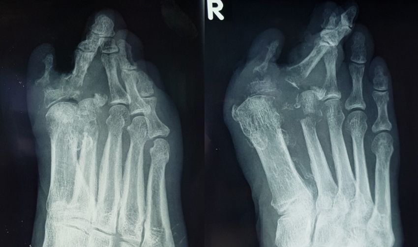
Case reports
 Clinical practice

                     spreading soft-tissue infection, progressive bone                   Retrospective analysis of diabetic foot osteomyelitis
                     destruction or bone exposure. The operative                         management and outcome at a tertiary care hospital
                                                                                         in the UK. PLoS ONE 14(5):e0216701. https://doi.
                     aim is to reset the affected bone, avoid leaving                    org/10.1371/journal.pone.0216701
                     residual disease and to insert antibiotic cement/               Asmar BI (1992) Osteomyelitis in the neonate. Infect Dis Clin
                     spacer beads. As Lázaro-Martínez et al (2014;                       North Am 6(1): 117–32
                     2019) have shown, on top of preventing minor                    Berendt AR, Peters EJG, Embil JM, Eneroth M et al (2008)
                     and major amputations, conservative surgery                         Diabetic foot osteomyelitis: a progress report on
                                                                                         diagnosis and a systemic review of treatment. Diabetes
                     also leads to a reduction in the duration of                        Metab Res Rev 24(1):145–161. https://doi.org/10.1002/
                     antibiotic therapy. Other advantages of surgical                    dmrr.836
                     management are a high rate of limb salvage, low                 Bond H, Metcalf L, Gouni R, Snape S (2019) Diabetic foot
                     risk of recurrence through surgical offloading                      osteomyelitis treatment: an audit of success rates in
                                                                                         differing circumstances. Diabetic Foot Journal 22(4):
                     and obtaining samples for culture and                               17–21
                     histological analysis. The downside of surgical                 Cierny G, Mader JT (1984) Adult chronic osteomyelitis.
                     interventions are biomechanical changes, re-                        Orthopedics 7:1557–64. https://doi.org/10.3928/0147-
                     ulceration due to pressure transfer syndrome,                       7447-19841001-07
                     foot instability, higher costs and increased                    Cierny G, Mader JT, Penninck JJ (1985) A clinical stage
                                                                                         system for adult osteomyelitis. Contemp Orthop 10:
                     operative morbidity. As for adjuvant therapies,                     17–37
                     there is no conclusive evidence to support the                  Giurato L, Meloi M, Izzo V, Uccilio L (2017) Osteomyelitis
                     use of hyperbaric oxygen therapy, granulocyte                       in diabetic foot: a comprehensive overview. World J
                     growth factors, local antibiotic-delivery systems,                  Diabetes 8(4):135–42. https://doi.org/10.4239/wjd.
                                                                                         v8.i4.135
                     or maggot therapy.
                                                                                     Gold RH, Hawkins RA, Katz RD (1991) Bacterial
                        As Gold et al (1991) have shown, conservative                    osteomyelitis: findings on plain radiography, CT, MR,
                     surgery is being practiced where there is a                         and scintigraphy. AJR Am J Roentgenol 157(2):365–70.
                     high percentage of healing rate (78%) and less                      https://doi.org/10.2214/ajr.157.2.1853823
                     recovery time needed compared with medical                      Gustilo RB, Merkow RL, Templeman D (1990) The
                                                                                         management of open fractures. J Bone Joint Surg Am 72:
                     treatment alone (57%) and some of the findings
                                                                                         299–304.
                     claim that medical therapy alone could also                     Hartemann-Heurtier A, Sennivel E (2008) Diabetic foot
                     achieve good results of remission. Otherwise,                       osteomyelitis. Diabetes Metab 34:87–95. https://doi.
                     surgery in chronic osteomyelitis mainly                             org/10.1016/j.diabet.2007.09.005
                     focuses on a few procedures such as radical                     The International Working Group on the Diabetic Foot
                                                                                         (2019) IWGDF Guideline on offloading foot ulcers in
                     sequestrectomy, dead space management
                                                                                         persons with diabetes. http://www.iwgdfguidelines.org
                     (antibiotic-impregnated cement spacers with                         (assessed 20J anuary 2021)
                     vancomycin), soft tissue reconstruction and                     Jeffcoate WJ, Lipsky BA (2004) Controversies in diagnosing
                     restoration of bone stability to stop the infection                 and managing osteomyelitis of the foot in diabetes. Clin
                                                                                         Infect Dis 39(2):S115–20. https://doi.org/10.1086/383272
                     and retain limb and function. It is believed
                                                                                     Lew DP, Waldvogel FA (2004) Osteomyelitis. Lancet
                     both medical and surgical treatment play their                      364(9431):369–79. https://doi.org/10.1016/s0140-
                     respective roles and it depends on the patient’s                    6736(04)16727-5
                     condition, physiological state and support to                   Liao SL, Lai SH, Lin TY, Chou YH et al (2005). Premature
                     decide which option is better.                                      rupture of the membranes: a cause for neonatal
                                                                                         osteomyelitis? Am J Perinatol 22(2):63–6
                        In summary, although complex, early diagnosis
                                                                                     Lavery LA, Armstrong DG, Peters EJG, Lipsky BA (2007)
                     and timely management of diabetic foot                              Probe-to-bone test for diagnosing diabetic foot
                     osteomyelitis is crucial to avoid potential limb                    osteomyelitis. Diabetes Care 30(2):270–274. https://doi.
                     loss. Foot care education also plays a major role                   org/10.2337/dc06-1572
                     in prevention, as diabetic foot osteomyelitis can               Lavery LA, Peters EJ, Armstrong DG, Wendel CS et al (2009)
                                                                                         Risk factors for developing osteomyelitis in patients with
                     normally be prevented by footwear, footcare, and
                                                                                         diabetic foot wounds. Diabetes Res Clin Pract 83(3):347–
                     prompt treatment to any wounds of the foot. WAS                     52. https://doi.org/10.1016/j.diabres.2008.11.030
                                                                                     Lázaro-Martínez JL, Aragón-Sánchez J, García-Morales
                     References                                                          E (2014) antibiotics versus conservative surgery for
                     Aragón-Sánchez J, Lipsky BA, Lázaro-Martínez JL                     treating diabetic foot osteomyelitis: A randomized
                        (2011) Diagnosing diabetic foot osteomyelitis: is                comparative trial. Diabetes Care 34: 789–95. https://doi.
                        the combination of probe-to-bone test and plain                  org/10.2337/dc13-1526
                        radiography sufficient for high-risk inpatients?             Lázaro-Martínez JL, Álvarez YG, Tardáguila-Garcia A,
                        Diabet Med 28:191–4. https://doi.org/10.1111/j.1464-             Morales EG (2019) Optimal management of diabetic
                        5491.2010.03150.x                                                foot osteomyelitis: challenges and solutions. Diabetes
                     Arciola CR, An Y, Campoccia D, Donati M et al (2005)                Metab Syndr Obes12:947–59. https://doi.org/10.2147/
                        Etiology of implant orthopedic infections: a survey on           dmso.s181198
                        1027 clinical isolates. Int J Artif Organs 28:1091–1100.     Lipsky BA (2014) Treating diabetic foot osteomyelitis
                        https://doi.org/10.1177/039139880502801106                       primarily with surgery or antibiotics: Have we answered
                     Arias M, Hassan-Reshat S, Newsholme W (2019)                        the question? Diabetes Care 34(3):593–95. https://doi.

24                                                                     Wounds Asia 2021 | Vol 4 Issue 1 | ©Wounds Asia 2021 | www.woundsasia.com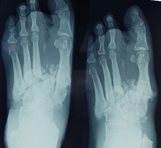
Case reports

                                               org/10.2337/dc13-2510                                                   Philadelphia
                                            Malhotra R, Chan CS, Nather A (2014) Osteomyelitis in the              Saavedra-Lozano J, Mejías A, Ahmad N, et al (2008)
                                               diabetic foot. Diabetic Foot & Ankle. 5:1. https://doi.                 Changing trends in acute osteomyelitis in children:
                                               org/10.3402/dfa.v5.24445                                                impact of methicillin-resistant Staphylococcus aureus
                                            McPherson DM (2002) Osteomyelitis in the neonate.                          infections. J Pediatr Orthop 28(5):569–75. https://doi.
                                               Neonatal Netw 21(1):9–22. https://doi.org/10.1891/0730-                 org/10.1097/bpo.0b013e31817bb816
                                               0832.21.1.9                                                         Senneville E, Lombart A, Beltrand E et al (2008) Outcome
                                            Morales Lozano R, González Fernández ML, Martinez                          of diabetic foot osteomyelitis treated nonsurgically: a
                                               Hernández D et al (2010) Validating the probe-to-bone                   retrospective cohort study. Diabetes Care 31: 637–42.
                                               test and other tests for diagnosing chronic osteomyelitis               10.2337/dc07-1744. https://doi.org/10.2337/dc07-1744
                                               in the diabetic foot. Diabetes Care 33:2140–5. https://             Sia IG, Berbari EF (2006) Osteomyelitis. Best Pract Res Clin
                                               doi.org/10.2337/dc09-2309                                               Rheumatol 20(6):1065–81
                                            Mutluoglu M, Lipsky BA (2016) Five things to kn0ow about               Smith IM, Austin OMB, Batchelor AG (2006) The treatment
                                               diabetic foot osteomyelitis. CMAJ, 118(17–18):E535                      of chronic osteomyelitis: A 10 year audit. J Plast
                                               https://doi.org/10.1503/cmaj.160228                                     Reconstr Aesthet Surg 59:11–5. https://doi.org/10.1016/j.
                                            Nair HKR (2017) Diabetic foot infection. In: Nathen A (ed)                 bjps.2005.07.002
                                               Diabetic foot: The Asian perspective, 2nd ed. Selangor:             Thomas-Ramoutar C, Tierney E, Frykberg R (2010)
                                               Uniquelink dot print Sdn Bhd: 58-67                                     Osteomyelitis and Lower Extremity Amputations in
                                            Parvizi J, Ghanem E, Azzam K, Davis E et al (2008)                         the Diabetic Population. The Journal of Diabetic Foot
                                               Periprosthetic infection: are current treatment strategies              Complications 2(1):18–24
                                               adequate? Acta Orthop Belg 74(6):793–800                            Tone A, Nguyen S, Devemy F et al (2014) Six-week versus
                                            Qadir M, Ali SR, Lakhani M, Hashmi P et al (2010)                          twelve-week antibiotic therapy for nonsurgically treated
                                               Osteomyelitis of the right humerus involving the right                  diabetic foot osteomyelitis: A multicenter open-label
                                               shoulder joint in an infant. J Pak Med Assoc 60(9):769–71               controlled randomized study. Diabetes Care 38(2):302–7.
                                            Ramsey SD, Newton K, Blough D, McCulloch DK et al (1999)                   https://doi.org/10.2337/dc14-1514
                                               Incidence, outcomes, and cost of foot ulcers in patients            Trampuz A, Zimmerli W (2006) Diagnosis and treatment
                                               with diabetes. Diabetes Care 22(3):382–7. https://doi.                  of infections associated with fracture-fixation devices.
                                               org/10.2337/diacare.22.3.382                                            Injury 37(2):S59–66. https://doi.org/10.1016/j.
                                            Rao N, Ziran BH, Lipsky BA (2011) Treating osteomyelitis:                  injury.2006.04.010
                                               antibiotics and surgery. Plast Reconstr Surg                        Waldvogel FA, Medoff G, Swartz MN (1970) Osteomyelitis:
                                               127(1): 177S–87S. https://doi.org/10.1097/                              a review of clinical features, therapeutic considerations,
                                               prs.0b013e3182001f0f                                                    and unusual aspects. N Engl J Med 282(4):198–206.
                                            Rosenberg AE (2010) Bones, joints, and soft-tissue                         https://doi.org/10.1056/nejm197001222820406
                                               tumors. In: Kumar V, Abbas AK, Fausto N et al. (eds)                Whyte NS, Bielski RJ (2016) Acute hematogenous
                                               Pathologic Basis of Disease. (8th edn) Saunders Elsevier,               osteomyelitis in children. Pediatr Ann 45(6): 204–208

                                                                                                            Call for abstracts
                                                                                                          Wounds UK are pleased to announce the call for abstracts for the
                                                                                                          2021 Annual Conference, which we fully expect to be a face-to-face
                                                                                                          event this year. It will be held at the Harrogate Convention Centre
                                                                                                          on 8-10 November. Following such a challenging period, this will be
                                                                                                          a long awaited celebration of all that is good in Tissue Viability.

                                                                                                          Entries for the e-poster exhibition require you to submit an abstract.
                                                                                                          Every entry received will automatically be considered for the
                                                                                                          Wounds UK Award for Excellence 2021.

                                                                                                          All abstracts will be reviewed by our judging panel, who will be
                                                                                                          looking to accept submissions that display high levels of innovation,
                                                                                                          relevance to current and/or best practice and provide high-quality
                                                                                                          research/evidence.

                         To submit your abstract please use the following link                            This year’s categories are:
                         www. surveymonkey.co.uk/r/ WUKH21                                                COVID-19, CASE STUDY, COST, DIABETIC FOOT,
                                                                                                          INFECTION, PHD PRESENTATION, PRACTICE,
                                                                                                          RESEARCH, SCIENCE, SKIN INTEGRITY, OTHER
                          THE WINNER OF THE WOUNDS UK AWARD FOR                                           Deadline for submissions is
                        EXCELLENCE WILL RECEIVE A FREE 3-DAY DELEGATE
                           PASS WITH ENTRANCE TO THE GALA DINNER                                          1 AUGUST 2021
                                                                                                          All successful entries will be notified by 30 SEPTEMBER 2021

                       Please contact the events team on info@omniamed.com or 020 3735 8244 if you have
                       any questions or require further information                                       Poster presentations will be presented on electronic poster displays only, no
                                                                                                          hard copies will be on display

Wounds Asia 2021 | Vol 4 Issue 1 | ©Wounds Asia 2021 | www.woundsasia.com                                                                                                                 25

You can also read