Case Report Giant Posttraumatic Angiolipoma of the Forearm: A Case Report and Review of the Literature - Hindawi.com
←
→
Page content transcription
If your browser does not render page correctly, please read the page content below
Hindawi
Case Reports in Orthopedics
Volume 2021, Article ID 4047777, 6 pages
https://doi.org/10.1155/2021/4047777

Case Report
Giant Posttraumatic Angiolipoma of the Forearm: A Case Report
and Review of the Literature

          Athanasios Fotiadis ,1 Petros Ioannidis,1 Ioannis Skandalos ,2 Stergios Papastergiou,1
          Aristeidis Vrettakos,1 Theodoros Tzigkalidis ,3 and Themistoklis Vampertzis 4
          1
            Orthopaedic Surgery and Traumatology-Unit for Sport Injuries, Agios Pavlos General Hospital of Thessaloniki,
            55134 Thessaloniki, Greece
          2
            Surgical Department, Agios Pavlos General Hospital of Thessaloniki, 55134 Thessaloniki, Greece
          3
            Department of Pathology, Agios Pavlos General Hospital of Thessaloniki, 55134 Thessaloniki, Greece
          4
            Trauma and Orthopaedics, Bedford Hospital NHS Foundation Trust, MK42 9DJ Bedford, UK

          Correspondence should be addressed to Athanasios Fotiadis; thanos.fotiadis@hotmail.com

          Received 12 May 2021; Accepted 3 July 2021; Published 21 July 2021

          Academic Editor: Akio Sakamoto

          Copyright © 2021 Athanasios Fotiadis et al. This is an open access article distributed under the Creative Commons Attribution
          License, which permits unrestricted use, distribution, and reproduction in any medium, provided the original work is
          properly cited.

          Angiolipoma is a type of lipoma, a benign soft tissue tumor. It is distinguished by the excessive degree of vascular proliferation and
          the presence of mature adipocytes. It occurs commonly on the trunk and extremities. Angiolipomas larger than 4 cm are classified
          as “giant,” and due to their size, histological evaluation is necessary to exclude malignancy. We report a case of a male patient who
          suffered from a giant noninfiltrating intramuscular angiolipoma which formed after venipuncture in the antecubital fossa. Clinical
          examination showed a palpable painless soft mass. Computed tomography (CT) and magnetic resonance imaging (MRI)
          demonstrated a giant angiolipoma on the right forearm. Surgical removal of the mass was performed, and the biopsy was
          negative for malignancy. To the best of our knowledge, this is the first report in the literature of posttraumatic intramuscular
          angiolipoma. Physicians and orthopedic/general surgeons should be aware of the possibility of soft tissue masses in a
          posttrauma situation.

1. Introduction                                                               The authors present a case of a giant angiolipoma of the
                                                                          forearm in a male patient who reported a venipuncture in
Angiolipomas are benign soft tissue tumors of mesenchymal                 the affected area, 12 months before our clinical evaluation.
origin which are made of mature adipocytes with an exces-                 To our knowledge and after reviewing the current literature,
sive degree of vascular proliferation [1]. They can occur in              there are no confirmed cases of posttraumatic intramuscular
every part of the body, but they are more common on the                   angiolipoma.
extremities, trunk, head, and neck. They are often asymp-
tomatic and painless except when they cause a mass effect                  2. Case Presentation
[2]. Appropriate imaging is useful to depict the nature and
the margins of the lesion, while histological evaluation is nec-          A 62-year-old male farmer presented in the outpatient clinic
essary for the correct diagnosis [3]. The pathogenesis of                 with a large mass on the volar surface of his right forearm
angiolipomas is still unclear, but acute or recurrent trauma              (dominant arm). The patient reported that the mass started
has been suggested as a possible etiologic factor [1, 4, 5].              appearing right after a venipuncture in the region of the
2 Case Reports in Orthopedics
antecubital fossa, about 12 months ago. Since then, the mass
gradually increased in size with a more rapid growth over the
past 6 months. The patient complained of slight discomfort
during flexion of the elbow. No history of previous trauma in
the affected area was reported. Past medical history included
arterial hypertension. In addition, he was a social drinker and
a nonsmoker.
Physical examination revealed a large, palpable, and
painless soft solid mass on the upper half of the right forearm
(Figure 1). No overlying skin changes were found. Range of
motion of the elbow, forearm, and wrist was normal. There
were no sensory or motor defects, and peripheral circulation
was normal. X-rays were negative for bone pathology. Blood
tests were normal.
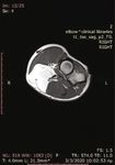
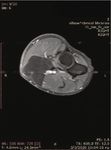
A CT and MRI with contrast demonstrated a well-encap- Figure 1: Preoperative view of the patient’s forearm.
sulated, solid multilobular lesion with lipoid content and the
presence of septa (Figures 2 and 3). The enhancement of the At present, the WHO Classification of Soft Tissue
septa demonstrated the rich vascularity of the lesion. There Tumors classifies angiolipomas in the group of adipocytic
were no signs of infiltration of the surrounding tissues. The tumors, the largest group of mesenchymal tumors (the same
dimensions of the lesion were 4:8 × 4:7 × 7:03 cm. The find- as lipomas, liposarcomas, etc.) [8]. It is described as a benign
ings suggested a lipoma, but the imaging could not exclude soft tissue tumor subdivided into infiltrative and noninfiltra-
a sarcomatous transformation due to the presence of septa tive types. The rare infiltrative, nonencapsulated type typi-
and of the dishomogeneity of the signal’s density. cally involves deep soft tissues and is separated from the
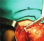
A lazy-S incision was performed, and the mass was cutaneous (encapsulated) lesion because of its growth pattern
removed surgically. The lesion occupied almost the entire and tendency to recur locally: this lesion is now best classified
upper half of the forearm including the antecubital fossa. It as intramuscular hemangioma [9].
was located subcutaneously above the radial artery near the Angiolipomas are more common in young adults (infil-
bifurcation of the brachial artery and between the brachiora- trating angiolipomas are usually diagnosed in older patients),
dialis and pronator teres muscles (Figure 4). The margins of they are equally distributed between sexes and occur mostly
the lesion were marked with metallic clips to determine the in the extremities (2/3 of the cases in the forearm), trunk,
margins in case of malignancy and radiation therapy. spinal axis, head, and neck, and their size almost never
The removed mass’s dimensions were 5 × 5 × 8 cm, and exceeds 4 cm [5, 10]. Multiple lesions are seen in approxi-
its weight was 68 grams (Figure 5). Histological examination mately 70-80% of cases, and 5% of these are familiar but with
showed mature adipose tissue with a vaguely lobular archi- an unclear genetic pattern [5]. A recent study suggests an
tecture, due to areas of excess fibrin deposition in the stroma involvement of chromosome 13 in angiolipomas [11]. Clini-
(Figure 6(a)). Additionally, in a few subcapsular areas, there cally, the encapsulated/noninfiltrative angiolipoma presents
were evident small groups of tiny thin-walled hyperplastic as a subcutaneous nodule: the lesions are commonly multi-
vascular vessels, most of which exhibited the formation of ple, typically firm, tender to palpation but often painful,
fibrous thrombi in their lumens (Figure 6(b)). These mor- and rarely associated with overlying skin changes [2, 5, 9,
phological characteristics were compatible with the diagnosis 10]. Pain and associated neuropathies are secondary to vascular
of angiolipoma [6]. engorgement and edema that can lead to compression of the
12 months postoperatively, the patient was asymptomatic. adjacent neural tissue [10]. Because of the subcutaneous loca-
tion, their—usually—small size, and their indolent clinical
3. Discussion appearance, angiolipomas are often misdiagnosed as ordinary
small lipomas and rarely imaged [12].
Although Bowen in 1912 first described a case of angiolipoma The diagnosis can be aided by ultrasonographic analysis
as a different entity from the generic diagnostic term “lipoma” (US), computed tomography (CT), or magnetic resonance
[7], it was Howard and Helwig who established angiolipoma imaging (MRI), but the microscopic examination is neces-
as an entity in 1960, describing it as a benign encapsulated sary for a conclusive diagnosis. US analysis is not very
and lobulated tumor that differs from lipoma by the presence specific but can be useful for differentiating subcutaneous
of an excessive degree of vascular proliferation [1]. In 1966, J. J. angiolipomas from ordinary lipomas by observing color
Lin and F. Lin described 2 entities in angiolipomas, the infil- Doppler flows [12]. CT contrast scans show a central low-
trating and noninfiltrating (encapsulated) types that should density mass (lipomatous component) surrounded by areas
be treated differently because of their different biological of contrast enhancement (vascular component); studies
behavior [2]. Their criteria to determine angiolipoma were as without contrast show the homogenous low attenuation of
follows: (1) gross evidence of tumor formation with or without a typical lipoma and may not define the margins and, there-
a capsule, (2) microscopic evidence of mature lipocytes as the fore, the exact extent of the lesion [13]. Recent studies have
major population (at least 50%) of the tumor, and (3) micro- shown that MRI with contrast defines well the margins of
scopic evidence of angiomatous proliferation inside the tumor. the lesion and demonstrates the presence of septa and theCase Reports in Orthopedics 3
Figure 2: CT scans.
(a) (b) (c)
Figure 3: MRI images (performed with a 1.5-Tesla unit): (a) axial T1-weighted image; (b) axial T1 fat suppression; (c) sagittal T1-weighted image.
(a) (b)
Figure 4: (a) Intraoperative view of the lesion and (b) residual cavity after the excision of the angiolipoma.4 Case Reports in Orthopedics
Figure 5: Macroscopic appearance of the surgical specimen.
(a) (b)
Figure 6: Histological examination of the lesion: (a) mature adipose and proliferated vascular tissue (hematoxylin and eosin; magnification
×40); (b) hyperplastic vessels with fibrous thrombi (hematoxylin and eosin; magnification ×100).
enhancement reveals the dense capillary proliferation. These lipoma, parosteal lipoma, lipoma of joint or tendon sheath,
results suggest that MRI is a useful noninvasive method in and lipoma arborescens), and liposarcoma [5, 10].
diagnosing and evaluating angiolipomas [3]. The treatment of noninfiltrating angiolipomas is simple
Microscopic examination of the surgical specimen is surgical excision, and they show no tendency to recur.
necessary for the conclusive diagnosis. The histopathologic Fine-needle aspiration is rarely diagnostic. Treatment of
characteristics were described by J. J. Lin and F. Lin as encap- infiltrating angiolipomas consists of wide surgical excision,
sulated (noninfiltrating angiolipomas) or nonencapsulated but there is a recurrence rate of 50%. When it is not associ-
(infiltrating angiolipomas) lesions with, at least, 50% evidence ated with pain or has a small size (under 5 cm), it can be
of mature adipocytes and angiomatous proliferation in the followed up without surgical treatment [3, 14].
tumor, in various ratios [2]. The presence of fibrinous micro- The pathogenesis of this lesion remains unclear, and the
thrombi is a distinctive feature that differentiates angiolipomas origin of angiolipomas is still controversial. Increased famil-
from other lipomas. Angiolipomas have not been found to iar incidence has been reported (approximately 5%) but does
undergo malignant transformation [1, 5], but histological eval- not exceed that of ordinary lipomas [5]. Howard and Helwig
uation is necessary if measuring larger than 5 cm in a single and J. J. Lin and F. Lin suggest that angiolipomas originate as
dimension [14, 15]. congenital lipomas that undergo vascular proliferation after
The differential diagnosis of angiolipomas include lipomas further stimuli such as trauma [1, 2].
and other lipoma variants (lipomatosis, myolipoma, chon- The relationship between trauma and formation of post-
droid lipoma, hibernoma, spindle-cell lipoma, atypical lipoma, traumatic “tumors” has been investigated since 1935, and
pleomorphic lipoma, and lipoblastoma), hemangiomas, Ewing suggested that repeated or acute trauma could, rarely,
benign lesions affecting bone, joints, or tendons (intraosseous cause a soft-tissue tumor or aggravate a preexisting one [4].Case Reports in Orthopedics 5
Since then, various authors have reported cases of posttrau- [2] J. J. Lin and F. Lin, “Two entities in angiolipoma. A study of
matic lipomas, and still to date, their genesis remains unclear. 459 cases of lipoma with review of literature on infiltrating
In their most recent study of 31 cases with posttraumatic angiolipoma,” Cancer, vol. 34, no. 3, pp. 720–727, 1974.
lipomas and review of the literature, Aust et al. found an [3] Y. Kitagawa, M. Miyamoto, S. Konno et al., “Subcutaneous
elevated PTT in their patients and reported a link between angiolipoma: magnetic resonance imaging features with histo-
manifest or subclinical coagulation disorders and develop- logical correlation,” Journal of Nippon Medical School, vol. 81,
ment of posttraumatic lipomas that are defined as adipose no. 5, pp. 313–319, 2014.
tissue tumors forming at the location of a precedent trauma, [4] J. Ewing, “The modern attitude toward traumatic cancer,” Bul-
with a marked gender predominance towards women. They letin of the New York Academy of Medicine, vol. 11, no. 5,
also reviewed precedent studies and possible theories on their pp. 281–333, 1935.
formation [16]. The first “mechanical effect” of trauma the- [5] J. Arenaz Búa, R. Luáces, F. Lorenzo Franco et al., “Angioli-
ory [17, 18], suggests that acute or repeated trauma leads to poma in head and neck: report of two cases and review of
a prolapse of adipose tissue through Scarpa’s fascia forming the literature,” International journal of oral and maxillofacial
surgery, vol. 39, no. 6, pp. 610–615, 2010.
a pseudolipoma (not a true lipoma because it is not encapsu-
lated). The second theory based on studies with encapsulated [6] C. D. M. Fletcher, J. A. Bridge, P. C. W. Hogendoorn, and
lipomas without lesions in Scarpa’s fascia [19–21] suggests F. Mertens, WHO Classification of Tumors of Soft Tissue and
Bone, IARC, Lyon, 2013.
that the induction of the differentiation of mesenchymal pre-
cursors (preadipocytes) to mature adipocytes and the effect [7] J. T. Bowen, “Multiple subcutaneous hemangiomas, together
with multiple lipomas, occurring in enormous numbers in an
of local/systemic inflammation and hormonal factors (such
otherwise healthy, muscular subject,” The American Journal
as growth hormones and sex steroids) can induce the forma-
of the Medical Sciences, vol. 144, no. 2, pp. 189–192, 1912.
tion of posttraumatic lipomas.
[8] J. C. Vilanova, WHO Classification of Soft Tissue Tumors,
Posttraumatic angiolipomas are a very rare clinical diag-
Springer International Publishing, 2017.
nosis. With their case report and after review of the literature,
Morgan et al. reported—in total—2 cases of cranial intraoss- [9] L. W. Bancroft, M. J. Kransdorf, J. J. Peterson, and M. I.
O’Connor, “Benign fatty tumors: classification, clinical course,
eous angiolipomas, a very rare benign tumor of the bone,
imaging appearance, and treatment,” Skeletal Radiology,
with a possible association with head trauma [22]. vol. 35, no. 10, pp. 719–733, 2006.
[10] T. B. Grivas, O. D. Savvidou, S. A. Psarakis et al., “Forefoot
4. Conclusion plantar multilobular noninfiltrating angiolipoma: a case report
and review of the literature,” World journal of surgical oncol-
Angiolipomas are benign tumors of soft tissue composed of ogy, vol. 6, no. 1, p. 11, 2008.
mature adipocytes with an excessive degree of vascular pro- [11] I. Panagopoulos, L. Gorunova, K. Andersen, I. Lobmaier,
liferation. They occur more commonly on the extremities B. Bjerkehagen, and S. Heim, “Consistent involvement of
and trunk/head of young adults, and their size rarely exceeds chromosome 13 in angiolipoma,” Cancer Genomics & Proteo-
4 cm. Among imaging tools, MRI with contrast is the most mics, vol. 15, no. 1, pp. 61–65, 2018.
sensitive imaging tool to better define the lesion. Histological [12] M. Bang, B. S. Kang, J. C. Hwang et al., “Ultrasonographic
analysis is necessary to define the diagnosis, and surgical analysis of subcutaneous angiolipoma,” Skeletal Radiology,
excision is the treatment of choice, especially in the presence vol. 41, no. 9, pp. 1055–1059, 2012.
of giant lesions. In the past years, more studies suggest that [13] Y. Matsuoka, K. Kurose, O. Nakagawa, and J. Katsuyama,
trauma can induce the formation of lipomas and various “Magnetic resonance imaging of infiltrating angiolipoma of
mechanisms were presented trying to explain the formation the neck,” Surgical Neurology, vol. 29, no. 1, pp. 62–66, 1988.
of posttraumatic lipomas. Physicians and surgeons should [14] C. J. Johnson, P. B. Pynsent, and R. J. Grimer, “Clinical features
be aware of the possibility and recognize the formation of soft of soft tissue sarcomas,” Annals of the Royal College of Sur-
tissue masses in a posttrauma situation. To our knowledge geons of England, vol. 83, no. 3, pp. 203–205, 2001.
and after reviewing the current literature, there are no con- [15] B. Allen, C. Rader, and A. Babigian, “Giant lipomas of the
firmed cases of posttraumatic intramuscular angiolipomas. upper extremity,” The Canadian Journal of Plastic Surgery,
vol. 15, no. 3, pp. 141–144, 2007.
Consent [16] M. C. Aust, M. Spies, S. Kall et al., “Lipomas after blunt soft tis-
sue trauma: are they real? Analysis of 31 cases,” The British
Informed consent was obtained from the patient for publica- Journal of Dermatology, vol. 157, no. 1, pp. 92–99, 2007.
tion of this case report and any accompanying images. [17] L. Rozner and G. W. Isaacs, “The traumatic pseudolipoma,”
The Australian and New Zealand Journal of Surgery, vol. 47,
Conflicts of Interest no. 6, pp. 779–782, 1977.
[18] N. I. Elsahy, “Posttraumatic fatty deformities,” European Jour-
The authors declare that there is no conflict of interest. nal of Plastic Surgery, vol. 12, no. 2, pp. 208–211, 1989.
[19] J. H. Penoff, “Traumatic lipomas/pseudolipomas,” The Journal
References of Trauma, vol. 22, no. 1, pp. 63–65, 1982.
[20] M. Signorini and G. L. Campiglio, “Posttraumatic lipomas:
[1] W. R. Howard and E. B. Helwig, “Angiolipoma,” Archives of where do they really come from?,” Plastic and Reconstructive
Dermatology, vol. 82, no. 6, pp. 924–931, 1960. Surgery, vol. 101, no. 3, pp. 699–705, 1998.6 Case Reports in Orthopedics
[21] E. Copcu and N. S. Sivrioglu, “Posttraumatic lipoma: analysis
of 10 cases and explanation of possible mechanisms,” Derma-
tologic Surgery, vol. 29, no. 3, pp. 215–220, 2003.
[22] K. M. Morgan, S. Hanft, and Z. Xiong, “Cranial intraosseous
angiolipoma: case report and literature review,” Intractable &
Rare Diseases Research, vol. 9, no. 3, pp. 175–178, 2020.You can also read


