Cardiac Fulcrum - Medtext Publications
←
→
Page content transcription
If your browser does not render page correctly, please read the page content below
Cardiovascular Surgery International                                                                                                                   ISSN 2692-7969

Research Article

                                                          Cardiac Fulcrum
Trainini Jorge1*, Beraudo Mario2, Wernicke Mario3, Trainini Alejandro1,2, Lowenstein Jorge4, Bastarrica María Elena2 and Lowenstein Diego4
Department of Cardiac Surgery, Hospital Presidente Perón, Argentina
1

Department of Cardiac Surgery, Clínica Güemes, Argentina
2

Department of Pathology, Clínica Güemes, Argentina
3

Department of Cardiology, Investigaciones Médicas, Argentina
4

Abstract
Objective: The cardiac muscle cannot be anatomically free in the thorax. Therefore, think and analyze that there could be a myocardial support point (lever
fulcrum).
Material and methods: They were used: 1) cardiac dissection in ten young (two years old) bovine hearts (800 g to 1000 g); 2) cardiac disection in eight human
hearts: one embryo, 4 g; one 10 years old, 250 g; and six adult, mean weight 300 g. The myocardial band was unrolled in its entirety. The extracted pieces were
analyzed by anatomy and histology.
Results: In anatomical investigations we have found in the all human and bovine hearts studied a nucleus underlying the right trigone of bone, chondroid or
tendon histological structure. The microscopic analysis revealed in bovine hearts a trabecular osteochondral matrix (fulcrum). In the ten year old human heart
and in the fetus, a central area of the fulcrum formed by chondroid tissue was found. Histology found a tendon matrix in adult human hearts. This fulcrum is
attached to the myocardium and would serve to support both the origin and the end of the myocardium.
Conclusion: The cardiac fulcrum found in the anatomical investigation of bovine and human hearts would clarify the point of support of the myocardial muscle
to complete its rotating function.
Keywords: Heart; Cardiac anatomy; Ventricular band; Cardiac structure

Introduction                                                                       distortion [8,9]. Therefore, we think and analyze that there could be a
                                                                                   myocardial support point (lever fulcrum).
     The development of the myocardial band exposed in 1970 by
Torrent Guasp [1,2] allows to see that it starts and ends at the origin            Material and Methods
of the great vessels, therefore the anchoring of the fibers is not done                The methods used were:
in the atrioventricular rings. The myocardium is attached to these
rings but not inserted into it. The myocardial band consists of a set of               1) Cardiac dissection in ten bovine hearts (two years old, 800 g
muscle fibers twisted on themselves like a string (theory of the string),                  to 1000 g).
flattened laterally as a band, which by turning two spirals defines a                  2) Cardiac disection in eight human hearts: one embryo, 4 g; one
helicoid that delimits the two ventricles and conforms its functionality                   10 years old, 250 g; and six adult, mean weight 300 g.
[3,4]. However, Torrent Guasp did not analyze the possibility of
supporting the heart muscle, as the muscle has for its function.                       3) Histology was performed with 10% formalin buffer
                                                                                           hematoxylin-eosin stain; Masson’s           trichrome      staining
    Later, Maclvear [5-7] considered that the ventricular walls we                         technique and four microns seccions.
are made up of an intricate Three-Dimensional (3D) network of
aggregated cardiomyocytes specifying: “None of the histological                        The hearts examined correspond to material from morgue
studies of the myocardium that we are aware, in contrast, have                     (human) and slaughterhouses (bovine). The study had the approval
provided any evidence for an origen and insertion as described for the             of the Ethics Committee of all the institutions involved. The key
alleged unique myocardial band” [5].                                               maneuver to achieve myocardial unwinding consists in severing
                                                                                   superficial fibers called interventricular or aberrant fibers [1] that
    In this research we consider that the muscle fibers are                        extend transversely through the anterior aspect of the ventricles to be
inevitably forced to "intertwine" with the cardiac fulcrum to fulfill              able to enter through the anterior interventricular groove. It should
its hemodynamic function of shortening-torsion and elongation-                     be understood that as the myocardial band is unfolded, separating
                                                                                   the pulmonary artery and the pulmo-tricuspid cord (anterior) from
Citation: Jorge T, Mario B, Mario W, Alejandro T, Jorge L, María Elena
                                                                                   the ascending segment (posterior), the vision of the homogeneous
B, et al. Cardiac Fulcrum. Cardiovasc Surg Int. 2021;2(1):1011.
                                                                                   anatomical reality is lost. This concurrence of the beginning and end
Copyright: © 2021 Trainini Jorge                                                   of the muscle band in the cardiac fulcrum constitutes a meeting point
Publisher Name: Medtext Publications LLC                                           between the right segment and the ascending segment, origin and end
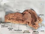
                                                                                   of the myocardial band (Figure 1).
Manuscript compiled: Mar 11th, 2021
*Corresponding author: Jorge Carlos Trainini, Hospital Presidente
                                                                                   Results
Perón, Avellaneda, Provincia de Buenos Aires, Argentina, Tel: + 5411                   This structure, which we have called cardiac fulcrum, was the
15 40817028; E-mail: jctrainini@hotmail.com                                        only perceptible edge where the muscle band fibers originate and end

    © 2021 - Medtext Publications. All Rights Reserved.                      031                                                2021 | Volume 2 | Article 1011
Cardiovascular Surgery International
Figure 2: Cardiac fulcrum (bovine heart). (A): Resected piece; (B): Mature
trabecular bone forming the cardiac fulcrum tissue. Hematoxylin-eosina stain
Figure 1: Unfolding of the myocardium. at low magnification (10x); (C): Cardiac fulcrum in other view.
(Figures 2 and 3). In analogy, as with skeletal muscle, we found in
the myocardial muscle that its contraction takes place between a fixed
point of support (insertion of the ascending segment in the fulcrum)
and a more mobile one (insertion of the right segment in the anterior
face of the fulcrum) (Figures 3 and 4). This last point was shown in the
dissection of a fragile character, totally opposite to the solidity of the
opposite end of the band in its attachment to the fulcrum.
Histology
In bovine, microscopy of the cardiac fulcrum finds an
osteochondral matrix (Figure 2). The size of the fulcrum was 45 mm
× 15 mm and its shape was triangular. The same structure was found
in chimpanzees [10].
In the 10-year-old human heart, Figure 5A shows that the central
area of the fulcrum is formed by chondroid tissue. It is logical, given
Figure 3: Cardiac fulcrum (human hearts). (A): 10-year-old; (B): 23 week
the age, that the fulcrum is smaller and has more chondroid tissue gestation human embryo heart; (C): fulcrum resected from an adult human
than bone. In the 23-week-old human fetus, this finding was repeated. heart.
Characteristic prechondroid bluish areas can be seen in a myxoid
stroma (Figure 5B).
The osseous structure in the bovine os cordis and its relationship
with the myxoid-chondroid cardiac fulcrum texture in human hearts,
even in gestational stages, is rational for the interpretation analysis.
This disparity is associated with the different age evolution from
chondroid to osseous material and by the greater force developed in
bovids requiring a more rigid supporting point.
However, the histological analysis of the fulcrum in adult human
hearts evidenced a tendinous collagenous matrix, needing an additional
clarification. In principle, there is constancy in the detection, site and
morphology of the fulcrum in all the hearts analyzed. This means that
from a functional point of view, its presence is akin to myocardial
band insertion, as established in the histological analysis, becoming
a solid point of interpretation to achieve its biomechanical function.
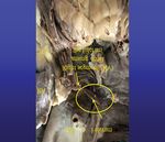
In this supporting point, the muscle fibers are inevitably forced to Figure 4: Fulcrum in the adult human heart. Note the right segment corre-
“intertwine” with the connective, chondroid or osseous fulcrum, and sponding to the Right Ventricle (RV) arising from the fulcrum.
our anatomical and histological investigations have shown that this
insertion attaches both the origin and end of the myocardial band anterior septal band are born, both belonging to the right segment of
(Figures 6 and 7). the myocardial band, forming a muscular raphe, where precisely the
point of origin of the myocardial band is considered [11-13].
Discussion
We are located in front of the aortic valve at the level of the origin The pulmo-tricuspid cord, where the myocardial band begins
of the right coronary artery. In this place the bridge band and the with its right segment, is located in front of a compact area of
© 2021 - Medtext Publications. All Rights Reserved. 032 2021 | Volume 2 | Article 1011Cardiovascular Surgery International
fibrous connective tissue that surrounds the anterior two thirds
of the circumference of the U-shaped aortic ring, whose open end
(posterior) is occupied by the anterior leaflet of the mitral valve. At
its ends this tissue has two trigones. The right fibrous trigone has the
tricuspid valve on its right, the aortic ring behind it and the pulmo-
tricuspid cord anteriorly. The less prominent left trigones lies between
the left mitral valve on the left and the aorta. Both trigones are
connected medially by collagen fibers. In the continuity of the aortic
orifice with the posterior leaflet of the mitral there is no connective
tissue, since laterally, the two fibrous bodies are continued by a band
of connective tissue that surrounds the orifice of the mitral valve
partially to gradually fade. The septal valve of the mitral is located
between both trigones as a wedge.
Adjacent to the right trigone (anterior and inferior) we found
a structure with solidity to palpation and homogeneous osteo-
Figure 5: (A): Ten year old human heart. Central area of the fulcrum formed chondroid histology where the fibers of the right segment and
by chondroid tissue. Hematoxylin-eosin stain (15x). (B): Cardiac fulcrum in ascending segment are tied. This insertion is the fulcrum of the
a 23-week gestation fetus showing prechondroid bluish areas in a myxoid
myocardium both at the level of its origin and that of its termination.
stroma. masson´s trichrome staining technique (15x).
In relation to this finding, research on this fixation point, which
we call cardiac fulcrum, becomes a piece that supports and allows
the band to exert with the necessary force the fundamental rotatory
movements of the left ventricle [14,15]. The fact that the band is
anchored to the cardiac fulcrum in its finalization corresponds to
the active movements of the cardiac cycle (systole and suction) that
involves the apical loop (descending and ascending segments).
Conclusion
The cardiac fulcrum found in the anatomical investigation would
clarify the point of support of the myocardial band to complete its
rotating function. Without its presence, the heart could not meet the
hemodynamic efficiency of ejecting blood at a speed of 300 cm/s.
References
1. Trainini JC, Elencwajg B, López Cabanillas N, Herreros J, Lago N, Lowenstein
J, et al. Stimuli propagation, muscle torsión and cardiac suction effect through
electrophysiological research. In. Basis of the New Cardiac Mechanics. The Suction
Pump. Trainini JC and col. Lumen, Bs. 2015:39-61.
Figure 6: Insertion of the myocardium in the fulcrum (bovine heart). (1): Myo- 2. Guasp FT, Buckberg G, Carmine C, Cox J, Coghlan H, Gharib M. The structure and
cardial fibers and myxoid stroma; (2): Myocardial tapes in a chondroid stroma function of the helical heart and its buttress wrapping. I. The normal macroscopic
(insertion); (3): Bony cortical tissue of the fulcrum.
structure of the heart. Semin Thorac Cardiovasc Surg. 2001;13(4):301-19.
3. Buckberg GD, Coghlan HC, Torrent Guasp F. The structure and function of the
helical heart its buttress wrapping. VI. Geometrics conceps of heart failure and use for
structural correction. Sem Thorac Cardiovasc Surg. 2001;13(4):386-401.
4. Ballester M, Ferreira A, Carreras F. The myocardial band. Heart Fail Clin.
2008;4(3):261-72.
5. MacIver DH, Stephenson RS, Jensen B, Agger P, Sanchez-Quintana D, Jarvis JC, et
al. The end of the unique myocardial band: Part I. Anatomical considerations. Eur J
Cardiothorac Surg. 2018;53(1):112-19.
6. MacIver DH, John B, Partridge JB, Agger P, Stephenson RS, Boukens BJD, et al. The
end of the unique myocardial band: Part II. Clinical and functional considerations.
Eur J Cardiothorac Surg. 2018;53(1):120-28.
7. Anderson R, Ho S, Redman K, Sanchez-Quintana D, Punkenheimer P. The
anatomical arrangement of the myocardial cells making up the ventricular mass. Eur
J Cardiothoracic Surg. 2005;28(4):517-25.
8. Trainini JC, Trainini A, Valle Cabezas J, Cabo J. Left Ventricular Suction in Right
Ventricular Dysfunction. EC Cardiol. 2019;2(1):572-7.
Figure 7: Cardiomyocytes penetrating the fibro-colagenous tissue (adult 9. Streeter DD, Spotnitz HM, Patel J, Ross J, Sonnenblick EH. Fiber orientation in the
human heart). (1): Cardiomyocytes; (2): Fibrocolagenous matrix. The circle canine left ventricle during diastole and systole. Biophysical J. 1969;24:339-47.
details the insertion site.
© 2021 - Medtext Publications. All Rights Reserved. 033 2021 | Volume 2 | Article 1011Cardiovascular Surgery International
10. Moittié S, Baiker K, Strong V, Cousins E, White K, Liptovszky M, et al. Discovery of os 13. Mora V, Roldán I, Romero E, Saurí A, Romero D, Perez-Gozabo J, et al. Myocardial
cordis in the cardiac skeleton of chimpanzees (Pan troglodytes). Sci Rep. 2020;10:9417. contraction during the diastolic isovolumetric period: analysis of longitudinal strain
by means of speckle tracking echocardiography. J Cardiovasc Dev Dis. 2018;5(3):41.
11. Carreras F, Ballester M, Pujadas S, Leta R, Pons-Lladó G. Morphological and
functional evidences of the helical heart from non-invasive cardiac imaging. Eur J 14. Kocica MJ, Corno AF, Carreras-Costa F, Ballester-Rodes M, Moghbel MC, Cueva CN,
Cardiothoracic Surg. 2006;29(Suppl 1):S50-5. et al. The helical ventricular myocardial band: global, three-dimensional, functional
architecture of the ventricular myocardium. Eur J Cardiothorac Surg. 2006;29(Suppl
12. Trainini JC, Elencwajg B, López-Cabanillas N, Herreros J, Lowenstein J,
1):S21-40.
Bustamante-Munguira J, et al. Ventricular torsion and cardiac suction effect: The
electrophysiological analysis of the cardiac band muscle. Interventional Cardiol. 15. Henson RE, Song SK, Pastorek JS, Ackerman JH, Lorenz CH. Left ventricular torsion
2017;9(1):45-51. is equal mice and humans. Am J Physiol Heart Circu Physiol. 2000;278:H1117-23.
© 2021 - Medtext Publications. All Rights Reserved. 034 2021 | Volume 2 | Article 1011You can also read


