Can biophysical models give insight into the synaptic changes associated with addiction?
←
→
Page content transcription
If your browser does not render page correctly, please read the page content below
Can biophysical models give insight into the synaptic changes
                                                                      associated with addiction?
                                                                      Mayte Bonilla-Quintana and Padmini Rangamani*
                                                                       Department of Mechanical and Aerospace Engineering,
                                                                       University of California San Diego, La Jolla CA 92093.
                                                                ∗ To whom correspondence must be addressed: prangamani@ucsd.edu

                                                                                                  Abstract
arXiv:2201.01735v1 [physics.bio-ph] 5 Jan 2022

                                                           Effective treatments that prevent or reduce drug relapse vulnerability should be developed to
                                                       relieve the high burden of drug addiction to society. This will only be possible by enhancing the
                                                       understanding of the molecular mechanisms underlying the neurobiology of addiction. Recent
                                                       experimental data have shown that dendritic spines, small protrusions from the dendrites that
                                                       receive input from excitatory neurons, from spiny neurons in the nucleus accumbens exhibit
                                                       morphological changes during drug exposure and withdrawal. Moreover, these changes relate
                                                       to the characteristic drug-seeking behavior of addiction. However, due to the complexity of
                                                       the dendritic spines, we do not yet fully understand the processes underlying their structural
                                                       changes in response to different inputs. We propose that biophysical models can enhance the
                                                       current understanding of these processes by incorporating different, and sometimes, discrepant
                                                       experimental data to identify the shared underlying mechanisms and generate experimentally
                                                       testable hypotheses. This review aims to give an up-to-date report on biophysical models of
                                                       dendritic spines, focusing on those models that describe their shape changes, which are well-
                                                       known to relate to learning and memory. Moreover, it examines how these models can enhance
                                                       our understanding of the effect of the drugs and the synaptic changes during disease progression.

                                                 1    Introduction
                                                 The highest burden of drug addiction to all societies is the immense loss of human capital – from
                                                 unfulfilled personal ambitions to loss of family structure. According to the National Institute on
                                                 Drug Abuse (1 ), in 2019 more than 70,000 Americas died from a drug-involved overdose, including
                                                 illicit drugs and prescription opioids. However, this number only represents a small percentage of
                                                 drug-related deaths. According to a statistical study (2 ), in 2016 the drug-associated deaths was
                                                 2.2 times the number of drug-coded deaths. Besides these losses, the estimated medical cost in
                                                 hospitals of substance use disorder in 2017 was $13.7 billion (3 ).
                                                      A continued medical aspiration for relieving the high burden of drug addiction in society is
                                                 to develop and tailor effective treatment to prevent relapse. Assuming that drug addiction is a
                                                 neuropsychiatric disease whose behavioral pathology consists in the propensity to relapse, even after
                                                 periods of abstinence, then an effective treatment should prevent or reduce relapse vulnerability (4 ).
                                                 Because current behavioral and pharmacological therapy only helps a small percentage of patients
                                                 (4 ), we need to understand the molecular mechanisms underlying the neurobiology of addiction.
                                                 In the past decade, studies have focused on the neurons in the nucleus accumbens (NAc), a brain
                                                 region that mediates goal-directed behavior, and their structural changes after drug exposure (4 –
                                                 7 ). These studies have noted that structural changes in neurons could be an important readout
                                                 of addictive behavior. For example, when animals are exposed to an environment associated with
                                                 previous cocaine consumption, it triggers cocaine-seeking behavior that is induced by structural
                                                 changes in NAc neurons (7 ). Another study showed that structural alterations to these neurons
                                                 are only present in animals that show a persistent increase in the psychomotor activating effects
                                                 of cocaine after repeated exposure to it, i.e., psychomotor sensitization (8 ). However, there is
                                                 no consensus on whether and how the structural changes in neurons derived from drug exposure

                                                                                                      1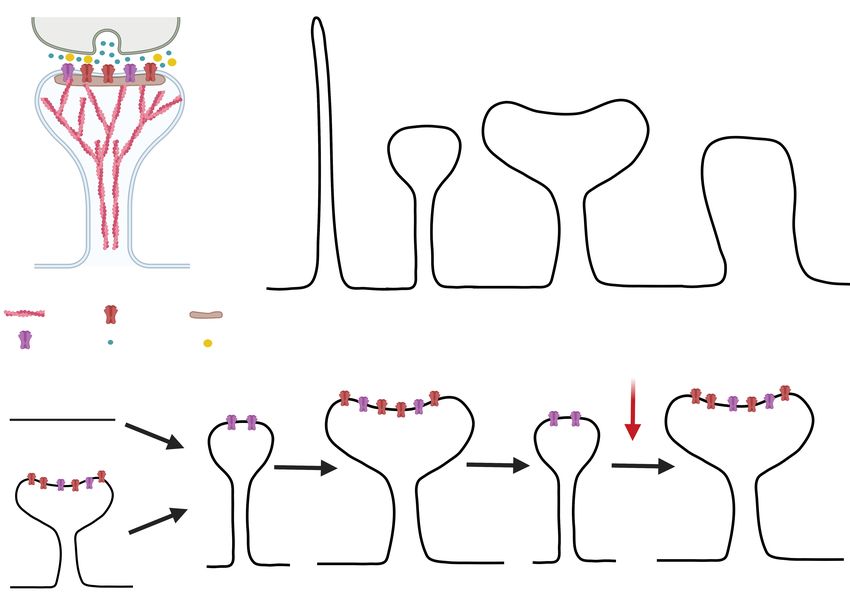
drive addictive behavior (4 , 5 ) due to opposing experimental data which show that the structural
changes in NAc neurons after cocaine exposure are not required for locomotor sensitization and
suggest that, instead, these changes may represent a compensatory process (9 ). Complicating
matters, studies have also shown that different drugs have different effects on these neurons (4 ,
5 ). For example, stimulants, such as cocaine, amphetamine, and nicotine, enhance the structural
complexity of the neurons, while narcotics, like morphine, decrease it (10 ).
    In this review, we explore the idea that experimentally-constrained biophysical computational
models that relate structural changes of neurons to different drug conditions may be able to narrow
down the possible mechanisms that may underlie the neurobiology of drug addiction. Although
there are a repertoire of theoretical models of drug use and addiction, they rely on theories of how
the brain solves computational, information-processing, and control problems (11 , 12 ). Some of
these models link these computations with specific brain areas (12 ). However, to our knowledge,
they do not consider the structural and molecular changes of the neurons and, instead, focus
on drug use behavior. Recently, biophysical models have informed the structural and molecular
mechanisms related to learning and memory (13 –19 ). Here, we review such models and describe
how they can broaden our understanding of the mechanisms behind addiction and relapse to aid the
treatments for drug addicts. We focus on dendritic spines, small protrusions from the dendrites that
receive excitatory input, and hence, form the postsynaptic part of the synapse. Dendritic spines are
thought to be standalone biochemical computational units (20 –22 ) and it is well-documented that
they undergo structural plasticity during learning and memory formation (23 ) and during drug
consumption and withdrawal (5 , 7 , 24 ). Therefore, biophysical analyses of spines may provide us
with the key links between these fundamental neurological processes.
    This work begins by giving an overview of the structure and dynamics of dendritic spines. Then,
we introduce the most studied forms of synaptic changes, namely, long-term potentiation (LTP)
and long-term depression (LTD), and relate them with the modifications associated with drug
addiction. After this biological background, we review different types of biophysical models and
discuss how they can enhance our understanding of drug addiction and other neurological diseases.
We finish by describing how these biophysical models can be extended to study memory formation
and storage, and identifying the opportunities for predictive model development.

2     Dendritic Spines
2.1   Morphology
Although dendritic spines were first described by Santiago Ramón y Cajal over a century ago,
it has only recently become possible, through the development of novel imaging techniques, to
investigate their structure-function relationships. The dendritic spine is a bulbous protrusion from
the dendritic shaft often connected by a thin neck, creating a spine head. The postsynaptic density
(PSD) is located at the tip of the spine head, close to the presynaptic terminal, and it is enriched
with receptors, proteins, and signaling molecules (Fig. 1A). The quantitative characteristics of
the spine morphology are broadly described by: the length and width of its neck; the volume of
its head; and the surface area of the PSD. These geometric features alone have been studied in
terms of correlations between the morphology and function of the spine (25 –27 ). For example, the
spine head volume correlates with the area of the PSD, the number of postsynaptic receptors, the
readily-releasable pool of transmitters, and the neck width (25 –27 ). Moreover, dendritic spines
that connect to the same axon have similar size and head volume demonstrating that they share
the same activation history (28 ).
    Dendritic spines also have diverse shapes. Thus, early studies classified them into three cate-

                                                 2
gories: 1) spines with long, thin necks and small heads (thin spines), 2) spines with thicker necks
and large heads (mushroom spines), and 3) short spines without well-defined necks (stubby spines)
(29 ) (Fig.1B). Experiments found that spine shapes were more distinct in adults than in young
animals, which suggested that stubby spines form from the dendritic shaft when the axon contacts
the membrane (shaft synapse) and then turn into thin or mushroom spines (30 ). However, the
current view is that a filopodia-like structure that emerges from the dendritic shaft may connect to
the axon to form a synapse (31 , 32 ) evolving to a thin spine which later matures to a mushroom
spine (33 ). Moreover, thin spines are more prone to respond to synaptic activity and change their
morphology accordingly, suggesting that they are learning spines that can mature to mushroom-
like spines, which are more stable and thus, thought to be memory spines (33 ). While mushroom
spines can persist for months, thin spines only last a few days (33 ). Dendritic spines have also
been divided depending on the size of their heads (34 ). On one hand, spines with small heads
(filopodia and thin spines) are motile and unstable. On the other hand, spines with large heads
(mushroom and stubby spines) are stable and have stronger connections. Thus, if we see spines as
memory devices, large-headed spines are “write protected”: they maintain preexisting connections
and prevent new memory formation, while small-headed spines are “write-enabled” because they
allow the acquisition of new memories (34 ).
In addition to their unique morphological features, spines have attracted considerable attention
of the signaling community because it has been demonstrated they compartmentalize calcium and
that spine necks can filter membrane potentials, thereby isolating biochemical and electrical synap-
tic input from each other (35 ). These findings have led to the hypothesis that spines implement
input-specific learning rules (25 ). The shape changes in spines, in response to synaptic input, are
possible by a reconfiguration of the spine cytoskeleton, mainly composed of actin filaments; actin
remodeling is triggered by a cascade of chemical reactions due to calcium influx into the spine
(36 –41 ). Changes in spine shape and size not only occur in development but also in adulthood
(42 ), for example, during motor skill learning (43 ) and fear learning and extinction (44 ). This
dynamic change to spine morphology is known as structural plasticity (45 ). Note that due to this
plasticity, the shapes of spines form a continuum instead of distinct categories (26 ).
2.2 Structural Plasticity
The connection between neurons can be strengthened or weakened in a process called synaptic
plasticity, and this change in the connections has a direct impact on learning and memory forma-
tion (42 , 46 –48 ). The most studied forms of these synaptic changes are long-term potentiation
(LTP), which strengthens the synapse, and long-term depression (LTD), which weakens it. In
LTP, glutamate released due to the high-frequency stimulation of the presynaptic neuron is cap-
tured by the α-amino-3-hydroxy-5-methyl-4-isoxazolepropionic acid receptors (AMPARs) located
at the PSD of the dendritic spine (Fig. 1A). Due to this glutamate influx, the spine depolarizes
and releases the Magnesium (Mg2+ ) ion of the N-methyl-D-aspartate receptors (NMDARs), which
allows the calcium (Ca2+ ) influx into the spine and triggers a cascade of reactions that increase
the number of AMPARs at the PSD and AMPARs mediated current. Such increase facilitates the
subsequent uptake of glutamate, and hence, the synapse strengthens. Moreover, dendritic spines
show an associated increase in size, thus, linking their function and morphology (22 , 45 –47 ). LTD
also depends on the activation of NMDARs, but it is induced by low-frequency stimulation of the
presynaptic cell, causing glutamate release. However, the amount of glutamate is not enough to
depolarize the postsynaptic cell and remove all the Mg2+ ions blocking the NMDARs, but only a
few. Hence, the Ca2+ influx is lower than in LTP and triggers a different set of chemical reactions
resulting in a decrease in the number of AMPARs at the PSD, and spine shrinkage (48 ).
3How is plasticity impacted by drugs? It has been shown that cocaine use impairs the induction
of LTP in spines of medium spiny neurons (MSNs) in the NAc (7 ). These spines are depressed after
exposure to cocaine, which generates silent synapses that lack AMPARs but have NMDARs (4 , 5 , 7 ,
24 ). The lack of AMPARs impairs LTP because these spines cannot respond to glutamate release.
Because silent synapses are highly abundant during development, it has been hypothesized that the
brain returns to a more juvenile state after drug exposure, which may underlie pathological drug-
seeking behaviors (49 ). Interestingly, AMPARs are reinserted into these silent synapses only when a
stimulus related to cocaine consumption is present (7 , 24 ). This synaptic potentiation induces drug
craving, and thus, may explain the high rates of relapse (7 ). Moreover, re-silencing these synapses
via optogenetic removal of AMPARs inhibits relapse-like behaviors (49 ). A recent study shows that
there is a time window in which previously silent synapses that are potentiated due to exposure
to cues related to drug consumption can be temporally destabilized and vulnerable to disruptions
before being consolidated (becoming mature) (24 ). Moreover, this window relates to the dynamics
of signaling molecules that alter the spine cytoskeleton. Preventing spine maturation during this
window decreases cue-induced cocaine seeking, which can be used for anti-relapse treatments (24 )
(Fig. 1C).
However, the precise neuronal and molecular substrates encoding the dynamics of drug memories
have not been fully identified (4 , 24 ). Moreover, until recently, new imaging techniques allowed for
the study of the mechanisms underlying the structural and molecular changes of the synapses (42 ),
but they remain incompletely understood due to the complexity of the synapse and the diverse
temporal and spatial scales of the experiments and underlying events.
A Axon B
Presynapse
Head
Postsynapse
Dendritic
Spine
Neck
Dendrite
Actin Filopodia Thin spine Mushroom spine Stubby spine
filament AMPAR PSD
NMDAR Glutamate Calcium
Intervention
C
No spine
Cocaine
Administration Drug Reconsolidation
Withdrawal
Challenge
Silent Spine Mature Spine Silent Spine Mature Spine
Mature Spine
Figure 1: Dendritic Spine. A) Schematic depiction of a synapse. The dendritic spine (postsynapse) is in blue
and the presynaptic bouton is in green. B) Different types of dendritic spines. C) Hypothesized evolution upon
cocaine administration, withdrawal, and drug challenge, proposed by (24 ). The red arrow signals the window when
the synapse is destabilized and treatments can take place to avoid reconsolidation. Created with BioRender.com
42.3 Signaling Pathways associated with structural plasticity
Upon LTP induction, there is an increase of the number of AMPARs in the PSD due to the
Ca2+ / calmodulin-dependent protein kinase II (CaMKII) mediated phosphorylation of AMPAR
auxiliary protein stargazin, which allows extrasynaptic diffusive receptors to bind to PSD95, a
PSD molecule (50 –52 ). The exocytosis of extrasynaptic AMPARs into the plasma membrane
depends on the rat sarcoma virus-extracellular signal-regulated kinase (RAS–ERK) pathway, RAB-
GTPase proteins, soluble N-ethylmaleimide-sensitive-factor attachment protein receptors (SNARE
proteins), syntaxin 4 and 13, and the motor protein myosin V (50 , 51 , 53 , 54 ). The RAS-ERK
pathway is stimulated by CaMKII upon LTP induction (54 , 55 ). Thus, CaMKII has a dual
function upon LTP: it immobilizes extracellular AMPARs in the PSD and replenishes the pool of
extrasynaptic receptors via exocytosis. Moreover, CaMKII is one of the most important molecules
for LTP. It is activated by calcium-calmodulin, which in turn is activated by Ca2+ influx into the
spine through NMDARs and it can act as a protein switch due to its autophosphorylation property.
Such a state lasts longer than calcium elevation, thus CAMKII acts as a biochemical integrator of
the multiple calcium pulses during LTP induction (50 ).
In addition to its involvement with AMPARs, CaMKII also affects the dendritic spine cytoskele-
ton by stabilizing actin filaments (56 ). Actin is a globular protein (G-actin) that forms filaments
(F-actin) and it is the main component of the spine cytoskeleton. F-actin is a polar structure
that continuously polymerizes G-actin at its plus end and it is depolymerized at its minus end.
In the spine, there is a dynamic equilibrium between G-actin and F-actin that is modulated by
actin-binding proteins (ABPs) which promote F-actin depolymerization and G-actin polymeriza-
tion (57 ). Moreover, the actin cytoskeleton is restructured upon LTP by an orchestrated interplay
between actin and ABPs (58 , 59 ), which allows for spine enlargement. In the basal state, F-actin
is bundled by CaMKII, which stabilizes the bundle. Upon NMDARs activation, CaMKII autophos-
phorylates and detaches from actin filaments, which allows CaMKII to interact with other molecules
and remodel the actin cytoskeleton (56 , 60 ). Furthermore, CaMKII regulates actin dynamics by
signaling pathways involving the Rho family of small GTPases, such as RhoA, Rac1, or Cdc42
(56 ). The activity of these GTPases is triggered by guanine-nucleotide-exchange factors (GEFs)
and suppressed by GTPase activating proteins (GAPs), and they coordinately regulate ABPs ac-
tivity. For example, RhoA controls profilin II and cofilin (56 , 61 , 62 ). The former binds to G-actin
and facilitates the polymerization of F-actin at its plus end. Cofilin, which is also controlled by
Rac1, has a dual function depending on its concentration. At low concentrations, cofilin severes
F-actin and at high concentrations, it promotes F-actin nucleation (63 ). Note that the nucleation
and severing events produce more actin filaments, and the combination of both events is likely to
be responsible for keeping a steady distribution of the F-actin length. Thus, cofilin regulation is
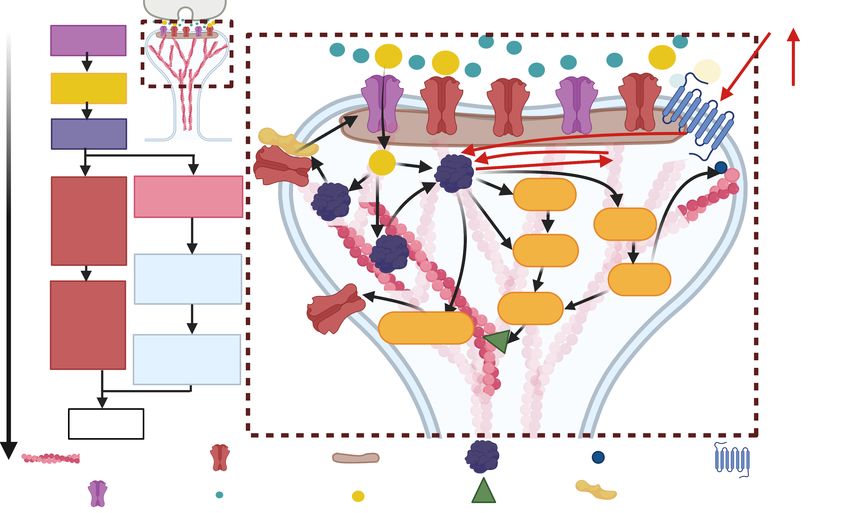
critical for actin dynamics. These pathways are depicted in Figure 2.
It has been shown that cocaine decreases active RhoA in the NAc MSNs, which could lead to a
decrease in cofilin (64 ), affecting the spine cytoskeleton. Moreover, activation of CaMKII by D1-
like dopamine receptors in the NAc reinstates cocaine-seeking behavior; this is associated with an
increase of AMPARs in the membrane (65 ). CaMKII also phosphorylates ∆Fos and it is required for
the cocaine-mediated accumulation of ∆Fos in NAc (66 ) (red lines in Fig. 2). ∆Fos is a Fos family
transcription factor that shows long-lasting accumulation in the NAc after chronic administration
of any drug of abuse, thereby supporting the view that changes in gene expression contribute to
drug addiction. Interestingly, it has been shown that ∆Fos upregulates the transcription of the
CaMKII gene selectively in the NAc shell and that CaMKII stabilizes ∆Fos, allowing for a greater
accumulation of ∆Fos that induces more CaMKII (66 ). However, how this positive feedback alters
the spines is not fully understood, because of the complexity of the signaling pathways. Moreover,
5the signaling pathways activated by LTP induction have diverse temporal and spatial extents (46 ),
and the coupling between the signaling pathways and spine structure is also complex (Fig.2). In
order to disentangle the multiple spatial and temporal complexities involved in LTP induction
and effects of drugs, we propose the theoretical models that incorporate biophysical details at
various levels can shed light into the emergent behaviors of spines. These modeling approaches
can elucidate the mechanisms underlying adaptation to addiction by allowing the incorporation of
different experimental observations, eliminate possibilities that are not physicochemically feasible,
and generate experimentally testable hypotheses.
time
Dopamine
0.1 s NMDAR
10 s Calcium
Cocaine
1 min CaMKII
ΔFos
4-5 AMPAR Actin
min laterial reorganization
Ras
diffusion RhoA
and
exocytosis transient
Rac1
spine ROCK
AMPAR enlargement
insertion LIMK
10
and Ras-ERK
clustering sustained
min at the PSD spine
enlargement
1 hr LTP
Actin filament AMPAR PSD CaMKII Profilin Dopamine
Cofilin Stargazin Receptor
NMDAR Glutamate Calcium
Figure 2: Signaling pathways associated with LTP in the dendritic spine. Right: events time-scale. Left:
scheme of the signaling events in the dendritic spine. CaMKII is activated upon the Ca2+ influx into the spine during
LTP induction, which triggers the Ras, Rac1, and RhoA pathways. CaMKII also induces AMPARs exocytosis via
activation of the Ras-ERK pathway and capture of AMPARs into the PSD. During cocaine consumption, dopamine
receptors trigger CaMKII activity and ∆Fos is increased. Figure created with BioRender.com
3 Biophysical Models of Dendritic Spines
Addictive behaviors share common features with learning models (67 ), and hence, learning theories
(68 ) can be adapted to investigate these behaviors. However, in this work, we focus on the structural
and functional changes in dendritic spines that can inform relapse propensity. We first present a
review of the models that investigate signaling pathways in the dendritic spine relevant to the
mechanisms involved in drug addiction, particularly those pathways that result in structural LTP
or LTD due to an increase or decrease of AMPARs and CaMKII dynamics. Then, we revise models
that have incorporated idealized and realistic spine shapes linking the structural and functional
characteristics of dendritic spines.
63.1 Signaling Models
Early models of synaptic plasticity, based on Ca+2 influx through the NMDARs, describe how
different levels of Ca+2 (69 , 70 ) or induction protocols (71 ) lead to an increase or decrease in the
synaptic weight, which describes the strength of the synaptic connection. Later models provide
a more detailed description of the biochemical reactions leading to either LTP or LTD (72 ), the
volume regulation of dentric spines upon LTP (73 ), or the change in AMPARs mediated current
promoting synaptic modifications (74 ). These signaling models use ordinary differential equations
(ODEs) to describe the biochemical reactions. They span from simple descriptions based on key
assumptions (71 ) to detailed descriptions of the different chemical pathways (74 ). As technological
advances allow for the study of specific signaling pathways, models have incorporated these findings
to test synaptic plasticity hypotheses (75 ). The increase in computational power has also allowed
the use of agent-based models (76 ), in which the molecules are agents that follow a set of rules,
mimicking chemical reactions leading to LTP. Some of these models include the CaMKII activity
(70 , 72 , 76 , 77 ) and GTPases (73 , 74 ).
These models test different hypotheses based on the available experimental findings and create
theories about how long-term information can be stored in the brain. For example, early models
test the idea that CaMKII provides a bistable switch (69 ) where “off” CaMKII is turned “on”
when there is a synaptic event and stays on for long periods due to the autophosphorylation
property of the kinase. Furthermore, this hypothesized switch can stay on for longer times than
protein turnover because the newly synthesized unphosphorylated CaMKII subunit could become
phosphorylated (78 ). The mathematical models show the feasibility of this hypothesis (69 ), and
expand it to add a phosphatase switch involved in LTD, providing a tristable system to examine
how a synapse can be bidirectionally modified (72 ). Models have also been used to systematically
investigate how the interaction between CaMKII and other molecules can control the sensitivity
of synapses to calcium signals (73 , 76 ). Moreover, the incorporation of actin remodeling revealed
that it added robustness to the dendritic spine response upon LTP (73 ). One common difficulty
for these models is parameter fitting since not all the reaction rates or protein concentrations are
known in the spines, due to experimental difficulties. Thus, informed assumptions and decisions are
made. One common assumption is that reaction rates are the same across cell types, and protein
concentrations are cell-type dependent; and hence, protein concentrations are fitted in the models
(74 ). Although parameters are fitted when unknown and varied to see their effects in the model
dynamics, they have to be within physiological regimes (73 , 74 ).
As experiments began to investigate the source of the AMPARs that incorporate into the PSD
upon LTP, i.e., whether they are exocytosed or laterally diffused into the PSD (51 , 79 –81 ), theoret-
ical studies examined the implications of the different sources to plasticity (15 , 82 –85 ). Moreover,
models started to explore how synapses are maintained over time periods that are longer than the
average lifetime of these receptors and other molecules (86 –88 ), and aided the study of AMPARs
clusters formation (88 , 89 ). These models propose different hypotheses for memory maintenance.
Instead of relying on a bistable molecular switch, like that of CaMKII, a model of clusters of AM-
PARs show that they are metastable with longer lifetimes than that of a single receptor, and that
synaptic weights, which depend on the number of AMPARs, can be bidirectionally changed (88 ).
An alternative hypothesis is that AMPARs exhibit a self-sustained switch related to their movement
to and from the membrane (86 ). In this model, the presence of receptors at the synapse enhances
the recruitment of more AMPARs, resembling the conversion from silent to active synapses (86 ).
Moreover, a model based on simplified biophysical assumptions shows cooperative binding and un-
binding of proteins at the PSD, replicating experimental observations (87 ). By performing model
simulations, the interplay between AMPARs and CaMKII dynamics has been examined (85 , 86 ).
7Another important player of structural plasticity, the actin filaments dynamics (36 , 38 ) and its
dynamic equilibrium (57 ), has also been investigated providing a quantitative description of the
change of the filamentous to globular actin ratio upon LTP observed experimentally (90 ). More-
over, computer simulations have aid the understanding of the F-actin binding and unbinding to
CaMKII (60 ).
3.2 Models Incorporating Spine Shape
Given the structural and morphological complexity of spine shapes, the incorporation of geometrical
detail is a critical aspect of computational modeling for LTP. Models started examining the role
of the spatial localization of signaling molecules in synaptic plasticity (19 , 91 , 92 ), the effect of
their diffusion on nearby spines (18 , 92 , 93 ) or the localization of other Ca2+ sources, for example
the endoplasmatic reticulum or the mitochondria (17 , 18 , 28 , 94 ), or astrocytes, a glia type cell
of the central nervous system that contacts the synapse (95 ). Many of these models use idealized,
simplified geometries where the spine head is represented by an ellipsoid or a sphere and the spine
neck by a cylinder connecting the spine head with the dendrite (17 , 18 , 75 , 91 , 92 ). The dynamics
of the signaling molecules are described by reaction-diffusion models that use partial differential
equations (PDEs). Because some molecules are scarce in the dendritic spines, they cannot be
described by their concentration amount, as is done for deterministic reaction-diffusion models,
and thus, stochastic simulations were implemented to better represent their dynamics (91 , 92 ).
Also, the localization and diffusion of AMPARs were examined in idealized geometries (83 , 96 ).
Recent models have used reconstructed dendritic segments with spines from electron micrographs
to give an insight into how the shape of dendritic spines influences calcium dynamics (28 , 97 , 98 ).
Overall, the incorporation of spatial detail in the models allowed examination of how molecu-
lar organization in the dendritic spines affect the response of signaling molecules to calcium (19 ),
and the function of proteins (91 ). Moreover, different hypotheses, that are not possible to exper-
imentally test yet, about how organelle size and contact between organelles affect Ca2+ dynamics
have been tested (17 , 18 ). Although including the spatial component in the models increases
their complexity and the computational burden of the simulations, it is necessary to shed light on
mechanisms that rely on the protein distribution and translocation, as structural plasticity.
Models started to explore the role of actin cytoskeleton remodeling into spine maturation, how
different configurations of actin filaments derive in the different types of spines due to a balance
between a force generated by actin polymerization and the resistance offered by the spine membrane
(16 , 99 ), and show the importance of the spine-neck constriction by structures like actin rings
(100 ) for spine stability. These models assumed isotropic forces generated by actin pushing the
lipid membrane forward, which is represented as a thin elastic shell that minimizes its bending
energy (101 ), to investigate the equilibrium configurations of symmetric spine shapes.
However, models that incorporate spatio-temporal dynamics of signaling often neglect the dy-
namics of actin remodeling because of the inherently different types of governing equations. Sig-
naling models usually focus on reaction and diffusion in a spatio-temporal manner (17 –19 , 91 ,
92 ). Models incorporating actin remodeling usually involve force balances and movement of the
boundary (spine membrane in this case) in response to those forces (16 , 99 ). A model coupling the
signaling spatio-temporal dynamics and the membrane dynamics resulting from a force imbalance
is hard to analyze because it is not always clear how its steady state is defined: the protein location
affects the spine shape that, in turn, affects the protein dynamics. Moreover, in basal conditions,
proteins are not steady, they diffuse and react with other molecules, and the spine shape fluctuates
(102 , 103 ). This increases the complexity of computational simulations because, at each time step,
the chemical reactions and spine shape have to be updated. Nonetheless, some efforts have been
8made to push the boundaries on these biophysical models forward. New models were proposed to
investigate these spontaneous spine shape fluctuations (104 , 105 ) and their implications in LTP
(14 , 105 ) by continuously changing the membrane according to the actin dynamics. These models
couple the biochemical and mechanical properties of dendritic spines, and thus, their function and
structure. This allows the comparison with time-lapse microscopy data and shed light into the un-
derlying mechanisms for spine maintenance and changes upon LTP. Moreover, it promotes testing
different hypothesis regarding to the most efficient mechanisms behind synaptic function.
3.3 Design considerations for model building
Although we focus on models of dendritic spines, they span across different temporal scales. For
example, Ca+2 dynamics are fast (tens to hundreds of milliseconds) (17 , 28 , 71 , 94 , 98 ) while
kinases like CaMKII or more complex signaling dynamics are slower (seconds to minutes) (18 , 19 ,
72 , 74 –76 , 86 , 91 , 92 ). AMPARs dynamics also span from seconds, in studies of AMPARs lateral
diffusion (83 ), to minutes, when investigating different sources of AMPARs (15 , 96 ). Moreover,
models investigating spontaneous fluctuations of spine shape (104 , 105 ) or AMPARs (87 ) can span
from seconds to minutes, respectively (Fig. 2).
The level of detail also varies across studies, from simple models based on a few assumptions
(71 ) to detailed models accounting for all the signaling pathways (74 ). Most of the models look at
the reaction induced by Ca2+ influx through NMDARs, but also other sources of calcium such as
organelles (17 , 18 ). Moreover, the spatial representation of the dendritic spine also expands from
a volume (73 ) or number of AMPARs readout (74 ) to the embodiment in static idealized (16 –18 ,
28 , 83 ) and real geometries (97 ) or motile shapes (14 , 104 , 105 ).
The dynamics are described in different ways depending on the studied phenomena. To select
which type of model is the most suited to investigate a biological phenomenon, it is critical to know
the readout of the model and whether it is comparable with experiments. If there are no available
experiments, at least there has to be evidence that such experiments can be performed with the
current technology. This way, the model predictions can be tested, and the model can be used to
motivate more experiments. Most signaling pathways use ODEs to describe chemical reactions (75 )
or PDEs when accounting for molecular diffusion (75 ). When the number of the studied molecules
inside the spine is low, stochastic ODEs or PDEs are used instead (91 , 92 ). Other models take
molecules (76 ) or filaments (90 ) as agents that follow experimentally verified rules that mimic their
reactions.
Overall, there is always a trade-off between the level of detail, the spatial and temporal scale,
and computational burden when proposing theoretical models. For example, for modeling the
relapse propensity that lasts from hours to days (24 ), most of the fast molecular dynamics can
be assumed to reach a semi-steady state. Hence, the model would only account for deviations
of this state due to drug exposure. In this top-down approach, only chemical pathways affected
by drug consumption are considered, and the rest are assumed to be unaffected. The affected
pathways involve cytoskeleton proteins which induce remodeling. Thus, not only the variation of
the AMPARs should be included, but the spine size and shape changes. Moreover, mechanical
properties, such as surface area tension that regulates cell functions and triggers signals (106 ),
should be considered in the models.
94 Perspective: Can we use models to enhance our understanding
of synaptic changes during disease progression and to study the
effect of drugs?
We consider that biophysical models can act as a computational microscope that incorporates
diverse experimental findings to perform in silico experiments and test hypotheses that are not
yet accessible experimentally. Therefore, they provide a platform to study the molecular effects of
drug consumption in dendritic spines and simulate the impact of relapse treatments that inhibits or
enhance certain signaling pathways. Although currently there is vast experimental measurements
that can be use to restrict these models, these are sensitive to the the different experimental
setups of drug administration: whether the drug is self-administrated or administrated by an
experimenter (4 , 5 ). Thus, the models should also consider these discrepancies. Models should be
suited to incorporate different spatial and temporal extents of the signal transduction underlying
LTP (46 ) that is affected by cocaine. As a modeling community, this can be achieved by building
modular, shareable models for different time- and spatial scales that can be coupled. By developing
biophysical models to understand the changes in the synapse during drug abuse, we can gain insight
into mechanisms that may be involved in other neurological diseases. For example, dendritic
spine alterations have been found in autism spectrum disorders, schizophrenia, and Alzheimer’s
disease (49 , 107 ). Such alterations correlate with the expression of proteins associated with the
cytoskeleton (108 ), or the number of AMPARs (109 ).
The models presented here can be extended to study the complex interplay between membrane
mechanics, cytoskeleton dynamics, and protein interaction during endo- and exocytosis of extracel-
lular vesicles (EVs) containing AMPARs that are inserted and removed from the spine membrane,
respectively. Although such mechanisms have been modeled for other types of cells (110 –112 ), they
have not been incorporated into dendritic spine studies. Furthermore, EVs containing proteins and
RNAs are also endo- and exocytosed. Recent evidence shows EVs are involved in pathologies of
neurological diseases (113 , 114 ), and that they can potentially serve as diagnostic tools (113 –115 )
or as vehicles for medicine delivery (116 ). Therefore, biophysical models can be used to comple-
ment the efforts in understanding the underlying mechanisms to propose new avenues to diagnose
and treat neurological diseases.
5 Outlook
We have assumed that dendritic spines are capable of storing memories: they protrude from den-
drites and then connect to presynaptic sites. After LTP, the number of AMPARs at the PSD
increase, enhancing the AMPAR-mediated conductivity and enlarging the spine. These mature
spines can last over days or disappear to promote the creation of new memories, allowing for dy-
namic memory (117 ). But the idea that memories reside on synapses has not been conclusively
proven yet (118 ). Moreover, synaptic proteins have half-lives of a few days and there is a con-
tinuous turnover of spines (118 , 119 ), but memories can last years. This leads to an alternative
hypothesis stating that memories are rather stored in engrams, that are populations of neurons
activated by an experience, across different brain regions (120 ). There have been modeling efforts
to understand these different mechanisms, from the study of different learning rules that dictate
the strength changes of the connection between neurons (68 , 121 ) to investigate how memories can
be stored and recalled in different cell assemblies (122 ) or how these networks can learn without
disrupting memories (123 ). Besides spanning across the spatial scale of the synapse and cell as-
semblies, the mechanisms underlying learning and memory storage span across different time scales
10(124 ). Although models are addressing how memories can be stored despite high spine turnover
(13 ) or how multiple spines in the dendrite contribute to single neuron computations and how it
is affected by dendritic morphology (125 , 126 ), more studies are needed to address the pressing
problems facing society.
Acknowledgements
We thank members of the Rangamani Lab for discussion about dendritic spines. This work was
supported by an Air Force Office of Scientific Research Grant FA9550-18-1-0051 to P.R.
References
1. N. I. o. D. Abuse, Overdose Death Rates, National Institute on Drug Abuse (2021; https:
//www.drugabuse.gov/drug-topics/trends-statistics/overdose-death-rates).
2. D. A. Glei, S. H. Preston, PLOS ONE 15, e0226732, issn: 1932-6203, DOI 10 . 1371 /
journal . pone . 0226732, (2021; https : / / journals . plos . org / plosone / article ? id =
10.1371/journal.pone.0226732) (Jan. 15, 2020).
3. C. Peterson, M. Li, L. Xu, C. A. Mikosz, F. Luo, JAMA Network Open 4, e210242, issn:
2574-3805, DOI 10.1001/jamanetworkopen.2021.0242, (2021; https://doi.org/10.
1001/jamanetworkopen.2021.0242) (Mar. 5, 2021).
4. M. D. Scofield et al., Pharmacological Reviews 68, ed. by J. M. Witkin, 816–871, issn: 0031-
6997, 1521-0081, DOI 10.1124/pr.116.012484, (2021; https://pharmrev.aspetjournals.
org/content/68/3/816) (July 1, 2016).
5. S. J. Russo et al., Trends in Neurosciences 33, 267–276, issn: 0166-2236, DOI 10.1016/j.
tins.2010.02.002, (2021; https://www.sciencedirect.com/science/article/pii/
S0166223610000202) (June 1, 2010).
6. S. Spiga, G. Mulas, F. Piras, M. Diana, Frontiers in Neuroanatomy 8, 110, issn: 1662-5129,
DOI 10.3389/fnana.2014.00110, (2021; https://www.frontiersin.org/article/10.
3389/fnana.2014.00110) (2014).
7. C. D. Gipson et al., Neuron 77, 867–872, issn: 0896-6273, DOI 10.1016/j.neuron.2013.01.
005, (2021; https://www.sciencedirect.com/science/article/pii/S0896627313000494)
(Mar. 6, 2013).
8. Y. Li, M. J. Acerbo, T. E. Robinson, European Journal of Neuroscience 20, 1647–1654, issn:
1460-9568, DOI 10.1111/j.1460-9568.2004.03612.x, (2021; https://onlinelibrary.
wiley.com/doi/abs/10.1111/j.1460-9568.2004.03612.x) (2004).
9. S. Pulipparacharuvil et al., Neuron 59, 621–633, issn: 1097-4199, DOI 10.1016/j.neuron.
2008.06.020 (Aug. 28, 2008).
10. T. E. Robinson, B. Kolb, Neuropharmacology, Frontiers in Addiction Research: Celebrating
the 30th Anniversary of the National Institute on Drug Abuse. 47, 33–46, issn: 0028-3908,
DOI 10.1016/j.neuropharm.2004.06.025, (2021; https://www.sciencedirect.com/
science/article/pii/S0028390804001959) (Jan. 1, 2004).
11. B. Gutkin, S. H. Ahmed, Computational Neuroscience of Drug Addiction (Springer Science
& Business Media, Oct. 27, 2011), 345 pp., isbn: 978-1-4614-0751-5.
12. J. A. Mollick, H. Kober, Journal of Abnormal Psychology 129, 544, issn: 1939-1846, DOI
10.1037/abn0000503, (2021; https://psycnet.apa.org/fulltext/2020- 57436- 002.
pdf).
1113. M. Fauth, F. Wörgötter, C. Tetzlaff, PLOS Computational Biology 11, e1004684, issn: 1553-
7358, DOI 10 . 1371 / journal . pcbi . 1004684, (2021; https : / / journals . plos . org /
ploscompbiol/article?id=10.1371/journal.pcbi.1004684) (Dec. 29, 2015).
14. M. Bonilla-Quintana, F. Wörgötter, Scientific Reports 11, 7072, issn: 2045-2322, DOI 10.
1038/s41598- 021- 86367- z, (2021; https://www.nature.com/articles/s41598- 021-
86367-z) (Mar. 29, 2021).
15. M. F. P. Becker, C. Tetzlaff, PLOS Computational Biology 17, e1008813, issn: 1553-7358,
DOI 10.1371/journal.pcbi.1008813, (2021; https://journals.plos.org/ploscompbiol/
article?id=10.1371/journal.pcbi.1008813) (Mar. 22, 2021).
16. C. A. Miermans, R. P. T. Kusters, C. C. Hoogenraad, C. Storm, PLOS ONE 12, e0170113,
issn: 1932-6203, DOI 10.1371/journal.pone.0170113, (2021; https://journals.plos.
org/plosone/article?id=10.1371/journal.pone.0170113) (Feb. 3, 2017).
17. M. Bell, T. Bartol, T. Sejnowski, P. Rangamani, The Journal of General Physiology 151,
1017–1034, issn: 1540-7748, DOI 10.1085/jgp.201812261 (Aug. 5, 2019).
18. A. Leung, D. Ohadi, G. Pekkurnaz, P. Rangamani, npj Systems Biology and Applications 7,
1–14, issn: 2056-7189, DOI 10.1038/s41540-021-00185-7, (2021; https://www.nature.
com/articles/s41540-021-00185-7) (June 2, 2021).
19. D. Ohadi, P. Rangamani, Biophysical Journal 117, 1981–1994, issn: 0006-3495, DOI 10.
1016/j.bpj.2019.10.004, (2021; https://www.sciencedirect.com/science/article/
pii/S000634951930832X) (Nov. 19, 2019).
20. K. Svoboda, D. W. Tank, W. Denk, Science (New York, N.Y.) 272, 716–719, issn: 1095-
9203, DOI 10.1126/science.272.5262.716, (2021; https://doi.org/10.1126/science.
272.5262.716) (May 1, 1996).
21. R. Yuste, W. Denk, Nature 375, 682–684, issn: 1476-4687, DOI 10.1038/375682a0, (2021;
https://www.nature.com/articles/375682a0) (June 1995).
22. R. Yuste, T. Bonhoeffer, Annual Review of Neuroscience 24, 1071–1089, DOI 10 . 1146 /
annurev.neuro.24.1.1071, (2021; https://doi.org/10.1146/annurev.neuro.24.1.
1071) (2001).
23. R. Lamprecht, J. LeDoux, Nature Reviews Neuroscience 5, 45–54, issn: 1471-0048, DOI
10.1038/nrn1301, (2021; https://www.nature.com/articles/nrn1301) (Jan. 2004).
24. W. J. Wright et al., Nature Neuroscience 23, 32–46, issn: 1546-1726, DOI 10.1038/s41593-
019- 0537- 6, (2021; https://www.nature.com/articles/s41593- 019- 0537- 6) (Jan.
2020).
25. J. Arellano, R. Benavides-Piccione, J. DeFelipe, R. Yuste, Frontiers in Neuroscience 1,
10, issn: 1662-453X, DOI 10 . 3389 / neuro . 01 . 1 . 1 . 010 . 2007, (2021; https : / / www .
frontiersin.org/article/10.3389/neuro.01.1.1.010.2007) (2007).
26. N. Ofer, D. R. Berger, N. Kasthuri, J. W. Lichtman, R. Yuste, Developmental Neurobiology
81, 746–757, issn: 1932-846X, DOI 10.1002/dneu.22829, (2021; https://onlinelibrary.
wiley.com/doi/abs/10.1002/dneu.22829) (2021).
27. J. Tønnesen, G. Katona, B. Rózsa, U. V. Nägerl, Nature Neuroscience 17, 678–685, issn:
1546-1726, DOI 10.1038/nn.3682, (2021; https://www.nature.com/articles/nn.3682)
(May 2014).
28. T. M. Bartol et al., Frontiers in Synaptic Neuroscience 7, 17, issn: 1663-3563, DOI 10.3389/
fnsyn.2015.00017, (2021; https://www.frontiersin.org/article/10.3389/fnsyn.
2015.00017) (2015).
1229. A. Peters, I. R. Kaiserman-Abramof, American Journal of Anatomy 127, 321–355, issn:
1553-0795, DOI 10.1002/aja.1001270402, (2021; https://onlinelibrary.wiley.com/
doi/abs/10.1002/aja.1001270402) (1970).
30. K. M. Harris, F. E. Jensen, B. Tsao, Journal of Neuroscience 12, 2685–2705, issn: 0270-6474,
1529-2401, DOI 10.1523/JNEUROSCI.12-07-02685.1992, (2021; https://www.jneurosci.
org/content/12/7/2685) (July 1, 1992).
31. Y. Zuo, A. Lin, P. Chang, W.-B. Gan, Neuron 46, 181–189, issn: 0896-6273, DOI 10.1016/
j.neuron.2005.04.001, (2021; https://www.sciencedirect.com/science/article/
pii/S0896627305003090) (Apr. 21, 2005).
32. N. E. Ziv, S. J. Smith, Neuron 17, 91–102, issn: 0896-6273, DOI 10.1016/S0896-6273(00)
80283-4, (2021; https://www.sciencedirect.com/science/article/pii/S0896627300802834)
(July 1, 1996).
33. J. Bourne, K. M. Harris, Current Opinion in Neurobiology, Signalling mechanisms 17, 381–
386, issn: 0959-4388, DOI 10 . 1016 / j . conb . 2007 . 04 . 009, (2021; https : / / www .
sciencedirect.com/science/article/pii/S0959438807000633) (June 1, 2007).
34. H. Kasai, M. Matsuzaki, J. Noguchi, N. Yasumatsu, H. Nakahara, Trends in Neurosciences
26, 360–368, issn: 0166-2236, DOI 10 . 1016 / S0166 - 2236(03 ) 00162 - 0, (2021; https :
//www.sciencedirect.com/science/article/pii/S0166223603001620) (July 1, 2003).
35. B. L. Bloodgood, B. L. Sabatini, Science (New York, N.Y.) 310, 866–869, issn: 1095-9203,
DOI 10.1126/science.1114816 (Nov. 4, 2005).
36. L. A. Cingolani, Y. Goda, Nature Reviews Neuroscience 9, 344–356, issn: 1471-0048, DOI
10.1038/nrn2373, (2021; https://www.nature.com/articles/nrn2373) (May 2008).
37. N. Honkura, M. Matsuzaki, J. Noguchi, G. C. R. Ellis-Davies, H. Kasai, Neuron 57, 719–
729, issn: 0896-6273, DOI 10 . 1016 / j . neuron . 2008 . 01 . 013, (2021; https : / / www .
sciencedirect.com/science/article/pii/S0896627308000743) (Mar. 13, 2008).
38. J. Borovac, M. Bosch, K. Okamoto, Molecular and Cellular Neuroscience, Membrane Traf-
ficking and Cytoskeletal Dynamics in Neuronal Function 91, 122–130, issn: 1044-7431, DOI
10 . 1016 / j . mcn . 2018 . 07 . 001, (2021; https : / / www . sciencedirect . com / science /
article/pii/S1044743117304177) (Sept. 1, 2018).
39. A. Konietzny, J. Bär, M. Mikhaylova, Frontiers in Cellular Neuroscience 11, 147, issn: 1662-
5102, DOI 10.3389/fncel.2017.00147, (2021; https://www.frontiersin.org/article/
10.3389/fncel.2017.00147) (2017).
40. F. Korobova, T. Svitkina, Molecular Biology of the Cell 21, 165–176, issn: 1059-1524, DOI
10.1091/mbc.e09-07-0596, (2021; https://www.molbiolcell.org/doi/full/10.1091/
mbc.e09-07-0596) (Jan. 1, 2010).
41. P. Hotulainen, C. C. Hoogenraad, Journal of Cell Biology 189, 619–629, issn: 0021-9525,
DOI 10 . 1083 / jcb . 201003008, (2021; https : / / doi . org / 10 . 1083 / jcb . 201003008)
(May 10, 2010).
42. N. L. Rochefort, A. Konnerth, EMBO Reports 13, 699–708, issn: 1469-221X, DOI 10.1038/
embor.2012.102, (2021; https://www.ncbi.nlm.nih.gov/pmc/articles/PMC3410382/)
(Aug. 2012).
43. X. Yu, Y. Zuo, Current Opinion in Neurobiology, Developmental neuroscience 21, 169–174,
issn: 0959-4388, DOI 10.1016/j.conb.2010.07.010, (2021; https://www.sciencedirect.
com/science/article/pii/S0959438810001182) (Feb. 1, 2011).
44. C. S. W. Lai, T. F. Franke, W.-B. Gan, Nature 483, 87–91, issn: 1476-4687, DOI 10.1038/
nature10792 (Feb. 19, 2012).
1345. Y. Nakahata, R. Yasuda, Frontiers in Synaptic Neuroscience 10, 29, issn: 1663-3563, DOI
10.3389/fnsyn.2018.00029, (2021; https://www.frontiersin.org/article/10.3389/
fnsyn.2018.00029) (2018).
46. H. Murakoshi, R. Yasuda, Trends in Neurosciences 35, 135–143, issn: 0166-2236, DOI 10.
1016/j.tins.2011.12.002, (2021; https://www.sciencedirect.com/science/article/
pii/S0166223611001998) (Feb. 1, 2012).
47. M. Matsuzaki, N. Honkura, G. C. R. Ellis-Davies, H. Kasai, Nature 429, 761–766, issn:
1476-4687, DOI 10 . 1038 / nature02617, (2021; https : / / www . nature . com / articles /
nature02617) (June 2004).
48. Q. Zhou, K. J. Homma, M.-m. Poo, Neuron 44, 749–757, issn: 0896-6273, DOI 10.1016/j.
neuron.2004.11.011, (2021; https://www.sciencedirect.com/science/article/pii/
S0896627304007226) (Dec. 2, 2004).
49. C. D. Gipson, M. F. Olive, Genes, Brain and Behavior 16, 101–117, issn: 1601-183X, DOI
10.1111/gbb.12324, (2021; https://onlinelibrary.wiley.com/doi/abs/10.1111/gbb.
12324) (2017).
50. J. Lisman, R. Yasuda, S. Raghavachari, Nature reviews. Neuroscience 13, 169–182, issn:
1471-003X, DOI 10 . 1038 / nrn3192, (2021; https : / / www . ncbi . nlm . nih . gov / pmc /
articles/PMC4050655/) (Feb. 15, 2012).
51. D. Choquet, Journal of Neuroscience 38, 9318–9329, issn: 0270-6474, 1529-2401, DOI 10.
1523/JNEUROSCI.2119-18.2018, (2021; https://www.jneurosci.org/content/38/44/
9318) (Oct. 31, 2018).
52. C. Bats, L. Groc, D. Choquet, Neuron 53, 719–734, issn: 0896-6273, DOI 10 . 1016 / j .
neuron.2007.01.030, (2021; https://www.cell.com/neuron/abstract/S0896-6273(07)
00071-2) (Mar. 1, 2007).
53. M. A. Patterson, E. M. Szatmari, R. Yasuda, Proceedings of the National Academy of Sciences
107, 15951–15956, issn: 0027-8424, 1091-6490, DOI 10 . 1073 / pnas . 0913875107, (2021;
https://www.pnas.org/content/107/36/15951) (Sept. 7, 2010).
54. S.-J. R. Lee, Y. Escobedo-Lozoya, E. M. Szatmari, R. Yasuda, Nature 458, 299–304, issn:
1476-4687, DOI 10 . 1038 / nature07842, (2021; https : / / www . nature . com / articles /
nature07842) (Mar. 2009).
55. J. J. Zhu, Y. Qin, M. Zhao, L. Van Aelst, R. Malinow, Cell 110, 443–455, issn: 0092-8674,
DOI 10 . 1016 / S0092 - 8674(02 ) 00897 - 8, (2021; https : / / www . sciencedirect . com /
science/article/pii/S0092867402008978) (Aug. 23, 2002).
56. K. Okamoto, M. Bosch, Y. Hayashi, Physiology 24, 357–366, issn: 1548-9213, DOI 10.1152/
physiol.00029.2009, (2021; https://journals.physiology.org/doi/full/10.1152/
physiol.00029.2009) (Dec. 1, 2009).
57. K.-I. Okamoto, T. Nagai, A. Miyawaki, Y. Hayashi, Nature Neuroscience 7, 1104–1112, issn:
1546-1726, DOI 10.1038/nn1311, (2021; https://www.nature.com/articles/nn1311)
(Oct. 2004).
58. M. Bosch et al., Neuron 82, 444–459, issn: 0896-6273, DOI 10.1016/j.neuron.2014.03.
021, (2021; https://www.sciencedirect.com/science/article/pii/S0896627314002517)
(Apr. 16, 2014).
59. S. Okabe, Molecular and Cellular Neurosciences 109, 103564, issn: 1095-9327, DOI 10 .
1016/j.mcn.2020.103564 (Dec. 2020).
1460. Q. Wang et al., Proceedings of the National Academy of Sciences 116, 18937–18942, issn:
0027-8424, 1091-6490, DOI 10.1073/pnas.1911452116, (2021; https://www.pnas.org/
content/116/38/18937) (Sept. 17, 2019).
61. M. Maekawa et al., Science, DOI 10.1126/science.285.5429.895, (2021; https://www.
science.org/doi/abs/10.1126/science.285.5429.895) (Aug. 6, 1999).
62. W. Witke, Trends in Cell Biology 14, 461–469, issn: 0962-8924, DOI 10.1016/j.tcb.2004.
07.003, (2021; https://www.sciencedirect.com/science/article/pii/S096289240400162X)
(Aug. 1, 2004).
63. E. Andrianantoandro, T. D. Pollard, Molecular Cell 24, 13–23, issn: 1097-2765, DOI 10.
1016 / j . molcel . 2006 . 08 . 006, (2021; https : / / www . sciencedirect . com / science /
article/pii/S109727650600565X) (Oct. 6, 2006).
64. W. Y. Kim, S. R. Shin, S. Kim, S. Jeon, J. .-. Kim, Neuroscience 163, 501–505, issn: 0306-
4522, DOI 10.1016/j.neuroscience.2009.06.067, (2021; https://www.sciencedirect.
com/science/article/pii/S0306452209011178) (Oct. 6, 2009).
65. S. M. Anderson et al., Nature Neuroscience 11, 344–353, issn: 1546-1726, DOI 10.1038/
nn2054, (2021; https://www.nature.com/articles/nn2054) (Mar. 2008).
66. A. J. Robison et al., The Journal of Neuroscience 33, 4295–4307, issn: 0270-6474, DOI
10 . 1523 / JNEUROSCI . 5192 - 12 . 2013, (2021; https : / / www . ncbi . nlm . nih . gov / pmc /
articles/PMC3658178/) (Mar. 6, 2013).
67. S. Jones, A. Bonci, Current Opinion in Pharmacology 5, 20–25, issn: 1471-4892, DOI 10.
1016/j.coph.2004.08.011, (2021; https://www.sciencedirect.com/science/article/
pii/S1471489204001985) (Feb. 1, 2005).
68. J. C. Magee, C. Grienberger, Annual Review of Neuroscience 43, 95–117, DOI 10.1146/
annurev- neuro- 090919- 022842, (2021; https://doi.org/10.1146/annurev- neuro-
090919-022842) (2020).
69. J. E. Lisman, M. A. Goldring, Proceedings of the National Academy of Sciences 85, 5320–
5324, issn: 0027-8424, 1091-6490, DOI 10.1073/pnas.85.14.5320, (2022; https://www.
pnas.org/content/85/14/5320) (July 1, 1988).
70. J. Lisman, Proceedings of the National Academy of Sciences 86, 9574–9578, issn: 0027-8424,
1091-6490, DOI 10.1073/pnas.86.23.9574, (2021; https://www.pnas.org/content/86/
23/9574) (Dec. 1, 1989).
71. H. Z. Shouval, M. F. Bear, L. N. Cooper, Proceedings of the National Academy of Sciences
99, 10831–10836, issn: 0027-8424, 1091-6490, DOI 10.1073/pnas.152343099, (2021; https:
//www.pnas.org/content/99/16/10831) (Aug. 6, 2002).
72. H. J. Pi, J. E. Lisman, Journal of Neuroscience 28, 13132–13138, issn: 0270-6474, 1529-2401,
DOI 10.1523/JNEUROSCI.2348-08.2008, (2021; https://www.jneurosci.org/content/
28/49/13132) (Dec. 3, 2008).
73. P. Rangamani, M. G. Levy, S. Khan, G. Oster, Proceedings of the National Academy of
Sciences 113, E5298–E5307, issn: 0027-8424, 1091-6490, DOI 10.1073/pnas.1610391113,
(2021; https://www.pnas.org/content/113/36/E5298) (Sept. 6, 2016).
74. T. Mäki-Marttunen, N. Iannella, A. G. Edwards, G. T. Einevoll, K. T. Blackwell, eLife 9,
ed. by H. Z. Shouval, M. J. Frank, H. Z. Shouval, e55714, issn: 2050-084X, DOI 10.7554/
eLife.55714, (2021; https://doi.org/10.7554/eLife.55714) (July 30, 2020).
75. D. Ohadi et al., Biophysical Journal 117, 1963–1980, issn: 0006-3495, DOI 10.1016/j.
bpj . 2019 . 10 . 003, (2021; https : / / www . sciencedirect . com / science / article / pii /
S0006349519308318) (Nov. 19, 2019).
1576. M. Ordyan, T. Bartol, M. Kennedy, P. Rangamani, T. Sejnowski, PLOS Computational
Biology 16, e1008015, issn: 1553-7358, DOI 10.1371/journal.pcbi.1008015, (2021; https:
//journals.plos.org/ploscompbiol/article?id=10.1371/journal.pcbi.1008015)
(July 17, 2020).
77. Y. E. Rodrigues, C. M. Tigaret, H. Marie, C. O’Donnell, R. Veltz, “A stochastic model of hip-
pocampal synaptic plasticity with geometrical readout of enzyme dynamics”, p. 2021.03.30.437703,
DOI 10.1101/2021.03.30.437703, (2021; https://www.biorxiv.org/content/10.1101/
2021.03.30.437703v2).
78. S. G. Miller, M. B. Kennedy, Cell 44, 861–870, issn: 0092-8674, DOI 10 . 1016 / 0092 -
8674(86 ) 90008 - 5, (2022; https : / / www . sciencedirect . com / science / article / pii /
0092867486900085) (Mar. 28, 1986).
79. M. Park, Frontiers in Cellular Neuroscience 12, 361, issn: 1662-5102, DOI 10.3389/fncel.
2018 . 00361, (2021; https : / / www . frontiersin . org / article / 10 . 3389 / fncel . 2018 .
00361) (2018).
80. H. Makino, R. Malinow, Neuron 64, 381–390, issn: 0896-6273, DOI 10.1016/j.neuron.
2009.08.035, (2021; https://www.sciencedirect.com/science/article/pii/S0896627309006758)
(Nov. 12, 2009).
81. M. Park, E. C. Penick, J. G. Edwards, J. A. Kauer, M. D. Ehlers, Science 305, 1972–1975,
DOI 10 . 1126 / science . 1102026, (2021; https : / / www . science . org / doi / 10 . 1126 /
science.1102026) (Sept. 24, 2004).
82. V. M. Burlakov, N. Emptage, A. Goriely, P. C. Bressloff, Physical Review Letters 108,
028101, DOI 10.1103/PhysRevLett.108.028101, (2021; https://link.aps.org/doi/10.
1103/PhysRevLett.108.028101) (Jan. 10, 2012).
83. M. Adrian, R. Kusters, C. Storm, C. C. Hoogenraad, L. C. Kapitein, Biophysical Journal
113, 2261–2270, issn: 0006-3495, DOI 10 . 1016 / j . bpj . 2017 . 06 . 048, (2021; https :
//www.sciencedirect.com/science/article/pii/S0006349517306987) (Nov. 21, 2017).
84. D. Choquet, A. Triller, Nature Reviews Neuroscience 4, 251–265, issn: 1471-0048, DOI 10.
1038/nrn1077, (2021; https://www.nature.com/articles/nrn1077) (Apr. 2003).
85. M. K. Bell, P. Rangamani, “Crosstalk between biochemical signaling and trafficking deter-
mines AMPAR dynamics in synaptic plasticity”, p. 2021.12.23.473965, DOI 10.1101/2021.
12 . 23 . 473965, (2021; https : / / www . biorxiv . org / content / 10 . 1101 / 2021 . 12 . 23 .
473965v1).
86. A. Hayer, U. S. Bhalla, PLOS Computational Biology 1, e20, issn: 1553-7358, DOI 10.1371/
journal.pcbi.0010020, (2021; https://journals.plos.org/ploscompbiol/article?
id=10.1371/journal.pcbi.0010020) (July 29, 2005).
87. A. Shomar, L. Geyrhofer, N. E. Ziv, N. Brenner, PLOS Computational Biology 13, e1005668,
issn: 1553-7358, DOI 10.1371/journal.pcbi.1005668, (2021; https://journals.plos.
org/ploscompbiol/article?id=10.1371/journal.pcbi.1005668) (July 13, 2017).
88. H. Z. Shouval, Proceedings of the National Academy of Sciences 102, 14440–14445, issn:
0027-8424, 1091-6490, DOI 10.1073/pnas.0506934102, (2021; https://www.pnas.org/
content/102/40/14440) (Oct. 4, 2005).
89. D. Nair et al., Journal of Neuroscience 33, 13204–13224, issn: 0270-6474, 1529-2401, DOI
10.1523/JNEUROSCI.2381-12.2013, (2021; https://www.jneurosci.org/content/33/
32/13204) (Aug. 7, 2013).
16You can also read