C. elegans TAT-6, a putative aminophospholipid translocase, is expressed in sujc cells in the hermaphrodite gonad.
←
→
Page content transcription
If your browser does not render page correctly, please read the page content below
11/4/2021 - Open Access

   C. elegans TAT-6, a putative aminophospholipid translocase, is expressed
   in sujc cells in the hermaphrodite gonad.
   Lars Nilsson1, Shapour Rahmani1 and Simon Tuck1§
   1UCMM,   Umeå University, Sweden
   §
    To whom correspondence should be addressed: simon.tuck@umu.se

   Abstract
   In healthy eukaryotic cells, the two leaflets that make up plasma membranes are highly asymmetric with respect to the lipids
   they contain. In both unicellular eukaryotes and metazoans, the asymmetry in the distribution of aminophospholipids is
   maintained by P4-family transmembrane ATPases, which catalyze the movement of selected phospholipids from the outer
   leaflet to the inner. C. elegans has six P4-family ATPases, TAT-1 – TAT-6. TAT-1 – TAT-5 are expressed in many tissues and
   cells. Here we report that, in contrast, TAT-6 is much less broadly expressed and that, within the somatic gonad, expression of
   TAT-6 reporters is restricted to the spermathecal-uterine core cell (sujc) cells.
11/4/2021 - Open Access
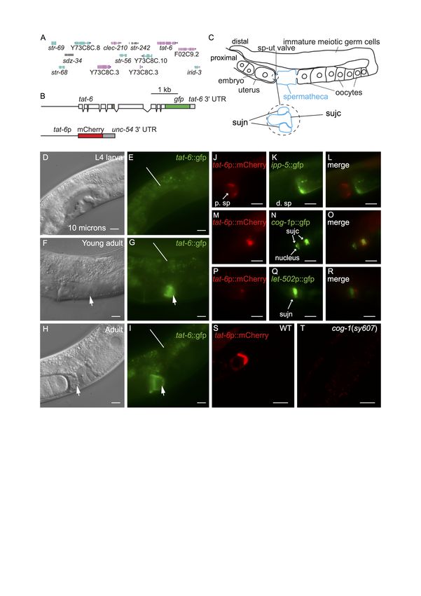
11/4/2021 - Open Access
   Figure 1. tat-6 is expressed in sujc cells: A. The tat-6 chromosomal region. The genes shown are those present in the fosmid
   used to generate the tat-6::gfp reporter shown in B. B. Schematics of the tat-6 reporters used in this study. Boxes represent
   exons. The upper construct encodes a TAT-6::GFP fusion protein in which GFP is fused to the whole of TAT-6. The beige-
   colored box indicates the part of TAT-6 containing the P-type ATPase motif. The reporter gene was generated by inserting gfp-
   encoding sequences into the fosmid (Sarov et al. 2012). The fosmid also contains the genes (shown in A) to the left and right
   of tat-6 in the C. elegans genome as well as intragenic sequences (Sarov et al. 2012). The tat-6::gfp reporter has the tat-6 3ʹ
   untranslated region (UTR). The lower construct is a transcriptional reporter in which mCherry expression is driven by a 1.4 Kb
   fragment from the tat-6 promoter. This construct contains the 3ʹ-UTR of the unc-54 gene. C. A schematic showing one arm of
   the gonad in an adult hermaphrodite. The spermatheca is highlighted in blue. ‘sp-ut valve’ indicates the position of the
   spermathecal-uterine valve. The expanded region shows a schematic cross section of the valve as it exists prior to ovulation. In
   such animals, the sujc syncytium occupies the core of the valve and is surrounded by a toroidal syncytium, sujn (Kimble and
   Hirsh 1979). During the first ovulation, sujc is displaced from the core of the valve. D-I, micrographs of the mid-body regions
   of hermaphrodite worms harboring the tat-6::GFP transgene shown in B. In D, F and H the worms were viewed with
   Nomarksi differential interference contrast (DIC) optics; E, G and I show the same worms viewed with fluorescence optics. In
   F and H the arrows indicate the position of the spermathecal-uterine valve. The uterus is to the left of the arrows, the
   spermatheca to the right. In G and I the arrows indicate GFP fluorescence in the gonad. The lines in E, G and I indicate
   background autofluorescence from the intestine. H shows a hermaphrodite that contained fertilized eggs in the uterus; the egg
   in closest proximity to the spermatheca is partially enveloped by material containing the TAT-6::GFP fusion protein. J,K,L.
   Fluorescent micrographs of an adult hermaphrodite carrying tat-6p::mCherry and ipp-5::GFP transgenes. The worms were
   mounted for photography so that the uterus was to the left. The ipp-5::GFP transgene is expressed in distal spermathecal cells
   (d. sp) at the junction with the ovary (Bui and Sternberg 2002). Note the lack of overlap between the mCherry and GFP
   fluorescence signals. M,N,O. Fluorescence micrographs of a hermaphrodite harboring tat-6p::mCherry and cog-1p::GFP
   transgenes. The cog-1 transgene is expressed in the sujc syncytium, which forms the core of the spermathecal-uterine valve
   (Palmer et al. 2002). To the right in the panels, the mCherry and GFP signals overlap. Note that while the distal-most part of
   the sujc syncytium is firmly within the center of the core, the sujc cell nuclei protrude into the uterus and are almost 10
   microns from the distal part of the cell (Palmer et al. 2002). While some GFP encoded by the cog-1p::GFP transgene is
   nuclear, the mCherry signal is strongest in the distal part of the syncytium. P,Q,R. Fluorescent micrographs of a hermaphrodite
   harboring tat-6p::mCherry and let-502p::GFP transgenes. The let-502p::GFP transgene is strongly expressed in sujn cells
   (Wissmann et al. 1999). S,T. Confocal fluorescence micrographs of adult hermaphrodites harboring the tat-6p::mCherry
   transgene. S shows an otherwise wild-type hermaphrodite; T shows a cog-1 mutant. Scale bars in all panels indicate 10
   microns.

   Description
   tat-1 and tat-5 are both very broadly expressed in C. elegans (Lyssenko et al. 2008, Ruaud et al. 2009, Chen et al. 2010,
   Wehman et al. 2011). tat-2, tat-3 and tat-4 are also expressed in many cells and tissues although apparently not ubiquitously
   (Lyssenko et al. 2008). No reports exist presently describing the pattern in which tat-6 is expressed. We generated multiple
   transgenic lines containing a construct encoding the entire TAT-6 protein fused to GFP (Fig. 1A,B). The construct contained
   tat-6 promoter sequences, all exons and introns as well as intragenic sequences to the left and right of the tat-6 coding region
   (Fig. 1A,B) (Sarov et al. 2012). Expression was seen in cells in the head and tail but in the center of the worm, expression was
   restricted to a proximal part of the gonad (Fig. 1C,F,G,H,I). Expression in the gonad was absent in early and mid-L4 stage
   worms but was robust in young adults (Fig. 1D,E,F,G). Prior to ovulation, fluorescence was seen in a region at the junction
   between the spermatheca and the uterus (Fig. 1C,F,G). In hermaphrodites in which ovulation had occurred, GFP fluorescence
   was seen partially surrounding the distal-most egg in the uterus (Fig.1H,I). The valve forming the junction between
   spermatheca and the uterus consists of a toroidal syncytium, sujn, and a core cell syncytium, sujc, which initially occupies the
   center of the valve (Fig. 1C) (Kimble and Hirsh 1979, Lints and Hall 2013). During the first ovulation, the core is displaced by
   the passage of the newly fertilized egg from the spermatheca to the uterus (Kimble and Hirsh 1979). The change in the
   distribution of TAT-6::GFP fluorescence we observed during the first ovulation suggested that TAT-6 might be expressed in
   sujc. Since existing GFP markers for cells in the spermatheca were available, to determine in which cells tat-6 was expressed,
   we first constructed strains containing a tat-6p::mCherry transcriptional reporter (Fig. 1B). The marker was expressed in the
   same way as the GFP reporter (Fig. 1S). In a strain containing the tat-6p::mCherry reporter and a reporter in which GFP
   expression was driven by promoter sequences from the cog-1 gene active in sujc (Palmer et al. 2002), the mCherry and GFP
   signals were seen in the same cell (Fig. 1M,N,O). In contrast, in a strain containing the tat-6p::mCherry reporter and an ipp-
   5p::gfp reporter, which is expressed in distal spermathecal cells that form part of the junction with the ovary (Bui and
   Sternberg 2002), the two fluorescent signals did not overlap (Fig. 1J,K,L). Similarly, in a strain containing the tat-6p::mCherry
   reporter and an let-502p::gfp reporter, which is strongly expressed in sujn cells (Wissmann et al. 1999), the mCherry signal
11/4/2021 - Open Access
   was adjacent to the strong GFP expression rather than coincident with it (Fig. 1P,Q,R). To further verify the identity of the
   cells expressing the tat-6 reporters, we crossed the tat-6p::mCherry transgene into a cog-1(sy607) mutant background. cog-1
   encodes a GTX/Nkx6.1 homeodomain transcription factor; in cog-1(sy607) mutant hermaphrodites, cells having the
   morphology of sujc cells are absent (Palmer et al. 2002). cog-1 is not expressed in sujn cells (Palmer et al. 2002). Consistent
   with the results with the fluorescent markers, no tat-6 reporter expression was seen in the gonad in the cog-1(sy607) mutant
   (Fig. 1S,T).
   We do not presently know the function of TAT-6 in sujc. Indeed, the function of the sujc cells themselves is not presently
   known: ablation of sujc cells in the mid-L4 stage causes only a weak effect on brood size (Palmer et al. 2002) (although an
   earlier function for sujc cells in morphogenesis of the spermathecal-uterine junction has not been ruled out (Palmer et al.
   2002)). Genetic research in Saccharomyces cerevisiae has revealed that all five P4-family ATPases in this organism, through
   their actions as phospholipid translocases, promote one or more vesicle transport events in the endosomal or secretory
   pathways (Hankins et al. 2015, Pomorski and Menon 2016, Yang et al. 2018). It is thought that aminophospholipid
   translocases promote membrane bending that occurs during the formation of transport vesicles (Hankins et al. 2015, Pomorski
   and Menon 2016, Yang et al. 2018). C. elegans tat-1 is required for correct vesicle transport within the endolysosomal system
   (Ruaud et al. 2009, Chen et al. 2010, Nilsson et al. 2011). C. elegans tat-5 is required for endosome to Golgi trafficking of
   MIG-14 (a C. elegans Wntless homologue) in the QL neuroblast (McGough et al. 2018), and to suppress the formation of
   extracellular vesicles in the embryo (Wehman et al. 2011, Beer et al. 2018). Thus, it is possible that TAT-6 promotes one or
   more vesicle transport event within sujc. It is worth noting that the plasma membrane of sujc cells is unusual in being highly
   convoluted (Kimble and Hirsh 1979, Lints and Hall 2013). Although it is not known how the extensive folding of the
   membrane arises, cell autonomous processes that promote membrane folding in sujc cells have not been ruled out. Finally, our
   studies also shed light on what happens to material from the sujc cells following ovulation. The fate of these cells after the
   core of the spermathecal-uterine valve has been displaced is presently not known. The fact that some TAT-6::GFP fusion
   protein (and mCherry) remains in the uterine epithelium even in older hermaphrodites indicates that at least a part of the sujc
   cells is retained following ovulation.

   Methods
   Request a detailed protocol
   Standard methods were used in the maintenance of C. elegans worms. Clone I16253250892533I A08 (Sarov et al. 2012) was
   used to generate svEx940, the extrachromosomal array from which the integrated array svIs144 was derived; pVB652 was
   used to generate svEx967 and svEx968. Transgenic strains were generated by microinjection (Fire 1986). The DNA clones
   were microinjected at a concentration of 50 ng/μl. svIs144 was derived from svEx940 by γ-irradiation. Micrographs were made
   with DM6000 B and DMRB compound microscopes (Leica); confocal micrographs were made with an A1 confocal
   microscope (Nikon).

   Reagents
    Strain     Genotype                                                                                      Available from
    VB3030     unc-119(ed3op); svIs144[tat-6p::tat-6::GFP unc-119(+)]                                        This work
    VB3041     unc-4(e120); svEx967[tat-6p::mCherry unc-4(+)]                                                This work
    PS3747     ipp-5(sy605); syEx429[ipp-5::GFP rol-6(su1006)]                                               CGC
    PS3662     syIs63[cog-1p::GFP]                                                                           CGC
    VB2238     svEx968[tat-6p::mCherry]; syEx429[ipp-5::GFP rol-6(su1006)]                                   This work
    VB3122     syIs63[cog-1p::GFP]; svEx968[tat-6p::mCherry unc-4(+)]                                        This work
    HR606      sbEx136[let-502p::GFP rol-6(su1006)]                                                          CGC
    VB3123     sbEx136[let-502p::GFP rol-6(su1006)]; svEx968[tat-6p::mCherry unc-4(+)]                       This work
    VB3357     cog-1(sy607); svEx967[tat-6p::mCherry unc-4(+)]                                               This work

   References
   Beer KB, Rivas-Castillo J, Kuhn K, Fazeli G, Karmann B, Nance JF, Stigloher C, Wehman AM. 2018. Extracellular vesicle
   budding is inhibited by redundant regulators of TAT-5 flippase localization and phospholipid asymmetry. Proc Natl Acad Sci U
11/4/2021 - Open Access S A 115: E1127-E1136. PMID: 29367422. Bui YK, Sternberg PW. 2002. Caenorhabditis elegans inositol 5-phosphatase homolog negatively regulates inositol 1,4,5- triphosphate signaling in ovulation. Mol Biol Cell 13: 1641-51. PMID: 12006659. Chen B, Jiang Y, Zeng S, Yan J, Li X, Zhang Y, Zou W, Wang X. 2010. Endocytic sorting and recycling require membrane phosphatidylserine asymmetry maintained by TAT-1/CHAT-1. PLoS Genet 6: e1001235. PMID: 21170358. Fire A. 1986. Integrative transformation of Caenorhabditis elegans. EMBO J 5: 2673-80. PMID: 16453714. Hankins HM, Baldridge RD, Xu P, Graham TR. 2015. Role of flippases, scramblases and transfer proteins in phosphatidylserine subcellular distribution. Traffic 16: 35-47. PMID: 25284293. Kimble J, Hirsh D. 1979. The postembryonic cell lineages of the hermaphrodite and male gonads in Caenorhabditis elegans. Dev Biol 70: 396-417. PMID: 478167. Lints R, Hall DH. Reproductive system, somatic gonad. (February 5, 2013), WormAtlas. ed. Laura Herndon, WormAtlas. DOI: doi:10.3908/wormatlas.1.22 Lyssenko NN, Miteva Y, Gilroy S, Hanna-Rose W, Schlegel RA. 2008. An unexpectedly high degree of specialization and a widespread involvement in sterol metabolism among the C. elegans putative aminophospholipid translocases. BMC Dev Biol 8: 96. PMID: 18831765. McGough IJ, de Groot REA, Jellett AP, Betist MC, Varandas KC, Danson CM, Heesom KJ, Korswagen HC, Cullen PJ. 2018. SNX3-retromer requires an evolutionary conserved MON2:DOPEY2:ATP9A complex to mediate Wntless sorting and Wnt secretion. Nat Commun 9: 3737. PMID: 30213940. Nilsson L, Jonsson E, Tuck S. 2011. Caenorhabditis elegans numb inhibits endocytic recycling by binding TAT-1 aminophospholipid translocase. Traffic 12: 1839-49. PMID: 21917090. Palmer RE, Inoue T, Sherwood DR, Jiang LI, Sternberg PW. 2002. Caenorhabditis elegans cog-1 locus encodes GTX/Nkx6.1 homeodomain proteins and regulates multiple aspects of reproductive system development. Dev Biol 252: 202-13. PMID: 12482710. Pomorski TG, Menon AK. 2016. Lipid somersaults: Uncovering the mechanisms of protein-mediated lipid flipping. Prog Lipid Res 64: 69-84. PMID: 27528189. Ruaud AF, Nilsson L, Richard F, Larsen MK, Bessereau JL, Tuck S. 2009. The C. elegans P4-ATPase TAT-1 regulates lysosome biogenesis and endocytosis. Traffic 10: 88-100. PMID: 18939953. Sarov M, Murray JI, Schanze K, Pozniakovski A, Niu W, Angermann K, Hasse S, Rupprecht M, Vinis E, Tinney M, Preston E, Zinke A, Enst S, Teichgraber T, Janette J, Reis K, Janosch S, Schloissnig S, Ejsmont RK, Slightam C, Xu X, Kim SK, Reinke V, Stewart AF, Snyder M, Waterston RH, Hyman AA. 2012. A genome-scale resource for in vivo tag-based protein function exploration in C. elegans. Cell 150: 855-66. PMID: 22901814. Wehman AM, Poggioli C, Schweinsberg P, Grant BD, Nance J. 2011. The P4-ATPase TAT-5 inhibits the budding of extracellular vesicles in C. elegans embryos. Curr Biol 21: 1951-9. PMID: 22100064. Wissmann A, Ingles J, Mains PE. 1999. The Caenorhabditis elegans mel-11 myosin phosphatase regulatory subunit affects tissue contraction in the somatic gonad and the embryonic epidermis and genetically interacts with the Rac signaling pathway. Dev Biol 209: 111-27. PMID: 10208747. Yang Y, Lee M, Fairn GD. 2018. Phospholipid subcellular localization and dynamics. J Biol Chem 293: 6230-6240. PMID: 29588369. Funding: Cancerfonden grant 14683 to ST Author Contributions: Lars Nilsson: Conceptualization, Formal analysis, Investigation, Visualization, Writing - original draft, Writing - review and editing. Shapour Rahmani: Investigation, Visualization, Writing - review and editing. Simon Tuck: Conceptualization, Funding acquisition, Investigation, Project administration, Supervision, Writing - original draft, Writing - review and editing. Reviewed By: Anonymous History: Received September 6, 2021 Revision received October 25, 2021 Accepted October 25, 2021 Published November 4, 2021
11/4/2021 - Open Access
   Copyright: © 2021 by the authors. This is an open-access article distributed under the terms of the Creative Commons
   Attribution 4.0 International (CC BY 4.0) License, which permits unrestricted use, distribution, and reproduction in any
   medium, provided the original author and source are credited.
   Citation: Nilsson, L; Rahmani, S; Tuck, S (2021). C. elegans TAT-6, a putative aminophospholipid translocase, is expressed
   in sujc cells in the hermaphrodite gonad.. microPublication Biology. https://doi.org/10.17912/micropub.biology.000495
You can also read