Brain network dysfunctions in addiction: a meta-analysis of resting-state functional connectivity - Nature
←
→
Page content transcription
If your browser does not render page correctly, please read the page content below
Translational Psychiatry                                                                                                                            www.nature.com/tp

                  ARTICLE                 OPEN

                  Brain network dysfunctions in addiction: a meta-analysis of
                  resting-state functional connectivity
                                           ✉                           2,3,4 ✉
                  Serenella Tolomeo1           and Rongjun Yu

                  © The Author(s) 2022

                      Resting-state functional connectivity (rsFC) provides novel insights into variabilities in neural networks associated with the use of
                      addictive drugs or with addictive behavioral repertoire. However, given the broad mix of inconsistent findings across studies,
                      identifying specific consistent patterns of network abnormalities is warranted. Here we aimed at integrating rsFC abnormalities and
                      systematically searching for large-scale functional brain networks in substance use disorder (SUD) and behavioral addictions (BA),
                      through a coordinate-based meta-analysis of seed-based rsFC studies. A total of fifty-two studies are eligible in the meta-analysis,
                      including 1911 SUD and BA patients and 1580 healthy controls. In addition, we performed multilevel kernel density analysis (MKDA)
                      for the brain regions reliably involved in hyperconnectivity and hypoconnectivity in SUD and BA. Data from fifty-two studies
                      showed that SUD was associated with putamen, caudate and middle frontal gyrus hyperconnectivity relative to healthy controls.
                      Eight BA studies showed hyperconnectivity clusters within the putamen and medio-temporal lobe relative to healthy controls.
1234567890();,:

                      Altered connectivity in salience or emotion-processing areas may be related to dysregulated affective and cognitive control-related
                      networks, such as deficits in regulating elevated sensitivity to drug-related stimuli. These findings confirm that SUD and BA might
                      be characterized by dysfunctions in specific brain networks, particularly those implicated in the core cognitive and affective
                      functions. These findings might provide insight into the development of neural mechanistic biomarkers for SUD and BA.

                      Translational Psychiatry (2022)12:41 ; https://doi.org/10.1038/s41398-022-01792-6

                  INTRODUCTION                                                                             functional magnetic resonance imaging (fMRI) to assess aberrant
                  Substance use disorder is characterized by excessive drug-seeking                        recruitment of brain regions in the context of different experi-
                  and taking [1]. Its core clinical symptoms comprise a chronically                        mental paradigms and stages of addiction [6–9].
                  relapsing cycle of binging, intoxication, withdrawal and craving,                           As an alternative approach to task-based fMRI, resting-state
                  despite the enormous adverse consequences. Behavioral addic-                             (rs) fMRI has been widely applied in both healthy participants
                  tions (BA) or non-substance addictions, such as gambling                                 and patients with neurological and psychiatric disorders [10–12].
                  addiction, are defined as a set of behavior that the individual                           Rs-fMRI is based on fluctuations of the blood-oxygenation-level-
                  becomes dependent on. Both disorders are characterized by a                              dependent (BOLD) signal, which characterize the intrinsic
                  persistent compulsion to seek and take a drug or perform a                               neuronal activity of the brain while subjects are in the awake
                  behavior, loss of control in limiting the intake, and they are often                     state [13]. The literature evaluating rs-fMRI in substance and no-
                  accompanied by negative emotions when the availability of the                            substance addiction is quite broad and includes seed-based
                  drug or behavior is prevented [2].                                                       functional connectivity (FC), regional homogeneity (ReHo),
                     Both SUD and BA are complex multifaceted and multistage                               independent component analysis (ICA), amplitude of low-
                  diseases. A previous rsfMRI systematic review reported that                              frequency oscillations (ALFF) and graph analysis under different
                  addictions would engage a range of brain networks, including                             types of addictions (for review see: Fedota and Stein, 2015;
                  the reward network, executive network and the habit and memory                           Ieong and Yuan, 2017, Pariyadath et al., 2016; Sutherland et al.,
                  networks [3], and are broadly linked with changes in many cortical                       2012) [14–17].
                  and subcortical brain regions. Among these addiction-related                                Previously, Tahmasian and colleagues concluded that seed-
                  networks, it is crucial to identify key brain regions/networks that                      based FC and effective connectivity are the standard methods to
                  specifically contribute to addiction, in order to shed light on                           detect disruption of specific brain areas, whereas graph- and
                  prevention and treatment. To assess the neural correlates of                             network-based analyses are valuable methods for assessing
                  addictions, a number of functional neuroimaging studies have                             alterations across the whole brain networks [12]. These different
                  examined abnormalities in local brain regions and in communica-                          methodological approaches have provided quite an ambiguous
                  tion between functionally distinct brain regions, as reported in two                     overview of the pathophysiological mechanisms underlying SUD
                  recent meta-analyses [4, 5]. These studies primarily used task-based                     and BA. For this reason, reviewing the literature, together with a

                  1
                   Institute of High Performance Computing, Agency for Science, Technology and Research (A*STAR), Singapore, Singapore. 2Department of Management, Hong Kong Baptist
                  University, Hong Kong, China. 3Department of Sport, Physical Education and Health, Hong Kong Baptist University, Hong Kong, China. 4Department of Physics, Hong Kong Baptist
                  University, Hong Kong, China. ✉email: Serenella_Tolomeo@ihpc.a-star.edu.sg; rongjunyu@hkbu.edu.hk

                  Received: 16 June 2021 Revised: 5 January 2022 Accepted: 12 January 2022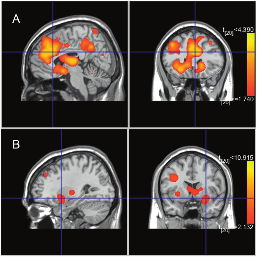
S. Tolomeo and R. Yu
2

    Fig. 1   PRISMA flowchart. PRISMA flowchart for the selection of eligible studies.

    quantitative meta-analysis, is needed to explain the inconsisten-       Gaussian distribution produces a statistical map that assesses the
    cies between previously published works.                                likelihood of activation for each voxel as determined by all studies
       SUD and BA might have common disease aetiologies [18, 19]. It        in the analysis [28]. Instead, the MKDA method establishes a
    is important to highlight the similarities and differences between      binary map for each study, which are averaged giving the
    these two types of addictions. For example, similar to drug             proportion of studies with any foci within a given radius from a
    addiction [20, 21] a number of studies concluded that pathological      voxel [29]. ALE focuses on the distribution of peak coordinates
    gambling is characterized by white matter abnormalities [22–24]         [28], while MKDA focuses on the distribution of statistical contrast
    and reductions of cortical thickness [25]. However, one study           maps [29]. We conducted a systematic review and three ALE meta-
    reported increased corticolimbic connectivity in cocaine depen-         analyses of resting-state functional connectivity (rsFC) studies.
    dence, and a decrease in pathological gambling [19], suggesting         Both hypo and hyper connectivities were examined delineate the
    that SUD and BA may also be associated with distinct brain              abnormality patterns among intrinsic functional networks in
    abnormalities.                                                          substance use disorders and behavioral addictions. Finally, using
       To the best of our knowledge, no previous rsFC studies have          the Multilevel Kerned Density Analysis (MKDA) meta-analytic
    directly compared SUD and BA to probe for neural specificity.            technique, we aimed to replicate the findings of our first meta-
    Individuals in general show unique patterns of addiction.               analyses employed using a different meta-analytic technique [28].
    Although a small proportion of addicts show addiction co-
    occurrence, many addicts struggle with one or more addictive
    behaviors but do not have difficulty with other types of addictive       METHODS
    behaviors. For example, gambling addiction is only weakly               Search strategy
    associated with drug abuse. Addiction specificity describes this         A comprehensive literature search was carried out using Pubmed (https://
                                                                            www.ncbi.nlm.nih.gov/pubmed/) and Web of Science (http://www.
    phenomenon: one addictive pattern may be acquired whereas               webofknowledge.com) in August 2021. This was performed by combining
    another is not [26]. To date, the neurobiological evidence on why       a total of 18 searches using the key terms: [“rest” OR “resting”] and
    some addictions may not co-occur within the same individual has         [“connect” OR “connectivity”], [“fMRI” OR “neuroimaging”] and various key
    not been conclusively quantified. Similarly, the neural basis of co-     terms corresponding to each search: “addiction”, “substance use disorder”,
    occurrence of addictions remain elusive as well [27]. We believe        “substance abuse”, “alcohol”, “cocaine”, “opioid”, “smokers”, “smoking”,
    that it is important to directly compare the neural substrates of       “heroin”, “stimulants”, “methamphetamine”, “gambling”, “gaming” and
    various types of addiction and examine the neural specificity of         “internet gaming”. The search resulted in 139 papers and after screening
    each addiction. Such an approach may help explain addiction             seventy-two papers were reached. Figure 1 displays a PRISMA diagram of
    specificity and addiction co-occurence.                                  the specific search method reported. Notably, most resting-state
                                                                            connectivity meta-analysis analyses are based on these seventy-two
       Here we conducted two coordinate-based meta-analysis                 studies, see Table 1 for further details.
    approaches activation likelihood estimation (ALE) and Multilevel           We included original functional magnetic resonance imaging (fMRI)
    Kernel Density Analysis (MKDA). Both ALE and MKDA are                   studies that used seed-based rsFC:
    coordinate-based meta-analysis (CBMA) approaches. Specifically,
    the ALE method involves the modeling of the reported loci of              1. To compare group differences in seed-based functional connectivity
    maximum activation as peaks of a 3D Gaussian probability, which              among SUD-HC were examined using the results of between-group
    is defined by a specified full-width half-maximum (FWHM). 3D                   contrasts (SUD < HC and SUD > HC).

                                                                                                  Translational Psychiatry (2022)12:41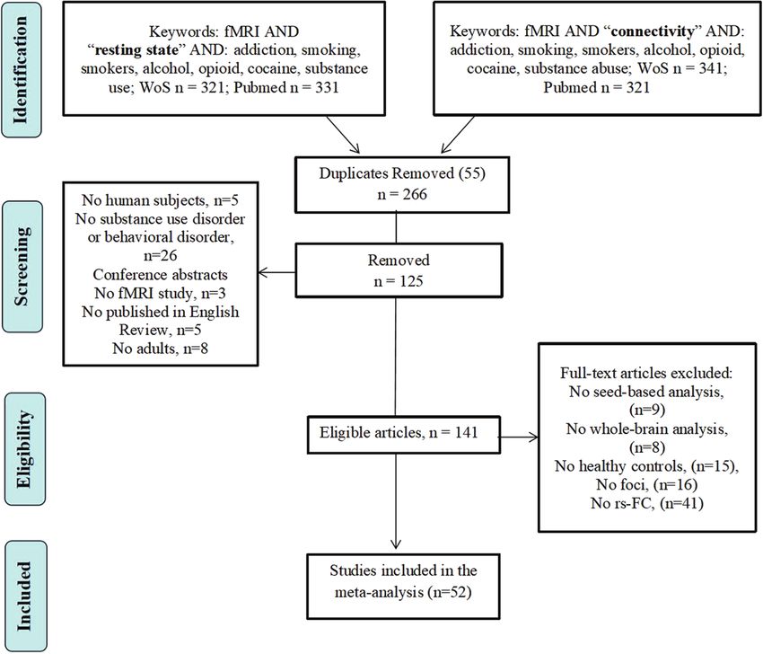
Table 1. Demographic and clinical characteristics of SUD and BA studies included in the FC meta-analysis.
Article HC (n) Mean age, HC SUD (n) Mean age, SUD Illness duration Illness severity SUD Phase Substance
Camchong et al., 2013a [45] 23 47.99 (6.7) 23 48.46 (7.1) – – Long-term abstinence Alcohol
Camchong et al., 2013b [97] 23 47.99 (6.7) 36 47.85 (7.30) – – Long-term abstinence Alcohol
Halcomb et al., 2019 [98] 21 35.3 (11.4) – 5.7 (3.5) Users Alcohol
16 37.3 (11.6) 2.5 (0.8)
Müller-Oehring et al., 2015 26 49 (11) 27 50 (9) – AUDIT:26.6 (9.5); ACQ-R: Abstinence Alcohol
[99, 100] 7.8 (2.6)
Wang et al., 2016 [101] 20 40.5 (8.2) 20 43.95 (6.3) – MAST: 31.50 (4.61) Dependence Alcohol
Translational Psychiatry (2022)12:41
Wang et al., 2018 [102] 33 42.88 (6.05) 35 41.80 (9.53) – MAST: 33.46 (4.55) Dependence Alcohol
Weiland et al., 2014 [103] 87 25.8 (8.3) 255 31.1 (8.3) – AUDIT:14.9 (7.0) Dependence Alcohol
Liu et al., 2019 15 47.3 (4.9) 15 47.3 (5.0) – ADS: 21.5 (10.8) Dependence Alcohol
AUDIT: 17.1 (10.0)
Blanco-Hinojo et al., 2017 [46] 29 22 (3) 28 21 (2) 6 years – Users Cannabis
Pujol et al., 2014 [48] 28 22 (3) 27 21 (2) – – Users Cannabis
Zhou et al., 2018 [49] 28 23.39 (2.86) 24 24 (3.46) – – Dependence Cannabis
Adinoff et al., 2015 [50] 20 42.2 (8.9) 22 44.7 (6.4) – CCQ-Brief: 2.6 (9) Dependence Cocaine
OCCS: 23.9 (9.0)
Contreras-Rodríguez et al., 2016 21 31 (4.6) 19 34.6 (6.8) – 18.4 g/month Dependence Cocaine
[19]
Geng et al., 2017 [51] 67 39.99 (5.7) 64 40.59 (6.01) 4.24 years – Dependence Cocaine
Gu et al., 2010 [52] 39 40 (5.1) 39 38 (6.2) 4.3 years Use: $200/week Dependence Cocaine
Hu et al., 2015 [53] 56 38.7 (7.82) 56 39.86 (6.71) 12.64 years Use: $246.70/week Dependence Cocaine
Kelly et al., 2011 [54] 24 35.1 (7.5) 25 35 (8.8) 11.43 years CSSA: 12.48 Dependence Cocaine
Martins et al., 2018 [55] 67 39.99 (5.7) 64 40.59 (6.01) 13 years Use: $198 Using and Cocaine
dependence
McHugh et al., 2014 [56] 22 42.05 (8.4) 21 43.10 (6.84) 7.72 years – Dependence Cocaine
43.75 (7.53) 8.88 years
S. Tolomeo and R. Yu
McHugh et al., 2017 [57] 22 42.05 (8.4) 21 43.10 (6.84) 7.72 years CCQ: 19.48 (10.73) Dependence Cocaine
Motzkin et al., 2014 [58] 18 31.7 (7.5) 22 32.0 (7.0) – ESI-SUB: 16 Dependence Cocaine and other
Verdejo-Garcia et al., 2014 [59] 14 30.1 (8.8) 10 35.1 (8.9) – – Dependence Cocaine
Zhang and Li, 2018 [60] 66 39.3 (9.2) 66 41.4 (7.3) 20.2 years CCQ: 23.8 (10.4) Dependence Cocaine
Li et al., 2013 [104] 15 31.9 (6.8) 14 35.4 (6.4) 7.44 years 0.6 g/day Dependence Heroin
Lin et al., 2018 [105] 30 41.47 (5.18) 30 42.44 (5) 7.66 years 52 mg/day Dependence Methadone
Wang et al., 2016 [68] 30 42.44 (5) 30 41.47 (5.18) 7.66 years 52 mg/day Dependence Heroin
Wang et al., 2016 [101] 30 38.9 (6.3) 30 40.7 (5.6) 14.4 years 2g Dependence Heroin
Zhai et al., 2014 [69] 15 28.9 (8.12) 22 33.05 (6.04) 6.59 years 0.96 g/day Dependence Heroin
Zhang et al., 2015 [70] 15 27.79 (7.81) 21 33.07 (5.99) 6.20 years 0.85 g/day Dependence Heroin
Zou et al., 2015 [71] 29 38.9 (6.33) 30 40.73 (5.61) 14.40 years – Abstinence Heroin
Kohno et al., 2014 [61] 27 33.8 (2.30) 25 35.68 (1.64) 8.59 years 3.57 g/week Dependence Methamphetamine
Kohno et al., 2016 [62] 18 38.9 (9.63) 20 37.0 (9.64) 6.82 years – Dependence Methamphetamine
Kohno et al., 2018 [63] 20 33.4 (11.11) 30 37.62 (9.7) 12.04 years – Dependence Methamphetamine
3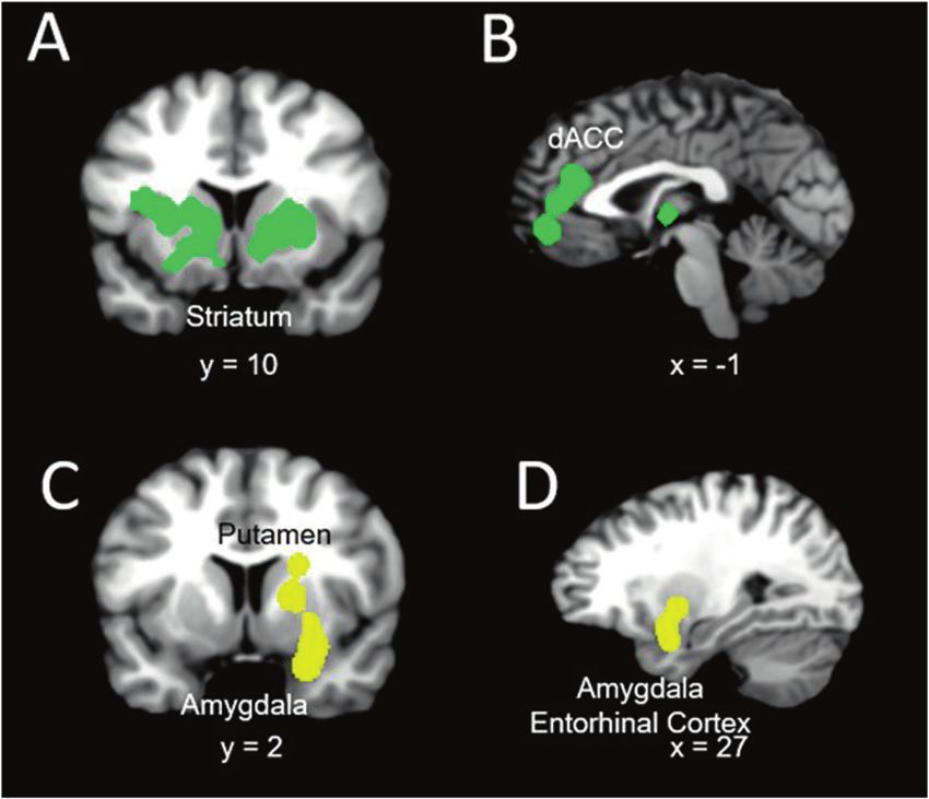
4
                                       Table 1.   continued
                                       Article                             HC (n)     Mean age, HC      SUD (n)     Mean age, SUD       Illness duration    Illness severity                   SUD Phase                Substance
                                       Li et al., 2020 [64]                31         34.48 (7.73)      34          32.15 (6.85)        6.59 years          –                                  Short-term Abstinence    Methamphetamine
                                       Wang et al., 2019 [65]              21         29.52 (2.54)      16          28 (4.24)           –                   –                                  Dependence               Amphetamine
                                       Huang et al., 2014 [38]             10         22.5 (6.78)       11          23.7 (1.98)         –                   FTND: 4.0                          Dependence               Nicotine
                                       Shen et al., 2017 [39]              41         39.46 (8.60)      84          38.23 (6.85)        20.70 years         FTND:5.23                          Dependence               Nicotine
                                       Shen et al., 2018 [40]              41         38.46 (8.60)      85          38.24 (6.81)        20.63 years         FTND: 5.18                         Dependence               Nicotine
                                       Um et al., 2019 [41]                62         35.31 (14.14)     34          34.15 (12.68)       16 years            FTND: 2.30                         Users                    Nicotine
                                       Yuan et al., 2016 [43]              60         19.95 (1.8)       60          20.0 (1.7)          4.4 years           FTND: 6.0                          Users                    Nicotine
                                       Zhang et al., 2017                  37         32.81 (9.57)      37          33.11 (9.58)        15.05 years         FTND: 7                            Users                    Nicotine
                                       Yu et al., 2017                     27         19.5 (2.3)        27          19.4 (2.3)          –                   FTND: 6.4                          Dependence               Nicotine
                                       Bi et al., 2017 [37]                40         19.8 (2.04)       40          19.62 (1.89)        4.20 years          FTND: 5,73                         Dependence               Nicotine
                                       Liu et al., 2016 [106, 107]         32         45.8 (9.3)        33          46.7 (9.4)          20.6 years          342 g/day                          Dependence               Betel quid
                                       Chen et al., 2016 [75]              30         24.14 (2.53)      30          23.64 (2.54)        –                   CIAS: 83.14 (10.26)                Dependence               Internet gaming
                                       Hong et al., 2015 [77]              11         14.81 (087)       12          13.41 (2.31)        –                   YIAT: 57.00 (17.39)                                         Internet gaming
                                       Lin et al., 2015 [78]               15         17.87             14          17.12 (2.73)        –                   YIAS: 65.07 (13.25)                Dependence               Internet addiction
                                       Yuan et al., 2017 [79]              44         19.5 (1.8)        43          19 (1.4)            –                   IAT:61.2 (11.1)                    Dependence               Internet gaming
                                       Zhang et al., 2015 [80]             24         23.13 (2.09)      35          22.46 (2.21)        –                   CIAS: 76.23 (7.67)                 Dependence               Internet gaming
                                       Zhang et al., 2016 [81]             41         23.02 (2.09)      74          22.28 (1.98)        7.28 years          CIAS: 78.36 (8.43)                 Dependence               Internet gaming
                                                                                                                                                                                                                                                   S. Tolomeo and R. Yu

                                       Contreras-Rodríguez et al., 2016    21         31 (4.6)          19          33.8 (7.5)          –                   40.8 h/month                       Pathological             Gambling
                                       [19]
                                       Jung et al., 2014 [73]              15         26.60 (4.29)      15          27.93 (3.59)        2.20 years          PG-YBOCS: 16.13 (7.28)             Dependence               Gambling
                                       Dependence = active users who are not abstinent; ACQ = Alcohol Craving Questionnaire; ADS = Alcohol Dependence Scale; CCQ-Brief = Cocaine Craving Questionnaire-Brief; CIAS = Chen Internet Addiction
                                       Scale; ESI-SUB = Externalizing Spectrum Inventory–Substance Abuse subscale; FTND = Fagerström Test for Nicotine Dependence; G-SAS = Gambling Symptom Assessment Scale; HC = healthy controls; IAT =
                                       Internet Addiction Test; MAST = Michigan Alcoholism Screening Test; n = sample size; OCCS = Obsessive-Compulsive Cocaine Scale; PG-YBOCS = Pathological Gambling Modification of Yale-Brown Obsessive
                                       Compulsive Scale; YIAS = Young’s Internet Addiction Scale; YIAST = Young’s Internet Addiction Test.

Translational Psychiatry (2022)12:41
S. Tolomeo and R. Yu
5
2. To compare group differences in seed-based functional connectivity For the SUD group, eight studies reported participants with
among BA-HC were examined using the results of between-group nicotine addiction [37–44] and eight articles reported participants
contrasts (BA < HC and BA > HC), respectively. with alcoholism and/or harmful drinking habits [36–39, 42, 44, 45].
Four studies reported participants [46–49] who were cannabis
Effects were categorized based on the direction of effect (hypercon- users and 13 studies reported participants who were cocaine users
nectivity or hypoconnectivity in SUD or BA). Hyperconnectivity has been
defined as larger positive or reduced negative rsFC and hypoconnectivity [19, 50–60]. The remaining articles on SUD reported a variety of
as larger negative or reduced positive rsFC compared with healthy stimulants including methamphetamine/amphetamine (n = 5)
controls. [61–65], and heroin/methadone (n = 12) [66–71]. For the BA
group, three articles reported participants with pathological
gambling disorder [72–74] and eight studies reported participants
Study eligibility criteria
Studies focusing on other psychiatric comorbidities, such as depression,
with internet gaming disorder [66, 75–81].
schizophrenia, anxiety, obsessive-compulsive disorder and neurological
conditions were excluded as they have been separated in the DSM-V [30]. Meta-analytic resting-state functional connectivity
In the first screening of articles, the titles and abstracts were considered, SUD results. A total of forty-four studies investigating rsFC
and the following exclusion criteria were applied: (1) non-empirical studies, abnormalities in SUD patients were identified. Table 3 shows a
(2) non-human studies, (3) non-fMRI studies, (4) non-rsFC studies, (5) non- complete list of the independent meta-analysis of rsFC on SUD
substance use disorder studies and (6) no adults. Subsequently, the full and HC only (ALE values are listed in Table 2).
text of every article was further evaluated for eligibility. Studies were also The rsFC meta-analysis in SUD, when compared with HC,
excluded due to (1) not in English, (2) no HC group, (3) entries having the revealed the largest hyperconnectivity cluster to be within the
same seed regions of interest reported in another publication. These
searches and exclusion criteria yielded a sample, n = 1911 for SUD + BA striatum (putamen, caudate) and middle frontal gyrus (dorsolat-
and n = 1580 for controls (Table 1). Coordinates were reported either in eral prefrontal cortex or DLPFC) (Fig. 2A and B). This was followed
Talairach or Montreal Neurology Institute (MNI) coordinate space. The final by a relatively large globus pallidus and anterior cingulate cluster
dataset included seventy-two articles for SUD and BA > HC (310 contrasts) and thalamus, as well as medial frontal gyrus (ventromedial
and seventy-two articles for the HC > SUD and BA (283 contrasts). prefrontal cortex or VMPFC) for the largest hypoconnectivity cluster
(See Table 2).
Meta-analysis
GingerALE is a freely available, quantitative meta-analysis method developed BA Results. A total of eight studies investigating rsFC abnorm-
by Turkeltaub et al [31] with the latest version described by Eickhoff and alities in BA patients were identified. The majority of the identified
colleagues [28, 32] and Turkeltaub and colleagues [33]. Here, the latest studies investigated pathological gambling addiction and the
version of GingerALE (3.0.2) was used (The BrainMap Database, www. remaining three studies investigated rsFC in internet gaming
brainmap.org; San Antonio, TX, USA), which relies on activation likelihood disorder. Table 2 shows a complete list of brain connectivity for
estimation (ALE) to compare coordinates compiled from multiple articles, independent meta-analyses on BA and HC (ALE values are listed in
estimate the magnitude of overlap, and yield clusters most statistically likely Table 2). The rsFC meta-analysis in BA included hyperconnectivity
to become active across studies. The algorithm minimizes within-group
effects and provides increased power by allowing for the inclusion of all in regions within the putamen, amygdala, and medial frontal
relevant experiments [28, 33]. Talairach coordinates were converted to MNI gyrus (Fig. 2C and D), and hypoconnectivity in the caudate,
with the Lancaster and colleagues (2007) transformation algorithm. cingulate, and thalamus.
Coordinates in MNI space were imported into the software. Imported foci
were modeled using a full-width at half-maximum (FWHM) kernel estimated Conjunction and contrast analyses: SUD and BA Results. Conjunc-
based on the corresponding experiment’s sample size as three-dimensional tion and contrast analyses were performed to assess whether
Gaussian spatial probability distributions [28, 33]. The resulting statistical addiction specificity was present, based on previous reviews
maps were thresholded at p < 0.05 using a cluster-level correction for [26, 27]. While the conjunction analysis revealed hyperconnectivity
multiple comparisons and a cluster threshold at p < 0.05 [28]. Group for both SUD and BA in the putamen, the contrast analysis
differences were examined using contrast analyses. The threshold for
group-contrasts was set to p < 0.05 uncorrected for multiple comparisons revealed hyperconnectivity in the claustrum, caudate, putamen and
with 5000 permutations [34]. Group differences in resting-state functional anterior cingulate for SUD (largest cluster size 8112) when
connectivity were examined using the following six contrasts in ALE analyses: compared with BA. The putamen/caudate and insula had the
SUD + BA > HC and SUD + BA < HC; SUD > HC and SUD < HC; BA > HC and greatest hyperconnectivity cluster size in BA when compared with
BA < HC; SUD > BA and SUD < BA. SUD (largest cluster size 1544). Hypoconnectivity for SUD and BA
was shown in the medial frontal gyrus and thalamus. Hypocon-
MKDA nectivity within the temporal lobe was shown in SUD when
Multilevel kernel density analysis (MKDA) was implemented through compared with BA. The medial frontal gyrus and anterior cingulate
Matlab toolbox NeuroElf (http://neuroelf.net/) consistently with MKDA revealed hypoconnectivity in BA when compared with SUD (ALE
neuroimaging meta-analytic procedures [35, 36]. Contrast coordinates in values are listed in Table 2).
Talaraich space were converted to MNI space. For all analyses, we used a
priori threshold of p < 0.05 (family-wise error-corrected for multiple SUD + BA Results. A total of seventy-two studies investigating
comparisons). Specifically, we investigated these meta-analytic contrasts rsFC abnormalities in SUD + BA were identified. Table 2 shows a
as follow: [SUD > HC and SUD < HC] and [BA > HC and BA < HC]. complete list of brain activities for independent meta-analyses on
SUD + BA and HC. Data from each cluster are listed in order of
cluster size in MNI space identified by all ALE meta-analyses.
RESULTS Higher ALE values are indicative of a greater likelihood of rsFC
Included studies and sample characteristics (ALE values are listed in Table 3).
Meta-analyses were performed using GingerALE and consist of For SUD + BA, the meta-analysis revealed that the largest
individuals with SUD (1911 subjects) and healthy controls (1580), hyperconnectivity cluster was within the amygdala, thalamus, and
both of which satisfied ALE power recommendations and include midbrain (Fig. 3) and relatively large parahippocampal gyrus,
a minimum of 17 contrasts [28]. The mean age for SUD individuals caudate, and putamen cluster, as well as hypoconnectivity in the
and for healthy controls was 33.24 (±6.16) and 34.28 (±5.99) years posterior lobe and parahippocampal gyrus. For HC, the rsFC meta-
old, respectively. See Supplementary Table S1 for more informa- analysis showed hyperconnectivity in the thalamus, midbrain,
tion regarding the rsFC methodology of the studies included in cingulate and frontal lobe and hypoconnectivity within the pa
the meta-analyses. rietal lobe and cerebellum. The contrast analysis revealed
Translational Psychiatry (2022)12:41S. Tolomeo and R. Yu
6
Table 2. Results of the meta-analysis of resting-state functional connectivity in SUD and BA.
Cluster Cluster size (mm3) Brain regions BA x y z ALE
SUD > HC
1 18,160 L Putamen −20 16 −6 0.0220
L Caudate −10 16 −2 0.0200
L Middle Frontal Gyrus 44 −44 18 26 0.0200
L Insula −34 −6 20 0.0100
2 7200 R Putamen 26 6 4 0.0200
R Globus Pallidus 16 8 2 0.0190
BA > HC
1 20,592 R Putamen 26 6 −12 0.0110
R Amygdala 21 48 −10 −12 0.0110
SUD < HC
1 10,000 L Thalamus −8 −14 2 0.0168
2 7312 R Medial Frontal Gyrus 4 44 38 0.0102
3 6432 R Parahippocampal Gyrus 28 28 −4 −20 0.0168
4 5152 L Insula 13 −42 12 10 0.0256
BA < HC
1 14,416 R Medial Frontal Gyrus 9 22 46 18 0.0100
R Anterior Cingulate 32 4 24 42 0.0090
2 12,328 L Thalamus −6 −8 12 0.0090
R Caudate 6 6 0 0.0090
SUD ∩ BA+
1 2584 R Putamen 18 4 8 0.0096
+
SUD > BA
1 8112 L Claustrum −30.2 17.2 4.1 0.1000
L Caudate −10.9 19.5 −5.6 0.1000
L Putamen −21.2 17 −9.2 0.0100
2 264 L Anterior Cingulate 24 −8 29 15 0.0100
BA > SUD+
1 1544 R Putamen 24.4 2.4 −10 0.100
2 1400 R Insula 45 −9 −9 0.100
3 192 R Caudate 20.3 8.5 17.3 0.040
−
SUD ∩ BA
1 3168 R Medial Frontal Gyrus 6 0 36 32 0.0090
2 1904 L Thalamus −24 −20 4 0.0090
SUD < BA−
1 272 L Temporal Lobe 38 −48.9 4.1 −19.8 0.0400
BA < SUD−
1 88 R Medial Frontal Gyrus 9 14.9 50.7 14.4 0.0500
2 24 R Anterior Cingulate 32 12 45.3 8.7 0.0500
MNI coordinates (x, y, z) of brain regions surviving a cluster-level threshold of p < 0.05 and a cluster forming threshold of p < 0.05 for single studies. Contrast
threshold was set to p = 0.05, 5000 permutations, >50 mm3, ALE = Activation Likelihood Estimate; BA = Brodmann Area; BA = Behavioral Addiction; HC =
Healthy Controls; + = Hyperconnectivity; − = Hypoconnectivity; SUD = Substance Use Disorders.
hyperconnectivity in the basal ganglia, thalamus, insula, amygdala, Nestor, 2017; Yip et al., 2017) [18, 82]. To our knowledge, no
and parahippocampal gyrus in SUD + BA in comparison with HC previous meta-analysis has examined how connections between
(See Table 3 for further details). MKDA results for SUD vs controls, different brain areas during rest are altered in addictive
and BA vs controls, are displayed in Fig. 4. neuropathologies. Here, we aimed to apply the ALE meta-
analysis method to estimate convergence in functional connec-
tivity rs-fMRI-based FC in substance use and behavioral disorders
DISCUSSION across studies.
The need to better describe the human brain connectivity in SUD We integrated findings from seventy-two rsFC studies and
and BA has long been recognized, whereby meta-analyses serve found convergent hyperconnectivity in individuals with SUD and/
as a crucial tool for consolidating evidence and streamlining the or BA: in the amygdala-basal ganglia, thalamus-midbrain and
prevalent narrative (for a recent perspective see Suckling and hypoconnectivity in the posterior lobe. In addition, basal ganglia,
Translational Psychiatry (2022)12:41S. Tolomeo and R. Yu
7
insula, amygdala and parahippocampal gyrus exhibited hyper-
connectivity in the SUD group compared with the healthy control
group. These findings show the enhancement of connectivity in
the reward and salience networks, suggesting that altered
physiology in the basal ganglia, midbrain, insula and medio-
temporal lobe might be evaluated as specific biomarkers for drug
addictions [3]. Interestingly, and importantly, our findings are
consistent with an earlier systematic review of rsFC brain
connectivity in drug addiction [17], which proposed that nicotine
addiction had salience and executive network altered. This was
confirmed by another recent review on chronic stimulant users
which found enhanced coupling of reward, salience, and memory
networks [83]. The hyperconnectivity of the reward and salience
network in our meta-analysis might suggest that the recruited
patients might be in a more chronic state of addiction [83]. The
current meta-analysis expands upon previous results on specific
brain regions by showing that abnormality in larger brain
networks might be present in addiction.
Fig. 2 Concordant activation across SUD and BA. A, B: regions Another novel finding of our ALE meta-analysis is the
concordant across studies for SUD (in green) and C, D: regions convergence of intrinsic functional patterns in the putamen in
concordant across studies for BA (in yellow). the SUD and BA, compared to HC. Numerous human and animal
studies have identified putamen as the key region in SUD and HC,
Table 3. Results of the meta-analysis of resting-state functional connectivity in SUD + BA and controls.
Cluster Cluster size (mm3) Brain regions BA x y z ALE
+
Healthy Control
1 55,056 L Thalamus −16 −10 12 0.0112
R Midbrain 6 −12 −10 0.0103
2 21,672 L Cingulate 24 −8 34 10 0.0101
R Medial Frontal Gyrus 32 6 32 44 0.0090
Healthy Control−
1 11,640 L Parietal Lobe 7 −14 −60 60 0.0093
2 10,256 R Cerebellum 0 −76 −24 0.0092
SUD + BA+
1 31,088 L Amygdala −22 −6 −14 0.0159
R Thalamus 10 −20 8 0.0141
R Midbrain 10 −22 −8 0.0095
2 13,088 R Caudate 10 14 −4 0.0157
R Putamen 30 −4 −10 0.0099
SUD + BA−
1 6832 R Posterior Lobe 44 −66 −20 0.0090
2 5536 R Parahippocampal Gyrus 42 −32 −18 0.0090
SUD + BA > Healthy Control+
1 20,864 L Caudate −12 2 12 0.0209
L Putamen −20 16 −6 0.0193
L Thalamus −10 −16 14 0.0134
L Insula 13 −34 12 14 0.0125
2 13,136 R Putamen 26 6 4 0.0244
R Caudate Body 16 8 4 0.0222
R Amygdala 26 −8 −28 0.0129
R Parahippocampal Gyrus 34 16 −4 −18 0.0103
SUD + BA < Healthy Control−
1 9832 R Dorsal Anterior Cingulate 32 8 36 22 0.0242
R Dorso Medial Frontal Cortex 32 4 30 36 0.0219
R Dorso Medial Frontal Cortex 6 2 24 44 0.0149
MNI coordinates (x, y, z) of brain regions surviving a cluster-level threshold of p < 0.05 and a cluster forming threshold of p < 0.05 for single studies. Contrast
threshold was set to p = 0.05, 5000 permutations, >50 mm3, ALE = Activation Likelihood Estimate; BA = Brodmann Area; + = Hyperconnectivity; − =
Hypoconnectivity.
Translational Psychiatry (2022)12:41S. Tolomeo and R. Yu
8
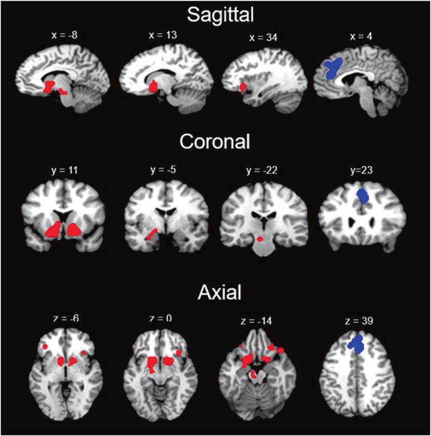
Fig. 3 Concordant connectivity across SUD and BA. Concordant hyperconnectivity (red) and hypoconnectivity (blue) across SUD and BA
(Sagittal, Coronal and Axial views).
putamen is part of the striatal “habit network” underlying learning
of automatic behavior [19, 72].
Interestingly, no previous rsFC study has compared SUD and BA to
probe the neural specificity. No studies have directly provide neural
evidence to support the idea of addiction specificity [26, 27]. Here, we
found that SUD showed hyperconnectivity in the basal ganglia
(putamen, caudate, and globus pallidus), claustrum, middle frontal
gyrus, and anterior cingulate compared with healthy controls. Instead,
BA revealed hyperconnectivity in the putamen, temporal and frontal
lobe. We suggest that the basal ganglia, claustrum and anterior
cingulate are neuroanatomical substrates linked with SUD, which
showed increased connectivity compared with BA. The importance of
these regions in encoding rewards and/or reward-seeking and
cognitive control has been demonstrated by functional imaging
and human lesion studies [52, 87–90]. These increased connectivities
for SUD adds further support to their central role specifically in the
chronic phase of the addiction cycle. Much evidence exists on the
capacity of drugs to enhance the mesolimbic dopamine system,
whilst there is much less evidence in BA [52, 91–93].
Although our findings integrated a remarkably large sample size
to establish consensus on the location of network disruptions in
drug and behavioral addictions, limitations should be considered.
First, the golden standard for directly detecting monosynaptic
axonal pathways is the chemical tracer technique which requires
Fig. 4 MKDA findings. Results from the MKDA analyses. A SUD vs ex-vivo tissue processing and can only be acquired in animal
controls and (B) BA vs controls. studies [94]. Thus, it is unclear how rsFC reflects the strength of
monosynaptic and polysynaptic pathways. Second, the seventy-
given its role in a variety of functions encompassing higher motor two experiments included in our meta-analysis differ in design,
control, impulsivity and inhibitory control [77, 84–86]. In BA, a methodology, age, gender of the population, illness severity and
selective involvement of putamen functional connectivity in duration of the use patterns (See Table 1 for further details). The
internet gaming disorder was revealed [77]. In addition, the wide variation of the substances used, and measures of quantity
Translational Psychiatry (2022)12:41S. Tolomeo and R. Yu
9
illustrate the need to report these factors more thoroughly and 8. Langleben DD, Ruparel K, Elman I, Busch-Winokur S, Pratiwadi R, Loughead J,
systematically in future studies. In addition, it highlights the need et al. Acute effect of methadone maintenance dose on brain fMRI response to
to standardize the reporting in future studies. Third, although the heroin-related cues. Am J Psychiatry. 2008;165:390–4.
functional significance of positive and negative rsFC remains 9. Liu S, Wang S, Zhang M, Xu Y, Shao Z, Chen L, et al. Brain responses to drug cues
predict craving changes in abstinent heroin users: a preliminary study. Neuro-
unclear, we lumped higher positive rsFC and lower negative rsFC
image. 2021;237:118169.
in the SUD/BA vs. HC contrast, making it difficult to differentiate 10. Brandl F, Avram M, Weise B, Shang J, Simões B, Bertram T, et al. Specific sub-
whether the differences were driven by higher rsFC in one group stantial dysconnectivity in schizophrenia: a transdiagnostic multimodal meta-
or lower rsFC in another group. The majority of studies only analysis of resting-state functional and structural magnetic resonance imaging
reported group differences without providing details about studies. Biol Psychiatry. 2019;85:573–83.
positive/negative rsFC in each group. Further studies may further 11. Kaiser RH, Andrews-Hanna JR, Wager TD, Pizzagalli DA. Large-scale network
explore the nature of group differences in rsFC. dysfunction in major depressive disorder: a meta-analysis of resting-state
Notably, there are several explanations for group differences in functional connectivity. JAMA Psychiatry. 2015;72:603–11.
functional connectivity. It is possible that both groups may show 12. Tahmasian M, Eickhoff SB, Giehl K, Schwartz F, Herz DM, Drzezga A, et al.
Resting-state functional reorganization in Parkinson’s disease: an activation
positive functional connectivity and one group exhibits stronger
likelihood estimation meta-analysis. Cortex 2017;92:119–38.
positive functional connectivity than the other group. It is also 13. Biswal BB. Resting state fMRI: a personal history. Neuroimage 2012;62:938–44.
possible that both groups show negative functional connectivity 14. Fedota JR, Stein EA. Resting-state functional connectivity and nicotine addiction:
and one group exhibits weaker negative connectivity than the prospects for biomarker development. Ann N. Y Acad Sci. 2015;1349:64–82.
other group. The third possibility is that one group shows positive 15. Ieong HFH, Yuan Z, Resting-state neuroimaging and neuropsychological find-
functional connectivity and the other group shows negative ings in opioid use disorder during abstinence: a review. Front Hum Neurosci.
functional connectivity and hence there is a significant group 2017; 11. https://doi.org/10.3389/fnhum.2017.00169.
difference. Unfortunately, in many of the original studies, the 16. Pariyadath V, Gowin JL, Stein EA, Resting state functional connectivity analysis
functional connectivity patterns in each group were not always for addiction medicine: from individual loci to complex networks. In: Progress in
brain research. Elsevier B.V., 2016, pp 155–73.
reported. Hence, the current study cannot do separate within-
17. Sutherland MT, McHugh MJ, Pariyadath V, Stein EA. Resting state functional
group ALE analyses, e.g., one ALE analysis for positive connectivity connectivity in addiction: lessons learned and a road ahead. Neuroimage
in SUD group and one ALE analysis for negative connectivity in 2012;62:2281–95.
SUD group. We suggest that future empirical studies should aim to 18. Yip SW, Morie KP, Xu J, Constable RT, Malison RT, Carroll KM, et al. Shared
routinely report functional connectivity for each group before microstructural features of behavioral and substance addictions revealed in areas
reporting group differences. Such practice might help researchers of crossing fibers. Biol Psychiatry Cogn Neurosci Neuroimaging. 2017;2:188–95.
understand the nature of group differences and the pathophysiol- 19. Contreras-Rodríguez O, Albein-Urios N, Vilar-López R, Perales JC, Martínez-
ogy of mental illnesses, including addictive disorders. Gonzalez JM, Fernández-Serrano MJ, et al. Increased corticolimbic connectivity
Fourth, due to the limited number of studies included we in cocaine dependence versus pathological gambling is associated with drug
severity and emotion-related impulsivity. Addict Biol. 2016;21:709–18.
aggregated studies with heterogeneous patients, ranging from initial
20. Lim KO, Choi SJ, Pomara N, Wolkin A, Rotrosen JP. Reduced frontal white matter
to abstinence stage, from short to long-term addictions. Analysis of integrity in cocaine dependence: a controlled diffusion tensor imaging study.
subtypes of addictions and their cognitive functions and behavioral Biol Psychiatry. 2002;51:890–5.
changes would be a strong supplement and would provide more 21. Moeller FG, Hasan KM, Steinberg JL, Kramer LA, Dougherty DM, Santos RM, et al.
context for each brain network. In the future, a meta-analysis with Reduced anterior corpus callosum white matter integrity is related to increased
ReHo [95], ICA [96], ALFF and graph analysis studies is warranted. impulsivity and reduced discriminability in cocaine-dependent subjects: diffu-
In conclusion, the findings of this meta-analysis suggest that sion tensor imaging. Neuropsychopharmacology. 2005;30:610–7.
rsFC connectivity in drug and behavioral addictions are disrupted. 22. Yip SW, Lacadie C, Xu J, Worhunsky PD, Fulbright RK, Constable RT, et al.
Altered hyperconnectivity within salience or emotion processing Reduced genual corpus callosal white matter integrity in pathological gambling
and its relationship to alcohol abuse or dependence. World J Biol Psychiatry.
may relate to deficits in regulating increased sensitivity to reward
2013;14:129–38.
and salience stimuli. These findings might be helpful when 23. Joutsa J, Saunavaara J, Parkkola R, Niemelä S, Kaasinen V. Extensive abnormality
attempting to identify potential putative markers for pharmaco- of brain white matter integrity in pathological gambling. Psychiatry Res-
logical interventions. A priority for future research would be to Neuroimaging. 2011;194:340–6.
further identify how these unbalanced networks impact different 24. Mohammadi B, Hammer A, Miedl SF, Wiswede D, Marco-Pallarés J, Herrmann M,
phases (inclusive of intoxication, withdrawal and dependence) of et al. Intertemporal choice behavior is constrained by brain structure in healthy
the addiction cycle. These results might be used as indicatives of participants and pathological gamblers. Brain Struct Funct. 2016;221:3157–70.
high risk in developing SUD or BA and might potentially guide 25. Grant JE, Odlaug BL, Chamberlain SR. Reduced cortical thickness in gambling
effective treatments at an early stage. disorder: a morphometric MRI study. Eur Arch Psychiatry Clin Neurosci.
2015;265:655–61.
26. Sussman S, Lisha N, Griffiths M. Prevalence of the addictions: a problem of the
majority or the minority? Eval Heal Prof. 2011;34:3–56.
REFERENCES 27. Sussman S, Leventhal A, Bluthenthal RN, Freimuth M, Forster M, Ames SL. A
1. Koob GF, Volkow ND. Neurocircuitry of addiction. Neuropsychopharmacology. framework for the specificity of addictions. Int J Environ Res Public Health.
2010;35:217–38. 2011;8:3399–415.
2. Koob GF, Volkow ND. Neurobiology of addiction: a neurocircuitry analysis. 28. Eickhoff SB, Laird AR, Fox PM, Lancaster JL, Fox PT. Implementation errors in the
Lancet Psychiatry. 2016;3:760–73. GingerALE software: description and recommendations. Hum Brain Mapp.
3. Zilverstand A, Huang AS, Alia-Klein N, Goldstein RZ, Review Neuroimaging 2017;38:7–11.
impaired response inhibition and salience attribution in human drug addiction: 29. TD Wager, MA Lindquist, TE Nichols, H Kober, JX Van Snellenberg, Evaluating the
a systematic review. 2018. https://doi.org/10.1016/j.neuron.2018.03.048. consistency and specificity of neuroimaging data using meta-analysis. Neuro-
4. Luijten M, Schellekens AF, Kühn S, Machielse MWJ, Sescousse G. Disruption of image 2009; 45. https://doi.org/10.1016/J.NEUROIMAGE.2008.10.061.
reward processing in addiction. JAMA Psychiatry. 2017;74:387–398. 30. American Psychiatric Association. DSM-V. 2013 https://doi.org/10.1176/appi.
5. Tolomeo S, Yaple ZA, Yu R, Neural representation of prediction error signals in books.9780890425596.744053.
substance users. Addict Biol. 2020. https://doi.org/10.1111/adb.12976. 31. Turkeltaub PE, Eden GF, Jones KM, Zeffiro TA. Meta-analysis of the functional
6. Kareken DA, Grahame N, Dzemidzic M, Walker MJ, Lehigh CA, O’Connor SJ. fMRI neuroanatomy of single-word reading: method and validation. Neuroimage.
of the brain’s response to stimuli experimentally paired with alcohol intoxica- 2002;16:765–80.
tion. Psychopharmacology. 2012;220:787–97. 32. Eickhoff SB, Laird AR, Grefkes C, Wang LE, Zilles K, Fox PT. Coordinate-based
7. Gu X, Lohrenz T, Salas R, Baldwin PR, Soltani A, Kirk U, et al. Belief about nicotine activation likelihood estimation meta-analysis of neuroimaging data: a random-
selectively modulates value and reward prediction error signals in smokers. Proc effects approach based on empirical estimates of spatial uncertainty. Hum Brain
Natl Acad Sci USA. 2015;112:2539–44. Mapp. 2009;30:2907–26.
Translational Psychiatry (2022)12:41S. Tolomeo and R. Yu
10
33. Turkeltaub PE, Eickhoff SB, Laird AR, Fox M, Wiener M, Fox P. Minimizing within- 57. McHugh MJ, Gu H, Yang Y, Adinoff B, Stein EA. Executive control network
experiment and within-group effects in activation likelihood estimation meta- connectivity strength protects against relapse to cocaine use. Addict Biol.
analyses. Hum Brain Mapp. 2012;33:1–13. 2017;22:1790–801.
34. Sokolowski HM, Fias W, Mousa A, Ansari D. Common and distinct brain regions 58. Motzkin JC, Baskin-Sommers A, Newman JP, Kiehl KA, Koenigs M. Neural cor-
in both parietal and frontal cortex support symbolic and nonsymbolic number relates of substance abuse: reduced functional connectivity between areas
processing in humans: a functional neuroimaging meta-analysis. Neuroimage. underlying reward and cognitive control. Hum Brain Mapp. 2014;35:4282–92.
2017;146:376–94. 59. Verdejo-Garcia A, Contreras-Rodríguez O, Fonseca F, Cuenca A, Soriano-Mas C,
35. Kober H, Wager TD. Meta-analysis of neuroimaging data. Wiley Interdiscip Rev Rodriguez J, et al. Functional alteration in frontolimbic systems relevant to moral
Cogn Sci. 2010;1:293–300. judgment in cocaine-dependent subjects. Addict Biol. 2014;19:272–81.
36. Wager TD, Lindquist M, Kaplan L. Meta-analysis of functional neuroimaging 60. Zhang S, Li CSR Ventral striatal dysfunction in cocaine dependence-difference
data: current and future directions. Soc Cogn Affect Neurosci. 2007;2:150–8. mapping for subregional resting state functional connectivity. Transl Psychiatry.
37. Bi Y, Yuan K, Guan Y, Cheng J, Zhang Y, Li Y, et al. Altered resting state functional 2018; 8. https://doi.org/10.1038/s41398-018-0164-0.
connectivity of anterior insula in young smokers. Brain Imaging Behav. 61. Kohno M, Morales AM, Ghahremani DG, Hellemann G, London ED. Risky decision
2017;11:155–65. making, prefrontal cortex, and mesocorticolimbic functional connectivity in
38. Huang W, King JA, Ursprung WWS, Zheng S, Zhang N, Kennedy DN, et al. The methamphetamine dependence. JAMA Psychiatry. 2014;71:812–20.
development and expression of physical nicotine dependence corresponds to 62. Kohno M, Okita K, Morales AM, Robertson CL, Dean AC, Ghahremani DG, et al.
structural and functional alterations in the anterior cingulate-precuneus path- Midbrain functional connectivity and ventral striatal dopamine D2-type recep-
way. Brain Behav. 2014;4:408–17. tors: link to impulsivity in methamphetamine users. Mol Psychiatry.
39. Shen Z, Huang P, Wang C, Qian W, Luo X, Guan X, et al. Altered function but not 2016;21:1554–60.
structure of the amygdala in nicotine-dependent individuals. Neuropsychologia. 63. Kohno M, Loftis JM, Huckans M, Dennis LE, McCready H, Hoffman WF. The
2017;107:102–7. relationship between interleukin-6 and functional connectivity in methamphe-
40. Shen Z, Huang P, Wang C, Qian W, Yang Y, Zhang M. Cerebellar gray matter tamine users. Neurosci Lett. 2018;677:49–54.
reductions associate with decreased functional connectivity in nicotine- 64. Li X, Su H, Zhong N, Chen T, Du J, Xiao K, et al. Aberrant resting-state cerebellar-
dependent individuals. Nicotine Tob Res. 2018;20:440. cerebral functional connectivity in methamphetamine-dependent individuals
41. Um M, Hummer TA, Cyders MA. Relationship of negative urgency to cingulo- after six months abstinence. Front Psychiatry. 2020;11:191.
insular and cortico-striatal resting state functional connectivity in tobacco use. 65. Wang Y, Yan KJ, Fan CX, Luo XN, Zhou Y. Altered functional connectivity of the
Brain Imaging Behav. 2020;14:1921–32. nucleus accumbens subdivisions in amphetamine-type stimulant abusers: a
42. Yu D, Yuan K, Bi Y, Luo L, Zhai J, Liu B, et al. Altered interhemispheric resting- resting-state fMRI study. BMC Neurosci. 2019;20:66.
state functional connectivity in young male smokers. Addict Biol. 66. Ding W-N, Sun JH, Sun YW, Zhou Y, Li L, Xu JR, et al. Altered default network
2018;23:772–80. resting-state functional connectivity in adolescents with internet gaming
43. Yuan K, Yu D, Bi Y, Li Y, Guan Y, Liu J, et al. The implication of frontostriatal addiction. PLoS ONE. 2013; 8. https://doi.org/10.1371/journal.pone.0059902.
circuits in young smokers: a resting-state study. Hum Brain Mapp. 67. Xiaonian W, Fenjuan L, Xianghe Q, Wenbin Y, Jie L, Chunjie L. Single-photon
2016;37:2013–26. emission computed tomography for the diagnosis of mandibular invasion
44. Sheng Z, Sien H, Lisa MF, Xingguang L, Carolyn MM, Laszlo Z, et al. Resting-state caused by oral cancers: a systematic review and meta-analysis. Hua Xi Kou
functional connectivity of the basal nucleus of meynert in cigarette smokers: Qiang Yi Xue Za Zhi. 2017;35:413–8.
dependence level and gender differences. Nicotine Tob Res. 2017; 19. https:// 68. Wang L, Zou F, Zhai T, Lei Y, Tan S, Jin X, et al. Abnormal gray matter volume
doi.org/10.1093/NTR/NTW209. and resting-state functional connectivity in former heroin-dependent indivi-
45. Camchong J, Stenger A, Fein G. Resting-state synchrony in long-term abstinent duals abstinent for multiple years. Addict Biol. 2016;21:646–56.
alcoholics. Alcohol Clin Exp Res. 2013;37:75–85. 69. Zhai TY, Shao YC, Xie CM, Ye EM, Zou F, Fu LP, et al. Altered intrinsic hippoc-
46. Blanco-Hinojo L, Pujol J, Harrison BJ, Macià D, Batalla A, Nogué S, et al. Atte- mapus declarative memory network and its association with impulsivity in
nuated frontal and sensory inputs to the basal ganglia in cannabis users. Addict abstinent heroin dependent subjects. Behav Brain Res. 2014;272:209–17.
Biol. 2017;22:1036–47. 70. Zhang Y, Gong J, Xie C, Ye EM, Jin X, Song H, et al. Alterations in brain con-
47. Orr C, Morioka R, Behan B, Datwani S, Doucet M, Ivanovic J, et al. Altered resting- nectivity in three sub-regions of the anterior cingulate cortex in heroin-
state connectivity in adolescent cannabis users. Am J Drug Alcohol Abus. dependent individuals: evidence from resting state fMRI. Neuroscience.
2013;39:372–81. 2015;284:998–1010.
48. Pujol J, Blanco-Hinojo L, Batalla A, López-Solà M, Harrison BJ, Soriano-Mas C, 71. Zou F, Wu X, Zhai T, Lei Y, Shao Y, Jin X, et al. Abnormal resting-state functional
et al. Functional connectivity alterations in brain networks relevant to self- connectivity of the nucleus accumbens in multi-year abstinent heroin addicts. J
awareness in chronic cannabis users. J Psychiatr Res. 2014;51:68–78. Neurosci Res. 2015;93:1693–702.
49. Zhou F, Zimmermann K, Xin F, Scheele D, Dau W, Banger M, et al. Shifted 72. Contreras-Rodríguez O, Albein-Urios N, Perales JC, Martínez-Gonzalez JM, Vilar-
balance of dorsal versus ventral striatal communication with frontal reward and López R, Fernández-Serrano MJ, et al. Cocaine-specific neuroplasticity in the
regulatory regions in cannabis-dependent males. Hum Brain Mapp. ventral striatum network is linked to delay discounting and drug relapse.
2018;39:5062–73. Addiction. 2015;110:1953–62.
50. Adinoff B, Gu H, Merrick C, McHugh M, Jeon-Slaughter H, Lu H, et al. Basal 73. Jung MH, Kim JH, Shin YC, Jung WH, Jang JH, Choi JS, et al. Decreased con-
hippocampal activity and its functional connectivity predicts cocaine relapse. nectivity of the default mode network in pathological gambling: a resting state
Biol Psychiatry. 2015;78:496–504. functional MRI study. Neurosci Lett. 2014;583:120–5.
51. Geng X, Hu Y, Gu H, Salmeron B, Adinoff B, Brain ES, et al. Salience and default 74. Koehler S, Ovadia-Caro S, van der Meer E, Villringer A, Heinz A, Romanczuk-
mode network dysregulation in chronic cocaine users predict treatment out- Seiferth N, et al. Increased functional connectivity between prefrontal cortex
come. academic.oup.com https://academic.oup.com/brain/article-abstract/140/ and reward system in pathological gambling. PLoS One. 2013;8:e84565.
5/1513/3038013 (Accessed 1 June 2020). 75. Chen CY, Yen JY, Wang PW, Liu GC, Yen CF, Ko CH. Altered functional con-
52. Gu H, Salmeron BJ, Ross TJ, Geng X, Zhan W, Stein EA, et al. Mesocorticolimbic nectivity of the insula and nucleus accumbens in internet gaming disorder: a
circuits are impaired in chronic cocaine users as demonstrated by resting-state resting state fMRI study. Eur Addict Res. 2016;22:192–200.
functional connectivity. Neuroimage. 2010;53:593–601. 76. Chen X, Wang Y, Zhou Y, Sun Y, Ding W, Zhuang Z et al., Different resting-state
53. Hu Y, Salmeron BJ, Gu H, Stein EA, Yang Y. Impaired functional connectivity functional connectivity alterations in smokers and nonsmokers with internet
within and between frontostriatal circuits and its association with compulsive gaming addiction. 2014. https://doi.org/10.1155/2014/825787.
drug use and trait impulsivity in cocaine addiction. JAMA Psychiatry. 77. Hong SB, Harrison BJ, Dandash O, Choi EJ, Kim SC, Kim HH, et al. A selective
2015;72:584–92. involvement of putamen functional connectivity in youth with internet gaming
54. Kelly C, Zuo XN, Gotimer K, Cox CL, Lynch L, Brock D, et al. Reduced inter- disorder. Brain Res. 2015;1602:85–95.
hemispheric resting state functional connectivity in cocaine addiction. Biol 78. Lin F, Zhou Y, Du Y, Zhao Z, Qin L, Xu J, et al. Aberrant corticostriatal functional
Psychiatry. 2011;69:684–92. circuits in adolescents with internet addiction disorder. Front Hum Neurosci.
55. Martins DLN, Valiatti TD, de S, D’Ávila J, Ferreira LF, Batista EK, et al. A con- 2015;9:356.
ectividade funcional extrínseca da rede de modo padrão em usuários de crack- 79. Yuan K, Yu D, Cai C, Feng D, Li Y, Bi Y, et al. Frontostriatal circuits, resting state
cocaína. Radio Bras. 2018;51:1–7. functional connectivity and cognitive control in internet gaming disorder.
56. McHugh MJ, Demers CH, Salmeron BJ, Devous MD, Stein EA, Adinoff B, Cortico- Addict Biol. 2017;22:813–22.
amygdala coupling as a marker of early relapse risk in cocaine-addicted indi- 80. Zhang JT, Ma SS, Yip SW, Wang LJ, Chen C, Yan CG, et al. Decreased functional
viduals. Front Psychiatry. 2014; 5. https://doi.org/10.3389/fpsyt.2014.00016. connectivity between ventral tegmental area and nucleus accumbens in
Translational Psychiatry (2022)12:41S. Tolomeo and R. Yu
11
Internet gaming disorder: evidence from resting state functional magnetic relapse in abstinent alcohol-dependent patients. PLoS ONE. 2018; 13. https://
resonance imaging. Behav Brain Funct. 2015; 11. https://doi.org/10.1186/ doi.org/10.1371/journal.pone.0196860.
s12993-015-0082-8. 103. Weiland BJ, Sabbineni A, Calhoun VD, Welsh RC, Bryan AD, Jung RE, et al.
81. Zhang JT, Yao YW, Li CSR, Zang YF, Shen ZJ, Liu L, et al. Altered resting-state Reduced left executive control network functional connectivity is associated
functional connectivity of the insula in young adults with Internet gaming with alcohol use disorders. Alcohol Clin Exp Res. 2014;38:2445–53.
disorder. Addict Biol. 2016;21:743–51. 104. Li Q, Yang WC, Wang YR, Huang YF, Li W, Zhu J, et al. Abnormal function of the
82. Suckling J, Nestor LJ. The neurobiology of addiction: the perspective from posterior cingulate cortex in heroin addicted users during resting-state and
magnetic resonance imaging present and future. Addiction. 2017;112:360–9. drug-cue stimulation task. Chin Med J. 2013;126:734–9.
83. Zilverstand A, O’Halloran R, Goldstein RZ Resting-state and structural brain 105. Lin HC, Wang PW, Wu HC, Ko CH, Yang YH, Yen CF. Altered gray matter volume
connectivity in individuals with stimulant addiction. In: The Routledge handbook and disrupted functional connectivity of dorsolateral prefrontal cortex in men
of philosophy and science of addiction. Routledge: 1 [edition]. | New York: Rou- with heroin dependence. Psychiatry Clin Neurosci. 2018;72:435–44.
tledge, 2018. | Series: Routledge handbooks in philosophy, 2019, pp 362–79. 106. Zhou S, Xiao D, Peng P, Wang S-K, Liu Z, Qin H-Y, et al. Effect of smoking on
84. Tolomeo S, Gray S, Matthews K, Steele JD, Baldacchino A. Multifaceted impair- resting-state functional connectivity in smokers: an fMRI study. Respirology.
ments in impulsivity and brain structural abnormalities in opioid dependence 2017;22:1118–24.
and abstinence. Psychol Med. 2016;46:2841–53. 107. Liu T, Li J, Zhao Z, Zhong Y, Zhang Z, Xu Q, et al. Betel quid dependence is
85. Groman SM, Morales AM, Lee B, London ED, Jentsch JD. Methamphetamine- associated with functional connectivity changes of the anterior cingulate cortex:
induced increases in putamen gray matter associate with inhibitory control. a resting-state fMRI study. J Transl Med. 2016;14:1–13.
Psychopharmacology. 2013;229:527–38.
86. Bahk JY, Li S, Park MS, Kim MO. Dopamine D1 and D2 receptor mRNA up-
regulation in the caudate–putamen and nucleus accumbens of rat brains by ACKNOWLEDGEMENTS
smoking. Prog Neuro-Psychopharmacol Biol Psychiatry. 2002;26:1095–104. This work was supported by Tier 2 Start Up Grant RC-OFSGT2/20-21/BUS/003. The
87. Gradin VB, Baldacchino A, Balfour D, Matthews K, Steele JD. Abnormal brain funders had no role in study design, data collection, analysis, and interpretation of
activity during a reward and loss task in opiate-dependent patients receiving the data; preparation, review, or approval of the manuscript; and decision to submit
methadone maintenance therapy. Neuropsychopharmacology. 2014;39:885–94. the manuscript for publication.
88. Schlagenhauf F, Rapp MA, Huys QJ, Beck A, Wustenberg T, Deserno L, et al.
Ventral striatal prediction error signaling is associated with dopamine synthesis
capacity and fluid intelligence. Hum Brain Mapp. 2013;34:1490–9.
89. Shackman AJ, Salomons TV, Slagter HA, Fox AS, Winter JJ, Davidson RJ. The AUTHOR CONTRIBUTIONS
integration of negative affect, pain and cognitive control in the cingulate cortex. ST: conceptualization, methodology, formal analysis, investigation, writing original
Nat Rev Neurosci. 2011;12:154–67. draft. RY: conceptualization. methodology, writing– review & editing.
90. Tolomeo S, Christmas D, Jentzsch I, Johnston B, Sprengelmeyer R, Matthews K,
et al. A causal role for the anterior mid-cingulate cortex in negative affect and
cognitive control. Brain. 2016;139:1844–54. COMPETING INTERESTS
91. Volkow ND, Wang GJ, Fowler JS, Logan J, Gatley SJ, Gifford A, et al. Prediction of The authors declare no competing interests.
reinforcing responses to psychostimulants in humans by brain dopamine D2
receptor levels. Am J Psychiatry. 1999;156:1440–3.
92. Volkow ND, Fowler JS, Wang GJ. Role of dopamine in drug reinforcement and ADDITIONAL INFORMATION
addiction in humans: results from imaging studies. Behav Pharm. 2002;13:355–66. Supplementary information The online version contains supplementary material
93. Volkow ND, Fowler JS, Wang GJ, Baler R, Telang F. Imaging dopamine’s role in available at https://doi.org/10.1038/s41398-022-01792-6.
drug abuse and addiction. Neuropharmacology. 2009;56:3–8.
94. Zeater N, Buzás P, Dreher B, Grünert U, Martin PR. Projections of three sub- Correspondence and requests for materials should be addressed to Serenella
cortical visual centers to marmoset lateral geniculate nucleus. J Comp Neurol. Tolomeo or Rongjun Yu.
2019;527:535–45.
95. Yu R, Zhao L, Tian J, Qin W, Wang W, Yuan K, et al. Regional homogeneity Reprints and permission information is available at http://www.nature.com/
changes in heavy male smokers: a resting-state functional magnetic resonance reprints
imaging study. Addict Biol. 2013;18:729–31.
96. Wang XF, Xue T, Dong F, Li YD, Xie DD, Liu C, et al. The changes of brain Publisher’s note Springer Nature remains neutral with regard to jurisdictional claims
functional networks in young adult smokers based on independent component in published maps and institutional affiliations.
analysis. Brain Imaging Behav. 2021;15:788–97.
97. Camchong J, Stenger VA, Fein G. Resting state synchrony in long-term abstinent
alcoholics with versus without comorbid drug dependence. Drug Alcohol
Depend. 2013;131:56–65.
98. Halcomb ME, Chumin EJ, Goñi J, Dzemidzic M, Yoder KK. Aberrations of anterior Open Access This article is licensed under a Creative Commons
insular cortex functional connectivity in nontreatment-seeking alcoholics. Psy- Attribution 4.0 International License, which permits use, sharing,
chiatry Res-Neuroimaging. 2019;284:21–28. adaptation, distribution and reproduction in any medium or format, as long as you give
99. Müller-Oehring E, Jung Y, AP-C, 2015 undefined. The resting brain of alcoholics. appropriate credit to the original author(s) and the source, provide a link to the Creative
academic.oup.com https://academic.oup.com/cercor/article-abstract/25/11/ Commons license, and indicate if changes were made. The images or other third party
4155/2366345 (Accessed 29 May 2020). material in this article are included in the article’s Creative Commons license, unless
100. Harrison PJ. The neuropathology of schizophrenia–A critical review of the data indicated otherwise in a credit line to the material. If material is not included in the
and their interpretation. Brain. 1999;122:593–624. article’s Creative Commons license and your intended use is not permitted by statutory
101. Wang J, Fan Y, Dong Y, Ma M, Ma Y, Dong Y, et al. Alterations in brain structure regulation or exceeds the permitted use, you will need to obtain permission directly
and functional connectivity in alcohol dependent patients and possible asso- from the copyright holder. To view a copy of this license, visit http://creativecommons.
ciation with impulsivity. PLoS ONE. 2016; 11. https://doi.org/10.1371/journal. org/licenses/by/4.0/.
pone.0161956.
102. Wang J, Fan Y, Dong Y, Ma M, Dong Y, Niu Y, et al. Combining gray matter
volume in the cuneus and the cuneus-prefrontal connectivity may predict early © The Author(s) 2022
Translational Psychiatry (2022)12:41You can also read