Bilirubin neurotoxicity: a Narrative Review on long lasting, insidious, and dangerous effects
←
→
Page content transcription
If your browser does not render page correctly, please read the page content below
Review Article
                                                                                                                                    Page 1 of 24

Bilirubin neurotoxicity: a Narrative Review on long lasting,
insidious, and dangerous effects
Dora Brites1,2, Rui F. M. Silva1,2
1
Instituto de Investigação do Medicamento (iMed.ULisboa), Faculdade de Farmácia, Universidade de Lisboa, Lisbon, Portugal; 2Departamento de
Ciências Farmacêuticas e do Medicamento, Faculdade de Farmácia, Universidade de Lisboa, Lisbon, Portugal
Contributions: (I) Conception and design: Both authors; (II) Administrative support: None; (III) Provision of study materials or patients: None;
(IV) Collection and assembly of data: Both authors; (V) Data analysis and interpretation: None; (VI) Manuscript writing: Both authors; (VII) Final
Approval of manuscript: Both authors.
Correspondence to: Dora Brites. Faculdade de Farmácia, Universidade de Lisboa, Lisbon, Portugal. Email: dbrites@ff.ulisboa.pt.

                Objective: In this review, we summarize the neuropathological effects of unconjugated bilirubin (UCB) and
                its free species (Bf) with a focus on the dysregulation of the central nervous system (CNS) cell homeostasis
                and subsequent toxic paracrine signaling effects. Direct or indirect actions of glial cells on UCB-induced
                neurodegeneration are also critically reviewed.
                Background: Elevated levels of UCB due to overproduction and/or defective clearance can severely impact
                the CNS leading to fatal encephalopathy or kernicterus spectrum disorders (KSD) associated with motor and
                auditory impairments. Still unknown is the preferential distribution of UCB in specific CNS regions and the
                long-lasting disabilities derived from severe neonatal hyperbilirubinemia. One of the aspects that remains
                uncertain is how unconjugated hyperbilirubinemia determines neural cell sequelae that may predispose to
                the development of neurodevelopmental, psychiatric, and neurodegenerative disorders. How UCB damages
                neurons and glial cells, and the injuries that can occur in more susceptible brain areas, thus potentially
                leading to permanent CNS dysfunction, is far from being clear.
                Methods: We conducted an exhaustive literature search using online databases (PubMed and Google
                Scholar). Search terms, besides UCB-induced neurodegeneration, were comprehensive (all years included)
                for all aspects related to glial cell dysregulation (e.g., oligodendrocytes, astrocytes, and microglia) by UCB
                and Bf.
                Conclusions: We specifically focused on the neurotoxic species of UCB and provided neuro- and
                gliocentric views in the context of neurodevelopmental alterations. Potential novel neuroprotective and
                regenerative strategies, including the use of extracellular vesicles (EVs) and their loading with medicines or
                microRNAs, were also addressed. Our perspectives on the future application of human advanced models
                and EVs to investigate UCB-induced neurotoxicity/KSD and subsequent pathological insults in early-life
                and lasting outcomes are outlined. We believe that this information could provide the next step for newborn
                screening using promising noninvasive biomarkers in the era of precision medicine to develop new and
                combinatorial therapeutic approaches at the forefront of translation.

                Keywords: Advanced BIND models and therapeutic opportunities; bilirubin-induced neuroinflammation and
                targets; intercellular (mis) communication by bilirubin; toxic bilirubin and neurodegeneration; UCB-induced glial
                aberrancies

                Received: 16 April 2021; Accepted: 30 May 2021; Published: 28 November 2021.
                doi: 10.21037/pm-21-37
                View this article at: https://dx.doi.org/10.21037/pm-21-37

© Pediatric Medicine. All rights reserved.                                            Pediatr Med 2021;4:34 | https://dx.doi.org/10.21037/pm-21-37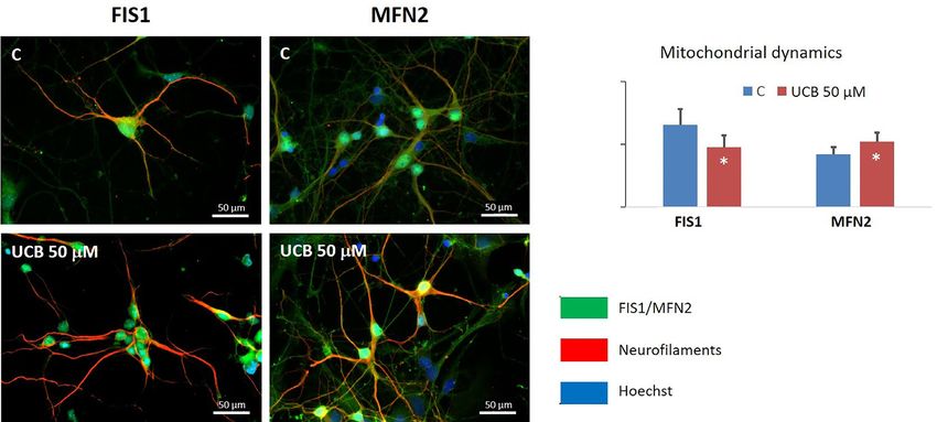
Page 2 of 24                                                                                              Pediatric Medicine, 2021

Introduction                                                     hepatocyte uptake due to sinusoidal protein polymorphisms
                                                                 (21,34). Low hepatic gene expression of the bilirubin uridine
The features of bilirubin deposition in the brain were
                                                                 diphospho-glucuronosyltransferase 1A1 (UGT1A1), as well
initially described by Orth (1) and later designated as
                                                                 as UGT1A1 enzyme deficiency in Gilbert’s disease (partial)
”kernicterus” by Schmorl (2) in the last quarter of the 19th
                                                                 and Crigler-Najjar types I (total, CN1) and II (almost
century. Today, more than a century later, and despite the
                                                                 total, CN2) syndromes, impairs bilirubin conjugation with
extensive research, multiple management recommendations
                                                                 (mostly) glucuronic acid (35-37), thus leading to increased
and guidelines (3-8), cases of acute bilirubin encephalopathy
                                                                 levels of UCB and Bf in circulation. Enzyme polymorphisms
(ABE) are still being described during the early neonatal
                                                                 may also play a role (35,38). Of note, CN1 syndrome
period (9,10), particularly in low- and/or middle-income
                                                                 leads to fatal outcomes with kernicteric features, unless
countries (11-14). Recent reviews recapitulate the spectrum
                                                                 liver transplantation is performed (39). Finally, excretion
of disorders associated with bilirubin neurotoxicity and
                                                                 of conjugated bilirubin into the bile, mainly mediated by
kernicterus, highlighting the toxic role of elevated free
                                                                 multidrug resistance-associated protein 2 (MRP2), is a
bilirubin (Bf) levels (15), i.e., unconjugated bilirubin not     key player for conjugated bilirubin elimination from the
bound to its main blood transporter, albumin. They also          liver (40) and stool output (41). MRP2 deficiencies may
emphasize several risk factors and co-morbidities that can       cause the re-uptake of conjugated bilirubin into circulation
lead to increased concentrations of serum albumin-bound          (Dubin-Johnson syndrome) and the presence of cholestasis
unconjugated bilirubin (UCB), accounting for elevated Bf         may lead to its elimination in urine (42).
levels and subsequent neurotoxicities (Figure 1). Moreover,         Other main risk factors for bilirubin-induced neurological
the available preventive and treatment options, as well as       damage (BIND) during neonatal hyperbilirubinemia
the recommendations are identified to manage ABE and             are: (I) prematurity that affects all the UCB clearance
kernicterus spectrum disorders (KSD) (16-20).                    mechanisms and increase neural cell susceptibilities to its
   With regards to the neurotoxic actions of UCB, four           harmful effects (10,43-47); and (II) hypoxia-ischemia (48),
main factors, acting alone or in combination, are implicated     sepsis (49), hypoalbuminemia (50) and acidosis (31,51,52)
as illustrated in Figure 1: increased bilirubin production,      that contribute to increase Bf concentrations and its
impaired hepatic uptake, reduced bilirubin conjugation,          entrance in the central nervous system (CNS) after crossing
and defective liver clearance (21). The excessive production     the blood-brain barrier (BBB) (Figure 1), causing neuronal
of UCB in the first days of life primarily derives from the      damage and glial activation.
relative polycythemia and breakdown of hemoglobin, as               Breastfeeding has also been associated with an increased
well as from the increased red blood cell (RBC) turnover         incidence of hyperbilirubinemia, but the causes for “breast
in neonates, with a rate of 6 to 8 mg/kg/day, more than          milk jaundice” or “breastfeeding failure jaundice” are
twice the production as adults (22). Bilirubin is generated      not completely clear (53,54). However, an association
from heme degradation, catalyzed by heme-oxygenase               with intestinal flora colonization status has been recently
(HO) to form biliverdin, which is then metabolized by            described (55,56). This may be important since it has been
biliverdin reductase (BVR) to Bf or UCB (if bound to             reported that the lack of microbiota in jaundiced babies may
albumin) (17). Other risk factors are implicated in UCB          lead to the reabsorption of non-polar UCB in the intestine
overproduction. This is the case for hemolytic diseases, e.g.,   and may contribute to the development of BIND (57).
glucose-6-phosphate dehydrogenase (G6PD) deficiency                 A less considered risk factor for UCB neurotoxicity
with increased erythrocyte fragility and hemolysis (23,24).      in neonates is the apparent lack of societal awareness for
The relative prevalence of G6PD deficiency (25), associated      this condition (58), together with early discharge policies
with neonatal hyperbilirubinemia (26) and prematurity (27),      practiced by birthing centers and maternity services that
makes both conditions significant risk factors. Notably,         impair early detection and timely therapeutics, which are
UCB can bind to RBCs (28,29) causing shape alterations           crucial to prevent UCB encephalopathies (59).
and increased fragility that culminate in increased                 In summary, newborn infants overproduce UCB and
hemolysis, further enhancing UCB and Bf production               have a decreased ability to eliminate UCB, thus increasing
(Figure 1) (30-33).                                              their susceptibility for UCB-induced neurodegeneration,
   UCB dissociates from albumin before entering the liver        oligodendrocyte dysfunction, astrocyte reactivity, and
and may be impacted by decreased delivery or by inefficient      microglia activation in specific brain regions, which can lead

© Pediatric Medicine. All rights reserved.                              Pediatr Med 2021;4:34 | https://dx.doi.org/10.21037/pm-21-37
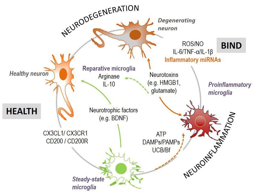
Pediatric Medicine, 2021                                                                                                          Page 3 of 24

Figure 1 Schematic representation of bilirubin production, transport, conjugation, and clearance, highlighting the distribution of the
circulating species, risk factors and passage across the blood-brain barrier into the brain. Bilirubin is mainly produced by the degradation
of hemoglobin prosthetic group, Heme from erythrocytes reaching its lifespan (old), by the enzymatic action of heme oxygenase (HO),
orignates biliverdin, which is then immediately converted to bilirubin by biliverdin reductase (BVR). Bilirubin not bound to albumin (free,
Bf) is in equilibrium with those bound to albumin (UCB), which is transported from the blood circulation into the liver for conjugation
with the glucuronic acid mediated by the bilirubin uridine diphosphoglucuronosyltransferase 1A1 (UGT1A1), to form conjugated bilirubin
(CB). CB is then excreted into bile and its final degradation products eliminated in feces. In cholestatic conditions, CB may return into the
circulation and be excreted in urine. When UCB is overproduced and exceeds the albumin binding capacity, Bf concentration raises, binds
to erythrocytes and causes hemolysis, as well as crosses the blood-brain barrier, mainly in the presence of risk factors like acidosis, hypoxia-
ischemia, sepsis, and hypoalbuminemia. In the brain, Bf interacts with neurons and glial cells (astrocytes, microglia, and oligodendrocytes)
causing several neuropathological sequelae.

to neurological sequelae with different severe long-term                   extracellular vesicles (EVs) to clarify pathological mechanisms
morbidities.                                                               associated with BIND and long-term sequelae are outlined,
   This review summarizes the concepts associated with UCB,                and the relevance of their use as new therapeutic tools in
Bf, and erythrocyte-linked neurotoxic species, using descriptive           personalized medicine are also addressed.
neuro- and gliocentric views, and addressing the key role of                  We present the following article in accordance with the
intercellular paracrine dysregulation to homeostatic imbalance             Narrative Review reporting checklist (available at https://
and BIND. Future research using human advanced models and                  dx.doi.org/10.21037/pm-21-37).

© Pediatric Medicine. All rights reserved.                                         Pediatr Med 2021;4:34 | https://dx.doi.org/10.21037/pm-21-37
Page 4 of 24                                                                                               Pediatric Medicine, 2021

               Protective                      Deleterious         issues, brain lesions and sequelae resulting from ABE and
          Antioxidant properties             Neurotoxic effects    chronic kernicterus most often associated with bilirubin
                                                                   levels of 19 mg/dL or higher (16), which have been mostly
                                                                   addressed (53). In contrast, the life-long consequences of
                                                                   moderate levels of total or UCB, such as those surpassing
              Low UCB/Bf levels        High UCB/Bf levels          5 mg/dL in the first 2 to 4 days and up to values below
                                                                   18 mg/dL (76,77) on the CNS are still unknown.
                                                                   One consequence is the association between neonatal
                                                                   hyperbilirubinemia and autism spectrum disorder (78),
Figure 2 The beneficial and harmful double-edge sword effects of   which has been only suspected before or even denied (53,79).
unconjugated bilirubin (UCB) in oxidative and neuroinflammatory    Auditory brainstem function is also impaired in neonates
conditions accordingly to its low and high concentrations. When    with hyperbilirubinemia (80), and hearing screening tests
at physiological or slightly elevated levels, UCB and its free     have shown a relevant association between bilirubin levels
species (Bf) not bound to human serum albumin exert antioxidant    and abnormal auditory activity in jaundiced newborns (81)
and anti-inflammatory protective mechanisms. In contrast, their    that have been associated with KSD (5,16,82,83). As would
increased concentrations are deleterious to the brain, involving   be expected, the risk for auditory damage is increased
neurodegeneration and glial activation or even causing death or    in preterm infants, where bilirubin levels considered
permanent severe outcomes.                                         “safe” for term babies can lead to irreversible lesions (84).
                                                                   Alterations of the sensorimotor system due to elevated
                                                                   UCB concentrations assessed at several developmental
Pathological implications of neurotoxic bilirubin                  ages have been reviewed by Lunsing (85). Abnormalities
species                                                            in the visuocortical function were observed at 3 months
                                                                   of age in children who had total bilirubin levels between
Before addressing the neuropathological effects of the
                                                                   10 and 25 mg/dL at postnatal (PN) day 3 (86). Delayed
increased concentrations of UCB and Bf, it is perhaps
                                                                   neurodevelopmental outcomes at 6 months (87) and 1
worthwhile to describe the controversies regarding the
                                                                   year of age (88) were also found in term newborns, even
beneficial and harmful effects of UCB, which directly
                                                                   with moderate hyperbilirubinemia accordingly to recent
depend on its “physiological” (slightly elevated) or
                                                                   prospective cohort studies performed in India.
markedly increased concentrations. The pleiotropic role
                                                                      Though usually not associated with cognitive
of UCB at low levels as an antioxidant (60-62), though
                                                                   abnormalities, the literature is divided on this issue
still controversial (63,64), and its anti-inflammatory effects     (89-93). Neurobehavioral disabilities with lower rates
(65-67), have contributed to a poor understanding, and             of school completion and full-time employment, as well
sometimes even a dismissive attitude towards the harmful           as reading difficulties, were related to the existence of
consequences of high UCB levels, either in neonatal                hyperbilirubinemia (94). Interestingly, a recent study using
life or as a consequence of inherited unconjugated                 hippocampal neurons and animal models revealed that
hyperbilirubinemias, such as Gilbert, CN1 and CN2                  UCB induces the deposition of the amyloid-β (Aβ) peptide
syndromes. As an example, a shift between antioxidant              and tau hyperphosphorylation, establishing a link between
and pro-oxidant actions may occur between intracellular            an early exposure to bilirubin and Alzheimer’s disease (AD)
UCB values of 7 ng/mg protein and those above                      features in later life (95), thus reinforcing its long-term
25 ng/mg protein, respectively (68). The dual effects of UCB       effects. In a later study, high UCB levels, together with
(Figure 2) are even more difficult to understand when              decreased serum concentrations of albumin, were found in
several therapeutic approaches have used low concentrations        dementia patients with Aβ and intravenous administration
of UCB to treat several pathologies based on its antioxidant       of albumin produced beneficial effects on daily function and
and anti-inflammatory properties (66,69-74). However,              dementia severity in AD patients (96).
the harmful effects of UCB at high concentrations and the             In conclusion, neonatal-associated UCB and BIND may
severe neurological consequences that unfortunately still          contribute to auditory and motor deficits (97), but also be
occur should not be disregarded (75).                              associated with developmental delay, cognitive impairment,
    For that reason, we will focus on neuropathological            behavioral problems as well as poor executive function, and

© Pediatric Medicine. All rights reserved.                               Pediatr Med 2021;4:34 | https://dx.doi.org/10.21037/pm-21-37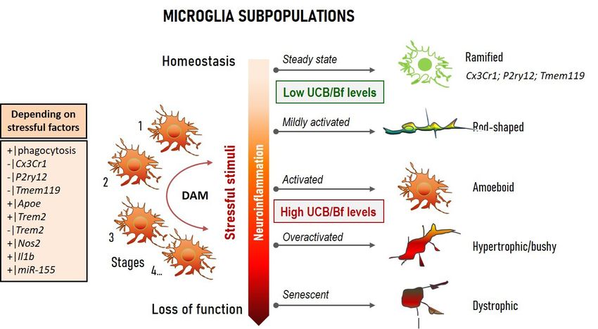
Pediatric Medicine, 2021 Page 5 of 24
psychiatric disorders (98,99). in the brain. Furthermore, UCB seemed not to be a passive
player in BBB dynamic properties, but it possibly could
trigger several damaging mechanisms that impair the barrier
Free and erythrocyte-bound bilirubin
function at the level of brain microvascular endothelial
Bf was first designated as the fraction of bilirubin that was cells (120) in a time-dependent manner (121). These
not conjugated and bound to albumin, distinct from the effects were observed both in vitro and in post-mortem brain
conjugated species. This concept was introduced in 1958 sections of infants with kernicterus (122).
with an Italian publication (100), followed by a French Once in the brain, UCB interacts with neurons and may
one (101) (and many others) until 1969 to 1972, when the cause irreversible damage. Initial studies in experimental
low concentration of the non-protein-bound bilirubin kernicterus already proposed that UCB diffuses through
species started to be estimated and was determined to the neuroplasm, interacting with the Golgi complex,
be around 10−10 or 10−9 mol/L (102,103). The authors, at neurotubules, and endoplasmic reticulum (ER) of
that time, designated this fraction as Bf or unbound and neurons, diffusing into the axoplasm and causing axonal
suggested that it could increase under some conditions destruction (123). Another long-recognized target for UCB is
to 10 −6 or 10 −5 mol/L. First determinations used the the mitochondria, where UCB damages respiration, uncouples
Sephadex G-25 elution technique for the separation oxidative phosphorylation, and induces brain mitochondrial
of Bf and albumin-bound bilirubin (104,105) and swelling, even at low micromolar concentrations (124,125).
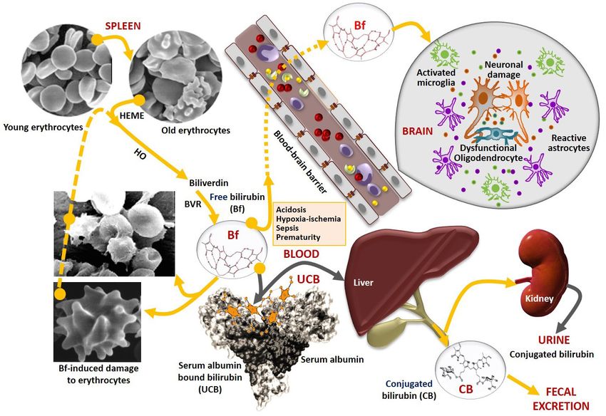
the enzymatic oxidation with hydrogen peroxide and We found that UCB also impairs the mechanisms associated
horseradish peroxidase (106). The former method was with mitochondrial fusion-fission dynamics as depicted in
even commercialized and recognized as a valuable aid to Figure 3 (unpublished data), which are associated with the
neonatologists in preventing bilirubin encephalopathy maintenance of cellular quality (126). Elevated mitochondrial
(107,108). Then, when the two processes were compared, fusion, here assessed by mitofusin 2 protein immunostaining,
the peroxidase method was found to require less volume favors the generation of interconnected mitochondria to
of serum and to be more sensitive for the assessment of Bf increase cell bioenergetics efficiency when facing an insult
concentration (109). All these studies were fundamental as a cell survival mechanism (127). In contrast, fission that
to finally separate two species of UCB, the one bound to we determined through the expression of the mitochondrial
albumin and the free species, which is the most toxic fraction fission 1 protein (FIS1) is associated with numerous
(15,110,111). Determination of Bf and the estimation of mitochondrial fragments and its decrease may lead to reduced
reserve albumin binding capacity was then complemented mitochondria motility (128). UCB also triggers mitochondrial
by the erythrocyte-bound bilirubin (29,112). All these membrane permeabilization, with the release of cytochrome c,
methods were thereafter reviewed (113). Now, some studies and activation of caspases 3 and 9, that culminate in neuronal
have assessed the modifications caused by the binding of apoptosis as described previously (129,130) and us as well (131).
bilirubin to erythrocytes, either for morphological changes Although the exact toxic mechanisms are still not clear, it
or induced hemolysis, and consequences that it could is becoming apparent that UCB impairs neuronal cells by a
have in aggravating the risk of BIND (Figure 1) (30,114). plethora of effects that eventually culminate in cell death by
Another important contribution to the relevance of the necrosis- and apoptosis-like mechanisms (132,133), which
toxic levels of Bf was the understanding about the bilirubin may involve glutamate excitotoxicity (134). In fact, several
displacement from albumin by competitive binding of reviews describe multiple neurotoxic mechanisms for UCB,
endogenous compounds and several drugs that promoted an like inhibition of neurite outgrowth and ramification (135),
increase of its levels (106,115-118). alteration of neuronal membrane microfluidity, impairment
of axonal arborization, and increased nitrosative stress (136)
that, together with glutamate, seem to mediate arborization
Neurocentric view of BIND
impairment (137) and to alter synaptic transmission (138).
The notion that UCB reaches the brain by crossing the Not surprisingly, immature cells appear to be more sensitive
BBB was probably concluded from studies performed in to UCB neurotoxicity (139,140), correlating with the
the mid-1960s using various animal models of experimental proposed age-related window of susceptibility to UCB
bilirubin encephalopathy (119). Such studies also suggested neurological damage (141). It is important to note that most
that neurologic damage was related to UCB concentration of these results were obtained in experimental conditions
© Pediatric Medicine. All rights reserved. Pediatr Med 2021;4:34 | https://dx.doi.org/10.21037/pm-21-37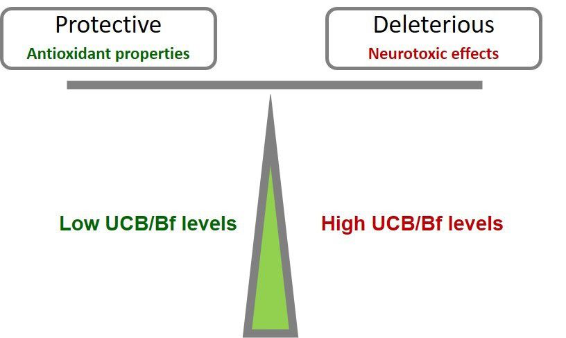
Page 6 of 24                                                                                                                                   Pediatric Medicine, 2021

                         FIS1                           MFN2

                                                                                                                             Mitochondrial dynamics

                                                                                   Fluorescence intensity (cell)
                                                                                                                   2
                                                                                                                                        C    UCB 50 μM

                                                                                                                   1

                                                                                                                   0
                                                                                                                             FIS1                     MFN2

                                                                                                                   FIS1/MFN2

                                                                                                                   Neurofilaments

                                                                                                                   Hoechst

Figure 3 Alterations in the mitochondrial mechanisms of fusion-fission dynamics induced by unconjugated bilirubin (UCB) in rat cortical
neurons. Neurons were incubated for 4 h at 37 ℃ with UCB at 50 μM plus 100-μM human serum albumin (HSA), and data compared
with controls (cells with albumin, but no UCB added). Bars represent the mean fluorescence values (± SEM) from at least four different
microphotographs and normalized by the number of cells in each photograph for fission 1 protein (FIS1) and mitofusin 2 protein (MFN2).
*, P
Pediatric Medicine, 2021 Page 7 of 24
Healthy OPCs UCB-injured OPCs
A2B5 + O4 A2B5 + O4
Figure 4 Unconjugated bilirubin (UCB) is harmful to oligodendrocyte progenitor cells (OPCs). Isolated OPCs were incubated for 8 h
at 37 ℃ in the absence (healthy) or in the presence of UCB at 50 μM plus 100-μM human serum albumin (HSA) (injured). Cells were
immunolabelled with specific antibodies, A2B5 for OPCs and O4 that stains the transition from OPCs to differentiated oligodendrocytes.
Representative pictures are shown. Magnification: 630×. Unpublished data obtained by A Barateiro at the D Brites laboratory.
function (154). Actually, glial cells, once considered as report on ultrastructural changes in the Gunn rat with
the glue between neurons, are presently acknowledged as bilirubin encephalopathy identified the presence of
key players in the brain immune system and in multiple myelin debris in the cytoplasm of neurons, which also
physiological processes linked to synaptic plasticity, presented mitochondrial alterations and glycogen-filled
energy metabolism, learning and memory formation, vacuoles (164). UCB was shown to bind to myelin and
among others (155). The intricate balance of homeostatic was suggested to be associated with its retention in the
and inflammatory functions influences the onset and the brain (165,166). However, UCB also caused cerebellar
progression of neurodegenerative diseases (156). Moreover, myelin fragmentation in in vitro cultures (167), and myelin
neurological disorders usually involve feedback loops loss was observed in biopsy samples from a kernicteric
that disseminate and perpetuate the disease (157), mostly preterm infant (168). Lesions in the myelin sheath of
mediated by the cell-secreted soluble factors and release of spiral ganglion cells were observed in neonatal guinea pigs
small (exosomes) and large EVs (158,159), already observed exposed to hyperbilirubinemia. Neuroimaging studies
in the cerebrospinal fluid (CSF) of patients with ABE (160). in infants at risk for kernicterus identified white matter
We propose that neuronal selectivity in BIND converges abnormalities (169). When assessed for in vitro effects,
with non-cell autonomous mechanisms involving signaling UCB was shown to impair OPCs (Figure 4) (146) and
mechanisms and non-neuronal cell types, thus requiring a oligodendrocytes (170), as well as to disturb the differentiation
better understanding. In this section, we will address data on of OPCs into myelinating oligodendrocytes (147). Further
glial sensitivity to UCB, i.e., the view of a more integrated studies, using rat organotypic cerebellar slices demonstrated
“gliocentric brain” (161), providing further information on that treatment with 20-nM Bf led to a reduction in
targets to unravel and prevent UCB brain lesions and their the number of myelinated fibers, together with the
sequela, and then assist in the insult recovery. gene expression of the myelin basic protein (171). The
data validate that concentrations mimicking neonatal
unconjugated hyperbilirubinemia impair myelination. Using
Myelin damage
a new kernicterus mouse model with Ugt1a1 gene deletion,
The myelinating cells of the CNS, the oligodendrocytes, it was possible to confirm the presence of cerebellum
are generated from bipolar oligodendrocyte progenitor cells atrophy by the elevated UCB concentrations, together with
(OPCs) that arise between 10 and 18 weeks of gestation axonal loss and decreased myelination, which was similarly
in humans (162,163). Maturation of oligodendrocytes noticed in the medulla oblongata and pons, but not in the
start at 28 to 40 weeks of gestation and proceeds during corpus callosum (172). In summary, deficits in myelination
the early postnatal period (141). Oligodendrocytes should be considered as targets when developing new
constitute 5% to 8% of total glial cells (163). The first therapeutic strategies for BIND.
© Pediatric Medicine. All rights reserved. Pediatr Med 2021;4:34 | https://dx.doi.org/10.21037/pm-21-37Page 8 of 24 Pediatric Medicine, 2021
MICROGLIA SUBPOPULATIONS
Figure 5 Simplified microglial phenotypic categorization in homeostatic and in inflammatory conditions, accordingly to intensity, type,
and duration of unconjugated bilirubin (UCB) and/or free bilirubin (Bf) treatment. Homeostatic microglia are known for their immune
surveillance and regulation of neural cell networks, with a ramified morphology and motile processes. The bipolarized or rod-shaped
microglia are highly proliferative, express both pro- and anti-inflammatory markers, and are associated to mild neurodegeneration and
repair. Phagocytic and activated microglia reveal an amoeboid shape with retracted processes. Hypertrophic and “bushy” microglia have
short and poorly ramified processes and are associated with cell activation/overactivation. While early microglia activation is important to
restore brain homeostasis, if chronically activated, they continuously release pro-inflammatory molecules that further increase tissue damage.
Dystrophic microglia relate to a less responsive or ineffective supportive cell showing loss of processes, cytoplasmic fragmentation, and
spheroid morphology. Evidence demonstrated that microglia may co-exist in different phenotypes (reparative, inflammatory, and senescent-
like). Diverse activation stages occur after the transition from the steady state into a disease-associated microglia (DAM) population.
Transcriptional signatures may vary in a context-dependent fashion and in the case of UCB/Bf stimulation, with the brain region, jaundice
severity, and presence of co-morbidities.
Microglia polarization Besides phagocytic and pruning functions, microglia
regulate myelin uptake, neurogenesis, and cerebral
Microglia are the resident macrophages of the CNS
angiogenesis (175). The first data on possible lipid droplet-
that are derived from the yolk sac and travel to the brain accumulating microglia were obtained in the Gunn rat
during early development (173). Microglia represent 5% cerebellum in 1986 (177), a model of CN1. In conditions
of total glial cells in the human cortical brain (174) and leading to HO-1 induction, producing biliverdin from
show phenotypical heterogeneity, regional diversity, and heme (Figure 1), this enzyme was found to be mainly
are highly complex and dynamic and with interchangeable localized in microglia and involved in their activation,
phenotypes (175) (Figure 5). Microglia release interleukin but it is unclear whether this might lead to beneficial or
(IL)-1β and tumor necrosis factor-alpha (TNF-α), harmful effects (178-180). A pioneer study showed that
among other cytokines (176), which regulate homeostasis UCB activates microglia leading to the release of the pro-
or are involved in neuroinflammation and pathology. inflammatory cytokines TNF-α, IL-1β, and IL-6, as well
© Pediatric Medicine. All rights reserved. Pediatr Med 2021;4:34 | https://dx.doi.org/10.21037/pm-21-37Pediatric Medicine, 2021 Page 9 of 24
as glutamate, while also inducing cell death by apoptosis aging and AD (192,206,207) show loss of function and
and necrosis (181), suggesting that these cells may have an chronic release of pro-inflammatory mediators. Caldeira
important role in BIND and, consequently, are promising et al. developed an in vitro microglia model able to mimic
targets to modulate excessive neuroinflammation. Certain “young/responsive” and “old/senescent” microglial
UCB photoproducts also produce neuro-inflammatory features (194). These authors using in vitro aging microglia
effects that may even surpass those of 140-nM Bf (182). were able to discriminate age-dependent responses by
Therefore, it is not surprising that microglia are activated Aβ (195). When such a model was used to assess Bf-
after intracerebral hemorrhage and can be associated induced responses in each of the conditions, increased
with bilirubin production in the CNS and its oxidation sickness prevailed in the younger microglia, as compared
products (183), while also facilitating early inflammation by with the older cells (208), and included enhanced amoeboid
neutrophil brain infiltration (184). morphology, NO release, and elevated high mobility group
Interaction of UCB with microglia first impacts on box protein 1 (HMGB1), TNF-α, and IL-6 gene expression
protective mechanisms associated with the activation levels. Among the vast number of small non-coding
of mitogen-activated protein kinases (MAPKs) and short RNAs (miRNAs) controlling post-transcriptional
nuclear factor kappa B (NF-κB), together with increased expression of target genes, some were recognized as
phagocytosis, and later release of pro-inflammatory inflammatory associated miRNAs (inflamma-miRNAs),
cytokines (185). In early responses to UCB, microglia may and were accepted as key players in microglia function/
then have a protective intervention (185,186). However, in dysfunction, polarization, and restoration (209). Among
chronic or long-lasting hyperbilirubinemia, such benefits those, upregulation of miRNA(miR)-155, miR-125b, miR-
may no longer be supported (187-189). Therefore, the good 21, and miR-146a by Bf was only observed in the “young”
may turn bad with the release of excessive inflammatory microglia, pushing the cell phenotype to an immune-
mediators. Actually, microglia are known by their dual polarized state, and indicating their propensity to be
neuroprotective and neuroinflammatory roles among the stimulated by Bf. However, Bf seemed to also induce a sort
kaleidoscope of polarized phenotypes (190) (Figure 5). In of microglia activation, independent of the age of the cells,
the steady-state, microglia have a ramified morphology based on an induced increased of CD11b staining (associated
with highly motile processes constantly surveying the with a proinflammatory status) and on the elevation of
neighboring environment. Changes in brain homeostasis inducible nitric oxide synthase (iNOS) gene expression at
leads to alterations in microglia shape and process motility. a 100-nM concentration. On the contrary, cells behaved
The acquired amoeboid morphology is associated with differently with early apoptosis exclusively noticed in
phagocytic ability and mild inflammation, the rod- “younger” microglia, and late apoptosis/necrosis only in
shape with activation by mild neurodegeneration, and “older” cells. Data have shown that microglia reveal age-
the hypertrophic with excessive immune reaction. When dependent performance when stimulated by bilirubin, with
damaged by chronic insults or senescence, microglia beneficial and pathological properties that may vary with
become dystrophic and are ineffective in supporting neural co-morbidities, CNS region, neurodevelopmental stage,
cell homeostasis (191-199). Using transcriptional single-cell cell maturation, jaundice duration, and hyperbilirubinemia
sorting, it was possible to identify several immune-related intensity.
classes and disease-associated microglia (DAM) phenotypes,
based on a specific set of genes found in AD models and
Astrocyte aberrancies
patients (200-202).
Activation of microglia was observed in the hippocampus Astrocytes comprise nearly 35% of the total CNS
and cerebellum of mice with hyperbilirubinemia population, and like microglia, they may be found in all
(172,203,204) and in rat cerebellar slice cultures treated CNS regions. Astrocytes participate in neuroinflammatory
with UCB (171), where the induction of excitotoxic and responses and show diverse subtypes that are disorder-
neurodegenerative processes were identified. However, and context-specific (210,211). Some of the biomarkers
we still need to better understand microglial population more often used in their characterization are glial fibrillary
diversity, in which each member may perform unique acidic protein (GFAP), S100B, glutamine synthetase,
functions in a disease-context-dependent fashion (205). or the glutamate transporters, GLT1 and GLAST
As already mentioned, senescent microglia associated with (210,212). One of the first studies using mixed fetal rat
© Pediatric Medicine. All rights reserved. Pediatr Med 2021;4:34 | https://dx.doi.org/10.21037/pm-21-37Page 10 of 24 Pediatric Medicine, 2021
glial cells, in which 80% to 95% of cells were astrocytes, at 50 µM plus 100-µM HSA was shown to peak at 2 to 4 h
identified morphological and cytotoxic alterations, as well of interaction (230) (Figure 6), much later than in microglia,
as age-in-culture-dependent sensitivity, when working where the peak was observed 30 min after exposure (185),
with UCB/HSA ratios of 2, i.e., recapitulating severe evidencing the early activation of microglia by UCB. We
hyperbilirubinemia in neonates (213). The study called also noticed an induced release of IL-1β and TNF-α from
attention for the higher susceptibility of immature neural UCB-treated cortical astrocytes with increased expression
cells to UCB harmful effects. The idea was later reinforced of the TNF-α receptor (TNFR)1 and IL-1β receptor
by several studies in primary cultures of astrocytes, as well as (IL-1R)1 (231). Moreover, UCB reduced the cytokine
in neurons and microglia (46,47,140,208,214,215). By using pro-forms while activated their converting enzymes, ICE
the MTT assay for cell viability and mitochondrial activity and TACE, respectively (231,232). ICE or caspase-1
assessments, Chuniaud et al. attributed the cytotoxicity activation that leads to pyroptotic cell death (233),
of UCB to mitochondria failure (216). Astrocytes have pro-inflammatory processes (234), and inflammasome
a wide range of relevant functions in the brain that activation, including the regulator of innate immunity
contribute to maintain extracellular homeostasis (217), NLR family pyrin domain containing 3 (NLRP3) (235),
such as their ability to uptake glutamate, thus preventing its was demonstrated in cultured rat cortical astrocytes after
accumulation at synapses and resulting excitotoxicity (218). exposure to UCB (236). Astrocytes, acquire phenotypic
UCB inhibited glutamate uptake and cell endocytosis, when aberrancies in neurodegenerative diseases and are activated
rat cortical astrocytes were used (219,220). Interestingly, by neuroinflammation and stressful factors, such as UCB,
while the inhibition of glutamate was higher in astrocytes contributing to pathophysiological paracrine signaling
than in neurons, cell death and changes in redox stress events, mainly mediated by EVs containing miRNAs
were prominent in neurons (133,221), suggesting cell (237-240). In such a way, dysfunctional astrocytes actively
pathological susceptibilities. Astrocyte increased resistance contribute to cell homeostatic imbalance in the brain, as
may derive from the elevated expression of the multidrug will be further explained in the next section.
resistance-associated protein 1 (MRP1) that was shown to
be promoted by UCB (222).
Dysregulated neuron-glia interplay: the gearbox?
Astrocytes detect infection and injury by neurons,
microglia, oligodendrocytes, and endothelial cells, with Brain function depends on coordinated interactions and
the secretion of cytokines and growth factors that may intercellular signals between neurons and glial cells that
act as immune regulators, following the activation of sustain cell homeostatic balance (241,242). In disease,
NF-κB. They are accepted as initiators and responders secretion of pathological signaling molecules and EVs
to inflammation, namely to mediators released by the from donor cells determine autocrine and paracrine
activated microglia, such as IL-1β, TNF-α, and interferon signaling dysregulation (243,244). Changes in neuro-
gamma (IFN-γ), and they react to lipopolysaccharide (LPS) immune homeostasis and neuroinflammation may start at
( 2 2 3 - 2 2 6 ) . I n t e r e s t i n g l y, U C B a t 5 0 - µ M ( U C B / the neurovascular unit composed by the BBB elements:
HSA=0.5) caused apoptotic cell death and TNF-α secretion (I) on one side, the endothelial cells, pericytes, and the
from rat cortical astrocytes, in a similar way to that of astrocyte end foots; and (II) on the other side the glial
10 ng/mL of LPS. They reacted strongly to UCB, then to LPS, cells, neurons, and the extracellular matrix. Disruption
in terms of necrosis, as well as to the secretion of IL-1β and of BBB by UCB is well documented (120,245-247) and
glutamate, and less to the release of IL-6 (227). The cytotoxicity facilitate the entrance of elevated Bf levels into the brain,
of UCB in astrocytes was also observed with serum from infants its interaction with neuronal cells and the emergence of
with unconjugated hyperbilirubinemia (228). As expected, BIND (Figure 1). Disruption of the BBB by bilirubin and
UCB activated astrocyte signaling pathways associated with hypercarbia/hyperosmolarity (52,248) may also allow the
MAPKs, i.e., p38, Jun N-terminal kinase (JNK)1⁄2 and passage of albumin-bound bilirubin (249), though the
extracellular signal-regulated kinase (ERK)1⁄2 pathways, as permeability is higher for the Bf species (250). Within
well as the key player NF-κB (229), which further increased the brain, the binding of Bf to cells is facilitated by
in hypoxia and oxygen-glucose deprivation preconditioning acidosis (251), increasing the risk of BIND. Pathological
conditions (48). Translocation of NF-κB from the cytoplasm synapse loss and dysfunction depends on the maturation of
to the nucleus in the cortical astrocytes treated with UCB neuronal circuits, proper function of glial cells, and synaptic
© Pediatric Medicine. All rights reserved. Pediatr Med 2021;4:34 | https://dx.doi.org/10.21037/pm-21-37Pediatric Medicine, 2021 Page 11 of 24
A B
0h 1h
2.0
Cytoplasm
NF-κB (fold change)
Nucleus
1.5
1.0
0.5
0.0
2h 4h 0 1 2 4
Time (h)
C
0h 1h 2h 4h
Cytoplasm
NF-κB
Nucleus
Figure 6 Unconjugated bilirubin (UCB) induces the translocation of nuclear factor kappa B (NF-κB) from the cytoplasm to the nucleus of
cortical astrocytes. Astrocytes were incubated for 1, 2, and 4 h at 37 ℃ with UCB at 50 μM plus 100-μM human serum albumin (HSA), and
data compared with controls (cells with no UCB added, time =0 h). (A) Representative images of NF-κB immunoreactivity using an anti-
NF-κB primary antibody. Magnification: ×400. (B) Bars represent the NF-κB-fold change values (±SD) from at least three independent
experiments. (C) Cytosolic and nuclear protein extracts were processed for Slot blot analysis of p65 NF-kB expression. *, PPage 12 of 24 Pediatric Medicine, 2021 Figure 7 Neuron-microglia interactions in bilirubin-induced neurological damage (BIND) associated to neuroinflammation and neurodegeneration. Microglia-neuronal signaling involves several secretome-associated mediators, including cytokines, chemokines, growth factors, and extracellular vesicles (EVs), among others, that sustain cell homeostasis. Stimulation of neuroinflammation by injury, as that caused by unconjugated bilirubin (UCB)/free bilirubin (Bf) and by infection, leads to neuronal degeneration and microglial activation, which may then switch to reparative microglia or be overactivated. DAMPs, damage-associated molecular patterns; HMGB1, high mobility group box protein 1; IL, interleukin; NO, nitric oxide; PAMPS, pathogen-associated molecular patterns; TNF, tumor necrosis factor; ROS, reactive oxygen species. (267,268), intervenes in balancing health and disease. Thus, culture. When the effects of microglia in the hippocampus good or bad cellular environmental conditions may affect response to UCB was assessed by using microglia-depleted the cell response to the UCB stimulus. Indeed, astrocytes and non-depleted organotypic cultures (PN7-10 plus 72-h may either reduce microglia inflammatory reaction (269), or slice functional recovery), release of glutamate and NO, as become more reactive when receiving specific inflammatory well as cell demise, were higher in the presence of microglia mediators from the activated microglia (224), aggravating after treatment with 50-µM UCB (at a UCB/HSA =0.5), neuroinflammation, and neurodegeneration. Secretome indicating the joint action of neurons and glial cells in from UCB-treated astrocytes or neurons, added to UCB- overall nerve cell toxicity surplus (137). treated microglia modulated IL-1β secretion and enhanced Lately, EVs were shown to be key players in cell-to- phagocytosis (269), highlighting the benefits of homeostatic cell signaling (209,242,271). These vesicles, besides lipids, cell drivers over the UCB immediate microglia cytotoxicity. proteins, and genetic material, include miRNAs (243) and Thus, cells establish a very strong communication until may release their contents into the extracellular space or attaining homeostasis; but also, their crosstalk after UCB into a neighboring cell after fusion or uptake (271), thus insult may act in a synergistic way to cause a neurotoxic sustaining homeostasis or propagating the disease. The environment. In hippocampal organotypic cultures from small EVs/exosomes produced by neural cells easily cross Wistar rats at 2 and 8 PN days, harmful effects at PN8 were the BBB and allow reciprocal communication between higher than at PN2 after treatment with 140-nM Bf (144), the CNS and the peripheral circulation, being considered as well as after 14 days in culture relatively to that observed promising biomarkers (272). Profiling of exosomal proteins after only 7 days (270). However, we cannot dismiss the or miRNAs in serum and CSF showed promise as relevant fact that cells may become more susceptible with time in markers in several brain disorders (273-276). In a recent © Pediatric Medicine. All rights reserved. Pediatr Med 2021;4:34 | https://dx.doi.org/10.21037/pm-21-37
Pediatric Medicine, 2021                                                                                            Page 13 of 24

study, quantitative proteomic characterization of EVs from       regenerative strategies (e.g., autologous transplantation).
the CSF of infants with ABE identified the involvement           Of note, induced hepatocytes from iPSCs transplanted into
of four proteins associated to immune-inflammation and           Gunn rats produced a decline of 30% to 60% of UCB and
signaling pathways (160).                                        biliary excretion of bilirubin glucuronides, ameliorating
   In conclusion, neurons and glial cells establish              hyperbilirubinemia and showing promise in the treatment
concerted actions that preserve brain function from              of unconjugated inherited liver diseases (289), namely in
injury, but also work in a synergistic fashion when the          CN1.
cell homeostatic balance is severely damaged, accounting            Given the complexity and the multiple factors
for the time-dependent aggravating effects of a sustained        associated to the risk of BIND or KSD in severe neonatal
hyperbilirubinemia. EVs are important players in                 hyperbilirubinemia, combination of therapeutic strategies
supporting health or in contributing to disease, and may         might be considered. Circulating exosomes may not only
turn out to be important tools to identify infants at risk of    then be used as noninvasive novel biomarkers to help in
BIND.                                                            the clinic, but also for personalized medicine. Engineered
                                                                 exosomes (290), biomimetic exosomes (291), and miRNA-
                                                                 enriched EVs (292), could also contribute to a better cell
Perspectives
                                                                 survival and revival, when used together with the traditional
Bilirubin encephalopathy and associated KSD have been            therapeutic interventions, hopefully facilitating neuro-
neglected conditions with limited funding and unsatisfactory     regeneration in infants with bilirubin-associated brain
health interventions due to insufficient knowledge of the        injury.
underlying pathological mechanisms. Current available
treatment options and potential therapies were recently
                                                                 Acknowledgments
reviewed (18). Some of the tested interventions include
ursodeoxycholic acid (UDCA) or its glycoconjugate                We thank Dr. Adelaide Fernandes Borralho and Dr.
(GUDCA) (129,130,139,232,247,277-279), minocycline               Andreia Barateiro for providing images and data included in
(280,281), bioactive compounds (282-284), and small-             Figures 4 and 6.
molecule activators (285,286).                                   Funding: This work was funded by Fundação para
   Though a few of the strategies were assessed for efficacy     a Ciência e a Tecnologia (FCT) to iMed.ULisboa
in clinical trials (278,279), most were tested in pre-clinical   (UIDB/04138/2020 and UIDP/04138/2020) and to DB
models, from cell cultures to organotypic systems and            (PDTC/MED-NEU/31395/2017 and LISBOA-01-0145-
animal models. Translation of such data to the clinic is         FEDER-031395).
a critical challenge that frequently disappoints due to
biological discrepancies and different response mechanisms
                                                                 Footnote
to perturbations among species (287). Reprogramming
of dysfunctional neural cells toward pro-regenerative            Provenance and Peer Review: This article was commissioned
functions, as suggested for microglia (288), may also            by the Guest Editors (David K. Stevenson and Ronald J
provide new therapeutic opportunities to prevent excessive       Wong) for the series “Neonatal Jaundice” published in
UCB-induced neuroinflammation and neurodegeneration.             Pediatric Medicine. The article has undergone external peer
However, strategies able to produce cell revival with            review.
modulatory medicines, such as the incorporation of
medicines in exosomes, miRNA-based therapies, or cell            Reporting Checklist: The authors have completed the
replacement strategies are innovative approaches that            Narrative Review checklist. Available at https://dx.doi.
are yet far from being developed or tested in the field of       org/10.21037/pm-21-37
hyperbilirubinemia. The use of fibroblasts from jaundiced
infants that can be differentiated into neural cells according   Conflicts of Interest: Both authors have completed the
to brain regions by reprogramming techniques (induced            ICMJE uniform disclosure form (available at https://dx.doi.
pluripotent stem cells or iPSCs), or by direct conversion,       org/10.21037/pm-21-37). The series “Neonatal Jaundice”
may also bring new opportunities. These advanced                 was commissioned by the editorial office without any
models will be important tools for drug testing, or use in       funding or sponsorship. DB and RFMS report funding

© Pediatric Medicine. All rights reserved.                             Pediatr Med 2021;4:34 | https://dx.doi.org/10.21037/pm-21-37
Page 14 of 24 Pediatric Medicine, 2021
from Fundação para a Ciência e Tecnologia and Fundação encephalopathy and its associated risk factors in a tertiary
para a Ciência e Tecnologia and LISBOA-01-0145- care hospital, Pakistan. Pak J Med Sci 2020;36:1189-92.
FEDER-031395. The authors have no other conflicts of 11. Farouk ZL, Slusher TM, Danzomo AA, et al. Knowledge,
interest to declare. Observation and practices related to neonatal jaundice
in a rural community in Kano, Nigeria. J Trop Pediatr
Ethical Statement: The authors are accountable for all 2021;67:fmaa134.
aspects of the work in ensuring that questions related 12. Erdeve O. Management of neonatal jaundice in low-
to the accuracy or integrity of any part of the work are income and middle-income countries. BMJ Paediatr Open
appropriately investigated and resolved. 2020;4:e000845.
13. Mir SE, van der Geest BAM, Been JV. Management
Open Access Statement: This is an Open Access article of neonatal jaundice in low- and lower-middle-income
distributed in accordance with the Creative Commons countries. BMJ Paediatr Open 2019;3:e000408.
Attribution-NonCommercial-NoDerivs 4.0 International 14. Slusher TM, Vaucher YE. Management of neonatal
License (CC BY-NC-ND 4.0), which permits the non- jaundice in low- and middle-income countries. Paediatr
commercial replication and distribution of the article with Int Child Health 2020;40:7-10.
the strict proviso that no changes or edits are made and the 15. Ahlfors CE, Wennberg RP, Ostrow JD, et al. Unbound
original work is properly cited (including links to both the (free) bilirubin: improving the paradigm for evaluating
formal publication through the relevant DOI and the license). neonatal jaundice. Clin Chem 2009;55:1288-99.
See: https://creativecommons.org/licenses/by-nc-nd/4.0/. 16. Riordan SM, Shapiro SM. Review of bilirubin
neurotoxicity I: molecular biology and neuropathology of
disease. Pediatr Res 2020;87:327-31.
References
17. Hansen TWR, Wong RJ, Stevenson DK. Molecular
1. Orth J. Ueber das Vorkommen von Bilirubinkrystallen physiology and pathophysiology of bilirubin handling
bei neugeborenen Kindern. Virchows Arch Pathol Anat by the blood, liver, intestine, and brain in the newborn.
1875;63:15. Physiol Rev 2020;100:1291-346.
2. Schmorl G. Zur kenntnis des ikterus neonatorum. Verh 18. Shapiro SM, Riordan SM. Review of bilirubin
Dtsch Pathol Ges 1904;6:109-15. neurotoxicity II: preventing and treating acute bilirubin
3. Qu Y, Huang S, Fu X, et al. Nomogram for acute encephalopathy and kernicterus spectrum disorders.
bilirubin encephalopathy risk in newborns with extreme Pediatr Res 2020;87:332-7.
hyperbilirubinemia. Front Neurol 2020;11:592254. 19. Arain Y, Banda JM, Faulkenberry J, et al. Clinical decision
4. Pediatrics AAo. Management of hyperbilirubinemia in the support tool for phototherapy initiation in preterm infants.
newborn infant 35 or more weeks of gestation. Pediatrics J Perinatol 2020;40:1518-23.
2004;114:297-316. 20. Anderson NB, Calkins KL. Neonatal indirect
5. Hameed NN, Hussein MA. BIND score: A system to hyperbilirubinemia. Neoreviews 2020;21:e749-60.
triage infants readmitted for extreme hyperbilirubinemia. 21. Singh A, Jialal I. Unconjugated hyperbilirubinemia.
Semin Perinatol 2021;45:151354. StatPearls. Treasure Island (FL) 2020.
6. Capasso L, Palma M, Coppola C, et al. Neonatal 22. Porter ML, Dennis BL. Hyperbilirubinemia in the term
hyperbilirubinemia: an updated appraisal of national newborn. Am Fam Physician 2002;65:599-606.
guidelines. Curr Pediatr Rev 2020;16:298-306. 23. Cappellini MD, Fiorelli G. Glucose-6-phosphate
7. Brown SA, Waldrop J, D'Auria J, et al. Improving dehydrogenase deficiency. Lancet 2008;371:64-74.
evaluation and treatment of hyperbilirubinemia in late 24. Kaplan M. Genetic interactions in the pathogenesis of
preterm infants. J Perinat Neonatal Nurs 2020;34:346-51. neonatal hyperbilirubinemia: Gilbert's Syndrome and
8. Zhang M, Tang J, He Y, et al. Systematic review of global glucose-6-phosphate dehydrogenase deficiency. J Perinatol
clinical practice guidelines for neonatal hyperbilirubinemia. 2001;21 Suppl 1:S30-4; discussion S35-9.
BMJ Open 2021;11:e040182. 25. Luzzatto L, Ally M, Notaro R. Glucose-6-phosphate
9. Iskander I, Gamaleldin R. Acute bilirubin encephalopathy: dehydrogenase deficiency. Blood 2020;136:1225-40.
some lessons learned. Semin Perinatol 2021;45:151353. 26. Kaplan M, Wong RJ, Stevenson DK. Hemolysis
10. Ahmad M, Rehman A, Adnan M, et al. Acute bilirubin and glucose-6-phosphate dehydrogenase deficiency-
© Pediatric Medicine. All rights reserved. Pediatr Med 2021;4:34 | https://dx.doi.org/10.21037/pm-21-37Pediatric Medicine, 2021 Page 15 of 24
related neonatal hyperbilirubinemia. Neonatology Perinatol 2004;28:348-55.
2018;114:223-5. 43. Watchko JF. Bilirubin-induced neurotoxicity in the
27. Kaplan M, Hammerman C, Bhutani VK. The Preterm preterm neonate. Clin Perinatol 2016;43:297-311.
infant: a high-risk situation for neonatal hyperbilirubinemia 44. Stevenson DK, Bhutani VK. Preterm neonates: beyond the
due to glucose-6-phosphate dehydrogenase deficiency. guidelines for neonatal hyperbilirubinemia. Clin Perinatol
Clin Perinatol 2016;43:325-40. 2016;43:xvii-xviii.
28. Tayyab S, Ali MK. Binding of bilirubin to mammalian 45. Bhutani VK, Wong RJ, Stevenson DK. Hyperbilirubinemia
erythrocytes. Comp Biochem Physiol B Biochem Mol Biol in preterm neonates. Clin Perinatol 2016;43:215-32.
1997;118:97-103. 46. Falcão AS, Fernandes A, Brito MA, et al. Bilirubin-induced
29. Bratlid D. Bilirubin binding by human erythrocytes. Scand inflammatory response, glutamate release, and cell death
J Clin Lab Invest 1972;29:91-7. in rat cortical astrocytes are enhanced in younger cells.
30. Brites D, Silva R, Brito A. Effect of bilirubin on Neurobiol Dis 2005;20:199-206.
erythrocyte shape and haemolysis, under hypotonic, 47. Falcão AS, Fernandes A, Brito MA, et al. Bilirubin-
aggregating or non-aggregating conditions, and correlation induced immunostimulant effects and toxicity vary with
with cell age. Scand J Clin Lab Invest 1997;57:337-49. neural cell type and maturation state. Acta Neuropathol
31. Brito MA, Brites D. Effect of acidosis on bilirubin- 2006;112:95-105.
induced toxicity to human erythrocytes. Mol Cell Biochem 48. Falcão AS, Silva RF, Fernandes A, et al. Influence of
2003;247:155-62. hypoxia and ischemia preconditioning on bilirubin damage
32. Brito MA, Silva RF, Brites D. Bilirubin induces loss of to astrocytes. Brain Res 2007;1149:191-9.
membrane lipids and exposure of phosphatidylserine in 49. Odutolu Y, Emmerson AJ. Low bilirubin kernicterus
human erythrocytes. Cell Biol Toxicol 2002;18:181-92. with sepsis and hypoalbuminaemia. BMJ Case Rep
33. Brito MA, Silva R, Tiribelli C, et al. Assessment of 2013;2013:bcr2012008042.
bilirubin toxicity to erythrocytes. Implication in neonatal 50. Watchko JF, Spitzer AR, Clark RH. Prevalence of
jaundice management. Eur J Clin Invest 2000;30:239-47. hypoalbuminemia and elevated bilirubin/albumin ratios in
34. Memon N, Weinberger BI, Hegyi T, et al. Inherited a large cohort of infants in the neonatal intensive care unit.
disorders of bilirubin clearance. Pediatr Res J Pediatr 2017;188:280-6.e4.
2016;79:378-86. 51. Perlman M, Kapitulnik J, Blondheim SH, et al. Bilirubin
35. Li Z, Song L, Hao L. The role of UGT1A1 (c.-3279 T > binding and neonatal acidosis. Clin Chem 1981;27:1872-4.
G) gene polymorphisms in neonatal hyperbilirubinemia 52. Bratlid D, Cashore WJ, Oh W. Effect of acidosis on
susceptibility. BMC Med Genet 2020;21:218. bilirubin deposition in rat brain. Pediatrics 1984;73:431-4.
36. Bhandari J, Thada PK, Yadav D. Crigler Najjar Syndrome. 53. Maisels JMW, Jaundice JF. In: MacDonald MGS, Mary
StatPearls. Treasure Island (FL) 2020. MK. editors. Avery's Neonatology: Pathophysiology &
37. Strauss KA, Ahlfors CE, Soltys K, et al. Crigler-Najjar Management of the Newborn, 7th ed. Lippincott Williams
Syndrome Type 1: pathophysiology, natural history, and & Wilkins, 2015:1216.
therapeutic frontier. Hepatology 2020;71:1923-39. 54. Bratton S, Cantu RM, Stern M. Breast milk jaundice.
38. Bartlett MG, Gourley GR. Assessment of UGT StatPearls. Treasure Island (FL) 2020.
polymorphisms and neonatal jaundice. Semin Perinatol 55. Li Y, Shen N, Li J, et al. Changes in intestinal flora and
2011;35:127-33. metabolites in neonates with breast milk jaundice. Front
39. van der Veere CN, Sinaasappel M, McDonagh AF, et Pediatr 2020;8:177.
al. Current therapy for Crigler-Najjar syndrome type 1: 56. Duan M, Han ZH, Huang T, et al. Characterization of gut
report of a world registry. Hepatology 1996;24:311-5. microbiota and short-chain fatty acid in breastfed infants
40. Čvorović J, Passamonti S. Membrane transporters for with or without breast milk jaundice. Lett Appl Microbiol
bilirubin and its conjugates: a systematic review. Front 2021;72:60-7.
Pharmacol 2017;8:887. 57. Chen K, Yuan T. The role of microbiota in neonatal
41. De Carvalho M, Robertson S, Klaus M. Fecal bilirubin hyperbilirubinemia. Am J Transl Res 2020;12:7459-74.
excretion and serum bilirubin concentrations in breast-fed 58. Farouk ZL, Usman F, Musa BM, et al. Societal awareness
and bottle-fed infants. J Pediatr 1985;107:786-90. on neonatal hyperbilirubinemia: a systematic review and
42. Venigalla S, Gourley GR. Neonatal cholestasis. Semin meta-analysis. Semin Perinatol 2021;45:151361.
© Pediatric Medicine. All rights reserved. Pediatr Med 2021;4:34 | https://dx.doi.org/10.21037/pm-21-37You can also read