Benign ovarian teratoma in the dog with predominantly nervous tissue: A case report
←
→
Page content transcription
If your browser does not render page correctly, please read the page content below
Veterinarni Medicina, 67, 2022 (02): 99–104                                                           Case Report

https://doi.org/10.17221/55/2021-VETMED

Benign ovarian teratoma in the dog with predominantly
nervous tissue: A case report
Peter Makovicky1, Alexander Vladimirovic Makarevich2, Pavol
Makovicky3, Alireza Seidavi4, Luca Vannucci5, Kvetoslava Rimarova6*
1
  Infectious Diseases and Preventive Medicine, Veterinary Research Institute, v. v. i.,
  Brno, Czech Republic
2
  National Agricultural and Food Centre, Research Institute for Animal Production,
  Lužianky, Slovak Republic
3
  Department of Biology, Faculty of Education, J. Selye University, Komárno, Slovak Republic
4
  Department of Animal Science, Rasht Branch, Islamic Azad University, Rasht, Iran
5
  Laboratory of Immunotherapy, Institute of Microbiology of the Czech Academy
  of Sciences, v. v. i., Prague, Czech Republic
6
  Department of Public Health and Hygiene, Faculty of Medicine, P. J. Šafárik University,
  Košice, Slovak republic
*Corresponding author: kvetoslava.rimarova@upjs.sk

Citation: Makovicky P, Makarevich AV, Makovicky P, Seidavi A, Vannucci L, Rimarova K (2022): Benign ovarian teratoma
in the dog with predominantly neuronal tissue: A case report. Vet Med-Czech 67, 99–104.

Abstract: Ovarian teratomas are rare neoplasms in female dogs, and they are characterised by the proliferation
of tissues of embryonic origin. Most teratomas are benign, but a histological diagnosis is important for clinicians.
The objective of this article is to describe a benign ovarian teratoma in a dog, which was found on the street and
was appearing like pregnant. A veterinary inspection by palpation documented an enlarged abdomen with a mass
of tough matter located on the right side in the abdominal-pelvic part. An ultrasound examination presumed
neoplastic mass in region of ovary. A bilateral ovariohysterectomy was performed and the subsequent histologi-
cal evaluation revealed a benign ovarian teratoma with a histochemically and immunohistochemically verified
nervous tissue. After one year, no distant metastases were found and the dog was recognised as being clinically
healthy without problems. On the basis of the ultrasonography diagnostics and histopathological analyses, we have
demonstrated the occurrence of a benign ovarian teratoma in a dog.

Keywords: germ cell neoplasm; ovary; veterinary pathology

  Teratomas are neoplasms, “composed of abnor-             described most common in the bitch (Blaszak et al.
mal tissue derived from at last two, and often all         2009; Pegas et al. 2020). Histologically, benign
three, germinal layers. They presumably arise from         and malignant forms are distinguishable. They are
pluripotent germ cells that undergone differentia-         also classified as germ cell neoplasms that exhibit
tion” (Linder et al. 1975). Ovarian teratomas are          differentiation to mature tissues of the germ cell
uncommon in domestic animals. They have been               layers. There is some dispute about what classifies

Supported with grants KEGA of The Ministry of Education, Science, Research and Sport of the Slovak Repub-
lic (No. 007UPJŠ-4/2018, No. 008UPJŠ-4/2020, No. 010UPJŠ-4/2021) and internal grant of University of Pavol Jozef
Safarik (No. IPEL VVGS-2020-1485).

                                                                                                                 99
Case Report                                                    Veterinarni Medicina, 67, 2022 (02): 99–104

                                                                  https://doi.org/10.17221/55/2021-VETMED

a true teratoma. “Immature” refers to a neoplasm          cal laboratory. The laboratory received the entire
that is only comprised of embryonic tissue, with          reproductive tract consisting of a part of the nor-
little to no differentiation to the fully developed       mally appearing vagina, uterus, left oviduct, left
tissue. Other classifications include the tissue ar-      ovary and, on the right side, the massive neoplasm
rangement (cystic, solid, mixed) and the grading.         localised on the ovary.
There are several reports documenting a terato-
ma’s presence in dog ovaries (Gulcubuk et al. 2012;
Yoshimura et al. 2017). Most of them are benign           MACROSCOPY
and composed of several tissue types. Even though
the terms “immature” and “mature” have fallen out           The observed structure on the ovary was defined
of favour, they are still used to distinguish between     as an ovoid neoplasm, approximately 25 × 20 ×
benign and malignant teratomas. In human medical          10 cm in size. It appeared as an encapsulated mass
pathology, a pre-pubertal and post-pubertal tera-         of tissue consisting of several differently-sized cavi-
toma is the preferred definition. Benign ovarian          ties, filled by some serous fluid, which flowed after
teratomas composing of predominantly one tissue           cutting. Part of the neoplasm (approximately 1/3)
are less frequent. Nervous tissue is a usual part         was calcified; the other part consisted of a mass
of benign ovarian teratomas, but there are not many       of pale, soft and crumbling tissue with individual
reports documenting benign ovarian teratomas              blood spaces (Figure 1E,F). Several samples from
with predominantly neuronal tissue in dogs. On the        the different parts were taken for further histologi-
other hand, neuronal tissue in a mature and im-           cal investigation.
mature human ovarian teratoma was documented
by Chai et al. (2017) and Iemura et al. (2018). In this
article, we describe the case of a dog with an ovar-      HISTOLOGICAL AND
ian benign teratoma composed of predominantly             IMMUNOHISTOCHEMICAL ASSAYS
nervous tissue. This tissue was detected in all parts
of the neoplasm subjected to the histological in-           Selected samples were standardly processed and
vestigation, and the case was defined as a benign         embedded into paraffin blocks. The samples were
ovarian teratoma.                                         cut on a microtome and the sections were placed
                                                          onto special slides (DAKO, Glosturp, Denmark).
                                                          The first slices were stained with haematoxylin-eo-
Case description                                          sin [(H&E); Bamed, s.r.o., České Budějovice, Czech
                                                          Republic], the second slices were stained with
CLINICS                                                   periodic acid-Schiff [(PAS); Bamed, s.r.o., České
                                                          Budějovice, Czech Republic] to detect the polysac-
  A dog, which was found on the street, was brought       charides, then with a Masson trichrome (Bamed,
to the veterinary ambulance. It was a female cross-       s.r.o., České Budějovice, Czech Republic) to ver-
breed neglected dog, which was found on the street        ify the collagen, and finally with Luxol fast blue
and appeared to be pregnant. A physical veterinary        (Bamed, s.r.o., České Budějovice, Czech Republic)
examination proved proved malnourished dog with           to visualise the myelin. The other slices were pro-
an enlarged abdomen with a mass of tough matter           cessed for the immunohistochemistry with an anti-
localized on the right side in the abdominal-pelvic       neurofilament (Zytomed Systems, Berlin, Germany)
part. An ultrasound-guided (USG) investigation            to visualise the neurofilaments, with an anti-GFAP
revealed an unbounded mass of the material and            (glial fibrillary acidic protein, RBK037; Zytomed
the finding was interpreted as a reproductive organ       Systems, Berlin, Germany) to visualise the nerv-
neoplasm. A supportive therapy was prescribed and         ous cells and finally with Ki-67 (DAKO, Glosturp,
a surgery was planned.                                    Denmark) to reveal the cell proliferation. Before the
  During the surgery, a massive neoplasm on the           immunostaining, heat-induced antigen retrieval
right ovary was found and a complete bilateral            was performed by a 20 min treatment in a mi-
ovariohysterectomy was performed (Figure 1A–              crowave, using a pH 6.0 retrieval buffer (target
D). The post-surgical material was fixed in a 10%         retrieval solution, high pH; DAKO, Glosturp,
formalin solution and sent to the histopathologi-         Denmark). Afterwards, the slices were incubated

100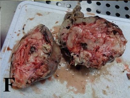
Veterinarni Medicina, 67, 2022 (02): 99–104                                                        Case Report

https://doi.org/10.17221/55/2021-VETMED

 Figure 1A                                                 Figure 1B

   (A)                                                       (B)

 Figure 1C                                                 Figure 1D

   (C)                                                       (D)

 Figure 1E                                                 Figure 1F

   (E)                                                       (F)

Figure 1. Macroscopy
(A–D) Macroscopic view to the neoplasm during surgery. (E, F) Macroscopic views to the tumour during trimming and
sample selection for the histological processing

for 1 h at room temperature. For visualisation,           DAKO, Glosturp, Denmark). Finally, the slices
an LSAB + System HRP kit (streptavidin-biotin per-        were stained with Mayer haematoxylin (DiaPath,
oxidase detection kit; DAKO, Glosturp, Denmark)           Martinengo, Italy). All the samples were analysed
was applied according to the product manual. The          from the light microscope images obtained using
reaction was visualised with DAB + chromogen              an Olympus AX70 Provis microscope (Olympus,
kit (Liquid DAB + Substrate Chromogen System;             Tokyo, Japan).

                                                                                                             101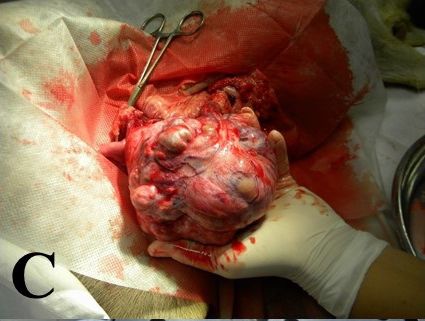
Case Report Veterinarni Medicina, 67, 2022 (02): 99–104
https://doi.org/10.17221/55/2021-VETMED
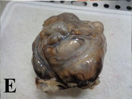
RESULTS ing parts composed of completely differentiated
nervous tissue containing diffusely arranged nerv-
The histological images are shown in Figure 2. ous (Figure 2A) and glial (Figure 2B) cells, which
Almost in the entire material there are alternat- were located on the richly vascularised fine pinkish
Figure 2A Figure 2B
(A) (B)
Figure 2C Figure 2D
(C) (D)
Figure 2E Figure 2F
(E) (F)
Figure 2. Histology and immunohistochemistry
(A) Histological image of the neoplasm consisting of nervous tissue with several well visible neuronal cells. H&E; × 200.
(B) Well-developed nervous tissue in the entire sample. H&E; × 200. (C) Cyst formation, which is layered by a monolayer
of epithelium resembling ependymal cells with superficially localised cilia. H&E; × 200. (D) GFAP-positivity in the neo-
plasm. GFAP; × 200. (E) Neurofilament-positivity in the neoplasm. Neurofilament; × 200. (F) Proliferation negativity
in the neoplastic cells. Ki-67; × 200
102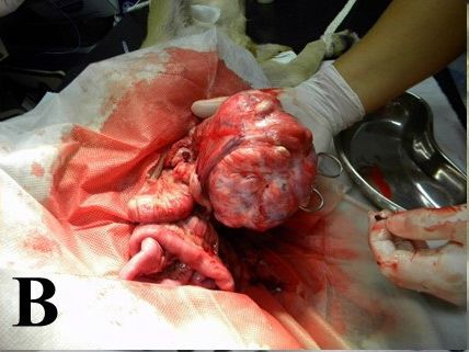
Veterinarni Medicina, 67, 2022 (02): 99–104                                                      Case Report

https://doi.org/10.17221/55/2021-VETMED

vacuolated interstitial tissue. Part of the neoplasm    with a mature ovarian teratoma consisting of high-
was compressed, organised in shapeless, variable-       ly differentiated nervous tissue, confirmed by the
sized nervous tissue nodules, which were separated      immunohistochemistry. On the other hand, there
by fibro-nervous septa containing spindle cells, re-    is an article describing an ovarian mature cystic
sembling fibroblasts with some minimal collagenous      teratoma in 25-year-old woman, which was mostly
fibres and some ovoid cells, resembling interstitial    composed of neurogenic elements (Akbulut et al.
nervous cells. The complete finding was accompa-        2006). This report documents that although benign
nied by several solid formations resembling perito-     teratomas contain variable tissues, they can be also
neal implants and isolated cystic structures, or only   monophasic. We suppose that benign teratoma al-
a remnant of rete ovarii, which were lined by a mon-    ways contains other tissues, which are not processed
olayer epithelium with superficially localised cilia    for histological investigation. Usually, these types
formations (Figure 2C) resembling ependymal cells,      of neoplasms grow into the largest size, creating
including individual pigmented cells with some re-      a large abdominal mass of tissue, when a diagnostic
sidual sparse serous secretion into the lumen. From     imaging procedure is sufficient for the visualisa-
place to place, there were some miniature shapeless     tion (Stussi et al. 2008). Although the enormous
structures corresponding to a differentiated hyaline    size of a benign teratoma with expansive growth
cartilage and epidermoid cysts. The histochemi-         and benign behaviour is evident without any clini-
cal assay revealed a stromal Masson trichrome           cal signs, several authors have documented that
reaction, some isolated PAS positivity and Luxol        it can be malignant and have a metastasis potential
fast blue negativity. Immunohistochemically, well       (Coggeshall et al. 2012; Da Costa et al. 2017). For ex-
visible GFAP positivity (Figure 2D) and neurofila-      ample, Patnaik and Greenlee (1987) found that 29%
ment positivity (Figure 2E) in the nervous tissue       of canine ovarian neoplasms were metastasised;
and negative proliferation activity (Figure 2F) were    adenocarcinomas showed 48% metastases and ma-
observed in the neoplasm samples.                       lignant teratomas showed 50% metastases, and
                                                        a distant metastasis was more common in a ma-
                                                        lignant teratoma. In dogs, it is assumed that one-
DISCUSSION AND CONCLUSIONS                              third of teratomas are malignant having a metastatic
                                                        potential and poor prognosis (De Bosschere et al.
   Although canine teratomas are not a frequently       1999). On the other hand, several cases showed
occurring neoplasm, their benign variant is most        no teratoma recurrence or other complications over
often encountered. It is a germ cell neoplasm his-      years of follow up (Lopez et al. 2017; Sarrau 2018).
tologically composed of several tissues. The variety    This concurs with our finding.
of tissues depends on the embryonic germ layers.           Ultrasonography is a useful tool when examin-
A variety of tissue may be present including hair,      ing intact bitches in diagnosing ovarian tumors
bone, cartilage, teeth and the like. Most teratomas     such as teratomas, which have metastatic poten-
are benign and composed of well-differentiated          tial in some cases (Oviedo-Penata et al. 2020).
mature tissues, but any of the tissue that make         Teratomas are tumours that have a higher preva-
up a teratoma may be malignant (Nagashima et al.        lence in advanced age bitches, but in some cases,
2000; Yamaguchi et al. 2004; Xiang et al. 2018).        they can arise in younger animals. As it is docu-
Differential diagnosis should include dysgermino-       mented in the above citations, they can occur and
ma, sex cord stromal tumors, vascular hamartoma,        be diagnosed also in young women. This is probably
fibroma, leiomyoma, rhabdomyosarcoma, as well           in the context of prepubertal aggressive malignant
as metastases from internal organs. However, the        tumours than of benign tumours arising in adult-
co-existence of an ovarian teratoma and uterine         hoods. It is likely that gene mutations play here
adenocarcinoma in a female dog was also reported        important role (Makovicky and Svecova 2016).
(Pires et al. 2019).                                    This is in relationships with prognosis, including
   Our case report shows that, in a neoplasm, one       survival, age and can be applied also to teratomas.
tissue can dominate over the others with a benign          On the basis of the USG diagnostics and histo-
morphology. To the best of our knowledge, only          pathological analyses, we demonstrated the occur-
one article with a similar finding in a dog was pub-    rence of benign ovarian teratoma, with prevalent
lished thus far. Rota et al. (2013) described a dog     nervous tissue, in a dog.

                                                                                                           103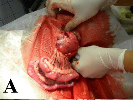
Case Report                                                        Veterinarni Medicina, 67, 2022 (02): 99–104

                                                                      https://doi.org/10.17221/55/2021-VETMED

Conflict of interest                                         Nagashima Y, Hoshi K, Tanaka R, Shibazaki A, Fujiwara K,
                                                               Konno K, Machida N, Yamane Y. Ovarian and retroperi-
  The authors declare no conflict of interest.                 toneal teratomas in a dog. J Vet Med Sci. 2000 Jul;62
                                                               (7):793-5.
                                                             Oviedo-Penata CA, Hincapie L, Benavides CR, Maldonado
REFERENCES                                                     JG. Concomitant presence of ovarian tumors (teratoma
                                                               and granulosa cell tumor), and pyometra in an English
Akbulut M, Kelten EC, Ege CB. Mature cystic teratoma with      Bulldog female dog: A case report. Front Vet Sci. 2020
  predominantly neurogenic elements – Case report. Ae-         Jan;14(6):500.
  gean Pathol J. 2006 Aug;22(3):18-20.                       Patnaik AK, Greenlee PG. Canine ovarian neoplasms:
Blaszak B, Walkowski M, Ibbs M, Jaskowski JM. Teratoma         A clinic-pathologic study of 71 cases, including histology
  adultum in a bitch: A case report. Vet Med-Czech. 2009       of 12 granulosa cell tumors. Vet Pathol. 1987 Nov;24(6):
  Aug;54(8):379-81.                                            509-14.
Chai Y, Woo CG, Kim JY, Kim CJ, Khang SK, Kim J, Park        Pegas GRA, Monteiro LN, Cassali GD. Extragonadal ma-
  IA, Kim EN, Kim KR. Diagnostic significance of cellular      lignant teratoma in a dog – Case report. Arq Bras Med
  neuroglial tissue in ovarian immature teratoma. J Pathol     Vet Zootec. 2020 Jan-Feb;72(1):115-8.
  Transl Med. 2017 Jan;51(1):49-55.                          Pires MDA, Catarino JC, Vilhena H, Faim S, Neves T,
Coggeshall JD, Franks JN, Wilson DU, Wiley JP. Primary         Freire A, Seixas F, Orge L, Payan-Carreira R. Co-existing
  ovarian teratoma and GCT with intra-abdominal metas-         monophasic teratoma and uterine adenocarcinoma in a fe-
  tasis in a dog. J Am Anim Hosp Assoc. 2012 Nov-Dec;48        male dog. Reprod Domest Anim. 2019 Jul;54(7):1044-9.
  (6):424-8.                                                 Rota A, Tursi M, Zabarino S, Appino S. Monophasic tera-
Da Costa DA, Da Silva MRM, De Souza NF, Pereira WLA,           toma of the ovarian remnant in a bitch. Reprod Domest
  Cardoso AMC. Giant canine ovarian teratoma: Case re-         Anim. 2013 Apr;48(2):26-8.
  port. J Cytol Histol. 2017 Aug;8(3):1-3.                   Sarrau S. Iatrogenic ureteral lesion following a bulky ovar-
De Bosschere H, Durnez V, Ducatelle R. Malignant ovarian       ian teratoma excision in a 3-year bitch. Rev Veterinaire
  teratoma in a Golden Retriever. Vlaams Diergeneesk Ti-       Clin. 2018 Apr;53(4):115-20.
  jdcchr. 1999 Feb;68(2):96-100.                             Stussi A, Bohler A, Zednik T, Shiblz S, Zednik P. Teratoma –
Gulcubuk A, Altun ED, Bozkurt ER, Sontas BS, Haktanir D.       A rare ovarian tumour in a bitch. Wien Tierärztl Monatss-
  Ovarian teratoma in a dog. Turkish J Vet Anim Sci. 2012      chr. 2008 Mar;95(3):85-90.
  May;36(5):573-6.                                           Xiang H, Han J, Ridley WE, Ridley LJ. Canine tooth: Mature
Iemura Y, Yamada Y, Hirata M, Kataoka TR, Minamiguchi S,       cystic teratoma. J Med Imaging Radiat Oncol. 2018 Oct;
  Haga H. Histopathological characterization of the neu-       62(S1):61.
  roglial tissue in ovarian teratoma associated with anti-   Yamaguchi Y, Sato T, Shibuya H, Tsumagari S, Suzuki T.
  N-methyl-d-aspartate (NMDA) receptor encephalitis.           Ovarian teratoma with a formed lens and non-supportive
  Pathol Int. 2018 Dec;68(12):677-84.                          inflammation in an old dog. J Vet Med Sci. 2004 Jul;66(7):
Linder D, McCaw BK, Hecht F. Parthenogenic origin of be-       861-4.
  nign ovarian teratomas. N Engl J Med. 1975 Jan 9;292       Yoshimura, Yamamoto M, Moriya M, Endo T, Sugiura N,
  (2):63-6.                                                    Kato T, Matsuda Y, Ishiwata T, Kajigaya H, Kamiiya S.
Lopez D, Singh A, Wright TF, Gartley C, Walker M. Single       Teratoma of the ovary in a free-ranging Japanese Raccoon
  incision laparoscopic-assisted ovariohysterectomy for        dog (Nyctereutes Procyonoides Viverrinus). J Zoo Wildl
  an ovarian tumour in a dog. Can Vet J. 2017 Sep;58(9):       Med. 2017 Mar;48(1):265-8.
  975-9.
Makovicky P, Svecova I. Veterinary pathology: The past,                                       Received: April 14, 2021
  present and the future. Phenogenomic Newsletter. 2016                                  Accepted: September 22, 2021
  April;2(2):22-3.

104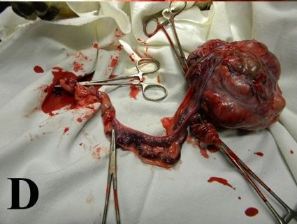
You can also read