Anti-Inflammatory and Healing Activity of the Hydroalcoholic Fruit Extract of Solanum diploconos (Mart.) Bohs
←
→
Page content transcription
If your browser does not render page correctly, please read the page content below

Hindawi
Journal of Immunology Research
Volume 2021, Article ID 9957451, 13 pages
https://doi.org/10.1155/2021/9957451

Research Article
Anti-Inflammatory and Healing Activity of the Hydroalcoholic
Fruit Extract of Solanum diploconos (Mart.) Bohs

 Larissa Benvenutti,1 Roberta Nunes,1 Ivonilce Venturi,1 Silvia Aparecida Ramos,1
 Milena Fronza Broering,1 Fernanda Capitanio Goldoni,2 Sarah Eskelsen Pavan,2
 Maria Verônica Dávila Pastor,2 Angela Malheiros,1,2 Nara Lins Meira Quintão,1,2
 Elizabeth Soares Fernandes ,3,4 and José Roberto Santin 1,2
 1
 Postgraduate Program in Pharmaceutical Science, Universidade do Vale do Itajaí, Rua Uruguai, 458, Itajaí, SC 88302-202, Brazil
 2
 School of Health Sciences, Biomedicine Course, Universidade do Vale do Itajaí, Rua Uruguai, 458, Itajaí, SC 88302-202, Brazil
 3
 Programa de Pós-graduação em Biotecnologia Aplicada à Saúde da Criança e do Adolescente, Faculdades Pequeno Príncipe,
 Av. Iguaçu, 333, Curitiba, PR 80230-020, Brazil
 4
 Instituto de Pesquisa Pelé Pequeno Príncipe, Av. Silva Jardim, 1632, Curitiba, PR 80250-060, Brazil

 Correspondence should be addressed to Elizabeth Soares Fernandes; elizabeth.fernandes@pelepequenoprincipe.org.br
 and José Roberto Santin; jrs.santin@univali.br

 Received 22 March 2021; Revised 15 June 2021; Accepted 6 July 2021; Published 20 July 2021

 Academic Editor: Michael Conlon

 Copyright © 2021 Larissa Benvenutti et al. This is an open access article distributed under the Creative Commons Attribution
 License, which permits unrestricted use, distribution, and reproduction in any medium, provided the original work is
 properly cited.

 Background. Solanum diploconos (Mart.) Bohs is a native Brazilian plant belonging to the Solanaceae family, popularly known as
 “tomatinho do mato” and poorly investigated. Herein, we presented for the first time evidence for the anti-inflammatory and
 wound healing activities of S. diploconos fruit hydroalcoholic extract. Material and Methods. In vitro fMLP-induced chemotaxis,
 LPS-induced inflammatory mediator levels (cytokines by ELISA and NO release by Griess reaction), and adhesion molecule
 expression (CD62L, CD49d, and CD18, by flow-cytometry) were assessed in neutrophils treated with different concentrations of
 the extract. Inflammation resolution was measured by the efferocytosis assay and the healing activity by in vivo and in vitro
 assays. The air pouch model of carrageenan-induced inflammation in Swiss mice was used to investigate the in vivo anti-
 inflammatory effects of the extract. Leukocyte influx (by optical microscopy) and cytokine release were quantified in the pouch
 exudates. Additionally, the acute and subacute toxic and genotoxic effects of the extract were evaluated. Results. In vitro, the
 extract impaired neutrophil chemotaxis and its ability to produce and/or release cytokines (TNFα, IL-1β, and IL-6) and NO
 upon LPS stimuli (p < 0:01). LPS-treated neutrophils incubated with the extract presented increased CD62L expression (p < 0:01),
 indicating a reduced activation. An enhanced efferocytosis of apoptotic neutrophils by macrophages was observed and
 accompanied by higher IL-10 and decreased TNFα secretion (p < 0:01). In vivo, similar results were noted, including reduction
 of neutrophil migration, protein exudation, and cytokine release (p < 0:01). Also, the extract increased fibroblast proliferation
 and promoted skin wound healing (p < 0:01). No signs of toxicity or genotoxicity were observed for the extract. Conclusion. S.
 diploconos fruit extract is anti-inflammatory by modulating neutrophil migration/activation as well macrophage-dependent
 efferocytosis and inflammatory mediator release. It also indicates its potential use as a healing agent. Finally, the absence of
 acute toxic and genotoxic effects reinforces its possible use as medicinal product.

1. Introduction migration of immune cells to the injury site to prevent the
 invasion of microorganisms and damage by hazardous sub-
Inflammation is a physiological response to injury character- stances in the absence of tissue integrity [1]. In this context,
ized by complex processes which are aimed at restoring tissue neutrophils play an important role, arriving at the site of
homeostasis. Its first stage comprises the quick activation and inflammation in a multistep controlled process which
2 Journal of Immunology Research

encompasses marginalization, slow rolling, adhesion, and aimed at investigating the anti-inflammatory effects, wound
transendothelial or abluminal migration [2, 3]. All these pro- healing activity, and the acute toxicity profile of the hydroal-
cesses are dependent on the specific interactions between coholic extract obtained from S. diploconos fruits using
proteins expressed on endothelial cells and leukocytes, such in vivo and in vitro protocols.
as integrins and selectins [2, 4]. The CD62L selectin is
expressed on the neutrophil membrane and mediates its
binding to endothelial cell adhesion molecules (P and E- 2. Material and Methods
selectins and integrins); it is also responsible for the neutro- 2.1. Plant Material and Phytochemical Analysis. S. diploconos
phil rolling behaviour [3, 5]. On the other hand, integrins (Mart) Bohs fruits were harvested from Itaiópolis, SC, Brazil
such as CD18 and CD49a, are involved in the neutrophil
 (latitude 60° 66 ′ 27″ S and longitude 70° 68 ′ 53″ W, eleva-
adhesion to the endothelium, allowing its transmigration to
 tion: 800.33 m; SisGen protocol A478BBA) in April 2014,
the injury site [6].
 and voucher specimens were deposited in the Herbarium
 Once at the injury site, neutrophils carry out an efficient
 Barbosa Rodrigues (HBR55276) and the Herbarium Dr.
local response, marked by the production of nitric oxide
 Roberto Miguel Klein (FURB49359). Whole fresh ripe fruits
(NO), reactive oxygen species (ROS), and cytokines; release
 (peel and pulp with seeds) were ground and subjected to
of cytotoxic granular contents; formation of neutrophil
 extraction by dynamic maceration with 95% ethanol in the
extracellular traps; and phagocytosis. Neutrophil migration
 proportion 1 : 10 (m/v), for 6 h at room temperature (24-
can become continuous in some cases, contributing to an
 28°C). The extract was concentrated and dried under reduced
uncontrolled inflammation, thus, leading to tissue damage
 pressure on a rotary evaporator with temperature below
and/or loss of function and chronification of the injury [1].
 50°C.
 In this context, neutrophil clearance is an important
 The chromatographic analysis was carried out on a
step of the resolution of inflammation and ultimate tissue
 HPLC Shimadzu® LC 20-AT system, consisting of a quater-
repair [7]. This process comprises the limitation of neutro-
 nary pump and a Shimadzu SPD-M20A photodiode array
phil migration, downregulation of proinflammatory media-
 detector. A SIL-20A HT autosampler with a Shimadzu
tors, induction of neutrophil apoptosis, and its subsequent
 CTO-10AS VT column oven equilibrated at 35°C and soft-
phagocytosis (efferocytosis) by resident macrophages [7,
 ware LC Solution were used. The chromatographic column
8]. Following efferocytosis, the macrophages switch from
 used was Phenomenex® C18 (250 × 4:6 mm) with particles
the inflammatory to the anti-inflammatory phenotype,
 of 5 μm. For the method development, different solvent sys-
characterized by the release of anti-inflammatory cytokines
 tems were assayed in gradient conditions using acetonitrile
and growth factors. In turn, these changes contribute
 (A) and ultrapure water acidified with acetic acid (pH 2.5;
towards angiogenesis, wound healing, tissue repair, and
 B). The chosen gradient was (A : B): 05 : 95 at 0-10 min,
homeostasis [7].
 15 : 85 at 10-20 min, 30 : 70 at 20-30 min, 40 : 60 at 30-
 Natural products have been used as sources of biological
 40 min, 55 : 45 at 40-45 min, and 05 : 95 at 4550 min. The flow
active substances and even of new drugs [9]. The Solanum
 rate was 1.0 mL/min. The detection of the compounds was
genus comprises approximately 1500 species, making this
 carried out at 320 nm. This method was chosen for the anal-
genus the largest and most complex of the Solanaceae family.
 ysis of phenolic compounds as previously established by
It is distributed in temperate and tropical regions, and it pre-
 Ribeiro et al. [13].
sents several classes of compounds such as alkaloids, triter-
penes, flavonoids, and saponins [10]. Some compounds
found in this genus have toxic effects; therefore, toxicological 2.2. Animals. Experiments were performed in male Swiss
studies are needed to ensure safe usage [11]. mice and female Wistar rats (exclusively for the toxicological
 S. diploconos (Mart.) Bohs is a poorly investigated Brazil- evaluation). All animals were obtained from the Central Ani-
ian native plant, popularly known as “tomatinho-do-mato” mal Facility of the Universidade do Vale do Itajaí (UNIVALI)
which belongs to the same genus of important food crops and kept in a climate-controlled room at 22 ± 2°C, under
such as tomato (S. lycorpersicum) and potato (S. tuberosum). light/dark (12 : 12 h) cycle, with water and food ad libitum.
Its fruits are edible and slightly acidic, consumed either in All experiments were approved by the local ethics committee
natura or as a juice [12]. Despite its consumption as food, (CEUA/UNIVALI: 028/16 and 019/18).
the biological activities of S. diploconos fruits remain unclear.
Phenolic compounds, carotenoids, tocopherols, and ascorbic 2.3. In Vitro Anti-Inflammatory Effects
acid were the main classes of secondary metabolites found in
the freeze-dried pulp and peel and in the extract obtained 2.3.1. Neutrophil Isolation. For this, male Swiss mice received
from the whole fruit of S. diploconos. Furthermore, the whole an intraperitoneal (i.p.) injection of 1% sterile oyster glyco-
fruit extract presented antioxidant actions when assessed gen in phosphate-buffered saline (PBS; 3 mL). After 4 h, the
in vitro against ROS and reactive nitrogen intermediates [13]. animals were culled with an overdose of ketamine (80 mg/kg,
 Data from the literature indicate a great therapeutic i.p.) and xylazine (8 mg/kg, i.p.), and their peritoneal cells
potential for the Solanum genus, highlighting the anti- were collected by washing the peritoneal cavity with 3 mL
inflammatory activity of extracts [14, 15] and some of the of sterile PBS. The number of viable cells (98%) was counted
compounds also identified in S. diploconos [16], including in a Neubauer chamber using a light microscope (Nikon,
wound healing activity [17]. In this context, this study was Japan) by the trypan blue exclusion test.
Journal of Immunology Research 3

2.3.2. Lipopolysaccharide-Stimulated Neutrophils. Neutro- of nitrite (NO2-) using the Griess reaction [21]. For this, sam-
phils (1 × 10⁶ cells per well) were incubated with RPMI ples (100 μL/well) were incubated for 10 min at 37°C, with
medium (10% foetal bovine serum (FBS)) in the presence 100 μL of Griess reagent (1% sulfanilamide, 0.1% naphthy-
or absence of lipopolysaccharide (LPS; 5 μg/mL) and simul- lethylenediamine dihydrochloride in 5% phosphoric acid).
taneously treated with S. diploconos extract (1, 10, or The absorbances were then determined at 550 nm. All cyto-
100 μg/mL) for 18 h, at 37°C in a 5% CO2 atmosphere. Then, kine results are expressed in picograms per milliliter, and
the supernatant was collected for the subsequent analysis NO levels as micromolar of NO2-.
of cytokine and nitrite (NO2-) levels. Neutrophil viability
was assessed by the trypan blue exclusion test. None of 2.4. In Vivo Anti-Inflammatory Effects
the tested extract concentrations produced cell toxicity
 2.4.1. In Vivo Leukocyte Migration: Air Pouch Model. Air
(data not shown).
 pouches were produced on the back of the mice as previously
2.3.3. Expression of Adhesion Molecules on Neutrophils. Neu- described [22, 23]. Briefly, the animals were fasted for 4 h and
 then orally treated by gavage with S. diploconos extract
trophils (1 × 10⁶ cells per well) stimulated or not with LPS
 (100 mg/kg, n = 6), indomethacin (30 mg/kg, positive con-
(5 μg/mL) were coincubated with either vehicle (cell culture
 trol, n = 6), or vehicle (PBS; 10 mL/kg; n = 6). After 1 h, car-
medium) or the extract (1, 10, or 100 μg/mL). After 1 h, the
 rageenan (1%; 3 mL/cavity) was injected directly into the air
cells were collected and incubated with monoclonal anti-
 pouch chamber. Four hours later, a small incision was
CD62L (FITC), anti-CD18 (PE), or anti-CD49d (APC) anti-
 made in the air pouch. The cavity was washed with 5 mL
bodies in the dark for 20 minutes, at 4°C. Cells were then ana-
 of PBS, and the inflammatory infiltrate collected for analy-
lysed on an Accuri C6 Flow Cytometer (BD Bioscience), and
 sis. Total and differential cell counts were performed, and
data from 10,000 events was obtained. The results are
 the rest of the exudate was kept for the further quantifica-
expressed as mean fluorescence intensity (MFI) [18].
 tion of cytokines.
2.3.4. In Vitro Neutrophil Chemotaxis. Neutrophil chemo- The air pouch lining tissue was collected for histological
taxis was evaluated as described by Nelson et al. [19]. Briefly, analysis and preserved in formaldehyde solution until the
 blades were made. The preparation consisted of the insertion
neutrophils (1 × 10⁷ cells/mL) treated or not with S. diploco-
 of the tissue fragment into a cassette. The sample then under-
nos extract (1, 10, or 100 μg/mL, for 1 h) were placed into
 went a 1 h dehydration process in increasing concentrations
peripheral wells (10 μL) made in an agarose gel solidified in
 of ethanol (70%, 96%, and absolute ethanol). Subsequently,
a Petri dish. The chemotactic factor (N-Formyl-Met-Leu-
 each sample was placed in xylol followed by paraffin bath.
Phe (fMLP); 0.1 μM, 10 μL) was added to the central well,
 Then, the cassettes containing samples were placed in a
and the plates were incubated for 4 h, at 37°C and 5% CO2.
 microtome where serial sections of 3 or 4 μm were made.
After incubation, the number of cells that migrated towards
 The sections were placed onto slides and stained with haema-
the central well containing fMLP was counted.
 toxylin and eosin. Samples were analysed by microscopy
2.3.5. Efferocytosis Assay. Macrophages were obtained by under the 4, 10, and 40x objectives.
washing the mouse medullary cavity with sterile PBS. The 2.5. Wound Healing Assessment
cell suspension (1 × 10⁶ cells/well) was incubated in a 24-
well plate onto a round glass coverslip at 37°C and 5% CO2 2.5.1. In Vitro Scratch Assay. Murine L929 cells were
for up to 2 h to allow macrophage adherence. After this obtained from the Bank of Cells of Rio de Janeiro (Rio de
period, the wells were washed three times with sterile PBS Janeiro, RJ, Brazil) and maintained in DMEM supplemented
to remove nonadherent cells and then, the remaining ones with 10% FBS, 100 μg/L streptomycin, and 100 IU/mL peni-
were incubated with S. diploconos extract diluted in DMEM cillin, at 37°C in a 5% CO2 atmosphere. Briefly, L929 cells
(1, 10, or 100 μg/mL). After 1 h, senescent mouse neutrophils (5 × 10⁴ cells/mL) were seeded into each well of a 24-well
(1 × 10⁶ cells) were added to each well and 20 min later, the plate and incubated at 37°C with 5% CO2. After cell conflu-
supernatant was collected for cytokine analysis. The remain- ence, the culture medium was removed and a continuous
ing cells on the slides were analysed by microscopy with scratch was made in the medial surface of each well with a
immersion objective (100x). One hundred macrophages were 200 μL tip. Then, the wells were washed with PBS to remove
observed, and the phagocytosis of senescent neutrophils was cell debris. The remaining cells were incubated with DMEM
expressed as the percentage (%) of macrophages containing containing S. diploconos fruit extract (1, 10, or 100 μg/mL).
neutrophils [20]. The analysis was performed by measuring the scratched area
 in square millimeter at 0 (immediately after mechanical
2.3.6. Cytokine and Nitrite Analysis. The levels of IL-1β, trauma) and 24 h after incubation with the extract, by
TNFα, IL-6, and IL-10 were analysed in the supernatants microscopy (Olympus CKX 41), using ImageJ1.46r software.
obtained from the cell cultures, including those from the The results are expressed as the percent of the coverage area
efferocytosis assay, or in exudate samples from the air pouch at 24 h in relation to time 0 [24].
model. One hundred μL of each sample was used for the
assays performed in accordance to the manufacturer’s 2.5.2. In Vivo Wound Healing Assay. For topical application,
instructions (DuoSet R&D Systems, Minneapolis, MN, a semisolid formulation of the S. diploconos extract was
USA). NO levels were indirectly quantified by the detection made. An oil in water (O/W) emulsion-based cream was
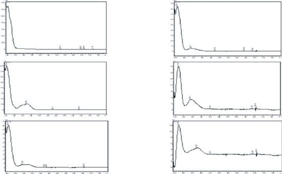
4 Journal of Immunology Research

 Peaks UV spectrum Peaks UV spectrum
 2; 3; 4; 14; 15;
 6; 7; 8; 17
 9; and 10
 50 320 nm 4 nm (1.00)

 5
 45
 40 5; 11; 23
 1

 35 12
 30
 25
 20
 15 13; 16; 24
 18; 19;
 1413 12
 15 16
 11

 10 2120
 23 22
 17 18
 20; 21;
 2

 24
 89
 3

 5 21; and 22
 4
 6
 7

 0
 0 10 20 30 40 min
 (a) (b)

 Figure 1: Chromatogram of the S. diploconos fruit extract at λ = 320 nm (a) and respective UV absorption spectra (b).

prepared by mixing Sepigel 305, vaseline, crodalan, isopropyl vivors were autopsied. The blood was collected for
myristate, BHT, cetostearyl alcohol, Phenonip®, EDTA, and biochemical (aspartate aminotransferase (AST), alanine ami-
water with slight warming. The oil-soluble components were notransferase (ALT), alkaline phosphatase (ALP), urea, and
dissolved in the oil phase and heated to 75°C. The water- creatinine) and hematological (total and differential cell, ery-
soluble components were dissolved in the aqueous phase throgram, and platelet counts) analysis [27].
and heated to 75°C. After heating, the aqueous phase was In the repeated dose 28-day oral toxicity (subacute)
added in portions to the oil phase with continuous stirring study, female Wistar rats were divided into different groups
until cooling took place. Then, the hydroalcoholic extract (n = 5/group) and orally treated by gavage with S. diploconos
obtained from S. diploconos fruits (1%) was incorporated to extract (30, 100, or 300 mg/kg) or vehicle (distilled water,
the base cream. The formulation was packed in aluminium 10 mL/kg). The extract and vehicle were administered once
tubes, with a seal and polypropylene cover. a day, for 28 days. At the end of the observation period, the
 Mice were divided in two groups: group I (base cream, macroscopic characteristics of the lungs, kidneys, liver,
n = 6) and group II (1% S. diploconos, n = 6). The animals spleen, and heart were observed. In addition, the blood was
were anaesthetized, and their backs were shaved with an elec- collected for analysis of biochemical and hematological
tric clipper. Then, a surgical excision of the skin (1.5 cm of parameters.
diameter) was made on the dorsal area of each mouse, as pre-
viously described by Tonin et al. [25]. The extract was topi- 2.6.3. Micronucleus Test. The micronucleus test was per-
cally applied to each wound (once a day, for 7 days). The formed 24 h after S. diploconos extract (2 g/kg, by gavage)
wounds were photographed daily with a scale, and the administration to the mice. Vehicle- (10 mL/kg) and methyl
wounded areas were measured and calculated in square mil- methanesulfonate- (MMS; 50 mg/kg, i.p.) treated animals
limeter by using the ImageJ1.46r software. were used as negative and positive controls, respectively.
 After treatments, the mouse right femurs were collected,
2.6. Toxicity Evaluation and the epiphyses cut. The medullary cavity of each femur
 was washed using a syringe containing FBS (3 mL). The
2.6.1. Cytotoxicity Assays. Possible toxic effects of the extract lavage fluid was centrifuged at 1000 × g, for 10 min at 5°C;
were investigated in L929 cells. For this, the cells were cul- the cells were obtained; and their viability was assessed by
tured in DMEM (containing 10% FBS, 100 μg/L streptomy- using 0.4% trypan blue dye. Then, 50 μL of the cell suspen-
cin, and 100 IU/mL penicillin) at 37°C in a 5% CO2 sions was placed onto glass slides, fixed with cold methanol,
atmosphere and incubated with either S. diploconos extract and stained with Giemsa for 10 min. The slides were washed
(0.1, 1, 10, or 100 μg/mL) or vehicle (RPMI medium). After and dried for examination under a light microscope (1000x).
24 h, the cell viability was evaluated by the MTT method One thousand polychromatic cells were assessed per slide.
[26]. The results are expressed as percent of viable cells in The ratio between monochromatic and polychromatic eryth-
relation to the control (vehicle-treated wells). rocytes was established and analysed [28].

2.6.2. Pathophysiological Analysis. In the acute toxicity assay, 2.7. Statistics. Data is expressed as mean ± standard error of
rats (n = 5/group) received either S. diploconos extract the mean (SEM). The percentages of inhibition were individ-
(2 g/kg) or vehicle (distilled water, 10 mL/kg) by oral route ually calculated and expressed as mean ± SEM%. Statistical
(gavage). The animals were then observed at every 30 min differences between groups were assessed by Student’s t-test
during the first 4 h followed by a daily observation for 14 or one-way analysis of variance (ANOVA) followed by
days. The cumulative weight change was calculated as per- Tukey’s post hoc test, as appropriate. Values of p < 0:05 were
cent to baseline. At the end of the observation period, all sur- considered as significant.
Journal of Immunology Research 5

 0.8 600

 #### ####
 0.6
 NO2 ( M/mL)
 400

 TNF (pg/mL)
 ⁎ ⁎⁎ ⁎⁎
 0.4
 ⁎⁎ ⁎⁎
 ⁎⁎
 200
 0.2

 0.0 0
 Basal LPS 1 10 100 Basal LPS 1 10 100
 Basal Basal
 +LPS (5 g/mL) +LPS (5 g/mL)
 +S. diploconos ( g/mL) +S. diploconos ( g/mL)
 (a) (b)
 600 #### 2000
 ####

 ⁎⁎ ⁎⁎ 1500
 1L-1 (pg/mL)

 400 ⁎⁎
 1L-6 (pg/mL)
 ⁎⁎⁎

 1000 ⁎⁎⁎⁎

 200
 500 ⁎⁎⁎⁎

 0 0
 Basal LPS 1 10 100 Basal LPS 1 10 100
 Basal Basal
 +LPS (5 g/mL) +LPS (5 g/mL)
 +S. diploconos ( g/mL) +S. diploconos ( g/mL)
 (c) (d)
 12000
 ⁎⁎⁎⁎
 100

 10000
 ⁎⁎⁎⁎

 ⁎⁎⁎
 MFI CD62L

 ⁎⁎⁎
 10

 8000
 ⁎⁎⁎⁎
 1

 6000
 ####
 Basal

 ####

 4000

 0 50 100 150 Basal LPS 1 10 100
 Number of cells that migrated/4h
 Basal
 Basal +LPS (5 g/mL)
 +S. diploconos ( g/mL) +S. diploconos ( g/mL)
 (e) (f)

 Figure 2: Continued.
6 Journal of Immunology Research

 50000 10000

 8000
 45000

 MFI CD49d
 MFI CD18

 6000
 40000
 4000

 35000
 2000

 30000 0
 Basal LPS 1 10 100 Basal LPS 1 10 100
 Basal Basal
 +LPS (5 g/mL) +LPS (5 g/mL)
 +S. diploconos ( g/mL) +S. diploconos ( g/mL)
 (g) (h)

Figure 2: Effects of the S. diploconos fruit extract on neutrophils. Neutrophils obtained from the peritoneal cavity of Swiss mice (1% oyster
glycogen) were incubated in the presence or absence of LPS (5 μg/mL) and with S. diploconos (1, 10, or 100 μg/mL). Nitrite levels were
measured by the Griess reaction (a). The levels of (b) TNFα, (c) IL-1β, and (d) IL-6 were determined by ELISA. For the chemotaxis assay
(e), a neutrophil suspension (1 × 10⁷ ) was incubated with different concentrations of S. diploconos (1, 10, or 100 μg/mL) for 15 minutes
and placed in front of the fMLP (1 μM) on an agarose plate. Quantification was made from the margin of the peripheral perforations
towards the chemotactic agent (central perforation). Neutrophils (1 × 10⁶ ) were incubated in the presence or absence of LPS (5 μg/mL)
along with S. diploconos (1, 10, or 100 μg/mL) for evaluation of the neutrophil adhesion molecules. Adhesion molecule expression was
assessed by labeling with (f) CD62L, (g) CD18, and (h) CD49 detection antibodies by flow cytometry. Values express the mean ± SEM of
tests performed with cells obtained from 8 animals per group. ∗ p < 0:05; ∗∗ p < 0:01; ∗∗∗ p < 0:001; and ∗∗∗∗ p < 0:0001 vs. the LPS group
significantly different from the basal group ####p < 0:0001 (one-way ANOVA followed by Turkey’s post hoc test).

3. Results 3.3. S. diploconos Fruit Extract Impairs Neutrophil Migration.
 Figure 2 also shows the effect of S. diploconos extract on neu-
3.1. Chemical Profile of Solanum diploconos Fruit Extract. trophil migration. Vehicle-treated neutrophils presented a
The yield of the whole fresh ripe fruit hydroalcoholic extract significant migration towards fMLP, whilst those treated with
was 11.49%. Its chemical composition (main classes of S. diploconos exhibited impaired chemotaxis (maximum
metabolites) was analysed by HPLC with a photodiode detec- inhibition of 78:1 ± 1:2%; Figure 2(e)).
tor. The HPLC/UV profiles of the extract are shown in Once leukocyte migration is mediated by adhesion mole-
Figure 1. Six distinct absorption profiles were detected. The cules, the expression of L-selectin (CD62L), β2-integrin
first profile demonstrates a band with a maximum absorption (CD18), and CD49d on neutrophils was evaluated by flow
at 210 nm (peaks 2-4 and 6-10) which indicates compounds cytometry. LPS triggered the activation of neutrophils as they
with chromophore groups that present π − π ∗ electronic presented shedding of CD62L and increased expression of
transition such as those with olefinic groups. Many classes CD18 and CD49b on their surface. Incubation of LPS-
of compounds are absorbed in this region; making it difficult treated neutrophils with S. diploconos extract impaired neu-
to determine the major one. Other five profiles indicate trophil activation, denoted by increased expression of
bands with a maximum absorption between 270 and CD62L (1.7-fold increase for the concentrations of 10 and
330 nm (other peaks). This is a region in which n − π ∗ tran- 100 μg/mL), i.e., increased MFI in comparison with LPS con-
sitions are observed. Of note, carbonyl compounds as well as trols (Figures 2(f) and 2(g)).
aromatic groups present this transition (Figure 1).
 3.4. S. diploconos Fruit Extract Induces Efferocytosis of
3.2. S. diploconos Fruit Extract Affects the Secretion of Apoptotic Neutrophils. All tested concentrations of the S.
Proinflammatory Cytokines and Nitric Oxide by Neutrophils. diploconos fruit extract (1, 10, or 100 μg/mL) were able to sig-
In order to evaluate the direct effects of the fruit extract on nificantly increase the efferocytosis process (maximum
inflammatory mediator production (TNFα, IL-6, IL-1β, and increase of 2.0-fold; Figure 3(a)). The extract was also able
NO2- levels), the supernatants from the neutrophil culture to reduce TNFα (maximum inhibition of 80:7 ± 1:2%;
were assessed. Data depicted in Figures 2(a)–2(d) demon- Figure 3(b)) and increase IL-10 (maximum increase of
strate that LPS induces the production of inflammatory 11.5-fold; Figure 3(c)) levels in the supernatant samples
mediators by neutrophils. Treatment with S. diploconos obtained from the efferocytosis assay.
extract significantly reduced NO2- (maximum inhibition of
63:5 ± 7:3%), TNFα (maximum inhibition of 54:2 ± 2:1%), 3.5. S. diploconos Fruit Extract Presents In Vivo Anti-
IL-1β (maximum inhibition of 68:9 ± 3:1%), and IL-6 (max- Inflammatory Activity. The in vivo effects of S. diploconos
imum inhibition of 73:6 ± 0:9%) levels in comparison with fruit extract on neutrophil migration were evaluated in the
LPS-treated neutrophils (Figures 2(a)–2(d)). air pouch model, using carrageenan as a phlogistic agent.
Journal of Immunology Research 7

 50 300
 ⁎⁎⁎⁎
 % efferocytosis 40 ⁎⁎⁎⁎
 200

 TNF (pg/mL)
 30 ⁎

 20 ⁎⁎⁎⁎ ⁎⁎⁎⁎
 100
 10 ⁎⁎⁎⁎

 0 0
 Basal 1 10 100 Basal 1 10 100
 Basal Basal
 +S. diploconos ( g/mL) +S. diploconos ( g/mL)
 (a) (b)
 800

 600 ⁎⁎⁎⁎
 IL-10 pg/mL

 400 ⁎⁎⁎⁎
 ⁎⁎⁎⁎

 200

 0
 Basal 1 10 100

 Basal
 +S. diploconos ( g/mL)
 (c)

Figure 3: Effects of S. diploconos on efferocytosis. Quantification of efferocytosis (a) and supernatant levels of TNFα (b) and IL-10 (c). Data is
expressed as a mean ± SEM of cells obtained from four animals. Statistical analysis was performed using one-way ANOVA followed by
Tukey’s test. ∗ p < 0:05; ∗∗ p < 0:01; ∗∗∗ p < 0:001, and ∗∗∗∗ p < 0:0001 vs. basal.

The histological analysis (Figure 4) of air pouch tissue sec- along with fibroblasts and neutrophils (Figures 4(h) and
tions from naive animals indicated that they present an air 4(i)).
pouch wall with connective tissue, rich in blood vessels, and In the S. diploconos-treated group, the thickness of the air
with relatively low cellular content (Figures 4(a) and 4(b)). pouches challenged with carrageenan was visibly reduced,
Their cell lining was primarily formed by fibroblasts, and, and the walls were formed by loose connective tissue with
in some areas, scarce macrophages were observed within newly formed blood vessels. No signs of oedema were
the air pouch walls (Figures 4(b) and 4(c)). observed (Figures 4(j) and 4(k)). Neutrophil and macrophage
 Vehicle-treated animals challenged with carrageenan numbers were largely diminished in comparison with the
exhibited typical morphological changes of inflammation negative control group, and a lining of fibroblast-like cells
including increased thickness of the air pouch membrane with no directional orientation was observed. Additionally,
and intense oedema, with fibrin-haemorrhagic exudate, leu- under the hair follicles, there was a layer of fibroblasts on
kocyte accumulation, and tissue injury (Figures 4(d) and an augmented deposit of collagen (Figures 4(k) and 4(l)).
4(e)). Most of the inflammatory cells were neutrophils, as These histological findings suggest that the acute inflamma-
identified by their lobulated nuclei. These cells were exten- tory response caused by carrageenan in the pouch wall is sig-
sively infiltrated in the damaged tissue and distributed within nificantly suppressed by the treatment with S. diploconos fruit
the adjacent muscular fibres. Macrophages with a foamy extract.
appearance were also observed, possibly due to the phagocy- The data presented in Figure 4(m) demonstrate that
tosis of carrageenan (Figures 4(e) and 4(f)). similarly to indomethacin, the fruit extract at 100 mg/kg
 Indomethacin treatment reduced the thickness of was able to reduce cell counts in the exudate in comparison
carrageenan-challenged air pouch membranes in compari- with the control group. Inhibitions were of 73:5 ± 2:9 and
son with vehicle-injected mice (Figures 4(g) and 4(h)). 95:4 ± 0:8% for S. diploconos and indomethacin, respec-
Oedema was restricted to limited areas, and there was also tively. Differential cell analysis indicates that both S. diploco-
an evident reduction of the neutrophil infiltrate. Few normal nos fruit extract and indomethacin affect mainly neutrophils
and foamy macrophages were seen in the pouch membrane, (Figure 4(n)).
8 Journal of Immunology Research

 Total polymorphonuclear (104)
 1500 1500
 40× 100× 400×

 Total leukocytes (104)
 ####
 (a) (b) (c) ####
 1000 1000
 pm
 Naive

 500 500
 ⁎⁎⁎⁎ ⁎⁎⁎⁎
 pm
 pm a a ⁎⁎⁎⁎ ⁎⁎⁎⁎
 a 0 0
 Naive Control 100 30 Naive Conrol 100 30
 (d) (e) (f)
 Carrageenan 1%

 (m) (n)
 80 5000
 ####

 Total proteins ( g/mL)
 #### 4000

 TNF (pg/mL)
 60
 3000
 a a a 40 ⁎⁎
 2000
 (g) (h) (i) 20 ⁎⁎⁎⁎
 30 mg/kg (p.o.)
 Indomethacin

 1000

 0 0
 Naive Control 100 30 Naive Control 100 30
 (o) (p)
 a a 2000 5000 ####
 ####

 (j) (k) (l) 4000

 IL-1 (pg/mL)
 1500 ⁎⁎⁎⁎

 IL-6 (pg/mL)
 100 mg/kg (p.o.)
 S. diploconos

 3000
 1000
 ⁎⁎⁎ 2000
 500
 1000
 a a
 0 0
 Naive Conrol 100 30 Naive Control 100 30
 (q) (r)

 Naive
 Carrageneenan 1%
 S. diploconos (mg/kg p.o)
 Indomethacin (mg/kg p.o)

Figure 4: S. diploconos actions in mouse air pouches challenged with carrageenan. Air pouch was induced in the dorsal subcutaneous tissue of
Swiss mice. The animals were treated orally one hour before the injection of 2 mL of carrageenan (1%). Representative H.E. histological
sections of skin biopsies obtained from mice with air pouches (40, 100, and 400x). Black arrows indicate PMN leukocytes, dashed black
arrows indicate macrophages, and arrowheads indicate fibroblasts (a-l). Air space pouch “a”; pouch membrane “pm.” The lavage of the
inflammatory infiltrate was collected 4 hours after the injection of carrageenan into the air pouch. The determination of the (m) total
number of exudate cells was performed on a Neubauer chamber, and the (n) differential value was performed. (o) The protein
concentration was measured in a spectrophotometer at 590 nm using the Bradford reagent. The levels of (P) TNFα, (q) IL-1β, and (r) IL-6
were determined by the ELISA method. Values express the mean ± SEM of tests performed with the inflammatory exudate obtained from
6 animals per group. ∗ p < 0:05, ∗∗ p < 0:01, and ∗∗∗∗ p < 0:0001, vs. the control group significantly different from the naïve group
####
 p < 0:0001 (one-way ANOVA followed by Tukey’s post hoc test).

 The fruit extract also reduced by nearly half the exu- a significant reduction (~35%) of the wounded area as dem-
date protein concentration (Figure 4(o)). Additionally, onstrated in Figures 5(c) and 5(d).
carrageenan-challenged mice treated with S. diploconos
exhibited lower levels of TNFα (Figure 4(p), 76:0 ± 2:7%),
IL-1β (Figure 4(q), 63:3 ± 2:6%), and IL-6 (Figure 4(r), 3.7. S. diploconos Fruit Extract Does Not Present Acute or
25:3 ± 3:1%) in comparison with vehicle controls. Subacute Toxicity. The fruit extract did not present genotoxic
Indomethacin-treated animals presented similar results; how- effects since no significant differences in the ratio of poly-
ever, this compound did not affect IL-6 production chromatic and monochromatic erythrocytes were observed.
(Figure 4(r)). Indomethacin diminished TNFα and IL-1β con- On the other hand, MMS induced a significant increase in
centrations by 50:4 ± 6:2% and 63:8 ± 3:3%, respectively. the ratio of polychromatic to monochromatic erythrocytes
 (36.0-fold increase, Table 1). Additionally, the extract did
 not promote cell cytotoxicity in the MTT assay.
3.6. S. diploconos Fruit Extract Presents Wound Healing In the acute toxicity test, animals received a single admin-
Activity. Data obtained from the in vitro scratch model using istration of S. diploconos fruit extract at the dose of 2 g/kg.
L929 cells show that S. diploconos fruit extract is able to pro- The extract did not induce animal death or alterations in
mote an increase of fibroblast migration at all tested concen- the following parameters: vocalization, behaviour, grip, body
trations (maximum increase of 2.6-fold) in comparison with tonus, and twisting. Ataxia, tremors, convulsions, tail Straub
the control group (Figures 5(a) and 5(b)). Additionally, when sign, hypnosis, anaesthesia, ptosis, lacrimation, piloerection,
assessed in vivo as a semisolid cream, S. diploconos extract alterations of the respiratory rate, and cyanosis were not
(1%) promoted a faster healing of skin wounds in compari- observed. The characteristics of the skin, fur, and eyes
son with the control group (base cream). This is denoted by remained normal and unchanged during the observation
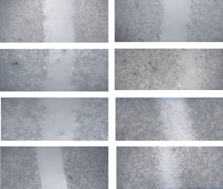
Journal of Immunology Research 9

 Time 0 24 hours

 Basal

 100
 1 g/mL
 S.d.

 80

 % cell migration
 ⁎⁎ ⁎⁎ ⁎⁎
 100 g/mL 10 g/mL

 60
 S.d.

 40

 20
 S.d.

 0
 Basal 1 10 100
 Basal
 + S. diploconos ( g/mL)
 (a) (b)
 80
 300

 60
 Area (mm2)

 200 ⁎⁎⁎⁎
 AUC

 40

 100
 20

 0 0
 1 2 3 4 5 6 7 Base cream +S. diploconos 1%
 Days
 Base cream
 Base cream +S. diploconos 1%
 +S. diploconos 1%
 (c) (d)

Figure 5: Analysis of the effects of the S. diploconos fruit extract in fibroblast migration and wound healing. (a) Representative image of the
in vitro scratch migration assay in fibroblasts (L929) and (b) percentage of fibroblast migration. Skin ulcers were treated individually with S.
diploconos 1% cream or vehicle (base cream). Progression of wound healing area over time after extract treatment (c) and area under the curve
(AUC) (d). Values express the mean ± SEM from 6 animals per group. Statistical analysis was performed using one-way ANOVA followed by
Tukey’s test (cell migration assay) with ∗∗ p < 0:01 vs. basal and t-test (AUC assessment) ∗∗∗∗ p < 0:0001 vs. base cream.

 period (12 h). Accordingly, no alterations were seen in the
Table 1: Percentage of polychromatic erythrocyte (PCE)
 food/water intake or body weight of S. diploconos-treated
micronuclei (PCEMN) in animals treated with S. diploconos fruit
extract. Percentage of polychromatic erythrocyte (PCE)
 animals in comparison with vehicle-treated rats.
micronuclei (PCEMN) in the micronucleus assay performed in Similarly, S. diploconos administration did not cause
erythrocytes obtained from the bone marrow (the values are any alterations to internal organs (Table 2). Additionally,
expressed as mean ± SEM (n = 5). ∗∗∗ p < 0:001 vs. the vehicle the histological analysis of the liver and kidney demon-
group). strated normal and conserved architecture, without degener-
 ative or inflammatory lesions in S. diploconos and vehicle
Group Dose (mg/kg) PCEMN/2000 PCE (%) groups.
Vehicle — 0:33 ± 0:33 No significant hematological parameter changes were
MMS 50 11:90 ± 1:09∗∗∗ found. In the platelet count, the high SEM presented for the
 extract-treated group refers to just one animal. Biochemical
S. diploconos 2000 1:75 ± 0:52
 analysis indicated that the extract promotes a decrease of
 39.8% in glucose and an increase of 1.3-fold in urea levels
 when compared with the vehicle group (Table 3).
10 Journal of Immunology Research

Table 2: Effects of Solanum diploconos extract treatment on the organ weights of mice following the acute oral toxicity experiments (values
expressed as mean ± SEM (n = 5)).

 Acute toxicity Subacute toxicity
Organ
 Vehicle 2000 (mg/kg) Vehicle 30 (mg/kg) 100 (mg/kg) 300 (mg/kg)
 Absolute (g) 0:853 ± 0:071 0:763 ± 0:053 0:676 ± 0:027 0:625 ± 0:012 0:623 ± 0:031 0:679 ± 0:039
Heart
 Relative (%) 0:326 ± 0:013 0:307 ± 0:017 0:351 ± 0:008 0:319 ± 0:008 0:335 ± 0:015 0:352 ± 0:020
 Absolute (g) 1:191 ± 0:097 1:098 ± 0:124 0:975 ± 0:023 0:936 ± 0:032 0:941 ± 0:020 0:962 ± 0:024
Lung
 Relative (%) 0:455 ± 0:029 0:442 ± 0:041 0:506 ± 0:007 0:477 ± 0:016 0:506 ± 0:012 0:499 ± 0:017
 Absolute (g) 9:182 ± 0:522 8:123 ± 0:840 5:300 ± 0:248 5:364 ± 0:141 5:071 ± 0:155 5:202 ± 0:122
Liver
 Relative (%) 3:515 ± 0:256 3:273 ± 0:316 2:751 ± 0:098 2:734 ± 0:058 2:725 ± 0:741 2,699 ± 0:070
 Absolute (g) 0:635 ± 0:047 0:593 ± 0:056 0:542 ± 0:038 0:554 ± 0:033 0:480 ± 0:008 0:496 ± 0:022
Spleen
 Relative (%) 0:242 ± 0:012 0:238 ± 0:014 0:280 ± 0:015 0:282 ± 0:017 0:258 ± 0:003 0:257 ± 0:012
 Absolute (g) 0:998 ± 0:076 0:914 ± 0:057 0:765 ± 0:034 0:862 ± 0:038 0:737 ± 0:017 0:779 ± 0:024
Left kidney
 Relative (%) 0:381 ± 0:024 0:368 ± 0:013 0:397 ± 0:012 0:439 ± 0:017 0:396 ± 0:008 0:404 ± 0:014
 Absolute (g) 1:021 ± 0:071 0:910 ± 0:100 0:802 ± 0:034 0:836 ± 0:043 0:753 ± 0:012 0:756 ± 0:029
Right kidney
 Relative (%) 0:391 ± 0:038 0:366 ± 0:030 0:416 ± 0:013 0:426 ± 0:019 0:405 ± 0:007 0:392 ± 0:015

Table 3: Biochemical markers and hematological parameters following the acute administration of the S. diploconos fruit extract (values
expressed as mean ± SEM (n = 5)). ∗ p < 0:05 vs. the vehicle group.

 Acute toxicity Subacute toxicity
Parameters
 Vehicle 2000 (mg/kg) Vehicle 30 (mg/kg) 100 (mg/kg) 300 (mg/kg)
Biochemical parameters
Glucose (mg/dL) 261:60 ± 16:93∗ 157:30 ± 16:23 131:6 ± 6:30 148:20 ± 11:10 125:20 ± 10:54 125:80 ± 3:48
 AST (U/L) 91:60 ± 5:97 128:81 ± 14:45 108:21 ± 10:79 100:69 ± 7:02 104:23 ± 6:16 89:22 ± 7:97
 ALT (U/L) 56:40 ± 5:14 38:61 ± 9:69 39:89 ± 4:39 37:86 ± 2:11 37:36 ± 1:89 33:07 ± 2:09
 ALP (U/L) 61:20 ± 7:94 73:80 ± 15:65 53:12 ± 4:30 60:59 ± 6:01 56:68 ± 2:06 56:30 ± 6:25
 Urea (mg/dL) 41:60 ± 1:21 53:50 ± 3:03∗ 35:00 ± 1:70 32:60 ± 1:50 36:80 ± 2:78 36:80 ± 1:39
 Creatinine (mg/dL) 0:53 ± 0:02 0:39 ± 0:053 0:30 ± 0:01 0:28 ± 0:01 0:25 ± 0:01 0:29 ± 0:02
Hematological parameters
 Leukocyte (mil/mm3) 1:120 ± 0:149 0:800 ± 0:152 3:425 ± 0:118 2:900 ± 0:207 4:020 ± 0:497 2:520 ± 0:323
 Neutrophil (%) 10:600 ± 1:805 13:333 ± 2:333 2:750 ± 0:250 2:400 ± 0:245 2:400 ± 0:245 3:600 ± 0:678
 Lymphocyte (%) 87:400 ± 2:014 84:667 ± 2:403 95:500 ± 0:288 95:800 ± 0:374 96:200 ± 0:374 94:400 ± 1:886
 Monocytes (%) 2:000 ± 0:707 1:333 ± 0:333 1:750 ± 0:250 1:800 ± 0:374 1:400 ± 0:245 2:000 ± 1:265
 Eosinophil (%) 0:000 ± 0:000 0:666 ± 0:333 0:000 ± 0:000 0:000 ± 0:000 0:000 ± 0:000 0:000 ± 0:000
 Basophil (%) 0:000 ± 0:000 0:000 ± 0:000 0:000 ± 0:000 0:000 ± 0:000 0:000 ± 0:000 0:000 ± 0:000
 Erythrocyte
 5:562 ± 0:067 4:960 ± 0:496 7:050 ± 0:131 7:000 ± 0:092 7:146 ± 0:129 6:698 ± 0:113
(millions/mm3)
 Hemoglobin (g/dL) 11:540 ± 0:153 10:266 ± 1:185 14:050 ± 0:342 13:940 ± 0:150 13:960 ± 0:265 13:220 ± 0:587
 Hematocrit (%) 31:100 ± 0:389 27:533 ± 2:936 40:600 ± 0:849 40:220 ± 0:414 40:560 ± 0:867 38:120 ± 0:587
 3 3
 Platelets (10 /mm ) 408:400 ± 34:993 293:000 ± 142:815 702:250 ± 46:424 657:400 ± 44:800 651:800 ± 39:328 715:600 ± 69:974

 The data obtained in the subacute toxicity test demon- 4. Discussion
strate that S. diploconos extract does not induce any alteration
in the biochemical and hematological parameters at the high- Neutrophils are cells with multiple functions which modulate
est evaluated dose (300 mg/kg). In this case, a no observed inflammation from its initial phase to tissue repair. Neutro-
adverse effect level (NOAEL) was established at 300 mg/kg. phils interact with other immune cells, directly or via
Journal of Immunology Research 11

inflammatory mediator production, affecting the course of may explain the reduced leukocyte recruitment and protein
both innate and adaptive immune responses [29]. We levels in the air pouch exudates obtained from mice chal-
showed for the first time that S. diploconos fruit extract is able lenged with carrageenan. A similar effect on neutrophils
to modulate neutrophil functions by reducing chemotaxis, was also observed in the skin tissue.
cytokine, and NO release and also promotes resolution of Neutrophils can indirectly contribute to tissue restora-
inflammation and wound healing. Additionally, the assays tion by modulating the functions of other cells. In this
performed indicate that the extract may directly affect intra- context, macrophages are moved to a state of tissue remodel-
cellular neutrophil pathways. ling by performing efferocytosis (phagocytosis of apoptotic
 Several inflammatory mediators are synthesized and neutrophils). Indeed, when neutrophils become apoptotic,
secreted during an inflammatory response with neutrophils they trigger a phagocytic activity in macrophages and stimu-
responding directly to these signals and producing different late their differentiation into M2 macrophages which are
inflammatory mediators such as cytokines that, in turn, mod- involved in tissue repair [38, 39]. The increased production
ulate inflammation and drive the immune response [29, 30]. of TGF-β and IL-10 is suggested to underlie these responses
In this context, it is well known that cytokines such as TNFα [7, 38, 40]. Herein, macrophages treated with S. diploconos
and IL-6 are increased in inflammation and may contribute presented increased efferocytosis, paralleled to decreased
to excessive neutrophil accumulation and subsequent tissue TNFα and enhanced IL-10 production.
damage [31]. Previous studies demonstrated that some Sola- These data indicate that S. diploconos may favour the res-
num species are able to reduce inflammatory mediator secre- olution of inflammation. In order to confirm this evidence,
tion by immune cells; however, their effects on neutrophils we assessed the effects of the extract in a scratch assay with
were unknown [18, 32]. In this context, our data clearly show L929 cells and in vivo, in animals with skin wounds. S. diplo-
the direct inhibitory actions of S. diploconos fruit extract on conos induced in vitro fibroblast proliferation and accelerated
LPS-induced neutrophil-mediated secretion of cytokines the healing of skin wounds in mice. Similar proliferative and
and NO. healing actions were previously observed for other Solanum
 Herein, the obtained phytochemical profile was com- species, such as S. incanum and S. xanthocarpum, which pre-
pared to that presented by Ribeiro et al. [13]. The authors sented healing activities in burn models and in the treatment
identified derivatives of caffeic, ferulic and coumaric acids of diabetic skin ulcers [32, 41].
in the freeze-dried pulp and peel of S. diploconos fruits, as Both the anti-inflammatory and healing activities of S.
well as in an extract obtained from its whole fruit using diploconos may be correlated to the presence of caffeic, feru-
HPLC-DAD-ESI-MS/MS [13]. Some phenolic compounds lic, and coumaric acids in its extract. It is known that caffeic
detected by the authors presented a similar UV absorption acid presents anti-inflammatory and wound healing effects in
profile also observed in this study for the peaks 5, 11-13, different experimental models [42, 43], and the same was
and 18-24. observed for ferulic acid [44, 45].
 The anti-inflammatory effect observed for S. diploconos Finally, an analysis of the possible toxic effects of the
may be due to the content of caffeic acid, lutein, and β-caro- extract was performed. No important toxic effects were
tene present in the extract [13], which were previously shown observed in vitro (in L929 cells) or in vivo, apart from a
to reduce the translocation of the transcription factor NF-ĸB reduction of glucose and an increase in urea levels in the
[16, 33, 34] necessary for the transcription of inflammatory acute but not in the subacute toxicity assay. Normal urea
mediators such as cytokines [35]. levels indicate that the metabolism of ammoniac to urea by
 We also found that S. diploconos fruit extract markedly the liver is not altered and that the detoxification and excre-
reduces fMLP-induced neutrophil chemotaxis in vitro. Inter- tion liver functions are preserved [45]. Despite observing
estingly, S. diploconos extract increased the expression of increased urea levels, the histopathological analysis showed
CD62L on LPS-stimulated neutrophils without altering no alterations in the renal tissue. These data indicate that
CD18 and CD49d profiles. It is important to highlight that the acute and subacute oral treatments with S. diploconos
CD62L mediates neutrophil rolling [36], whilst CD49d and fruit extract may be safe. Nonetheless, further studies are nec-
CD18 mediate adhesion to the endothelium [6, 37]. CD62L essary to address possible toxic effects of this extract when
becomes highly expressed on the cell membrane of leuko- administered repeatedly.
cytes when haemodynamic changes or production of inflam-
matory mediators occurs [3]. After the activation process,
however, CD62L is cleaved by the action of membrane metal- 5. Conclusion
loproteinases, allowing the activation of CD18 which is
responsible for the strong adhesion of inflammatory cells to Taken together, the data demonstrate the anti-inflammatory
endothelial cells [6, 36]. It is possible, thus, that the extract and prohealing activities of the S. diploconos fruit extract.
prevents L-selectin from being cleaved, allowing leukocytes These effects were linked to its ability to modulate neutrophil
to remain in rolling behaviour instead of adhering to the functions including the production of inflammatory media-
endothelium and migrating to the inflammatory focus. tors and chemotaxis, in addition to promoting efferocytosis
 S. diploconos inhibitory effects on neutrophil migration and the resolution of acute inflammation. The absence of
were confirmed in vivo. Our data demonstrate that the oral important acute and subacute toxic effects suggests the
treatment with S. diploconos decreases the production of promising application of the extract as a therapeutic aid for
inflammatory mediators (TNFα, IL-6, and IL-1β) which acute inflammatory reactions and wound healing.
12 Journal of Immunology Research

Data Availability [10] R. M. Hernández-Herrera, F. Santacruz-Ruvalcaba, M. A.
 Ruiz-López, J. Norrie, and G. Hernández-Carmona, “Effect
The datasets used to support this study will be made available of liquid seaweed extracts on growth of tomato seedlings (Sola-
upon request. Requests should be sent to JRS. num lycopersicum L.),” Journal of Applied Phycology, vol. 26,
 no. 1, pp. 619–628, 2014.
Conflicts of Interest [11] C. C. J. Almança, S. V. Saldanha, D. R. Sousa et al., “Toxicolog-
 ical evaluation of acute and sub-chronic ingestion of hydroal-
The authors declare no conflicts of interest. coholic extract of Solanum cernuum Vell. in mice,” Journal of
 Ethnopharmacology, vol. 138, no. 2, pp. 508–512, 2011.
Authors’ Contributions [12] S. R. I. JR, L. A. Mentz, M. F. Agra, M. Vignoli-Silva, and
 L. Giacomin, Solanaceae, Lista de Espécies da Flora do Brasil,
JRS and NLMQ conceived and designed research. LB, RN, 2015.
IV, SAP, MFB, FCG, and SEP conducted experiments. AM [13] A. B. Ribeiro, R. C. Chisté, J. L. F. C. Lima, and E. Fernandes,
contributed with analytical tools. JRS, MVDP, and NLMQ “Solanum diploconos fruits: profile of bioactive compounds
analysed the data. ESF, JRS, LB, and NLMQ drafted, critically and in vitro antioxidant capacity of different parts of the fruit,”
revised, and wrote the final manuscript. All authors read and Food & Function, vol. 7, no. 5, pp. 2249–2257, 2016.
approved the manuscript. [14] L. C. Lopes, J. E. de Carvalho, M. Kakimore et al., “Pharma-
 cological characterization of Solanum cernuum Vell.: 31-
Acknowledgments norcycloartanones with analgesic and anti-inflammatory
 properties,” Inflammopharmacology, vol. 22, pp. 179–185,
This study was supported by grants from the Conselho 2013.
Nacional de Desenvolvimento Científico e Tecnológico [15] L. Zhao, L. Wang, S. Di et al., “Steroidal alkaloid solanine A
(CNPq, grant numbers 429505/2018-3 to JRS, 305676/2019- from Solanum nigrum Linn. exhibits anti- inflammatory activ-
9 to ESF, and 305550/2018-7 to NLMQ) and the Coordenação ity in lipopolysaccharide/interferon γ-activated murine mac-
 rophages and animal models of inflammation,” Biomedicine
de Aperfeiçoamento de Pessoal de Nível Superior (CAPES)
 & Pharmacotherapy, vol. 105, pp. 606–615, 2018.
(Finance Code 001).
 [16] L. C. Wang, K. H. Chu, Y. C. Liang, Y. L. Lin, and B. L. Chiang,
 “Caffeic acid phenethyl ester inhibits nuclear factor-κB and
References protein kinase B signalling pathways and induces caspase-3
 expression in primary human CD4+ T cells,” Clinical and
 [1] M. Phillipson and P. Kubes, “The healing power of neutro-
 Experimental Immunology, vol. 160, no. 2, pp. 223–232, 2010.
 phils,” Trends in Immunology, vol. 40, no. 7, pp. 635–647,
 2019. [17] H. O. Kazancioglu, M. C. Bereket, S. Ezirganli, M. S. Aydin,
 and S. Aksakalli, “Effects of caffeic acid phenethyl ester on
 [2] S. Thome, D. Begandt, R. Pick, M. Salvermoser, and B. Walzog,
 wound healing in calvarial defects,” Acta Odontologica Scandi-
 “Intracellular β2 integrin (CD11/CD18) interacting partners in
 navica, vol. 73, no. 1, pp. 21–27, 2015.
 neutrophil trafficking,” European Journal of Clinical Investiga-
 tion, vol. 48, article e12966, 2018. [18] R. Nunes, M. F. Broering, R. De Faveri et al., “Effect of the
 [3] A. Dabrowski, J. Osada, M. I. Dabrowska, U. Wereszczynska- metanolic extract from the leaves of Garcinia humilis Vahl
 Siemiatkowska, and A. Siemiatkowski, “Increased expression (Clusiaceae) on acute inflammation,” Inflammopharmacology,
 of the intercellular adhesion molecule-1 (ICAM-1) on periph- vol. 29, pp. 423–438, 2021.
 eral blood neutrophils in acute pancreatitis,” Advances in Med- [19] R. D. Nelson, P. G. Quie, and R. L. Simmons, “Chemotaxis
 ical Sciences, vol. 59, no. 1, pp. 102–107, 2014. under agarose: a new and simple method for measuring che-
 [4] S. Schmidt, M. Moser, and M. Sperandio, “The molecular basis motaxis and spontaneous migration of human polymorpho-
 of leukocyte recruitment and its deficiencies,” Molecular nuclear leukocytes and monocytes,” Journal of Immunology,
 Immunology, vol. 55, no. 1, pp. 49–58, 2013. vol. 115, pp. 1650–1656, 1975.
 [5] D. N. Granger and E. Senchenkova, “Inflammation and the [20] M. F. Broering, R. Nunes, R. De Faveri et al., “Effects of Titho-
 microcirculation,” Colloquium Series on Integrated Systems nia diversifolia (Asteraceae) extract on innate inflammatory
 Physiology: From Molecule to Function, vol. 2, no. 1, pp. 1– responses,” Journal of Ethnopharmacology, vol. 242, article
 87, 2010. 112041, 2019.
 [6] K. Mastej and R. Adamiec, “Neutrophil surface expression of [21] L. C. Green, D. A. Wagner, J. Glogowski, P. L. Skipper, J. S.
 CD11b and CD62L in diabetic microangiopathy,” Acta Diabe- Wishnok, and S. R. Tannenbaum, “Analysis of nitrate, nitrite,
 tologica, vol. 45, no. 3, pp. 183–190, 2008. and [15N] nitrate in biological fluids,” Analytical Biochemistry,
 [7] S. E. Headland and L. V. Norling, “The resolution of inflam- vol. 126, pp. 131–138, 1982.
 mation: principles and challenges,” Seminars in Immunology, [22] A. D. Sedgwick and P. Lees, “Studies of eicosanoid production
 vol. 27, no. 3, pp. 149–160, 2015. in the air pouch model of synovial inflammation,” Agents and
 [8] M. A. Sugimoto, L. P. Sousa, V. Pinho, M. Perretti, and M. M. Actions, vol. 18, no. 3-4, pp. 429–438, 1986.
 Teixeira, “Resolution of inflammation: what controls its [23] M. Jain and H. S. Parmar, “Evaluation of antioxidative and
 onset?,” Frontiers in Immunology, vol. 7, p. 160, 2016. anti-inflammatory potential of hesperidin and naringin on
 [9] A. G. Atanasov, B. Waltenberger, E. M. Pferschy-Wenzig et al., the rat air pouch model of inflammation,” Inflammation
 “Discovery and resupply of pharmacologically active plant- Research, vol. 60, no. 5, pp. 483–491, 2011.
 derived natural products: a review,” Biotechnology Advances, [24] P. Y. K. Yue, E. P. Y. Leung, N. K. Mak, and R. N. S. Wong, “A
 vol. 33, no. 8, pp. 1582–1614, 2015. simplified method for quantifying cell migration/wound
Journal of Immunology Research 13

 healing in 96-well plates,” Journal of Biomolecular Screening, by metabolic reprogramming of macrophages,” Science,
 vol. 15, no. 4, pp. 427–433, 2010. vol. 356, no. 6337, pp. 513–519, 2017.
[25] T. D. Tonin, L. C. Thiesen, M. L. de Oliveira Nunes et al., [40] J. A. Marwick, R. Mills, O. Kay et al., “Neutrophils induce mac-
 “Rubus imperialis (Rosaceae) extract and pure compound rophage anti-inflammatory reprogramming by suppressing
 niga-ichigoside F1: wound healing and anti-inflammatory NF-κB activation,” Cell Death & Disease, vol. 9, no. 6, p. 665,
 effects,” Naunyn-Schmiedeberg's Archives of Pharmacology, 2018.
 vol. 389, no. 11, pp. 1235–1244, 2016. [41] K. M. Parmar, P. R. Shende, N. Katare, M. Dhobi, and S. K.
[26] F. Denizot and R. Lang, “Rapid colorimetric assay for cell Prasad, “Wound healing potential of Solanum xanthocarpum
 growth and survival: modifications to the tetrazolium dye pro- in streptozotocin-induced diabetic rats,” The Journal of Phar-
 cedure giving improved sensitivity and reliability,” Journal of macy and Pharmacology, vol. 70, no. 10, pp. 1389–1400, 2018.
 Immunological Methods, vol. 89, pp. 271–277, 1986. [42] F. M. Da Cunha, D. Duma, J. Assreuy et al., “Caffeic acid deriv-
[27] Organization For Economic Cooperation And Development, atives: in vitro and in vivo anti-inflammatory properties,” Free
 “Test No. 420: acute oral toxicity–fixed dose procedure,” in Radical Research, vol. 38, no. 11, pp. 1241–1253, 2004.
 OECD Guidel. Test, p. 14, OECD, Chem, 2002. [43] B. Romana-Souza, J. S. dos Santos, and A. Monte-Alto-Costa,
[28] Organization For Economic Cooperation And Development, “Caffeic acid phenethyl ester promotes wound healing of mice
 “Test No. 487: in vitro mammalian cell micronucleus test,” in pressure ulcers affecting NF-κB, NOS2 and NRF2 expression,”
 Guidel. Test, OECD, Ed., p. 23, Chem, 2010. Life Sciences, vol. 207, pp. 158–165, 2018.
[29] C. Rosales, “Neutrophil: a cell with many roles in inflamma- [44] K. Zduńska, A. Dana, A. Kolodziejczak, and H. Rotsztejn,
 tion or several cell types?,” Frontiers in Physiology, vol. 9, “Antioxidant properties of ferulic acid and its possible applica-
 p. 113, 2018. tion,” Skin Pharmacology and Physiology, vol. 31, no. 6,
[30] A. Azab, A. Nassar, and A. N. Azab, “Anti-inflammatory activ- pp. 332–336, 2018.
 ity of natural products,” Molecules, vol. 21, no. 10, p. 1321, [45] Z. N. Yin, W. J. Wu, C. Z. Sun et al., “Antioxidant and anti-
 2016. inflammatory capacity of ferulic acid released from wheat bran
[31] M. Fronza, C. Muhr, D. S. C. da Silveira et al., “Hyaluronidase by solid-state fermentation of Aspergillus niger,” Biomedical
 decreases neutrophils infiltration to the inflammatory site,” and Environmental Sciences, vol. 32, no. 1, pp. 11–21, 2019.
 Inflammation Research, vol. 65, no. 7, pp. 533–542, 2016.
[32] Z. Qureshi, T. Khan, A. J. Shah, and F. Wahid, “Solanum inca-
 num extract enhances wound healing and tissue regeneration
 in burn mice model,” Bangladesh Journal of Pharmacology,
 vol. 14, no. 2, pp. 101–106, 2019.
[33] J. H. Kim, H. J. Na, C. K. Kim et al., “The non-provitamin A
 carotenoid, lutein, inhibits NF-κB-dependent gene expression
 through redox-based regulation of the phosphatidylinositol
 3-kinase/PTEN/Akt and NF-κB-inducing kinase pathways:
 role of H2O2 in NF-κB activation,” Free Radical Biology &
 Medicine, vol. 45, no. 6, pp. 885–896, 2008.
[34] S. O. Cho, M.-H. Kim, and H. Kim, “β-Carotene inhibits acti-
 vation of NF-κB, activator protein-1, and STAT3 and regulates
 abnormal expression of some adipokines in 3T3-L1 adipo-
 cytes,” Journal of Cancer Prevention, vol. 23, no. 1, pp. 37–
 43, 2018.
[35] R. P. Kumar and A. Abraham, “Inhibition of LPS induced pro-
 inflammatory responses in RAW 264.7 macrophage cells by
 PVP-coated naringenin nanoparticle via down regulation of
 NF-κB/P38MAPK mediated stress signaling,” Pharmacologi-
 cal Reports, vol. 69, no. 5, pp. 908–915, 2017.
[36] K. Uchimura and S. D. Rosen, “Sulfated L-selectin ligands as a
 therapeutic target in chronic inflammation,” Trends in Immu-
 nology, vol. 27, no. 12, pp. 559–565, 2006.
[37] V. C. Ridger, B. E. Wagner, W. A. Wallace, and P. G. Hellewell,
 “Differential effects of CD18, CD29, and CD49 integrin sub-
 unit inhibition on neutrophil migration in pulmonary inflam-
 mation,” Journal of Immunology, vol. 166, no. 5, pp. 3484–
 3490, 2001.
[38] J. Bystrom, I. Evans, J. Newson et al., “Resolution-phase mac-
 rophages possess a unique inflammatory phenotype that is
 controlled by cAMP,” Blood, vol. 112, no. 10, pp. 4117–4127,
 2008.
[39] W. K. E. Ip, N. Hoshi, D. S. Shouval, S. Snapper, and
 R. Medzhitov, “Anti-inflammatory effect of IL-10 mediated
You can also read