Angiocrine Regulation of Epithelial Barrier Integrity in Inflammatory Bowel Disease - Frontiers
←
→
Page content transcription
If your browser does not render page correctly, please read the page content below
REVIEW
                                                                                                                                               published: 02 August 2021
                                                                                                                                         doi: 10.3389/fmed.2021.643607

                                              Angiocrine Regulation of Epithelial
                                              Barrier Integrity in Inflammatory
                                              Bowel Disease
                                              Michael Stürzl 1*, Meik Kunz 2 , Susanne M. Krug 3 and Elisabeth Naschberger 1
                                              1
                                                Division of Molecular and Experimental Surgery, Department of Surgery, Universitätsklinikum Erlangen, Friedrich-Alexander
                                              University (FAU) of Erlangen-Nürnberg, Erlangen, Germany, 2 Chair of Medical Informatics, Friedrich-Alexander-University
                                              (FAU) of Erlangen-Nürnberg, Erlangen, and Fraunhofer Institute of Toxicology and Experimental Medicine, Hannover,
                                              Germany, 3 Clinical Physiology/Nutritional Medicine, Charité-Universitätsmedizin Berlin, Berlin, Germany

                                              Inflammatory bowel disease describes chronic inflammatory disorders. The incidence
                                              of the disease is rising. A major step in disease development is the breakdown of the
                                              epithelial cell barrier. Numerous blood vessels are directly located underneath this barrier.
                                              Diseased tissues are heavily vascularized and blood vessels significantly contribute to
                                              disease progression. The gut-vascular barrier (GVB) is an additional barrier controlling
                                              the entry of substances into the portal circulation and to the liver after passing the
                                              first epithelial barrier. The presence of the GVB rises the question, whether the vascular
                                              and endothelial barriers may communicate bi-directionally in the regulation of selective
                                              barrier permeability. Communication from epithelial to endothelial cells is well-accepted.
                              Edited by:      In contrast, little is known on the respective backwards communication. Only recently,
                   Roberto Gramignoli,        perfusion-independent angiocrine functions of endothelial cells were recognized in a
      Karolinska Institutet (KI), Sweden
                                              way that endothelial cells release specific soluble factors that may directly act on the
                       Reviewed by:
                     Prashant Nighot,
                                              epithelial barrier. This review discusses the putative involvement of angiocrine inter-barrier
    The Pennsylvania State University,        communication in the pathogenesis of IBD.
                        United States
                   Linda Chia-Hui Yu,         Keywords: endothelial, angiocrine, barrier, inflammatory bowel disease, inflammation, angiogenesis, epithelial
    National Taiwan University, Taiwan

                  *Correspondence:
                        Michael Stürzl        CLINICAL PRESENTATION AND EPIDEMIOLOGY OF
       michael.stuerzl@uk-erlangen.de         INFLAMMATORY BOWEL DISEASE
                    Specialty section:        Inflammatory bowel disease (IBD) includes inflammatory diseases of the colon and small intestine
          This article was submitted to       with Crohn’s disease and ulcerative colitis being the major clinical presentations (1). Crohn’s disease
                      Gastroenterology,       affects the small intestine and large intestine, as well as the mouth, esophagus, stomach and the
                a section of the journal
                                              anus, whereas ulcerative colitis primarily affects the colon and the rectum (2). Crohn’s disease and
                   Frontiers in Medicine
                                              ulcerative colitis are different diseases, but commonly present with any of the following symptoms:
       Received: 18 December 2020             abdominal pain, diarrhea, rectal bleeding, severe internal cramps/muscle spasms in the region of
            Accepted: 07 July 2021
                                              the pelvis and weight loss. In addition, anemia is a common extra-intestinal complication of IBD.
         Published: 02 August 2021
                                                  IBD is classically considered as a disease of Westernized countries but has started to rise
                               Citation:      worldwide in the beginning of the twenty first century (3). The rise is population-dependent and
      Stürzl M, Kunz M, Krug SM and
                                              categorized into four different epidemiological stages: first, the Emergence Stage with sporadic cases
    Naschberger E (2021) Angiocrine
Regulation of Epithelial Barrier Integrity
                                              of IBD observed in developing countries, second, the Acceleration in Incidence Stage with rising
      in Inflammatory Bowel Disease.          incidence and relatively low prevalence in newly industrialized countries, third, the Compounding
               Front. Med. 8:643607.          Prevalence Stage with stable incidence and steeply rising prevalence in countries of the Western
    doi: 10.3389/fmed.2021.643607             world, and forth, the Prevalence Equilibrium Stage, which represents the opposing forces between

Frontiers in Medicine | www.frontiersin.org                                          1                                            August 2021 | Volume 8 | Article 643607
Stürzl et al. Angiocrine Regulation of Epithelial Barrier
an aging IBD population and the incidence of IBD. In Germany at of the epithelial integrity is a major aspect in order to preserve
present 620,085 persons are suffering from IBD with a predicted homeostasis and to avoid the progress of inflammation in
rise up to 815,200 patients in 2030. In the U.S. presently 2,489,362 mucosal tissues (18) [for review see: Lopez-Posadas et al. (11)].
patients are registered and a rise up to 3,544,480 is expected At the molecular level the intercellular barrier of the intestinal
within the next 10 years (4). epithelium is established by apical junction complexes comprised
IBD is characterized by a chronically relapsing intestinal of tight and adherens junctions. Adherens junctions consist of
inflammation that is thought to result from an exaggerated cadherins and nectins and are mainly important for the cell-cell-
immune response to the commensal microbiota. However, the adhesion (19, 20). Tight junctions are multiprotein-complexes
specific molecular mechanisms driving IBD pathogenesis are still consisting of several transmembrane proteins: tight junction
unclear. Many different putative susceptibility genes for IBD associated MARVEL proteins (TAMP) like occludin, marvelD3
are reported but all of these are associated with only low risk and tricellulin, junctional adhesion molecules (JAM), angulins
and differ in different countries of the world. At present, it and the family of claudins, which has in mammalia 27 members
is commonly accepted, that cytokines, such as, tumor necrosis that either possess barrier- or channel-forming properties
factor (TNF), interleukin (IL)-10, transforming growth factor affecting the overall permeability characteristics of the epithelia
(TGF)-β, IL-6, IL-12, IL-13, IL-17, IL-21, IL-23, interferon [for review see Günzel and Fromm (21)]. Adherens junctions
(IFN)-γ and C-X-C motif chemokine ligand (CXCL)10, are as well as tight junctions establish zipper-like structures, sealing
drivers of the excessive immune response, leading to leukocyte the paracellular space within the epithelial cell layer (22). These
infiltration and mucosal damage. In addition, there is agreement intercellular junctions are connected to the actin cytoskeleton
that IBD pathogenesis is closely associated with a loss of via cytoplasmic adaptors, such as zonula occludens proteins, and
intestinal epithelial barrier functions associated with bacterial catenins supporting the mechanical strength of the junctions
translocation, likely representing an initiating or early event in (23–25). Cell activation with molecules that induce permeability
the disease (5–10). causes actin reorganization into stress fibers. This is associated
Recently, it became evident that the intestinal barrier involves with increasing traction forces, which lead to the detachment
two sequential physical barriers. The first being the epithelial of adherens junctions from the cytoskeleton followed by the
barrier consisting of a single cell layer of epithelial cells and formation of gaps between adjacent cells (26, 27). Further
a mucus layer which physically separates the microbiota in mechanisms such as the removal of cell-cell interaction molecules
the gut lumen and epithelial cells (11). Directly below the from the cell surface by internalization and/or by proteolytic
epithelial barrier an additional barrier was identified, the gut- cleavage can regulate the intestinal barrier permeability (11, 28).
vascular barrier (GVB) controlling the entry of substances The epithelium is constantly renewed without an effect
into the portal circulation and their access to the liver after on its tightness. Within this process stem cells at the crypt
passage of the first epithelial barrier (12, 13). The discriminative bottom proliferate and differentiate into the different intestinal
control of nutrient uptake and tight sealing towards potentially epithelial cell subtypes with specialized biological functions (29).
pathological microorganisms requires a profound regulation of Subsequently, most of the differentiated epithelial cells migrate
the barrier permeability. upwards to the villus tip, where aged cells die and are shed
into the lumen (30, 31). The tightness of the epithelial layer
is maintained by the intercellular junctions during this process
STRUCTURE AND FUNCTION OF THE (23). During cell shedding, epithelial integrity is maintained
EPITHELIAL BARRIER IN IBD in cytoskeleton and membrane trafficking-dependent processes
regulating the redistribution of junctional proteins along lateral
The epithelial barrier allows the co-existence of commensal membranes (32, 33).
microbiota and mucosal immune cells in the gut. It consists Increased epithelial tight junction permeability is a hallmark
of a physical barrier established by the epithelial cells situated in the gut of IBD patients (34–38). It is believed that
on a basement membrane. Collagen type IV and laminins are the disruption of intercellular junctions and cytoskeleton
the predominant components of the basement membrane (14). rearrangements in the context of infection or inflammation
The basement membrane is subject of continuous remodeling. lead to a breakdown of epithelial integrity (39–41). Although a
Increased remodeling was observed under inflammatory correlation between epithelial barrier permeability and disease
conditions in association with decreased barrier functions activity has been observed in patients with Crohn’s disease, the
(14). At the cellular level barrier functions are established by (i) cause of this barrier collapse is still a matter of controversy
densely packed microvilli on the apical side of intestinal epithelial (42, 43). Experimental animal studies demonstrated that a
cells termed the brush border (15), (ii) tight cell-cell interactions deficiency of single tight junction proteins is not sufficient to
between the epithelial cells, (iii) the cellular resistance to cause pathology due to compensatory mechanisms (44, 45) with
bacterial transcytosis (16), and (iv) specialized epithelial cells, the exception of claudin-15 (46). However, agreement exists that
such as mucus-producing goblet cells and anti-microbial inflammation-derived soluble mediators such as IL-6 (47), IL-
peptide secreting Paneth cells (12). Altogether, the epithelium 13 (48, 49), TNF (50), and IFN-γ (51, 52) affect tight junctions
exerts manifold functions, establishing a physical barrier and may increase intestinal permeability in experimental colitis
against pathogen invasion and also performing innate immune models and IBD (53–55). These observations suggested that
functions and nutrient uptake (17). Thereby, the preservation the epithelial barrier breakdown occurs as a consequence of
Frontiers in Medicine | www.frontiersin.org 2 August 2021 | Volume 8 | Article 643607Stürzl et al. Angiocrine Regulation of Epithelial Barrier
proinflammatory cytokine stimulation. In contrast, recent studies results on the contribution of angiogenesis to disease activity.
in IBD patients demonstrated that an increase of epithelial Neutralization of VEGF-A resulted in a decreased vessel density
permeability precedes flares of inflammatory bowel pointing and improvement of the disease in dextran sulfate sodium (DSS)–
towards a causative role of epithelial barrier breakdown in the induced and 2,4,6-trinitrobenzenesulfonic acid (TNBS)–induced
development of intestinal inflammation (35, 56–58). The latter colitis (73, 76). In contrast, reduced angiogenic activity induced
is supported by reports that a decrease of epithelial permeability by deficiency of placental growth factor failed to ameliorate colitis
by application of vitamin D (59, 60), probiotics (61–63), IL-22- in the same experimental models (77). These results indicated
triggered mucus production (64), butyrate (65, 66), or an anti- that besides vessel density additional parameters such as vessel
TNF antibody caused clinical amelioration of chronic colitis (67, quality are of relevance in IBD pathogenesis. In fact, newly
68). Moreover, alternative portals for gut leakiness such as brush formed vessels in IBD tissues are strongly disorganized and leaky
border functions and intestinal bacterial endocytosis by epithelial as evident by associated edema (78).
cells have to be considered and may play important pathogenic The difficulties in determining the precise role of blood vessel
roles providing putative targets for therapy of inflammatory function in IBD may be due to the fact that the intestinal
bowel disease (15). Altogether, these results suggest that the endothelial cells are both, targets and regulators of inflammation
epithelial barrier function is important and its maintenance can (78). In this framework, IBD-associated inflammatory cytokines
counteract the development of inflammatory bowel disease. such as TNF-α, IL-1β and IFN-γ can activate endothelial cells
by inducing the expression of adhesion molecules for leukocytes
such as E-selectin, intercellular adhesion molecule (ICAM)-1 or
THE IMPACT OF BLOOD VESSELS ON IBD vascular cell adhesion molecule (VCAM)-1 (79). Macrophages
PATHOGENESIS are important drivers of IBD and are characteristically expressing
high amounts of TNF-α and IL-1β, which may amplify the
Capillaries are located in close proximity to the intestinal extravasation of these cells being responsible for the high
epithelial cell barrier (Figure 1A). Blood vessels in adult tissues numbers of macrophages present in IBD tissues (80). In
evolve through sprouting from preexisting vessels, a process addition, inflammation is associated with increased angiogenesis
termed angiogenesis (69). Angiogenic activity correlates with supporting immune cell recruitment by increase of blood flow
disease severity in IBD suggesting that blood vessels may and endothelial surface (81). As mentioned above the intestinal
contribute to pathogenesis (70–73). Moreover, elevated levels of endothelium also establishes an additional barrier in the gut,
angiogenic growth factors including vascular endothelial growth the GVB (12, 13). The GVB constitutes a semipermeable
factor (VEGF)-A and basic fibroblast growth factor (bFGF), barrier between the blood stream and the interstitium regulating
that synergize in angiogenesis activation, have been detected in the transport of nutrients, tissue fluid homeostasis and the
the inflamed mucosa and in the blood during active IBD (74, transmigration of immune cells but is non-permissive to bacterial
75). However, experimental colitis models provided conflicting penetration (13, 28, 78, 82). The latter is in agreement with
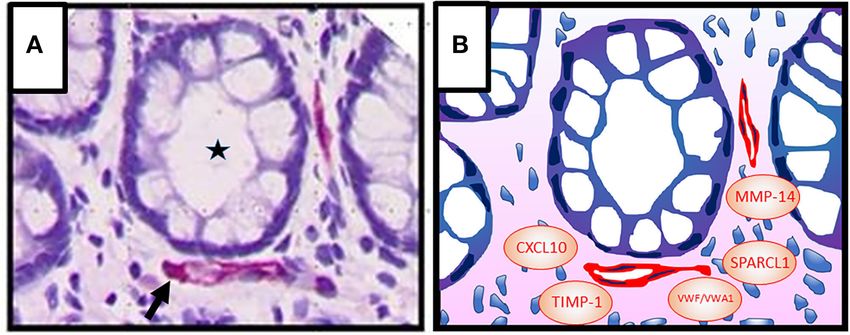
FIGURE 1 | (A) Colonic crypt (intestinal gland, asterisk) with vessels (red, arrow) in the lamina propria. Epithelial cells (1st barrier) and endothelial cells (2nd barrier) are
directly adjacent, indicating active inter-barrier communication. Vascular endothelial cells were stained immunohistochemically using an anti-CD31 antibody. Cell nuclei
(blue) were stained by haematoxylin. (B) Graphic presentation of (A) indicating possible factors that may be involved in angiocrine regulation of epithelial barrier
functions in IBD [von Willebrand factor A domain containing 1 (VWA1), von Willebrand factor (VWF), matrix metalloproteinase (MMP)-14, tissue inhibitor of
metalloproteinases (TIMP)-1, C-X-C motif chemokine ligand (CXCL) 10, secreted protein, acidic and rich in cysteine–like 1 (SPARCL1)].
Frontiers in Medicine | www.frontiersin.org 3 August 2021 | Volume 8 | Article 643607Stürzl et al. Angiocrine Regulation of Epithelial Barrier
the observation that bacterial lipopolysaccharides (LPS) in fenestrated blood endothelium to be distributed systemically
low concentrations are stabilizing the vascular barrier (83). (89, 90). Moreover, in response to pathogen invasion or
In contrast, high concentrations of LPS (>10 µg/ml) inhibit loss of barrier integrity, both intestinal epithelial cells and
endothelial cell migration, down-regulate intercellular junction tissue-resident leukocytes secrete cytokines, chemokines, reactive
molecules and increase the permeability of the vascular barrier oxygen species, and lipid mediators that activate endothelial cells
(83). Paracellular (i.e., in between the cells) or transcellular (i.e., to modulate the number and structure of vessels and to promote
across the cells) routes are available to cross the endothelial cell immune cell extravasation. For example, intestinal epithelial cells
monolayer. Transcellular exchange is accomplished via either in IBD were shown to secret the chemokines CXCL8/IL-8 and
solute transporters, or transcytosis via vesicular carriers (e.g., CCL20 (91, 92), both of which can activate angiogenesis (93, 94).
caveolae), or pore-like subcellular structures (i.e., fenestrae and In addition, these cells secrete the cytokine TNF-α (91), which
transendothelial channels) (84, 85). The paracellular route is regulates vessel remodeling and by directly acting on endothelial
controlled by adherens junctions and tight junction proteins cells may inhibit angiogenesis (95, 96). In addition, vascular
similar as in the epithelial barrier. In intestinal endothelial cells, permeability is increased by inflammatory mediators released
tight junctions are composed mainly of occludin, junctional from epithelial cells fostering both, inter- and trans-cellular
adhesion molecule (JAM)-A, zonula occludens (ZO)-1, and diapedesis (90, 97).
cingulin (13). Claudin-3, -5, and -12 from the claudin family are
known to be mainly expressed in endothelia (86, 87).
Adherens junctions are formed by vascular endothelial (VE)- ANGIOCRINE FUNCTIONS OF BLOOD
cadherin and β-catenin (13). Of note, the same cytokines VESSELS IN ORGAN DEVELOPMENT AND
regulating immune cell extravasation can also deregulate DISEASES
adherens and tight junction formation in endothelial cells
supporting translocation of bacteria thereby further amplifying The endothelium is not a passive response organ for nutrient
the inflammatory process [for review see: Lopez-Posadas supply, tissue entry of immune cells, and metabolite removal,
et al. (11)]. but actively regulates the tissue microenvironment in organ
The impact of the GVB in intestinal inflammation is development and diseases as indicated by novel results.
substantiated by mouse models of acute and chronic DSS- These perfusion-independent functions of endothelial cells were
colitis. In these models intestinal vessel perfusion remained recognized in experimental tumor models in mice for the first
constant during colitis whereas vessel permeability strongly time, where the inhibition of angiogenesis in certain instances
increased (5). Using experimental animal models with an did not abrogate tumor growth but instead enhanced tumor
endothelial cell specific knockout of the interferon-γ-receptor invasiveness (98). Based on this the hypothesis arose that
2 (IFNγR2) it was shown that the IBD-associated cytokine endothelial cells release specific soluble factors that may directly
IFN-γ induces a breakdown of the vascular barrier based regulate tumor growth in a perfusion-independent manner. This
on the disruption of the adherens junction protein VE- respective mechanism was termed as “angiocrine” regulation of
cadherin and this was significantly increasing DSS-induced tumorigenesis (98).
experimental colitis. Importantly, the disease-associated vascular Subsequent studies confirmed that endothelial cells may
barrier dysfunction could be confirmed in human IBD patients activate tumorigenesis by secreted factors (98, 99). For example,
indicating the clinical relevance of the findings. Imatinib angiocrine factors were reported to stimulate growth and
(brand name Gleevec) is a kinase inhibitor acting against migration of lymphoma tumor cells (100), to maintain stem cell
Abelson tyrosine kinase BCR–ABL, the KIT and PDGF like properties in colorectal carcinoma and glioblastoma cells
receptors and is used for therapy of chronic myeloid leukemia (101–103), to inhibit anoikis in head and neck cancer stem cells
(CML), gastrointestinal stromal tumors (GIST) and several (104) and, to activate proliferation, survival and epithelial to
other malignancies (88). Interestingly, treatment with imatinib mesenchymal transition of lung carcinoma cells (103) [for review
restored adherens junctions, inhibited vascular permeability, see: Lee et al. (105)].
and significantly reduced colonic inflammation in experimental Vice versa, it was noted that endothelial cells can also suppress
colitis. Altogether, these results highlighted the pathogenic cancer growth through angiocrine signaling. In this framework
impact of inflammation–associated vascular barrier defects in contact-dependent interactions between the endothelial cell
IBD and opens new avenues for vascular-directed treatment of surface receptor duffy antigen/receptor for chemokines and the
the disease (81). carcinoma cell surface receptor kang ai-1 were shown to suppress
The detection of an additional intestinal barrier rises the metastasis (106). In addition, in breast cancer endothelial cell-
question whether the epithelial and the vascular barriers may released slit homolog 2 protein (Slit 2), perlecan and additional
communicate in prevention or progression of the disease. as yet unknown factors were reported to inhibit proliferation,
Epithelial to endothelial cell communication is commonly invasion and pro-tumorigenic signaling of the cancer cells (107,
accepted. For example, the nutrient composition of the chyme 108). In addition, thrombospondin is regarded as a putative anti-
(partially digested food) and not simply gut distension modulates angiogenic factor secreted from endothelial cells (98). Angiocrine
blood flow. Specialized subsets of intestinal epithelial cells factors also exert key functions in physiologic condition
transport nutrients through the epithelial monolayer into the such as kidney development (109, 110), liver bud (111) and
lamina propria from where they are transported through the pancreatic bud formation (112), in neuronal development (113),
Frontiers in Medicine | www.frontiersin.org 4 August 2021 | Volume 8 | Article 643607Stürzl et al. Angiocrine Regulation of Epithelial Barrier
lung regeneration (114), osteogenesis (115) and hematopoiesis lymphoid structures similar to the human chronic disease.
(113). Altogether, these results indicated that intestinal angiocrine
Of note, a specific impact of angiocrine signaling on epithelial functions may establish a chemical barrier affecting both,
barrier functions was observed in retina development (116). epithelial and endothelial cell barrier functions in IBD (122).
Endothelial cells secrete factors that remodel the retinal pigment In a next step, we applied a meta-analysis to further
epithelium (RPE) basement membrane and integrin receptors investigate whether angiocrine signaling may impact barrier
sense these changes by triggering GTPase signals that modulate functions. To this goal, an in silico secretome screening
RPE tight junctions and enhance RPE barrier function (116). against the human proteome was performed using the VerSeDa
Similar parenchymal cell barrier regulatory mechanisms may be database [Vertebrate Secretome Database (125)]. Transcripts
active in other organs. with a prediction cut-off value > 0.8 (SignalP 4.1, TargetP
Altogether, angiocrine factors are involved in tumorigenic, 1.1, SecretomeP) were considered as secreted proteins. The
homeostatic, regenerative and morphogenetic processes in a resulting 1,050 genes (1,959 proteins; 1,959 gene transcripts)
paracrine or juxtacrine manner. The term “angiocrine” factors were used for a functional gene and phenotype annotation using
meanwhile includes secreted and membrane-bound inhibitory or the Ensembl BioMart database (http://www.ensembl.org/index.
stimulatory growth factors, trophogens, chemokines, cytokines, html). Next, candidates were selected based on data mining
extracellular matrix components, exosomes and other cellular (inflammatory, angiocrine, epithel, extracellular, endothelial,
products (117). The angiocrine profile of endothelial cells can barrier, cytokine, bowel, secreted). Subsequently, the resulting
differ between tissues, reflecting the diversity of cell types found 257 genes were mapped to profiles from human endothelial cells
adjacent to endothelial cells in organs (113, 117). of different origin, including human umbilical vein endothelial
cells (HUVEC) exposed to shear stress (126), under LPS-
stimulation (127), overexpressing γ-interferon-inducible protein
THE IMPACT OF ANGIOCRINE SIGNALING (IFI) 16 (128) and unstimulated (129), as well as endothelial
ON EPITHELIAL BARRIER FUNCTION IN cells from brain, lung, heart (130) and colorectal carcinoma
IBD (119, 131). This analysis identified in total 28 genes (Table 1). Six
of these may be of specific interest as candidates of angiocrine
Angiocrine functions in IBD have not been investigated barrier effects in IBD (Figure 1B). This includes components of
extensively as yet, despite the manifold effects of angiocrine the von Willebrand factor domain superfamily (VWA1, VWF)
signaling on epithelial cell functions in cancer, organ and tissue inhibitor of metalloproteinases (TIMP)-1, which were
development and tissue regeneration. However, first results retrieved from three different studies, respectively. vWF is a
indicating angiocrine activities in the colon have emerged. classical endothelial cell marker protein, that promotes adhesion
For example, endothelial cells release jagged 1, generated by of platelets to the sites of vascular injury by forming a molecular
proteolytic activity of ADAM metallopeptidase domain 17 bridge between sub-endothelial collagen matrix and the platelet-
(ADAM17) activating Notch in human colorectal cancer cells surface receptor complex (132). Its impact on the epithelial
and thereby promoting a cancer stem cell phenotype and barrier warrants further investigation. TIMP-1 is an inhibitor
chemo-resistance (103, 118). Moreover, it was shown that of the matrix metalloproteinases (MMPs). It is able to promote
selectively endothelial cells isolated from colorectal carcinomas cell proliferation in a wide range of cell types, has an anti-
with a prognostically favorable Th-1-like immune environment apoptotic function and can modulate the vascular barrier (133,
released the matricellular protein secreted protein, acidic 134). TIMP-1 may impact the epithelial cell barrier activity
and rich in cysteine–like 1 (SPARCL1), which autocrinely in the gut through these activities. In this framework, it is
and paracrinely inhibited angiogenesis and proliferation of interesting that MMP-14 was also identified by our meta-analyses
different cancer cell lines (119, 120). The latter indicated that as angiocrine mediator. MMP-14 was reported as an angiocrine
angiocrine activities in the colon may trigger the course of factor in lung regeneration and as a member of the membrane-
diseases in a microenvironment–dependent manner. A recent type matrix metalloproteinases that are not inhibited by TIMP-1
single cell RNAseq approach of intestinal cells and subsequent (114, 135). In addition, CXCL10, regarded as a major driver in
bioinformatics interaction analyses supported the molecular IBD pathogenesis (6), was also identified as angiocrine mediator
interaction between endothelial cells and epithelial cells in the in our meta analyses. In the DSS-model blockade of CXCL10
colon (121). enhanced crypt cell survival (136) and mice with a knock out
Specific support for angiocrine functions in IBD was obtained of the CXCL10 receptor CXCR3 showed considerably lower
from a recent report on an increased susceptibility for acute and crypt damage (137). Based on these findings it was suggested
chronic DSS-induced colonic inflammation in mice lacking the that CXCL10 may exert direct effects on epithelial cells in the
angiocrinely active SPARCL1 protein (122). SPARCL1 is almost gut (138).
exclusively expressed in vascular cells in the colon (119, 123, The bioinformatical analysis showed that the overlap of
124). In SPARCL1 (Sc1) KO animals colonic inflammation and genes retrieved from the different studies was low. This
colon vessel permeability were significantly increased and colon is well in agreement with the high variation of activation
length was shorter as compared to wildtype animals. Exaggerated and organ-dependent plasticity of endothelial cells. In this
inflammation in Sc1 KO animals was further supported by framework, the six genes identified in endothelial cells from
an increased detection of fibrosis and the presence of tertiary colorectal carcinoma may exhibit the highest relevance for IBD
Frontiers in Medicine | www.frontiersin.org 5 August 2021 | Volume 8 | Article 643607Frontiers in Medicine | www.frontiersin.org
Stürzl et al.
TABLE 1 | Angiocrine barrier-modulating candidate genes in inflammatory bowel disease.
Gene Full name Alias GeneID Burghoff Tunica Kwon Jambusaria Baggetta Naschberger
(according to (GeneBank) (GeneBank) et al. (126) et al. (129) et al. (127) et al. (130) et al. (128) et al. (119)
GeneBank)
CLU Clusterin AAG4, APO-J, APOJ, CLI, CLU1, CLU2, 1191 x
KUB1, NA1/NA2, SGP-2, SGP2, SP-40,
TRPM-2, TRPM2
CST3 Cystatin C ARMD11, HEL-S-2 1471 x
FBN2 Fibrillin 2 CCA, DA9, EOMD 2201 x
GDF15 Growth differentiation factor 15 GDF-15, MIC-1, MIC1, NAG-1, PDF, PLAB, 9518 x
PTGFB
MGP Matrix Gla protein GIG36, MGLAP, NTI 4256 x x
EDN1 Endothelin 1 ARCND3, ET1, HDLCQ7, PPET1, QME 1906 x
IGF2 Insulin like growth factor 2 C11orf43, GRDF, IGF-II, PP9974, SRS3 3481 x x
TIMP1 TIMP metallopeptidase inhibitor 1 CLGI, EPA, EPO, HCI, TIMP, TIMP-1 7076 x x x
LOXL2 Lysyl oxidase like 2 LOR, LOR2, WS9-14 4017 x x
CST1 Cystatin SN - 1469 x
A2M Alpha-2-macroglobulin A2MD, CPAMD5, FWP007, S863-7 2 x
MMP14 Matrix metallopeptidase 14 MMP-14, MMP-X1, MT-MMP, MT-MMP 1, 4323 x
MT1-MMP, MT1MMP, MTMMP1, WNCHRS
FBLN1 Fibulin 1 FBLN, FIBL1 2192 x
VWF Von Willebrand factor F8VWF, VWD 7450 x x
6
PDIA3 Protein disulfide isomerase family A ER60, ERp57, ERp60, ERp61, GRP57, 2923 x
member 3 GRP58, HEL-S-269, HEL-S-93n, HsT17083,
P58, PI-PLC
WFDC2 WAP four-disulfide core domain 2 EDDM4, HE4, WAP5, dJ461P17.6 10406 x
BSG Basigin (Ok blood group) 5F7, CD147, EMMPRIN, EMPRIN, HAb18G, 682 x
OK, SLC7A11, TCSF
CXCL10 C-X-C motif chemokine ligand 10 C7, IFI10, INP10, IP-10, SCYB10, crg-2, 3627 x
gIP-10, mob-1
PTGDS Prostaglandin D2 synthase L-PGDS, LPGDS, PDS, PGD2, PGDS, 5730 x
PGDS2
SAA2 Serum amyloid A2 SAA, SAA1 6289 x
SAA1 Serum amyloid A1 PIG4, SAA, SAA2, TP53I4 6288 x
ICAM1 Intercellular adhesion molecule 1 BB2, CD54, P3.58 3383 x x
Angiocrine Regulation of Epithelial Barrier
August 2021 | Volume 8 | Article 643607
SPARCL1 SPARC like 1 MAST 9, MAST9, PIG33, SC1, hevin 8404 x x
VWA1 Von Willebrand factor A domain WARP 64856 x x
containing 1
FGFR1 Fibroblast growth factor receptor 1 BFGFR, CD331, CEK, ECCL, FGFBR, 2260 x
FGFR-1, FLG, FLT-2, FLT2, HBGFR, HH2,
HRTFDS, KAL2, N-SAM, OGD, bFGF-R-1
PTGS1 Prostaglandin-endoperoxide COX1, COX3, PCOX1, PES-1, PGG/HS, 5742 x
synthase 1 PGHS-1, PGHS1, PHS1, PTGHS
CTSH Cathepsin H ACC-4, ACC-5, ACC4, ACC5, CPSB 1512 x
TNFRSF1B TNF receptor superfamily member CD120b, TBPII, TNF-R-II, TNF-R75, TNFBR, 7133 x
1B TNFR1B, TNFR2, TNFR80, p75, p75TNFRStürzl et al. Angiocrine Regulation of Epithelial Barrier
(see Table 1). Interestingly, SPARCL1, which has been shown FUNDING
to affect susceptibility to experimental colitis in mice was part
of this group (122). In summary, this analysis identified several The work of the authors was supported by grants from
interesting candidates, which may participate in the angiocrine the German Research Foundation (DFG) FOR 2438
inter-barrier communication in IBD. These factors may provide (subproject 2 to EN and MS); SFB/TRR 241 (subproject
putative new targets for treatment of the disease. The specific A06 to MS and B06 to SK), STU 238/10-1 (to MS), TRR
impact of most of these factors on the epithelial barrier functions 305 (subproject B08 to EN); the Interdisciplinary Center for
has to be determined in future studies. Clinical Research (IZKF) of the Clinical Center Erlangen
(to MS); the Programm zur Förderung von Corona-
CONCLUSION Forschungsprojekten, StMWK, München (to MS); W.
Lutz Stiftung (to MS); the Forschungsstiftung Medizin am
First evidence exists that the gut-vascular barrier (GVB) Universitätsklinikum Erlangen (to MS); the German Federal
communicates via angiocrine signals with the epithelial barrier Ministry of Education and Research (BMBF) Era-Net grant
during IBD. The molecules involved in this communication may 01KT1801 (to MK); and the CompLS program grant 031L0262C
provide new targets for clinical monitoring and treatment of the (to MK).
disease. In-depth elucidation of the underlying effects and the
specific mechanisms warrants further studies. ACKNOWLEDGMENTS
AUTHOR CONTRIBUTIONS We thank all colleagues cited in this review for their
inspiring work and we apologize at those colleagues
MS, SK, and EN: analyzed the literature and wrote the whose work was not integrated into this manuscript. We
manuscript. MK performed the bioinformatical analysis. All also thank the reviewers for their very thoughtful and
authors approved the final version of the manuscript. helpful comments.
REFERENCES barrier defects during gut inflammation. Front Immunol. (2017)
8:1240. doi: 10.3389/fimmu.2017.01240
1. Rutgeerts P, Vermeire S, Van Assche G. Biological therapies 12. Spadoni I, Fornasa G, Rescigno M. Organ-specific protection mediated by
for inflammatory bowel diseases. Gastroenterology. (2009) cooperation between vascular and epithelial barriers. Nat Rev Immunol.
136:1182–97. doi: 10.1053/j.gastro.2009.02.001 (2017) 17:761–73. doi: 10.1038/nri.2017.100
2. Xavier RJ, Podolsky DK. Unravelling the pathogenesis of inflammatory 13. Spadoni I, Zagato E, Bertocchi A, Paolinelli R, Hot E, Di Sabatino A, et al. A
bowel disease. Nature. (2007) 448:427–34. doi: 10.1038/nature06005 gut-vascular barrier controls the systemic dissemination of bacteria. Science.
3. Ng SC, Shi HY, Hamidi N, Underwood FE, Tang W, Benchimol EI, et al. (2015) 350:830–4. doi: 10.1126/science.aad0135
Worldwide incidence and prevalence of inflammatory bowel disease in the 14. Lindholm M, Manon-Jensen T, Madsen GI, Krag A, Karsdal MA, Kjeldsen
21st century: a systematic review of population-based studies. Lancet. (2018) J, et al. Extracellular matrix fragments of the basement membrane and the
390:2769–78. doi: 10.1016/S0140-6736(17)32448-0 interstitial matrix are serological markers of intestinal tissue remodeling
4. Kaplan GG, Windsor JW. The four epidemiological stages in the global and disease activity in dextran sulfate sodium colitis. Dig Dis Sci. (2019)
evolution of inflammatory bowel disease. Nat Rev Gastroenterol Hepatol. 64:3134–42. doi: 10.1007/s10620-019-05676-6
(2020) 18:56–66. doi: 10.1038/s41575-020-00360-x 15. Yu LC. Commensal bacterial internalization by epithelial cells:
5. Haep L, Britzen-Laurent N, Weber TG, Naschberger E, Schaefer A, an alternative portal for gut leakiness. Tissue Barriers. (2015)
Kremmer E, et al. Interferon gamma counteracts the angiogenic switch 3:e1008895. doi: 10.1080/21688370.2015.1008895
and induces vascular permeability in dextran sulfate sodium colitis in 16. Swidsinski A, Ladhoff A, Pernthaler A, Swidsinski S, Loening-Baucke
mice. Inflamm Bowel Dis. (2015) 21:2360–71. doi: 10.1097/MIB.00000000000 V, Ortner M, et al. Mucosal flora in inflammatory bowel disease.
00490 Gastroenterology. (2002) 122:44–54. doi: 10.1053/gast.2002.30294
6. Neurath MF. Cytokines in inflammatory bowel disease. Nat Rev Immunol. 17. Zhang K, Hornef MW, Dupont A. The intestinal epithelium
(2014) 14:329–42. doi: 10.1038/nri3661 as guardian of gut barrier integrity. Cell Microbiol. (2015)
7. Peterson LW, Artis D. Intestinal epithelial cells: regulators of barrier 17:1561–9. doi: 10.1111/cmi.12501
function and immune homeostasis. Nat Rev Immunol. (2014) 14:141– 18. Pastorelli L, De Salvo C, Mercado JR, Vecchi M, Pizarro TT. Central
53. doi: 10.1038/nri3608 role of the gut epithelial barrier in the pathogenesis of chronic intestinal
8. Uhlig HH, Powrie F. Dendritic cells and the intestinal bacterial flora: a inflammation: lessons learned from animal models and human genetics.
role for localized mucosal immune responses. J Clin Invest. (2003) 112:648– Front Immunol. (2013) 4:280. doi: 10.3389/fimmu.2013.00280
51. doi: 10.1172/JCI19545 19. Gates J, Peifer M. Can 1000 reviews be wrong? Actin, alpha-Catenin, and
9. Yamamoto-Furusho JK. Inflammatory bowel disease therapy: blockade of adherens junctions. Cell. (2005) 123:769–72. doi: 10.1016/j.cell.2005.11.009
cytokines and cytokine signaling pathways. Curr Opin Gastroenterol. (2018) 20. Perez-Moreno M, Jamora C, Fuchs E. Sticky business: orchestrating
34:187–93. doi: 10.1097/MOG.0000000000000444 cellular signals at adherens junctions. Cell. (2003) 112:535–
10. Pai YC, Weng LT, Wei SC, Wu LL, Shih DQ, Targan SR, et al. Gut microbial 48. doi: 10.1016/S0092-8674(03)00108-9
transcytosis induced by tumor necrosis factor-like 1A-dependent activation 21. Gunzel D, Fromm M. Claudins and other tight junction proteins. Compr
of a myosin light chain kinase splice variant contributes to IBD. J Crohns Physiol. (2012) 2:1819–52. doi: 10.1002/cphy.c110045
Colitis. (2020) 15:258–72. doi: 10.1093/ecco-jcc/jjaa165 22. Fries W, Belvedere A, Vetrano S. Sealing the broken barrier in IBD:
11. Lopez-Posadas R, Stürzl M, Atreya I, Neurath MF, Britzen- intestinal permeability, epithelial cells and junctions. Curr Drug Targets.
Laurent N. Interplay of GTPases and cytoskeleton in cellular (2013) 14:1460–70. doi: 10.2174/1389450111314120011
Frontiers in Medicine | www.frontiersin.org 7 August 2021 | Volume 8 | Article 643607Stürzl et al. Angiocrine Regulation of Epithelial Barrier
23. Niessen CM. Tight junctions/adherens junctions: basic structure and 42. D’Inca R, Di Leo V, Corrao G, Martines D, D’Odorico A,
function. J Invest Dermatol. (2007) 127:2525–32. doi: 10.1038/sj.jid.5700865 Mestriner C, et al. Intestinal permeability test as a predictor of
24. Harris TJ, Tepass U. Adherens junctions: from molecules to morphogenesis. clinical course in Crohn’s disease. Am J Gastroenterol. (1999)
Nat Rev Mol Cell Biol. (2010) 11:502–14. doi: 10.1038/nrm2927 94:2956–60. doi: 10.1111/j.1572-0241.1999.01444.x
25. Hartsock A, Nelson WJ. Adherens and tight junctions: structure, function 43. Yacyshyn BR, Meddings JB. CD45RO expression on circulating CD19+
and connections to the actin cytoskeleton. Biochim Biophys Acta. (2008) b cells in Crohn’s disease correlates with intestinal permeability.
1778:660–9. doi: 10.1016/j.bbamem.2007.07.012 Gastroenterology. (1995) 108:132–7. doi: 10.1016/0016-5085(95)90017-9
26. Mangold S, Wu SK, Norwood SJ, Collins BM, Hamilton NA, Thorn 44. Khounlotham M, Kim W, Peatman E, Nava P, Medina-Contreras O,
P, et al. Hepatocyte growth factor acutely perturbs actin filament Addis C, et al. Compromised intestinal epithelial barrier induces adaptive
anchorage at the epithelial zonula adherens. Curr Biol. (2011) 21:503– immune compensation that protects from colitis. Immunity. (2012) 37:563–
7. doi: 10.1016/j.cub.2011.02.018 73. doi: 10.1016/j.immuni.2012.06.017
27. Prasain N, Alexeyev M, Balczon R, Stevens T. Soluble adenylyl cyclase- 45. Pope JL, Bhat AA, Sharma A, Ahmad R, Krishnan M, Washington MK, et al.
dependent microtubule disassembly reveals a novel mechanism of Claudin-1 regulates intestinal epithelial homeostasis through the modulation
endothelial cell retraction. Am J Physiol Lung Cell Mol Physiol. (2009) of notch-signalling. Gut. (2014) 63:622–34. doi: 10.1136/gutjnl-2012-304241
297:L73–83. doi: 10.1152/ajplung.90577.2008 46. Tamura A, Kitano Y, Hata M, Katsuno T, Moriwaki K, Sasaki H,
28. Komarova YA, Kruse K, Mehta D, Malik AB. Protein et al. Megaintestine in claudin-15-deficient mice. Gastroenterology. (2008)
interactions at endothelial junctions and signaling mechanisms 134:523–34. doi: 10.1053/j.gastro.2007.11.040
regulating endothelial permeability. Circ Res. (2017) 120:179– 47. Al-Sadi R, Ye D, Boivin M, Guo S, Hashimi M, Ereifej L, et al.
206. doi: 10.1161/CIRCRESAHA.116.306534 Interleukin-6 modulation of intestinal epithelial tight junction permeability
29. van der Flier LG, Clevers H. Stem cells, self-renewal, and is mediated by JNK pathway activation of claudin-2 gene. PLoS ONE. (2014)
differentiation in the intestinal epithelium. Annu Rev Physiol. (2009) 9:e85345. doi: 10.1371/journal.pone.0085345
71:241–60. doi: 10.1146/annurev.physiol.010908.163145 48. Heller F, Florian P, Bojarski C, Richter J, Christ M, Hillenbrand B, et al.
30. Günther C, Neumann H, Neurath MF, Becker C. Apoptosis, necrosis and Interleukin-13 is the key effector Th2 cytokine in ulcerative colitis that affects
necroptosis: cell death regulation in the intestinal epithelium. Gut. (2013) epithelial tight junctions, apoptosis, and cell restitution. Gastroenterology.
62:1062–71. doi: 10.1136/gutjnl-2011-301364 (2005) 129:550–64. doi: 10.1053/j.gastro.2005.05.002
31. Watson AJ, Duckworth CA, Guan Y, Montrose MH. Mechanisms 49. Kawashima R, Kawamura YI, Oshio T, Son A, Yamazaki M, Hagiwara T,
of epithelial cell shedding in the mammalian intestine and et al. Interleukin-13 damages intestinal mucosa via TWEAK and Fn14 in
maintenance of barrier function. Ann N Y Acad Sci. (2009) mice-a pathway associated with ulcerative colitis. Gastroenterology. (2011)
1165:135–42. doi: 10.1111/j.1749-6632.2009.04027.x 141:2119–29.e8. doi: 10.1053/j.gastro.2011.08.040
32. Guan Y, Watson AJ, Marchiando AM, Bradford E, Shen L, Turner JR, 50. Ma TY, Iwamoto GK, Hoa NT, Akotia V, Pedram A, Boivin MA, et al. TNF-
et al. Redistribution of the tight junction protein ZO-1 during physiological alpha-induced increase in intestinal epithelial tight junction permeability
shedding of mouse intestinal epithelial cells. Am J Physiol Cell Physiol. (2011) requires NF-kappa B activation. Am J Physiol Gastrointest Liver Physiol.
300:C1404–14. doi: 10.1152/ajpcell.00270.2010 (2004) 286:G367–76. doi: 10.1152/ajpgi.00173.2003
33. Marchiando AM, Shen L, Graham WV, Edelblum KL, Duckworth 51. Bardenbacher M, Ruder B, Britzen-Laurent N, Naschberger E, Becker C,
CA, Guan Y, et al. The epithelial barrier is maintained by in vivo Palmisano R, et al. Investigating intestinal barrier breakdown in living
tight junction expansion during pathologic intestinal epithelial shedding. organoids. J Vis Exp. (2020) 157:1–9. doi: 10.3791/60546
Gastroenterology. (2011) 140:1208–18.e1–2. doi: 10.1053/j.gastro.2011. 52. Bardenbacher M, Ruder B, Britzen-Laurent N, Schmid B, Waldner M,
01.004 Naschberger E, et al. Permeability analyses and three dimensional imaging
34. Gitter AH, Wullstein F, Fromm M, Schulzke JD. Epithelial barrier defects in of interferon gamma-induced barrier disintegration in intestinal organoids.
ulcerative colitis: characterization and quantification by electrophysiological Stem Cell Res. (2019) 35:101383. doi: 10.1016/j.scr.2019.101383
imaging. Gastroenterology. (2001) 121:1320–8. doi: 10.1053/gast.2001.29694 53. Blum MS, Toninelli E, Anderson JM, Balda MS, Zhou J, O’Donnell L,
35. Hollander D. Permeability in Crohn’s disease: altered barrier et al. Cytoskeletal rearrangement mediates human microvascular endothelial
functions in healthy relatives? Gastroenterology. (1993) 104:1848– tight junction modulation by cytokines. Am J Physiol. (1997) 273:H286–
51. doi: 10.1016/0016-5085(93)90668-3 94. doi: 10.1152/ajpheart.1997.273.1.H286
36. Mankertz J, Schulzke JD. Altered permeability in inflammatory bowel 54. Capaldo CT, Farkas AE, Hilgarth RS, Krug SM, Wolf MF, Benedik
disease: pathophysiology and clinical implications. Curr Opin Gastroenterol. JK, et al. Proinflammatory cytokine-induced tight junction remodeling
(2007) 23:379–83. doi: 10.1097/MOG.0b013e32816aa392 through dynamic self-assembly of claudins. Mol Biol Cell. (2014) 25:2710–
37. Schmitz H, Barmeyer C, Fromm M, Runkel N, Foss HD, Bentzel CJ, 9. doi: 10.1091/mbc.e14-02-0773
et al. Altered tight junction structure contributes to the impaired epithelial 55. Capaldo CT, Nusrat A. Cytokine regulation of tight junctions. Biochim
barrier function in ulcerative colitis. Gastroenterology. (1999) 116:301– Biophys Acta. (2009) 1788:864–71. doi: 10.1016/j.bbamem.2008.08.027
9. doi: 10.1016/S0016-5085(99)70126-5 56. Kiesslich R, Duckworth CA, Moussata D, Gloeckner A, Lim LG, Goetz M,
38. Ukabam SO, Clamp JR, Cooper BT. Abnormal small intestinal permeability et al. Local barrier dysfunction identified by confocal laser endomicroscopy
to sugars in patients with crohn’s disease of the terminal ileum and colon. predicts relapse in inflammatory bowel disease. Gut. (2012) 61:1146–
Digestion. (1983) 27:70–4. doi: 10.1159/000198932 53. doi: 10.1136/gutjnl-2011-300695
39. Betanzos A, Javier-Reyna R, Garcia-Rivera G, Banuelos C, Gonzalez-Mariscal 57. Kiesslich R, Goetz M, Angus EM, Hu Q, Guan Y, Potten C,
L, Schnoor M, et al. The ehCPADH112 complex of Entamoeba histolytica et al. Identification of epithelial gaps in human small and large
interacts with tight junction proteins occludin and claudin-1 to produce intestine by confocal endomicroscopy. Gastroenterology. (2007)
epithelial damage. PLoS ONE. (2013) 8:e65100. doi: 10.1371/journal.pone.00 133:1769–78. doi: 10.1053/j.gastro.2007.09.011
65100 58. Lim LG, Neumann J, Hansen T, Goetz M, Hoffman A, Neurath MF, et al.
40. Nusrat A, von Eichel-Streiber C, Turner JR, Verkade P, Madara Confocal endomicroscopy identifies loss of local barrier function in the
JL, Parkos CA. Clostridium difficile toxins disrupt epithelial barrier duodenum of patients with Crohn’s disease and ulcerative colitis. Inflamm
function by altering membrane microdomain localization of tight junction Bowel Dis. (2014) 20:892–900. doi: 10.1097/MIB.0000000000000027
proteins. Infect Immun. (2001) 69:1329–36. doi: 10.1128/IAI.69.3.1329-133 59. Martinesi M, Ambrosini S, Treves C, Zuegel U, Steinmeyer A, Vito
6.2001 A, et al. Role of vitamin D derivatives in intestinal tissue of patients
41. Shifflett DE, Clayburgh DR, Koutsouris A, Turner JR, Hecht with inflammatory bowel diseases. J Crohns Colitis. (2014) 8:1062–
GA. Enteropathogenic E. coli disrupts tight junction barrier 71. doi: 10.1016/j.crohns.2014.02.005
function and structure in vivo. Lab Invest. (2005) 85:1308– 60. Zhao H, Zhang H, Wu H, Li H, Liu L, Guo J, et al. Protective role
24. doi: 10.1038/labinvest.3700330 of 1,25(OH)2 vitamin D3 in the mucosal injury and epithelial barrier
Frontiers in Medicine | www.frontiersin.org 8 August 2021 | Volume 8 | Article 643607Stürzl et al. Angiocrine Regulation of Epithelial Barrier
disruption in DSS-induced acute colitis in mice. BMC Gastroenterol. (2012) 79. Pober JS Sessa WC. Evolving functions of endothelial cells in inflammation.
12:57. doi: 10.1186/1471-230X-12-57 Nat Rev Immunol. (2007) 7:803–15. doi: 10.1038/nri2171
61. Corridoni D, Pastorelli L, Mattioli B, Locovei S, Ishikawa D, Arseneau 80. Ruder B, Becker C. At the forefront of the mucosal barrier: the role of
KO, et al. Probiotic bacteria regulate intestinal epithelial permeability in macrophages in the intestine. Cells. (2020) 9:2162. doi: 10.3390/cells9102162
experimental ileitis by a TNF-dependent mechanism. PLoS ONE. (2012) 81. Langer V, Vivi E, Regensburger D, Winkler TH, Waldner MJ, Rath T, et al.
7:e42067. doi: 10.1371/journal.pone.0042067 IFN-gamma drives inflammatory bowel disease pathogenesis through VE-
62. Mennigen R, Nolte K, Rijcken E, Utech M, Loeffler B, Senninger N, et al. cadherin-directed vascular barrier disruption. J Clin Invest. (2019) 129:4691–
Probiotic mixture VSL#3 protects the epithelial barrier by maintaining 707. doi: 10.1172/JCI124884
tight junction protein expression and preventing apoptosis in a murine 82. Habtezion A, Nguyen LP, Hadeiba H, Butcher EC. Leukocyte trafficking
model of colitis. Am J Physiol Gastrointest Liver Physiol. (2009) 296:G1140– to the small intestine and colon. Gastroenterology. (2016) 150:340–
9. doi: 10.1152/ajpgi.90534.2008 54. doi: 10.1053/j.gastro.2015.10.046
63. Pagnini C, Saeed R, Bamias G, Arseneau KO, Pizarro TT, 83. Zheng X, Zhang W, Hu X. Different concentrations of lipopolysaccharide
Cominelli F. Probiotics promote gut health through stimulation regulate barrier function through the PI3K/Akt signalling pathway
of epithelial innate immunity. Proc Natl Acad Sci USA. (2010) in human pulmonary microvascular endothelial cells. Sci Rep. (2018)
107:454–9. doi: 10.1073/pnas.0910307107 8:9963. doi: 10.1038/s41598-018-28089-3
64. Sugimoto K, Ogawa A, Mizoguchi E, Shimomura Y, Andoh A, Bhan AK, et al. 84. Aird WC. Phenotypic heterogeneity of the endothelium: i.
IL-22 ameliorates intestinal inflammation in a mouse model of ulcerative Structure, function, and mechanisms. Circ Res. (2007) 100:158–
colitis. J Clin Invest. (2008) 118:534–44. doi: 10.1172/JCI33194 73. doi: 10.1161/01.RES.0000255691.76142.4a
65. Steinhart AH, Hiruki T, Brzezinski A, Baker JP. Treatment of left-sided 85. Tse D, Stan RV. Morphological heterogeneity of endothelium. Semin Thromb
ulcerative colitis with butyrate enemas: a controlled trial. Aliment Pharmacol Hemost. (2010) 36:236–45. doi: 10.1055/s-0030-1253447
Ther. (1996) 10:729–36. doi: 10.1046/j.1365-2036.1996.d01-509.x 86. Wolburg H, Wolburg-Buchholz K, Kraus J, Rascher-Eggstein G, Liebner
66. Vernia P, Annese V, Bresci G, d’Albasio G, D’Inca R, Giaccari S, et al. S, Hamm S, et al. Localization of claudin-3 in tight junctions of the
Topical butyrate improves efficacy of 5-ASA in refractory distal ulcerative blood-brain barrier is selectively lost during experimental autoimmune
colitis: results of a multicentre trial. Eur J Clin Invest. (2003) 33:244– encephalomyelitis and human glioblastoma multiforme. Acta Neuropathol.
8. doi: 10.1046/j.1365-2362.2003.01130.x (2003) 105:586–92. doi: 10.1007/s00401-003-0688-z
67. Suenaert P, Bulteel V, Lemmens L, Noman M, Geypens B, Van 87. Morita K, Sasaki H, Furuse M, Tsukita S. Endothelial claudin: claudin-
Assche G, et al. Anti-tumor necrosis factor treatment restores the 5/TMVCF constitutes tight junction strands in endothelial cells. J Cell Biol.
gut barrier in Crohn’s disease. Am J Gastroenterol. (2002) 97:2000– (1999) 147:185–94. doi: 10.1083/jcb.147.1.185
4. doi: 10.1111/j.1572-0241.2002.05914.x 88. Cohen P, Cross D, Janne PA. Kinase drug discovery 20 years after imatinib:
68. Suenaert P, Bulteel V, Vermeire S, Noman M, Van Assche G, Rutgeerts progress and future directions. Nat Rev Drug Discov. (2021) 20(7):551–
P. Hyperresponsiveness of the mucosal barrier in Crohn’s disease is 69. doi: 10.1038/s41573-021-00195-4
not tumor necrosis factor-dependent. Inflamm Bowel Dis. (2005) 11:667– 89. Stan RV, Tse D, Deharvengt SJ, Smits NC, Xu Y, Luciano MR,
73. doi: 10.1097/01.MIB.0000168371.87283.4b et al. The diaphragms of fenestrated endothelia: gatekeepers of vascular
69. Cavallo T, Sade R, Folkman J, Cotran RS. Ultrastructural autoradiographic permeability and blood composition. Developmental Cell. (2012) 23:1203–
studies of the early vasoproliferative response in tumor angiogenesis. Am J 18. doi: 10.1016/j.devcel.2012.11.003
Pathol. (1973) 70:345–62. 90. Gentile ME King IL. Blood and guts: the intestinal vasculature
70. Alkim C, Alkim H, Koksal AR, Boga S, Sen I. Angiogenesis in inflammatory during health and helminth infection. PLoS Pathog. (2018)
bowel disease. Int J Inflam. (2015) 2015:970890. doi: 10.1155/2015/970890 14:e1007045. doi: 10.1371/journal.ppat.1007045
71. Danese S. VEGF in inflammatory bowel disease: a master regulator 91. Ferrari D, Cimino F, Fratantonio D, Molonia MS, Bashllari R, Busa R,
of mucosal immune-driven angiogenesis. Dig Liver Dis. (2008) 40:680– et al. Cyanidin-3-O-Glucoside modulates the in vitro inflammatory crosstalk
3. doi: 10.1016/j.dld.2008.02.036 between intestinal epithelial and endothelial cells. Mediators Inflamm. (2017)
72. Danese S, Sans M, de la Motte C, Graziani C, West G, 2017:3454023. doi: 10.1155/2017/3454023
Phillips MH, et al. Angiogenesis as a novel component of 92. Franze E, Marafini I, De Simone V, Monteleone I, Caprioli F, Colantoni
inflammatory bowel disease pathogenesis. Gastroenterology. (2006) A, et al. Interleukin-34 induces cc-chemokine ligand 20 in gut
130:2060–73. doi: 10.1053/j.gastro.2006.03.054 epithelial cells. J Crohns Colitis. (2016) 10:87–94. doi: 10.1093/ecco-jcc/
73. Scaldaferri F, Vetrano S, Sans M, Arena V, Straface G, Stigliano E, jjv181
et al. VEGF-A links angiogenesis and inflammation in inflammatory 93. Li A, Dubey S, Varney ML, Dave BJ, Singh RK. IL-8 directly enhanced
bowel disease pathogenesis. Gastroenterology. (2009) 136:585–95 endothelial cell survival, proliferation, and matrix metalloproteinases
e5. doi: 10.1053/j.gastro.2008.09.064 production and regulated angiogenesis. J Immunol. (2003) 170:3369–
74. Cornali E, Zietz C, Benelli R, Weninger W, Masiello L, Breier G, et al. 76. doi: 10.4049/jimmunol.170.6.3369
Vascular endothelial growth factor regulates angiogenesis and vascular 94. Benkheil M, Van Haele M, Roskams T, Laporte M, Noppen S, Abbasi K, et al.
permeability in Kaposi’s sarcoma. Am J Pathol. (1996) 149:1851–69. CCL20, a direct-acting pro-angiogenic chemokine induced by hepatitis C
75. Di Sabatino A, Ciccocioppo R, Armellini E, Morera R, Ricevuti L, Cazzola P, virus (HCV): potential role in HCV-related liver cancer. Exp Cell Res. (2018)
et al. Serum bFGF and vEGF correlate respectively with bowel wall thickness 372:168–77. doi: 10.1016/j.yexcr.2018.09.023
and intramural blood flow in Crohn’s disease. Inflamm Bowel Dis. (2004) 95. Baluk P, Yao LC, Feng J, Romano T, Jung SS, Schreiter JL, et al.
10:573–7. doi: 10.1097/00054725-200409000-00011 TNF-alpha drives remodeling of blood vessels and lymphatics
76. Cromer WE, Ganta CV, Patel M, Traylor J, Kevil CG, Alexander JS, et al. in sustained airway inflammation in mice. J Clin Invest. (2009)
VEGF-A isoform modulation in an preclinical TNBS model of ulcerative 119:2954–64. doi: 10.1172/JCI37626
colitis: protective effects of a VEGF164b therapy. J Transl Med. (2013) 96. Guenzi E, Töpolt K, Cornali E, Lubeseder-Martellato C, Jörg A, Matzen K,
11:207. doi: 10.1186/1479-5876-11-207 et al. The helical domain of GBP-1 mediates the inhibition of endothelial
77. Hindryckx P, Waeytens A, Laukens D, Peeters H, Van Huysse J, cell proliferation by inflammatory cytokines. EMBO J. (2001) 20:5568–
Ferdinande L, et al. Absence of placental growth factor blocks dextran 77. doi: 10.1093/emboj/20.20.5568
sodium sulfate-induced colonic mucosal angiogenesis, increases mucosal 97. Boueiz A, Hassoun PM. Regulation of endothelial barrier function by
hypoxia and aggravates acute colonic injury. Lab Invest. (2010) 90:566– reactive oxygen and nitrogen species. Microvasc Res. (2009) 77:26–
76. doi: 10.1038/labinvest.2010.37 34. doi: 10.1016/j.mvr.2008.10.005
78. Cromer WE, Mathis JM, Granger DN, Chaitanya GV, Alexander JS. Role 98. Butler JM, Kobayashi H, Rafii S. Instructive role of the vascular niche in
of the endothelium in inflammatory bowel diseases. World J Gastroenterol. promoting tumour growth and tissue repair by angiocrine factors. Nat Rev
(2011) 17:578–93. doi: 10.3748/wjg.v17.i5.578 Cancer. (2010) 10:138–46. doi: 10.1038/nrc2791
Frontiers in Medicine | www.frontiersin.org 9 August 2021 | Volume 8 | Article 643607Stürzl et al. Angiocrine Regulation of Epithelial Barrier
99. Franses JW, Drosu NC, Gibson WJ, Chitalia VC, Edelman ER. Dysfunctional 118. Wang R, Bhattacharya R, Ye X, Fan F, Boulbes DR, Xia L, et al.
endothelial cells directly stimulate cancer inflammation and metastasis. Int J Endothelial cells activate the cancer stem cell-associated NANOGP8 pathway
Cancer. (2013) 133:1334–44. doi: 10.1002/ijc.28146 in colorectal cancer cells in a paracrine fashion. Mol Oncol. (2017) 11:1023–
100. Hamada J, Cavanaugh PG, Lotan O, Nicolson GL. Separable growth and 34. doi: 10.1002/1878-0261.12071
migration factors for large-cell lymphoma cells secreted by microvascular 119. Naschberger E, Liebl A, Schellerer VS, Schutz M, Britzen-Laurent N, Kölbel
endothelial cells derived from target organs for metastasis. Br J Cancer. P, et al. Matricellular protein SPARCL1 regulates tumor microenvironment-
(1992) 66:349–54. doi: 10.1038/bjc.1992.269 dependent endothelial cell heterogeneity in colorectal carcinoma. J Clin
101. Pedrosa AR, Trindade A, Carvalho C, Graca J, Carvalho S, Peleteiro Invest. (2016) 126:4187–204. doi: 10.1172/JCI78260
MC, et al. Endothelial jagged1 promotes solid tumor growth through 120. Hu H, Zhang H, Ge W, Liu X, Loera S, Chu P, et al. Secreted protein
both pro-angiogenic and angiocrine functions. Oncotarget. (2015) 6:24404– acidic and rich in cysteines-like 1 suppresses aggressiveness and predicts
23. doi: 10.18632/oncotarget.4380 better survival in colorectal cancers. Clin Cancer Res. (2012) 18:5438–
102. Galan-Moya EM, Le Guelte A, Lima Fernandes E, Thirant C, Dwyer J, Bidere 48. doi: 10.1158/1078-0432.CCR-12-0124
N, et al. Secreted factors from brain endothelial cells maintain glioblastoma 121. Uhlitz F, Bischoff P, Peidli S, Sieber A, Obermayer B, Blanc E, et al. Mitogen-
stem-like cell expansion through the mTOR pathway. EMBO Rep. (2011) activated protein kinase activity drives cell trajectories in colorectal cancer.
12:470–6. doi: 10.1038/embor.2011.39 BioRxiv [Preprint]. (2020). doi: 10.1101/2020.01.10.901579
103. Lu J, Ye X, Fan F, Xia L, Bhattacharya R, Bellister S, et al. Endothelial cells 122. Regensburger D, Tenkerian C, Pürzer V, Schmid B, Wohlfahrt T, Stolzer
promote the colorectal cancer stem cell phenotype through a soluble form of I, et al. Matricellular protein SPARCL1 regulates blood vessel integrity
jagged-1. Cancer Cell. (2013) 23:171–85. doi: 10.1016/j.ccr.2012.12.021 and antagonizes inflammatory bowel disease. Inflamm Bowel Dis. (2021).
104. Campos MS, Neiva KG, Meyers KA, Krishnamurthy S, Nor JE. Endothelial doi: 10.1093/ibd/izaa346. [Epub ahead of print].
derived factors inhibit anoikis of head and neck cancer stem cells. Oral Oncol. 123. Klingler A, Regensburger D, Tenkerian C, Britzen-Laurent N, Hartmann
(2012) 48:26–32. doi: 10.1016/j.oraloncology.2011.09.010 A, Stürzl M, et al. Species-, organ- and cell-type-dependent expression
105. Lee E, Pandey NB, Popel AS. Crosstalk between cancer cells of SPARCL1 in human and mouse tissues. PLoS ONE. (2020)
and blood endothelial and lymphatic endothelial cells in tumour 15:e0233422. doi: 10.1371/journal.pone.0233422
and organ microenvironment. Expert Rev Mol Med. (2015) 124. St Croix B, Rago C, Velculescu V, Traverso G, Romans KE, Montgomery
17:e3. doi: 10.1017/erm.2015.2 E, et al. Genes expressed in human tumor endothelium. Science. (2000)
106. Bandyopadhyay S, Zhan R, Chaudhuri A, Watabe M, Pai SK, Hirota 289:1197–202. doi: 10.1126/science.289.5482.1197
S, et al. Interaction of KAI1 on tumor cells with DARC on vascular 125. Cortazar AR, Oguiza JA, Aransay AM, Lavin JL. VerSeDa:
endothelium leads to metastasis suppression. Nat Med. (2006) 12:933– vertebrate secretome database. Database (Oxford). (2017)
8. doi: 10.1038/nm1444 2017:baw171. doi: 10.1093/database/baw171
107. Brantley-Sieders DM, Dunaway CM, Rao M, Short S, Hwang Y, Gao Y, 126. Burghoff S Schrader J. Secretome of human endothelial cells under shear
et al. Angiocrine factors modulate tumor proliferation and motility through stress. J Proteome Res. (2011) 10:1160–9. doi: 10.1021/pr100937a
EphA2 repression of Slit2 tumor suppressor function in endothelium. Cancer 127. Kwon OK, Lee W, Kim SJ, Lee YM, Lee JY, Kim JY, et al. In-
Res. (2011) 71:976–87. doi: 10.1158/0008-5472.CAN-10-3396 depth proteomics approach of secretome to identify novel biomarker for
108. Franses JW, Baker AB, Chitalia VC, Edelman ER. Stromal endothelial sepsis in lPS-stimulated endothelial cells. Electrophoresis. (2015) 36:2851–
cells directly influence cancer progression. Sci Transl Med. (2011) 8. doi: 10.1002/elps.201500198
3:66ra5. doi: 10.1126/scitranslmed.3001542 128. Baggetta R, De Andrea M, Gariano GR, Mondini M, Ritta M, Caposio P,
109. Bjarnegard M, Enge M, Norlin J, Gustafsdottir S, Fredriksson S, Abramsson et al. The interferon-inducible gene IFI16 secretome of endothelial cells
A, et al. Endothelium-specific ablation of PDGFB leads to pericyte loss drives the early steps of the inflammatory response. Eur J Immunol. (2010)
and glomerular, cardiac and placental abnormalities. Development. (2004) 40:2182–9. doi: 10.1002/eji.200939995
131:1847–57. doi: 10.1242/dev.01080 129. Tunica DG, Yin X, Sidibe A, Stegemann C, Nissum M, Zeng L,
110. Serluca FC, Drummond IA, Fishman MC. Endothelial signaling in kidney et al. Proteomic analysis of the secretome of human umbilical vein
morphogenesis: a role for hemodynamic forces. Curr Biol. (2002) 12:492– endothelial cells using a combination of free-flow electrophoresis and
7. doi: 10.1016/S0960-9822(02)00694-2 nanoflow lC-MS/MS. Proteomics. (2009) 9:4991–6. doi: 10.1002/pmic.200
111. Hilscher MB, Sehrawat T, Arab JP, Zeng Z, Gao J, Liu M, et al. 900065
Mechanical stretch increases expression of CXCL1 in liver sinusoidal 130. Jambusaria A, Hong Z, Zhang L, Srivastava S, Jana A, Toth PT,
endothelial cells to recruit neutrophils, generate sinusoidal microthombi, et al. Endothelial heterogeneity across distinct vascular beds during
and promote portal hypertension. Gastroenterology. (2019) 157:193– homeostasis and inflammation. eLife. (2020) 9:e51413. doi: 10.7554/eLife.
209.e9. doi: 10.1053/j.gastro.2019.03.013 51413
112. Edsbagge J, Johansson JK, Esni F, Luo Y, Radice GL, Semb H. 131. Naschberger E, Regensburger D, Tenkerian C, Langheinrich M, Engel
Vascular function and sphingosine-1-phosphate regulate development FB, Geppert C, et al. Isolation of human endothelial cells from normal
of the dorsal pancreatic mesenchyme. Development. (2005) 132:1085– colon and colorectal carcinoma - an improved protocol. J Vis Exp. (2018)
92. doi: 10.1242/dev.01643 134:57400. doi: 10.3791/57400
113. Ramasamy SK, Kusumbe AP, Adams RH. Regulation of tissue 132. Rand JH, Glanville RW, Wu XX, Ross JM, Zangari M, Gordon RE, et al.
morphogenesis by endothelial cell-derived signals. Trends Cell Biol. The significance of subendothelial von Willebrand factor. Thromb Haemost.
(2015) 25:148–57. doi: 10.1016/j.tcb.2014.11.007 (1997) 78:445–50. doi: 10.1055/s-0038-1657567
114. Ding BS, Nolan DJ, Guo P, Babazadeh AO, Cao Z, Rosenwaks Z, et al. 133. Nalluri S, Ghoshal-Gupta S, Kutiyanawalla A, Gayatri S,
Endothelial-derived angiocrine signals induce and sustain regenerative lung Lee BR, Jiwani S, et al. TIMP-1 inhibits apoptosis in lung
alveolarization. Cell. (2011) 147:539–53. doi: 10.1016/j.cell.2011.10.003 adenocarcinoma cells via interaction with bcl-2. PLoS ONE. (2015)
115. Ramasamy SK, Kusumbe AP, Wang L, Adams RH. Endothelial notch activity 10:e0137673. doi: 10.1371/journal.pone.0137673
promotes angiogenesis and osteogenesis in bone. Nature. (2014) 507:376– 134. Duarte S, Hamada T, Kuriyama N, Busuttil RW, Coito AJ. TIMP-1 deficiency
80. doi: 10.1038/nature13146 leads to lethal partial hepatic ischemia and reperfusion injury. Hepatology.
116. Benedicto I, Lehmann GL, Ginsberg M, Nolan DJ, Bareja R, Elemento (2012) 56:1074–85. doi: 10.1002/hep.25710
O, et al. Concerted regulation of retinal pigment epithelium basement 135. Arpino V, Brock M, Gill SE. The role of tIMPs in regulation of
membrane and barrier function by angiocrine factors. Nat Commun. (2017) extracellular matrix proteolysis. Matrix Biol. (2015) 44–46:247–
8:15374. doi: 10.1038/ncomms15374 54. doi: 10.1016/j.matbio.2015.03.005
117. Rafii S, Butler JM, Ding BS. Angiocrine functions of organ-specific 136. Sasaki S, Yoneyama H, Suzuki K, Suriki H, Aiba T, Watanabe S, et al.
endothelial cells. Nature. (2016) 529:316–25. doi: 10.1038/nature17040 Blockade of cXCL10 protects mice from acute colitis and enhances crypt
Frontiers in Medicine | www.frontiersin.org 10 August 2021 | Volume 8 | Article 643607You can also read