Advanced ocular surface squamous cell carcinoma (OSSC): long term follow up
←
→
Page content transcription
If your browser does not render page correctly, please read the page content below
Graefe’s Archive for Clinical and Experimental Ophthalmology (2021) 259:3437–3443
https://doi.org/10.1007/s00417-021-05264-3

 ONCOLOGY

Advanced ocular surface squamous cell carcinoma (OSSC): long‑term
follow‑up
Gustavo Savino1,2 · Giovanni Cuffaro1,2 · Martina Maceroni1,2                  · Monica Maria Pagliara2 · Maria Grazia Sammarco2 ·
Luca Giraldi1,3 · Maria Antonietta Blasi1,2

Received: 20 December 2020 / Revised: 22 March 2021 / Accepted: 31 May 2021 / Published online: 20 July 2021
© The Author(s) 2021

Abstract
Purpose To analyze the clinical characteristics and long-term follow-up of patients with advanced ocular surface squamous
cell carcinoma (OSSC) involving periocular tissues and/or orbit. Primary outcomes were overall survival (OS), disease-free
survival (DFS), and overall recurrence rate (RR). Secondary outcomes were a correlation between primary outcomes and
tumor location, American Joint Committee on Cancer Classification (AJCC) staging system, histological results, surgical
margins, and type of treatment. Study design: a retrospective case series.
Methods The medical records of patients affected by OSSC involving periocular tissues and/or orbit referring, from 01/2011
to 01/2020, to our tertiary referral center were reviewed.
Results Thirty-six eyes of 36 patients were included. The mean age was 68.2 years; 18 (50%) patients were males. The mean
follow-up was 40 months. The RR was 64%. The OS at 12, 24, 36, and 60 months was respectively 97.1%, 92.7%, 92.7%,
and 92.7%. The DFS at 12, 24, 36, and 60 months was respectively 62.9%, 50.8%, 41.6%, and 29.7%. Multicentric disease
(p = 0.0039), inferior tarsus localization (p = 0.0428), histological diagnosis of high-risk SSCs (p = 0.0264), positive surgical
margins (p = 0.0434), and excisional biopsy (EB) alone (p = 0.0005) were associated with an increased risk of recurrence.
A shorter OS was observed in patients who underwent EB alone (p = 0.0049).
Conclusion OSCC involving periocular tissues and/or orbit is an aggressive disease with a high recurrence rate. Multicentric
disease, positive surgical margins, inferior tarsus localization, and surgery without adjuvant therapies are strong predictors
of recurrence and are the main factors affecting prognosis.

   Key Message:
         A minority of patients with Ocular Surface Squamous Carcinoma (OSCC) presents ocular, periocular and/or
         orbital invasion. In literature there are isolated case reports or small case series on this topic.

         In order to analyze a homogeneous sample, we selected 36 OSCC patients - out of 618 with histologically proven
         conjunctival OSCC – with periocular/orbital involvement and evaluated their prognosis, in terms of overall
         survival (OS), disease free survival (DFS) and overall recurrence rate (RR).

         When the inferior tarsus is involved and/or there is a multicentric disease, an aggressive and - where possible-
         radical surgery, is strongly advisable. Customized adjuvant treatments should always be planned, including
         EBRT, brachytherapy, local and systemic chemotherapy. Emerging treatments, such as the PD1 inhibitor
         Cemiplimab, could be considered in the therapeutic strategy.

Keywords Ocular surface squamous carcinoma · Advanced ocular surface squamous neoplasia · Orbit · Exenteration

* Martina Maceroni
  maceronimartina@gmail.com
Extended author information available on the last page of the article

                                                                                                                          13
                                                                                                                     Vol.:(0123456789)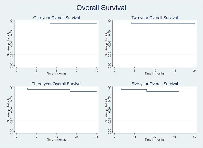
3438                                                    Graefe’s Archive for Clinical and Experimental Ophthalmology (2021) 259:3437–3443

Introduction                                                      was carried out with approval from the Head and Neck
                                                                  Institutional Review Board (approval ID; 18/2020) and
Ocular surface squamous neoplasia (OSSN) includes a               in adherence to the tenets of the Declaration of Helsinki.
spectrum of malignancy that ranges from mild epithelial           Photographs were obtained in selected cases with patients’
dysplastic changes (CIN) to more severe invasive carci-           permission.
noma, invading through the basement membrane into the
substantia propria (Ocular surface Squamous Cell Car-             Outcomes
cinoma - OSCC) [1]. The incidence of OSCC has been
reported from 0.03–1.9 per 100,000 persons/year in                Overall survival (OS) and overall recurrence rate (RR) were
patients in the USA and Australia, with a higher incidence        determined along with disease-free survival (DFS). Over-
in males and Whites [2, 3].                                       all survival was calculated as the time interval between the
   Orbital exenteration is probably the treatment of choice       diagnosis and the last follow-up or death for all causes, if
for tumors that have invaded the orbit or that exhibit com-       not differently specified. Overall recurrence rate (RR) was
plete involvement of the conjunctiva [4]. Orbital exentera-       defined considering the rate at which the disease recurred
tion therefore remains the ultimate therapeutic approach.         in the same or similar site of the original tumor after a com-
Nevertheless, very few reports exist on demography, sur-          plete resolution of the original tumor. DFS was defined as
vival disease free, overall survival, and predictive risk fac-    the time from the first treatment to relapse (local recurrence,
tors for OSCC invading periocular tissues and orbit [5].          lymph node metastasis, or distant metastasis) or all-cause
   The scope of this retrospective study was to analyze the       death, whichever came first. In addition, the following data
prevalence of diffuse OSCC with periocular involvement,           were collected: gender, age, site, laterality, size or extent of
the clinical outcomes in relation to the tumors staging,          the primary tumor (T), the presence or absence of lymph
and different medical and surgical approaches. Primary            node metastases (N) or distant metastases (M), histologic
outcomes were overall survival (OS), disease-free sur-            type (carcinoma in situ (CIS) or low-risk carcinoma and
vival (DFS) and overall recurrence rate (RR). Secondary           invasive squamous cell carcinoma or high-risk SCC), resec-
outcomes were a correlation between primary outcomes              tion boundaries, adjuvant therapies (radiotherapy, chemo-
and tumor location, eight editions of the American Joint          therapy, immunotherapy), and duration of follow-up. Tumor
Committee on Cancer Classification (AJCC) staging sys-            localization was defined considering the prevalent involved
tem stage, histological results, margins results, and type        site; when a prevalent site was not identifiable, the tumor
of treatment.                                                     was defined diffuse. The anatomic involvement has been
                                                                  defined as monocentric or multicentric according to whether
                                                                  only one or more anatomic sites were involved: superior
                                                                  tarsus, inferior tarsus, medial canthus, lateral canthus, or
Methods                                                           orbital tissues.

Patients                                                          Statistics

Patients suffering from OSCC demonstrating involvement            The Kaplan–Meier method was used to calculate survival
of conjunctiva and adjacent periocular tissues (e.g., eyelid      rates and to plot survival curves. Analyses were conducted
palpebral margin, superior tarsus, inferior tarsus, medial        using Stata software (StataCorp. 2015. Stata Statistical Soft-
canthus, lateral canthus, orbital tissues) were identified        ware: Release 15. College Station, TX, USA: StataCorp LP).
and included in a retrospective study. All patients were          Relations between categorical variables were analyzed using
treated at the Ocular Oncology Unit of the Fondazione             the log-rank test. Values of p < 0.05 were considered statisti-
Policlinico Universitario A. Gemelli, Rome, between Janu-         cally significant.
ary 01, 2011, and January 2020. Patients initially received
either surgical or topical treatment for their ocular sur-
face disease, followed by subsequent therapies for per-           Results
sistent or recurrent disease. Patients previously medically
or surgically treated or with a follow-up period of less          During the 9-year study period, there were a total of 36
than 6 months and or those with missing or incomplete             patients (5.8%), out of 618 total OSSN cases, with excised
records were excluded. A written informed consent for             histologically confirmed SCC involving ocular and perio-
data collection and analysis of collected data from their         cular tissues recorded. The mean follow-up was 40 months
medical records was obtained from all patients. The study         (median: 31 months, range: 6–120 months). Two deaths
                                                                  occurred during the follow-up period, unrelated to periocular

13
Graefe’s Archive for Clinical and Experimental Ophthalmology (2021) 259:3437–3443 3439
malignancy. The mean age at the time of first surgery was they were defined as a diffuse disease. Twenty (56%) patients
68.2 years (median: 73, range: 33–92). Eighteen (50%) were treated with excisional biopsy alone (EB), 10 (28%)
patients were male and 18 (50%) were female. All patients underwent EB plus adjuvant topical therapy (5 with sub-
were Caucasians. SCC occurred on the right side in 17 conjunctival interferon-alpha (INF-α), 4 with mitomycin C
(47%) cases and on the left in 19 (53%). Demographic data (MMC), 1 with both drugs); 3 (8%) cases received external
are listed in Table 1. beam radiotherapy (EBRT), MMC, and INF-α respectively
after an incisional biopsy; 3 (8%) patients were treated with
Risk categories exenteratio orbitae, one of which with an adjuvant EBRT.
Following histological examination, 5 (14%) cases were
Twenty-one patients (58%) showed a multicentric disease, classified as low-risk SCCs (carcinoma in situ (CIS)),
while 15 (42%) cases presented a monocentric disease. The whereas 24 (67%) were classified as high-risk SCCs (inva-
mainly involved site was the inferior eyelid tarsal plate in sive squamous carcinoma); 7 (19%) excised tissues were
10 cases (28%), the superior eyelid tarsal plate in 6 (17%), histologically classified as basaloid SCCs (BSCC). Seven
the eyelid palpebral margin in 6 (17%), orbit in 5 (14%), (19%) specimens presented positive margins at the histo-
internal canthus in 4 (11%), and external canthus in 2 (5%). logical examination, while 29 (81%) presented negative
In 3 patients (8%), no prevalent site was identifiable since surgical margins. The incompletely excised tumors were
high-risk SCCs variants in 5 (71%) cases, whereas 2 (29%)
were BSCC.
Table 1 Demographic, clinical, histological, and treatment features of According to the eighth edition of the AJCC, the most
OSSN patients common pT category was T3 with 31 (86%) cases, followed
Demographic features by T4a in 4 (11%) and T4b in 1 (3%). Table 1 lists clinical
Males (%)/females (%) 18 (50%)/18 (50%) and histological features of OSSN along with the types of
Age, mean (range) 68.2 (33–92) treatment.
Involved eye, RE (%)/LE (%) 17 (47%)/19 (53%) Overall recurrence rate was 64% (n = 23). Among the
Involved sites 23 recurred patients, 16 presented a multicentric disease
Multicentric 21 (58%) (p = 0.0039). The inferior tarsus was associated with the
Monocentric 15 (42%) highest recurrence rate (n = 8, p = 0.0428) and high-risk
Inferior eyelid TP 10 (28%) SSCs recurred more frequently than other histologic types
Superior eyelid TP 6 (17%) (n = 17, p = 0.0264). Positive surgical margins were associ-
Eyelid margin 6 (17%) ated with an increased risk of recurrence n = 5 (p = 0.0434).
Orbit 5 (14%) The highest recurrence rate was observed in the EB group
Internal cantus 4 (11%) (n = 9, p = 0.0005). Table 2 shows clinical and histological
External cantus 2 (5%) factors significantly associated with recurrence. Five (22%)
Diffuse 3 (8%) out of 23 recurred patients presented orbital invasion, mani-
Histology festing disease progression from the initial T3 stage.
Low-risk SCC 5 (14%) The overall survival rates at 12, 24, 36, and 60 months
High-risk SCC 24 (67%) were respectively 97.1%, 92.7%, 92.7%, and 92.7%.
Basaloid SCC 7 (19%) A short overall survival was observed in patients who
Positive margins 7 (19%) underwent EB alone (p = 0.0049).
Negative margins 29 (81%) The disease-free survival rates at 12, 24, 36, and
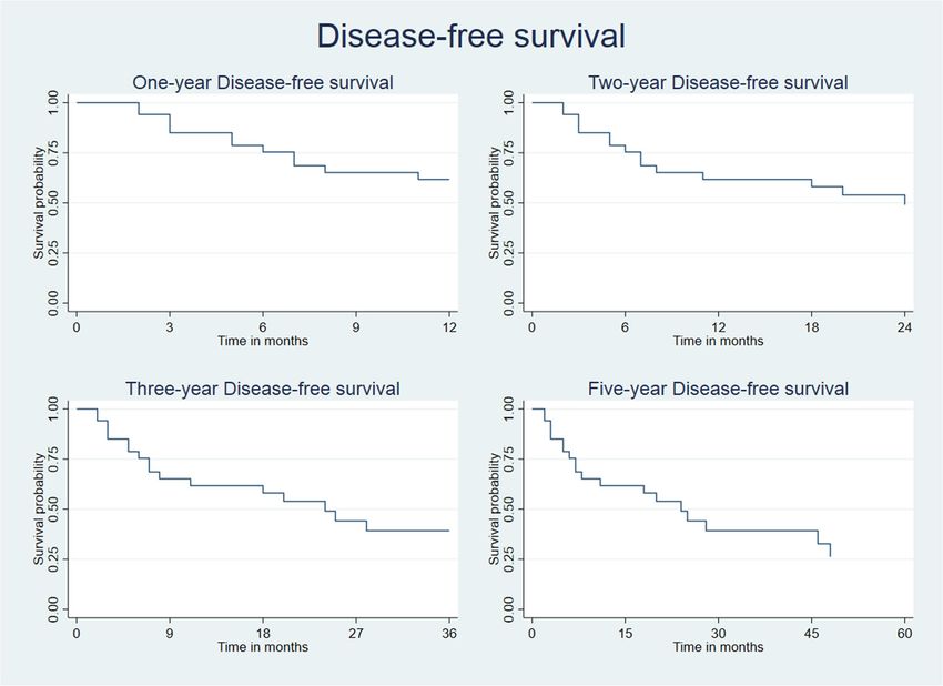
AJCC stage 60 months were respectively 62.9%, 50.8%, 41.6%, and
T3 31 (86%)
T4a 4 (11%)
T4b 1 (3%) Table 2 Clinical and histological factors significantly associated with
recurrence
Treatment
EB 20 (56%) Clinical and histological factors associated with recurrence P value
EB + (MMC, INF-α) 10 (28%)
Multicentric disease 0.0039
IB + (EBRT, MMC, INF-α) 3 (9%)
Inferior tarsus 0.0428
Exenteratio orbitae 3 (9%) High-risk SCC 0.0264
AJCC, American Joint Committee on Cancer; EB, excisional biopsy; Positive margins 0.0434
EBRT, external beam radiotherapy; IB, incisional biopsy; INF, inter- EB alone 0.0005
feron; LE, left eye; MMC, mitomycin C; RE, right eye; SCC, squa-
mous cell carcinoma; TP, tarsal plate EB, excisional biopsy; SCC, squamous cell carcinoma
133440 Graefe’s Archive for Clinical and Experimental Ophthalmology (2021) 259:3437–3443
Fig. 1 Kaplan–Meier analysis estimates overall survival rates over time
29.7%. Overall survival rates and disease-free survival rates and orbital invasion. In the current literature, only case
are represented in Fig. 1 and Fig. 2. reports or small case series of OSSC with periocular involve-
Five patients presented a second disease recurrence, but ment and/or orbital invasion are reported.
no statistically significant correlation with the abovemen- In order to analyze a homogeneous sample, we selected
tioned variables was found. 36 out of 618 patients with histologically proven conjunc-
tival SCC with periocular involvement (T3 and T4 AJCC
stage). Thus, 5.8% of cases represented advanced forms
Discussion of OSCC. This result agrees with other reports, consider-
ing that intraocular invasion has been observed in 3% to
Based on the series of 5002 conjunctival tumors referred to 9% of cases of OSSN reported in the literature, whereas
an ocular oncology tertiary care center, malignant conjunc- orbital invasion has been described in 1% to 6% [6–9]. Other
tival tumors were found to be 30%. In that series, the ocular authors reported that, among secondary orbital tumors origi-
surface squamous neoplasia (OSSN), including conjunctival nating from conjunctival malignancies, 81% of cases were
intraepithelial neoplasia (CIN) and squamous cell carcinoma identified as squamous cell carcinomas [10].
(SCC), represented 14% of the total of conjunctival tumors, Tarsal involvement (T3 AJCC stage) and more generally
with SCC accounting for 9% of malignancies [1]. higher AJCC stage, positive pathologic margins and higher
In the USA and Western Europe, most patients present pathologic grade were reported as strong predictors for more
with “classic OSSN” with typical limbal presentation and aggressive tumors [7, 11]. Although OSCC occurs predomi-
relatively benign clinical course. However, a minority of nantly in males [12, 13], we found an equal distribution in
patients present more aggressively behaving tumors, with a genders. None of the included cases showed HIV positivity,
higher rate of recurrence and associated ocular, periocular, albeit invasive and aggressive variants of OSSC are often
13Graefe’s Archive for Clinical and Experimental Ophthalmology (2021) 259:3437–3443 3441
Fig. 2 Kaplan–Meier analysis shows disease-free survival rates over time
observed in HIV patients [8, 11, 13]. However, there were development and thickness compared to similar submucosal
two cases of systemic immunosuppression, related to immu- superior eyelid structures and submucosal medial and lateral
nosuppressant therapy for organ transplantation and splenic canthal tendons [14].
disease respectively. The type of treatment is also correlated with the recur-
Analyzing 389 excised OSSN lesions, Galor et al. found rence rate [15–18]. Surgery alone (EB without adjuvant
that the 1-year recurrence rate was 10% and the 5-year treatment) is significantly associated with a higher recur-
recurrence rate was 21%, with a mean time to recurrence of rence rate. Moreover, disease recurrence tends often to
2.5 years. In our series, the overall recurrence rate was signifi- spread from the anatomical site of origin toward periocular
cantly higher (64% vs 21%) if compared with literature data, tissues and orbital structures. In fact, five (22%) out of 23
as would be expected for the kind of sample selected. Indeed, recurred patients presented orbital invasion, with disease
31 patients presented a T3 stage disease at baseline, while 5 progression from the initial T3 stage. It cannot be ruled out
of them referred with a T4a stage and 1 presented a T4b stage. that a further T3 stage stratification could better depict this
Our results show that tumor anatomical characteristics spread and could also be a useful tool to define treatments
at presentation, including multicentric pattern and inferior and prognosis. According to other reports [11, 18], adju-
tarsus involvement, histological diagnosis of high-risk SCC, vant topical therapy seems effective in decreasing recur-
and positive surgical margins are significantly related to a rence rates, particularly in patients with positive margins,
higher risk of local recurrence. In particular, inferior tarsus histological high-risk SCC, tarsal, and multicentric pattern
involvement can be considered as a high-risk factor because anatomical involvement.
the lower eyelid submucosal anatomical structures, tarsal In conclusion, we suggest that in advanced OSCC,
plate, retractors, and fascial fibers of Lockwood’s liga- particularly when the inferior tarsus is involved and/or
ment are probably more easily infiltrated, due to the lower there is a multicentric disease, an aggressive and—where
133442 Graefe’s Archive for Clinical and Experimental Ophthalmology (2021) 259:3437–3443
possible—radical surgery, consistent with the reconstruction 3. Sun EC, Fears TR, Goedert JJ (1997) Epidemiology of squamous
needs, is strongly advisable. Customized adjuvant treatments cell conjunctival cancer. Cancer Epidemiol Biomarkers Prev
6:73–77
should always be planned, including EBRT, brachytherapy, 4. Shields JA, Shields CL, Suvarnamani C, Tantisira M, Shah P
local, and also systemic chemotherapy. Cemiplimab, a PD1 (1991) Orbital exenteration with eyelid sparing: indications, tech-
inhibitor which was recently approved for the treatment of nique and results. Ophthalmic Surg 22:292–297
cutaneous SCC, has been suggested for OSCC patients and 5. Polski A, Sibug Saber M, Kim JW, Berry JL (2019) Extending
far and wide: the role of biopsy and staging in the management
could be considered in the therapeutic strategy [19–21]. of ocular surface squamous neoplasia. Clin Exp Ophthalmol
47(2):193–200
6. Arepalli S, Kaliki S, Shields CL, Emrich J, Komarnicky L, Shields
Author contributions All authors contributed to the study’s conception JA (2014) Plaque radiotherapy for scleralinvasive conjunctival
and design. Material preparation, data collection, and analysis were squamous cell carcinoma: analysis of 15 eyes. JAMA Ophthalmol
performed by Martina Maceroni, Giovanni Cuffaro, and Luca Giraldi. 132:691–696
The first draft of the manuscript was written by Gustavo Savino, Mar- 7. Yousef YA, Finger PT (2012) Squamous carcinoma and dysplasia
tina Maceroni, and Giovanni Cuffaro, and all authors commented on of the conjunctiva and cornea: an analysis of 101 cases. Ophthal-
previous versions of the manuscript. All authors read and approved mology 119(2):233–240
the final manuscript. 8. Erie JC, Campbell RJ, Liesegang TJ (1986) Conjunctival and
corneal intraepithelial and invasive neoplasia. Ophthalmology
Funding Open access funding provided by Università Cattolica del 93(2):176–183
Sacro Cuore within the CRUI-CARE Agreement. 9. Shields JA, Shields CL, Gunduz K, Eagle RC Jr (1999) The 1998
Pan American Lecture: intraocular invasion of squamous cell car-
cinoma of the conjunctiva in five patients. Ophthal Plast Reconstr
Availability of data and material Data and materials are available for Surg 15(3):153–160
consultation. 10 Soysal HG, Ardiç F (2008) Malignant conjunctival tumors invad-
ing the orbit. Ophthalmologica 222(5):338–343
Declarations 11. Galor A, Karp CL, Oellers P, Kao AA, Abdelaziz A, Feuer
W, Dubovy SR (2012) Predictors of ocular surface squamous
Ethics approval The study was carried out with approval from the Head neoplasia recurrence after excisional surgery. Ophthalmology
and Neck Institutional Review Board (approval ID; 18/2020) and in 119(10):1974–1981
adherence to the tenets of the Declaration of Helsinki. 12. Shields CL, Shields JA (2019) Tumors of the conjunctiva and
cornea. Indian J Ophthalmol 67(12):1930–1948
Consent to participate All patients gave their written informed consent 13. Lee GA, Hirst LW (1995) Ocular surface squamous neoplasia.
for data collection and analysis of collected data. Surv Ophthalmol 39:429–450
14. Rootman J, Stewart B, Goldberg RA (1995) Orbital surgery. A
Consent for publication All patients gave their written informed con- conceptual approach. Chapter 7, Orbital Anatomy. Philadelphia,
sent for data publication. Lippincott-Raven, pp 134–136
15. Shields JA, Shields CL, De Potter P (1997) Surgical management
of conjunctival tumors. Arch Ophthalmol 115(6):808–815
Conflict of interest The authors declare no competing interests. 16. Midena E, Angeli CD, Valenti M, de Belvis V, Boccato P (2000)
Treatment of conjunctival squamous cell carcinoma with topical
Open Access This article is licensed under a Creative Commons Attri- 5-fluorouracil. Br J Ophthalmol 84:268–272
bution 4.0 International License, which permits use, sharing, adapta- 17. Shields CL, Naseripour M, Shields JA (2002) Topical mitomy-
tion, distribution and reproduction in any medium or format, as long cin C for extensive, recurrent conjunctival-corneal squamous cell
as you give appropriate credit to the original author(s) and the source, carcinoma. Am J Ophthalmol 133:601–606
provide a link to the Creative Commons licence, and indicate if changes 18. Blasi MA, Maceroni M, Sammarco MG, Pagliara MM (2018)
were made. The images or other third party material in this article are Mitomycin C or interferon as adjuvant therapy to surgery for ocu-
included in the article’s Creative Commons licence, unless indicated lar surface squamous neoplasia: comparative study. Eur J Ophthal-
otherwise in a credit line to the material. If material is not included in mol 28(2):204–209
the article’s Creative Commons licence and your intended use is not 19. Nagarajan P, El-Hadad C, Gruschkus SK, Ning J, Hudgens CW,
permitted by statutory regulation or exceeds the permitted use, you will Sagiv O, Gross N, Tetzlaff MT, Esmaeli B (2019) PD-L1/PD1
need to obtain permission directly from the copyright holder. To view a expression, composition of tumor-associated immune infiltrate,
copy of this licence, visit http://creativecommons.org/licenses/by/4.0/. and HPV status in conjunctival squamous cell carcinoma. Invest
Ophthalmol Vis Sci 60(6):2388–2398
20. Wolkow N, Jakobiec FA, Afrogheh AH, Eagle RC Jr, Pai SI,
Faquin WC (2019) Programmed cell death 1 ligand 1 and pro-
References grammed cell death 1 ligand 2 are expressed in conjunctival
invasive squamous cell carcinoma: therapeutic implications. Am
1. Shields CL, Alset AE, Boal NS et al (2017) Conjunctival tumors J Ophthalmol 200:226–241
in 5002 cases. Comparative analysis of benign versus malignant 21. Demirci H, Elner VM, Demirci FY, Robinson DR, Chinnaiyan
counterparts. The 2016 James D. Allen Lecture Am J Ophthalmol A, Schlachter D, Joseph S, Worden F (2020) Immunotherapy for
173:106–133 conjunctival squamous cell carcinoma with orbital extension.
2. Emmanuel B, Ruder E, Lin SW, Abnet C, Hollenbeck AR, Mbu- Ophthalmology S0161–6420:30932–30935
laiteye SM (2012) Incidence of squamous-cell carcinoma of the
conjunctiva and other eye cancers in the NIH-Diet AARP and Publisher’s note Springer Nature remains neutral with regard to
Study Health. Ecancermedicalscience 6:254 jurisdictional claims in published maps and institutional affiliations.
13Graefe’s Archive for Clinical and Experimental Ophthalmology (2021) 259:3437–3443 3443
Authors and Affiliations
Gustavo Savino1,2 · Giovanni Cuffaro1,2 · Martina Maceroni1,2 · Monica Maria Pagliara2 · Maria Grazia Sammarco2 ·
Luca Giraldi1,3 · Maria Antonietta Blasi1,2
1 3
Università Cattolica del Sacro Cuore, Rome, Italy Sezione Di Igiene, Dipartimento Di Scienze Biologiche E
2 Sanità Pubblica, Università Cattolica del Sacro Cuore, Rome,
UOC Oncologia Oculare, Fondazione Policlinico
Italy
Universitario A. Gemelli - IRCCS, Largo Agostino Gemelli,
8, 00168 Rome, Italy
13You can also read