Acidic Microenvironment Aggravates the Severity of Hepatic Ischemia/ Reperfusion Injury by Modulating M1-Polarization Through Regulating PPAR-g ...
←
→
Page content transcription
If your browser does not render page correctly, please read the page content below
ORIGINAL RESEARCH
                                                                                                                                           published: 21 June 2021
                                                                                                                                  doi: 10.3389/fimmu.2021.697362

                                          Acidic Microenvironment Aggravates
                                          the Severity of Hepatic Ischemia/
                                          Reperfusion Injury by Modulating
                                          M1-Polarization Through Regulating
                                          PPAR-g Signal
                                          Wei Ding 1,2†, Yunfei Duan 1†, Zhen Qu 1†, Jiawei Feng 1, Rongsheng Zhang 3, Xiaodong Li 1,
                         Edited by:       Donglin Sun 1*, Xiaoying Zhang 1* and Yunjie Lu 1*
          Christoph Thiemermann,
                                          1Hepatopancreatobiliary Surgery Department, The Third Affiliated Hospital of Soochow University, Changzhou First People’s
   Queen Mary University of London,
                  United Kingdom          Hospital, Changzhou, China, 2 General Surgery Department, Wujin Hospital Affiliated with Jiangsu University, Changzhou,
                                          China, 3 Hepatobiliary Surgery Department, Nanjing Eight One Hospital, Nanjing, China
                       Reviewed by:
                     Zoran Todorovic,
        University of Belgrade, Serbia    Hepatic injury induced by ischemia and reperfusion (HIRI) is a major clinical problem after
                        Sura Al Zoubi,
  Al-Balqa Applied University, Jordan
                                          liver resection or transplantation. The polarization of macrophages plays an important role
                 *Correspondence:
                                          in regulating the severity of hepatic ischemia/reperfusion injury. Recent evidence had
                         Donglin Sun      indicated that the ischemia induces an acidic microenvironment by causing increased
                 sdldoctor@163.com
                                          anaerobic glycolysis and accumulation of lactic acid. We hypothesize that the acidic
                      Xiaoying Zhang
         zhangxy6689996@163.com           microenvironment might cause the imbalance of intrahepatic immunity which aggravated
                            Yunjie Lu     HIRI. The hepatic ischemia/reperfusion injury model was established to investigate the
           yjluforresearch@126.com
     †
                                          effect of the acidic microenvironment to liver injury. Liposomes were used to deplete
      These authors have contributed
             equally to this work and
                                          macrophages in vivo. Macrophages were cultured under low pH conditions to analyze the
                share first authorship     polarization of macrophages in vitro. Activation of the PPAR-g signal was determined by
                                          Western blot. PPAR-g agonist GW1929 was administrated to functionally test the role of
                   Specialty section:
         This article was submitted to
                                          PPAR-g in regulating macrophage-mediated effects in the acidic microenvironment during
                          Inflammation,    HIRI. We demonstrate that acidic microenvironment aggravated HIRI while NaHCO3
               a section of the journal   reduced liver injury through neutralizing the acid, besides, liposome abolished the
              Frontiers in Immunology
                                          protective ability of NaHCO3 through depleting the macrophages. In vivo and vitro
            Received: 19 April 2021
            Accepted: 28 May 2021         experiment showed that acidic microenvironment markedly promoted M1 polarization
           Published: 21 June 2021        but inhibited M2 polarization of macrophage. Furthermore, the mechanistic study proved
                            Citation:     that the PPAR-g signal was suppressed during the polarization of macrophages under
       Ding W, Duan Y, Qu Z, Feng J,
  Zhang R, Li X, Sun D, Zhang X and
                                          pH = 6.5 culture media. The addition of PPAR-g agonist GW1929 inhibited M1 polarization
 Lu Y (2021) Acidic Microenvironment      under acidic environment and reduced HIRI. Our results indicate that acidic
   Aggravates the Severity of Hepatic     microenvironment is a key regulator in HIRI which promoted M1 polarization of
       Ischemia/Reperfusion Injury by
 Modulating M1-Polarization Through       macrophages through regulating PPAR-g. Conversely, PPAR-g activation reduced liver
            Regulating PPAR-g Signal.     injury, which provides a novel therapeutic concept to prevent HIRI.
          Front. Immunol. 12:697362.
    doi: 10.3389/fimmu.2021.697362        Keywords: hepatic ischemia-reperfusion injury, macrophage, polarization, PPAR, acidic microenvironment, STAT6

Frontiers in Immunology | www.frontiersin.org                                   1                                         June 2021 | Volume 12 | Article 697362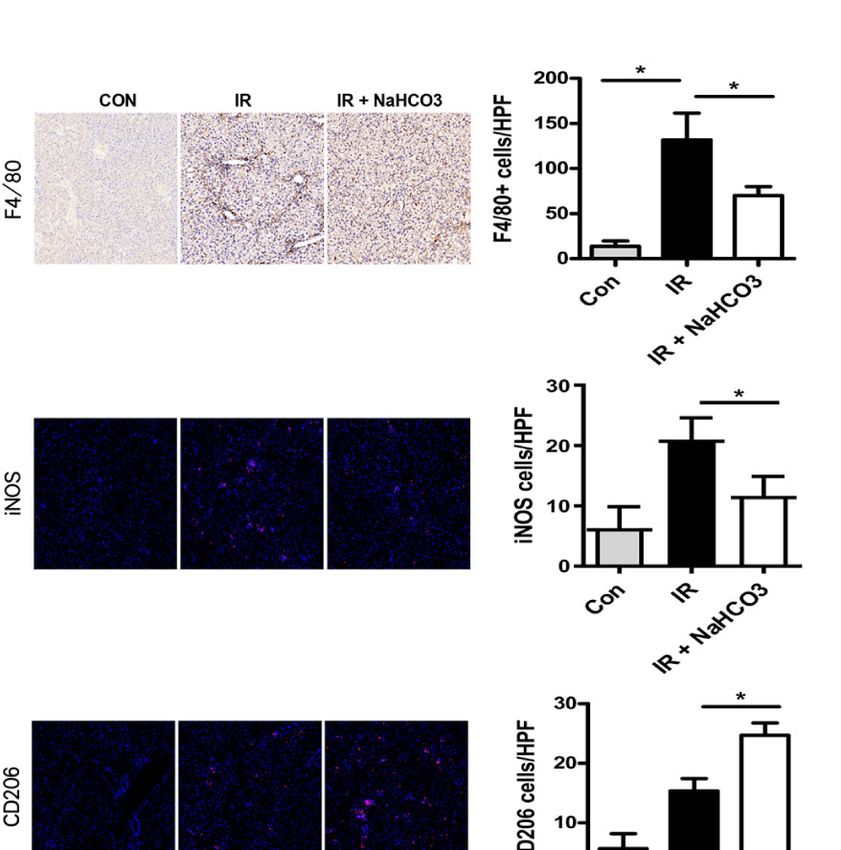
Ding et al.                                                                                              Acidic Microenvironment Aggravates HIRI by PPAR-g

INTRODUCTION                                                                      liposomes through tail vein (200 ml, Amsterdam, The
                                                                                  Netherlands) 24 h before the model was established. At the
Hepatic ischemia/reperfusion injury (HIRI) is the main cause of                   indicated time, mice liver tissue and blood were collected.
hepatic insufficiency and failure after liver surgery (1, 2), HIRI
causes a series of liver abnormalities, from liver insufficiency to                KCs Isolation and Cell Culture
liver nonfunction (3). As reported, the expression of immune-                     Mice were anesthetized, and livers were perfused with perfusion
related cytokines was abnormal when acute organ injury was                        buffer (HBSS) in situ via the portal vein, followed by 0.27%
caused by HIRI (4, 5).                                                            collagenase IV (Sigma, Saint Louis, MO, USA). Perfused livers
    The key role in the pathogenesis of HIRI is mainly the                        were dissected, and single-cell suspensions were generated
inflammation caused by the innate immune response of                               through a 70-mm cell filter. The suspension was added with
Kupffer cells (KC) (6). KCs, a type of macrophages, are                           40 ml of DMEM supplemented with 10% FBS, 10 mM HEPES,
resident in the liver, which can serve as outposts of hepatic                     2 mM GlutaMax, 100 U/ml penicillin, and 100 mg/ml
homeostasis (7). In the course of aseptic hepatitis, activated KCs                streptomycin and standing for 15 min at 37 °C. Then, the
secrete inflammatory cytokines and chemokines and attract                          adherent cells were left for further experiments while the non-
other inflammatory cells into the damaged tissue, resulting in                     adherent cells were removed. After 6 h of cultured, the KCs or the
increased inflammation (8). The pro-inflammatory M1 type and                        samples of supernatants were collected for further analysis.
anti-inflammatory M2 type are the different functional states of
macrophages. STAT1 and IRF5 control the M1 macrophage                             Immunohistochemical Staining
phenotype, whereas M2 macrophage polarization is regulated by                     Liver macrophages were identified with primary rat anti-mouse
STAT6, IRF4, and PPAR-g (9).                                                      F4/80 and CD11b, and neutrophils were identified with Ly6G
    Under ischemia conditions, the promotion of anaerobic                         mAb (BD Biosciences, San Jose, CA, USA). Then, the cells were
glycolysis and accumulation of lactic acid may cause the                          incubated with biotinylated anti-goat IgG, followed by
generation of acidic microenvironment while the pH values of                      immunoperoxidase (ABC Kit, Vector), according to the
blood or tissues go down to the PH of 6.0 to 6.5. A recent study                  specification. The numbers of positive-stained cells were
showed that such a change in the microenvironment may                             counted blindly in 10 high-power field (HPF) sections.
suppress the generation of T regulatory cells (Tregs) and cause
an immune imbalance which aggravated HIRI (10). Therefore,                        Immunofluorescence Staining
the acidic microenvironment is one of most important factors                      LC3B, iNOS, and CD206 in KCs were identified by indirect
leading to HIRI. However, the relationship between acidic                         immunofluorescence staining using rabbit anti-mouse LC3B
microenvironment and macrophages in HIRI has not yet been                         mAb, anti-mouse iNOS mAb, and anti-mouse CD206 mAb
investigated. So, the present study performed a series of                         (Cell Signaling Technology, MA, USA). After washing, the KCs
experiments to determine the function of acidic                                   which were premounted with VECTASHIELD medium with
microenvironment in regulating macrophages in the                                 DAPI (Vector) were incubated with secondary antibody goat
pathogenesis of HIRI.                                                             anti-mouse Texas Red-conjugated IgG (Sigma, Saint Louis, MO,
                                                                                  USA). The numbers of positive-stained cells were counted
                                                                                  blindly in 10 high-power field (HPF) sections.

MATERIALS AND METHODS                                                             Quantitative RT-PCR and Western
                                                                                  Blot Analysis
Animals                                                                           RNA and proteins were acquired following the manufacturer’s
We placed 8-week-old male mice (C57B/6J) under SPF                                instructions. Then the PCR and WB assays were run by the
conditions. These mice were treated and sacrificed at the                          standard protocol. Primers are shown as below:
specified time point according to the protocol approved by the
I n s t i tu ti o n a l A n im a l C a r e a n d U s e C o m m i t te e o f       iNOS, 5′- TCTTTCCGTCCGAAGACAGAT -3′
Soochow University.                                                               and 5′- TTGTAGTCGTAAGACCTCGATGA -3′;
                                                                                  TNF-a, 5′- TCTTCTCATTCCTGCTTGTGG-3′
Hepatic Ischemia/Reperfusion
                                                                                  and 5′- GGTCTGGGCCATAGAACTGA-3′;
Injury Model
Mice were as the previous study indicated (11). The specific                       IL-6, 5′- GGCGGATCGGATGTTGTGAT-3′
construction method was as follows. After anesthesia with 3.5%                    and 5′- GGACCCCAGACAATCGGTTG-3′;
chloral hydrate (0.1 ml/10 g), a midline incision was made on the                 Arg-1, 5′- CTGAGAGATTCAAGGCAAGAGG-3′
abdomen. Non-invasive vascular clamp was used to clamp the                        and 5′- GAACGCGCTATCTTACCCCAG-3′;
left and middle branches of portal vein and hepatic artery for
                                                                                  Mrc2, 5′- ATCCAGGGAAACTCACACGGA-3′
60 min, and the liver was 70% ischemia. Reperfusion was then
started by removing the clamp. The sham group goes through the                    and 5′- GCGCTCATCTTTGCCGTAGT-3′;
same procedure in addition to using vascular clamps. For some                     IL-10, 5′-GTTACTTGGGTTGCCAAG-3′
experiments, macrophages were depleted by injecting clodronate                    and 5′-TTGATCATCATGTATGCTTC-3′;

Frontiers in Immunology | www.frontiersin.org                                 2                                    June 2021 | Volume 12 | Article 697362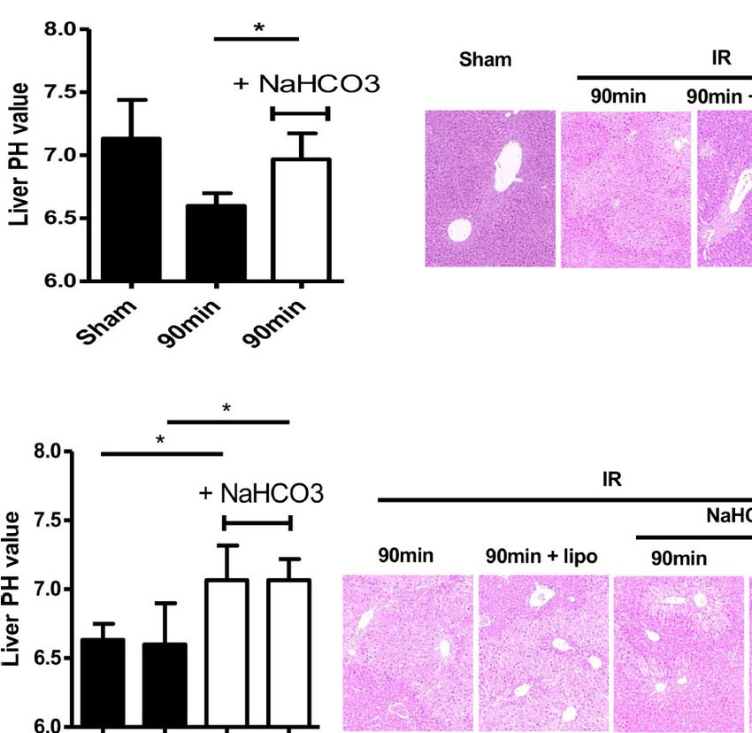
Ding et al.                                                                                                        Acidic Microenvironment Aggravates HIRI by PPAR-g

Gapdh, 5′-CCATCTTCCAGGAGCGAGATC-3′                                                     showed that the Suzuki scores of mouse livers were increased over
and 5′-GCCTTCTCCATGGTGGTGAA′.                                                          time, although the analysis of ANVOA showed no significance
                                                                                       (P=0.264) in the IR group (Figures 1B, C). The values of ALT and
                                                                                       AST were upregulated with the increase of ischemia time interval in
Statistical Analysis                                                                   the IR group (Figures 1D, E). Cytokine expression plays an
Stata software (version 11.0) was utilized to perform the analysis.                    important role to the pathogenesis of HIRI (12–14), so we
Data were expressed as mean ± standard deviation (SD), the                             evaluated the expression of the cytokine on CD4 T cells, data
differences between groups were analyzed by either the paired                          showed that compared with the 60-min ischemia model, 90 min
t-test or ANOVA test (both one-way ANOVA test and two-way                              ischemia/reperfusion model presented higher inflammatory
ANOVA test). In addition, we use Bonferroni or LSD as a post                           cytokine expression such as IL-4, IFN-g, IL-17, and IL-6
hoc test. P values less than 0.05 (two-tailed) was considered                          (Figure 1F). These results demonstrate that HIRI induced
statistically significant.                                                              acidification and dysfunction of the liver microenvironment.

                                                                                       Sodium Bicarbonate Injection Reverses
RESULTS                                                                                HIRI Through Regulating Acidic
                                                                                       Microenvironment by Moderating
HIRI Induced Acidification and                                                          Macrophages
Dysfunction of the Liver Microenvironment                                              Administration of sodium bicarbonate to increase the intra-
A mouse model of partial hepatic ischemia/reperfusion with                             tumoral pH had been evaluated as an effective anti-acidic
different ischemia time interval was established. After reperfusion,                   therapeutic strategy (15). Therefore, we tested the protective
injured liver lobe and blood specimens were collected at 60, 90, and                   ability of sodium bicarbonate on regulating intrahepatic pH
120 min. HE staining of liver tissue and detected ALT and AST in                       under conditions of HIRI. Sodium bicarbonate (100 ml/10 g,
the serum were performed to assess the degree of liver injury.                         pH 8.2) was injected through the tail vein at the beginning of
Meanwhile, the pH value of the ischemic liver lobe was also                            ischemia. NaHCO 3 injection recovered the decrease of
measured. Compared with the sham group, the PH value of                                intrahepatic pH comparing with the IR group (Figure 2A). HE
injured liver was significantly decreased in the IR group; besides,                     staining of liver tissue showed that NaHCO3 injection reduced
pH was getting lower accompanied with the increase of ischemia                         the degree of injury comparing with the IR group (Figure 2B).
time interval (Figure 1A). The pathological results of HE staining                     Consistent with this result, the values of ALT and AST were

              A                                                B                                                     C

                                                   E
              D                                                                              F

 FIGURE 1 | HIRI induced acidification and dysfunction of liver microenvironment. (A) pH value of injured liver lobe by time point after reperfusion in mice (one-way
 ANOVA with Bonferroni as a post hoc test, n=6). (B) Representative images of liver injury in mice [hematoxylin and eosin (HE) staining, 200×] by light microscopy.
 (C) Suzuki scores for liver tissues in each group (one-way ANOVA with Bonferroni as a post hoc test, n=3). Levels of ALT (D) and AST (E) in sham and IR groups
 (one-way ANOVA with Bonferroni as a post hoc test, n=4 for D, n=3 for E). (F) Cytokine expression on CD4+ T cells derived from blood was tested through flow
 cytometry (paired t test, n=4). *p < 0.05; ***p < 0.001.

Frontiers in Immunology | www.frontiersin.org                                      3                                           June 2021 | Volume 12 | Article 697362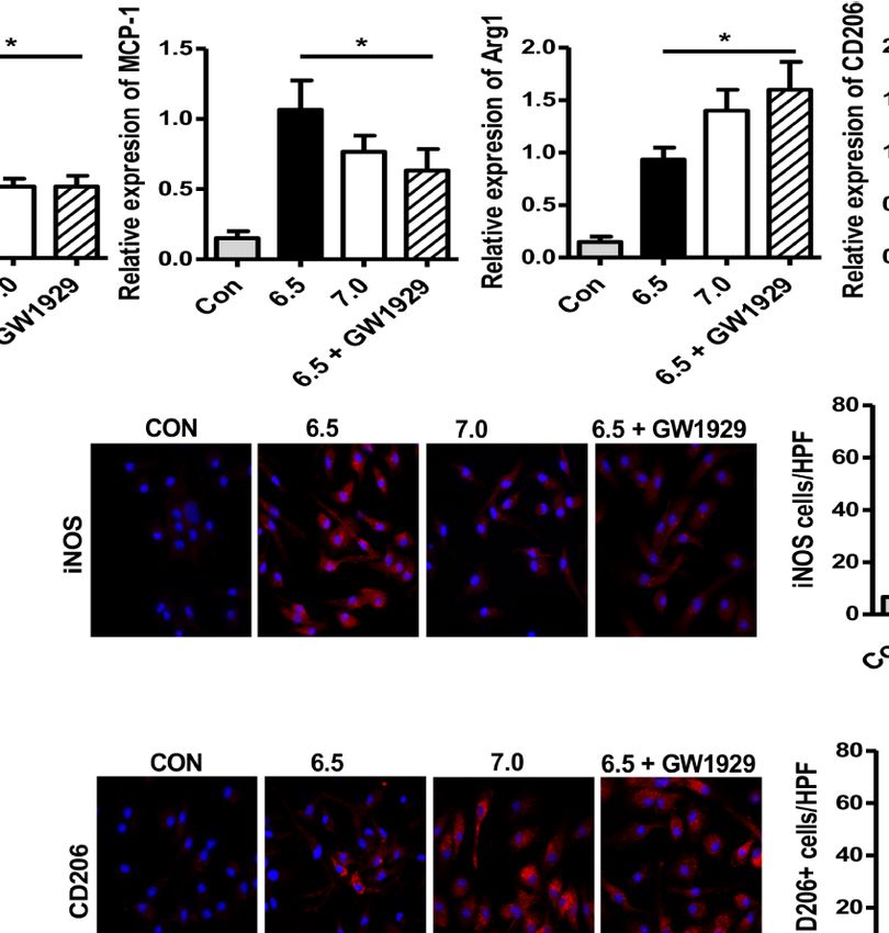
Ding et al.                                                                                                       Acidic Microenvironment Aggravates HIRI by PPAR-g

decreased with the injection of NaHCO3 which indicated that                           (F4/80) were increased in the IR group and reduced in the
NaHCO3 reduced liver injury through anti-acidic therapy                               NaHCO3 group. According to the function of macrophages,
(Figures 2C, D).                                                                      macrophages could be categorized into M1 (classical) and M2
    Next, we analyzed the role of macrophages during acidic                           (alternative) subtypes broadly (17). Thus, we concluded that the
microenvironment-mediated HIRI. In order to assess whether                            sodium bicarbonate has effect on regulating KCs M1/M2
NaHCO3 exerts a protective effect of HIRI depending on                                polarization. iNOS and CD206 were the key markers for M1
controlling KCs, we used the liposome to treat KCs (16).                              and M2 separately, so we detected the phenotype of macrophages
Liposome was injected 24 h before the model was established.                          through immunofluorescence staining, which showed that while
Then, we detected pH of the ischemic liver tissues, stained liver                     in the IR group, more macrophages presented M1 like
tissue by HE, and collected serum ALT and AST values. There is no                     macrophages, sodium bicarbonate moderated the macrophages
difference in pH value with or without liposome injection                             to polarize to M2 like macrophages (Figure 3A). To confirm the
(Figure 2E), HE staining showed that liposome treatment                               result, we derived intra-macrophages from each group, RT-PCR
abolished the protective effect of NaHCO3 in HIRI (Figure 2F).                        was used to evaluate the expression of M1 and M2 markers. In
The ALT and AST change also proved that while NaHCO3 reduced                          the IR model, iNOS, TNF-a, and IL-6 were upregulated in the
the injury of the liver, liposome injection aggravated the injury                     liver while sodium bicarbonate decreased the transcription of
(Figures 2G, H). Taken together, our data indicate that Sodium                        above mRNAs (Figure 3B). In contrast, Arg-1, Mrc2, and IL-10
bicarbonate injection reverses HIRI through regulating acidic                         were increased in the NaHCO3 group comparing with the IR
microenvironment by modulating macrophages.                                           group. At the same time, we observed that serum cytokine
                                                                                      expression for a different group. The results showed that
Sodium Bicarbonate Reduces Innate                                                     TNF-a and IL-6 were lower expressed, while IL-10 was
Immune and KC Polarization in HIRI                                                    higher expressed in the NaHCO 3 group (Figure 3C).
We further assessed the effects of sodium bicarbonate on                              Therefore, sodium bicarbonate reduces innate immune
regulating macrophage infiltration by immunohistochemical                              function by reducing pro-inflammatory TNF-a, IL-6 levels,
and immunofluorescence staining. Intrahepatic macrophages                              and KC polarization in HIRI.

       A                                        B                                       C                                   D

       E                              F                                                     G                                   H

 FIGURE 2 | Sodium bicarbonate injection reverse HIRI through regulating acidic microenvironment by moderating macrophages. Mice were injected with sodium
 bicarbonate the same time while the HIRI model were established. (A) pH value of injured liver lobe at each time point after reperfusion in mice (one-way ANOVA with
 Bonferroni as a post hoc test, n=3). (B) Representative images of liver injury in mice [hematoxylin and eosin (HE) staining, 200×] by light microscopy. Levels of ALT
 (C) and AST (D) in sham and IR groups (one-way ANOVA with Bonferroni as a post hoc test, n=3). Liposome was injected through tail vein 24 h before the HIRI
 model was established to abolish the effect of macrophages. Mice were injected with sodium bicarbonate the same time while the HIRI model were established.
 (E) pH value of injured liver lobe at each time point after reperfusion in mice (two-way ANOVA with Bonferroni as a post hoc test, n=3). (F) Representative images of
 liver injury in mice [hematoxylin and eosin (HE) staining, 200×] by light microscopy. Levels of ALT (G) and AST (H) in sham and IR groups (two-way ANOVA with LSD
 as a post hoc test, n=3). *p < 0.05.

Frontiers in Immunology | www.frontiersin.org                                     4                                             June 2021 | Volume 12 | Article 697362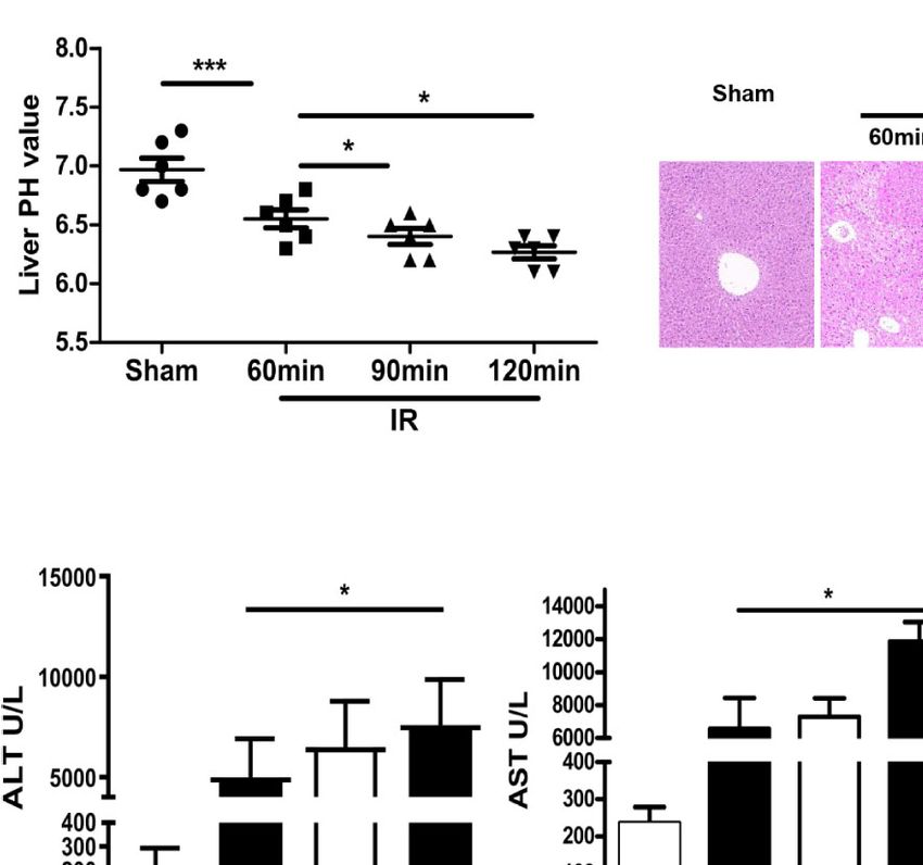
Ding et al.                                                                                                  Acidic Microenvironment Aggravates HIRI by PPAR-g

         A                                                                B

                                                                          C

 FIGURE 3 | Sodium bicarbonate reduce innate immune and KC polarization in HIRI. Mice were injected with sodium bicarbonate the same time while the HIRI
 model were established. (A) The infiltration of macrophages and M1/M2 cells was analyzed by immunohistochemical and immunofluorescence staining (one-way
 ANOVA with Bonferroni as a post hoc test, n=3). (B) The expressions of inflammatory genes in liver tissues were measured by quantitative RT-PCR (one-way
 ANOVA with Bonferroni as a post hoc test, n=3). (C) The levels of inflammatory cytokines in serum were measured by ELISA (one-way ANOVA with Bonferroni as a
 post hoc test, n=3). *p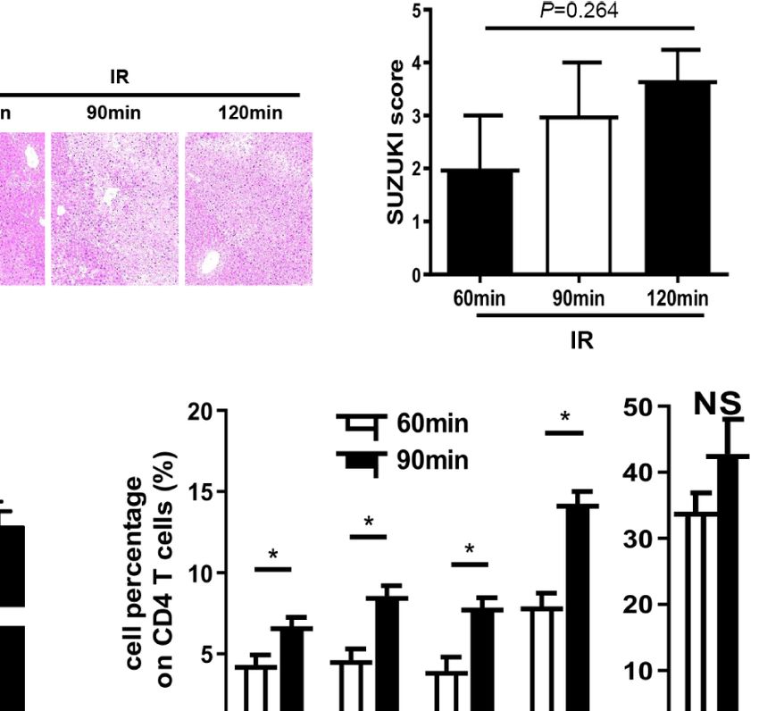
Ding et al.                                                                                                          Acidic Microenvironment Aggravates HIRI by PPAR-g

        A                                          B                                          C

        D                                                                                                        E

 FIGURE 4 | Acidic microenvironment regulates M1/M2 polarization in response to LPS condition by PPAR-g. Macrophages were isolated from B6 mice. M1 markers
 (iNOS and MCP-1) (A) and M2 markers (Arg-1 and CD206) (B) of gene induction were measured by quantitative RT-PCR after 6 h of acidic or normal condition
 treatment (paired t test, n=3). (C) Isolated macrophages from different experimental groups were cultured for 24 h. Then, the levels of TNF-a, IL-6, and IL-10 protein
 in the culture supernatant were detected using ELISA (paired t test, n=3). (D) Representative immunofluorescence staining images of iNOS and CD206 in
 macrophages (one-way ANOVA with Bonferroni as a post hoc test, n=3). (E) The expression of NF-kB and PPAR-g signaling in different groups as determined by
 western blot analysis. *p < 0.05.

(Figure 5C). Immunofluorescence staining indicated that                                 detected through immunofluorescence staining, which showed that
GW1929 inhibited iNOS expression of macrophages and                                    GW1929 treated macrophages presented a strong anti-acidic effect
upregulated the level of CD206 during acidic media treatment                           by suppressing the polarization of intrahepatic macrophages
(Figure 5D), which proved that GW1929 suppressed M1                                    (Figure 6D). To conclude, PPAR-g agonist GW1929 suppressed
polarization in the acidic microenvironment. We demonstrate                            M1 polarization induced by Acidic microenvironment in vivo.
that PPAR-g agonist GW1929 suppressed M1 polarization
induced by the acidic microenvironment in vitro.
                                                                                       DISCUSSION
PPAR-g Agonist GW1929 Suppressed
M1 Polarization Induced by Acidic                                                      Liver diseases, especially tumors or cirrhosis, were widely used the
Microenvironment In Vivo                                                               liver resection and liver transplantation to treat in recent years. HIRI
Finally, we set up an in vivo experiment to analyze the effect of                      most commonly occurs during liver resection or transplantation and
GW1929 in regulating the polarization of macrophages in the HIRI                       is still the major factor contributing to postsurgical liver dysfunction
murine model. Clodronate liposomes were injected through tail vein                     and liver failure. HIRI severely limits the use of marginal liver donors
24 h before the model was established to abolish the effect of                         and the development of extensive hepatectomy. The mechanism of
macrophages. Macrophages isolated from B6 mice were pretreated                         HIRI has been widely studied, but the mechanism was still unclear.
with GW1929 to enhance the expression of PPAR-g, the cells were                        The factors/pathways, including anaerobic glycolysis, oxidative stress,
injected through a tail vein the same day when the HIRI model was                      intracellular calcium overload, liver KCs, and neutrophil activation,
built. Data showed that group injected with GW1929 treated                             and secretion of cytokines and chemokines, were involved in
macrophages presented strong protective ability compared with                          this process.
IR group or macrophage alone injected group including the HE                               For a long time, people have recognized the vital physiological
staining for the liver lobe and ALT, AST level in the serum for each                   role of acid-base balance in maintaining normal cellular
group (Figures 6A–C). Additionally, enhanced iNOS levels were                          homeostasis (20). As the extracellular pH is lowered, numerous

Frontiers in Immunology | www.frontiersin.org                                      6                                           June 2021 | Volume 12 | Article 697362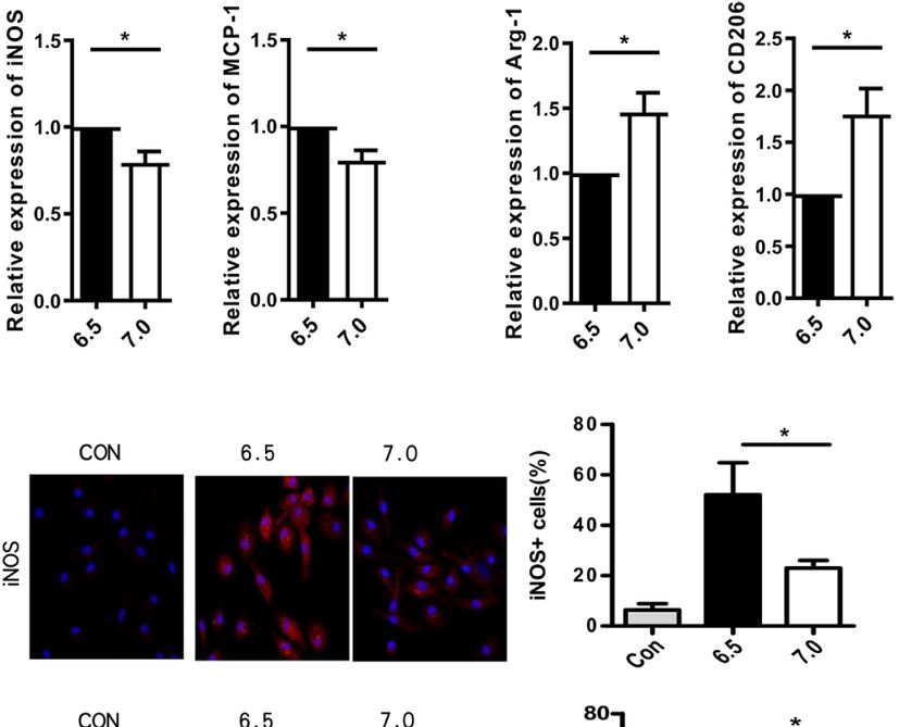
Ding et al.                                                                                                  Acidic Microenvironment Aggravates HIRI by PPAR-g

              A                                    B

              C

                                                                       D

 FIGURE 5 | PPAR-g agonist GW1929 suppressed M1 polarization induced by acidic microenvironment in vitro. Macrophages were isolated from B6 mice and
 GW1929 was added to enhance the expression of PPAR-g. M1 markers (iNOS and MCP-1) (A) and M2 markers (Arg-1 and CD206) (B) of gene induction were
 analyzed by quantitative RT-PCR after 6 h of acidic or normal condition treatment (one-way ANOVA with Bonferroni as a post hoc test, n=3). (C) Isolated
 macrophages from different experimental groups were cultured for 24 h, and TNF-a, IL-6, and IL-10 protein were measured in the culture supernatant by ELISA
 (one-way ANOVA with Bonferroni as a post hoc test, n=3). (D) Representative immunofluorescence staining images of iNOS and CD206 in macrophages (one-way
 ANOVA with Bonferroni as a post hoc test, n=3). *p < 0.05.

cellular reactions would decrease, including cytoplasmic- and                     protected HIRI through reverse the acidic microenvironment
membrane-related enzyme activities, ion transport activity,                       while liposome abolished the protective ability of NaHCO3
protein and DNA synthesis, and cAMP and calcium levels (21).                      through depleting the macrophages. In vivo and vitro
Previous researches have always focused on the effect of acid-base                experiment showed that acidic microenvironment markedly
environment on lymphocytes in tumors. It is interesting to find that               promoted M1-type polarization and inhibited M2-type
extracellular acidosis can both stimulate and suppress the activity of            polarization of macrophage. Furthermore, the mechanism
immune cells. For instance, while the lower PH promotes the                       study proved that the PPAR-g signal was suppressed during
expression of nitric oxide synthase in macrophages (22) and the                   the polarization of macrophages under PH=6.5 culture media.
activity of neutrophils (23), it also inhibits cytotoxic natural killer           The addition of PPAR-g agonist GW1929 inhibited M1
cells (24). Recently, Gan proved that acidic microenvironment                     polarization under acidic environment and reduced HIRI.
decreased Tregs and increased the severity of HIRI (11).                             PPAR-g is a type 2 nuclear receptor located on the human
Therefore, the extracellular acidic microenvironment may have                     chromosome, encoded by the PPARG gene. Initial studies have
different effects on macrophages.                                                 shown that this receptor has an important effect on fat cells and
    KC is the resident macrophages of the liver, accounting                       insulin, and is one of the research targets for the treatment of
approximately for 10-15% of all liver cells (25). KCs locate in the               diabetes (31, 32). At present, the study of PPAR-g on HIRI is not
liver sinusoids and engage in various liver diseases and injuries as              yet clear. Liver transplantation studies have shown that HIRI
the first-line defense against invading bacteria and virus. Many                   does not induce PPARg expression in the liver (33). However, in
studies have reported the role of KCs M1/M2 polarization in various               the mouse model of warm ischemia, the PPAR-g agonist
liver injuries (26). During HIRI, HO-1 suppressed M1 polarization                 pioglitazone reduces the accumulation of proinflammatory
and protects liver injury (27); Maspin regulated M1 polarization                  cytokines, chemokines and neutrophils to protect the liver
through activation of the NF-kB signal (28); besides, ATF6, ATF3                  injury. The liver damage was more severe in PPAR-g-KO mice
was also involved in HIRI and macrophage polarization (29, 30).                   generated HIRI models (34). Losartan (35), retinol-binding
    The results of the present study showed that HIRI induced                     protein 4 (36) have also been reported to reduce HIRI through
liver acidification which aggravated the liver injury, NaHCO3                      the PPARg pathway. Still, multiple studies have confirmed that

Frontiers in Immunology | www.frontiersin.org                                 7                                         June 2021 | Volume 12 | Article 697362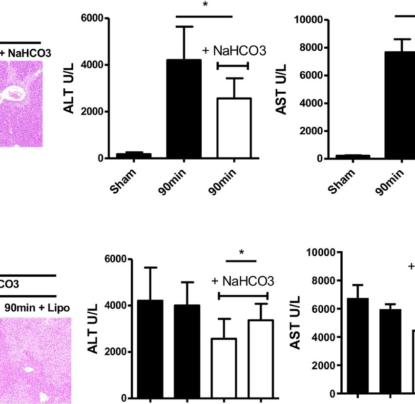
Ding et al.                                                                                                       Acidic Microenvironment Aggravates HIRI by PPAR-g

         A                                                                     B                                          C

         D

 FIGURE 6 | PPAR-g agonist GW1929 suppressed M1 polarization induced by Acidic microenvironment in vivo. (A) Representative images of liver injury in
 mice [hematoxylin and eosin (HE) staining, 200×] by light microscopy. Levels of ALT (B) and AST (C) in sham and IR groups (one-way ANOVA with
 Bonferroni as a post hoc test, n=3). (D) Representative immunofluorescence staining images of iNOS in macrophages (one-way ANOVA with Bonferroni as a post hoc test,
 n=3). *p < 0.05.

PPAR-g was involved in the regulation of macrophage function:                          AUTHOR CONTRIBUTIONS
PPARg activation inhibited the release of TNFa, IL-1, and IL-6
from macrophages (37, 38) and regulated polarization of                                Conception and design: WD, YD, and ZQ. Analysis and
macrophages to M1 (39).                                                                interpretation: WD and RZ. Data collection: WD and JF.
   In conclusion, the present study demonstrates that acidic                           Writing the article: WD, YD, ZQ, DS, XZ, and YL. Critical
microenvironment plays an important role in immune balance                             revision of the article: WD, YD, ZQ, DS, XZ, and YL. Final
modulation which directly leads to HIRI injury. PPAR-g signal                          approval of the article: WD, YD, ZQ, DS, XZ, and YL. Statistical
participated in the regulation effect of acidic microenvironment on                    analysis: WD, YL, and XL. Obtained funding: WD and YL.
macrophages. Our findings provided ideas for further confirming                          Overall responsibility: DS. All authors contributed to the article
the pathological relationship between HIRI and acidic                                  and approved the submitted version.
microenvironment and suggested that acidic microenvironment
should be considered as a potent target for HIRI.

                                                                                       FUNDING
DATA AVAILABILITY STATEMENT                                                            This work was supported by the National Natural Science Foundation
The data sets presented in this study can be found in online                           of China (81971504), Post-Doctoral Special Foundation of China
repositories. The names of the repository/repositories                                 (2020M670065ZX), Post-Doctoral Foundation of Jiangsu Province
and accession number(s) can be found in the article/                                   (2020Z021), Scientific and Technological Projects for Young Talents,
supplementary material.                                                                Changzhou Health and Family Planning Commission (QN201809),
                                                                                       Young Talent Development Plan of Changzhou Health Commission
                                                                                       (2020- 233), Young Talent Development Plan of Changzhou Health
                                                                                       Commission (CZQM2020118), the Development Foundation
ETHICS STATEMENT                                                                       of Affiliated Hospital of Xuzhou Medical University
                                                                                       (XYFY2020016), DAAD-K. C. WONG Funding (57501535),
The animal study was reviewed and approved by the institutional                        Changzhou Social Development Funding (CE20205038), and
animal care and use committee of Soochow University.                                   Lindau Follow-up Funding (GZ1600).

Frontiers in Immunology | www.frontiersin.org                                      8                                          June 2021 | Volume 12 | Article 697362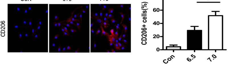
Ding et al.                                                                                                           Acidic Microenvironment Aggravates HIRI by PPAR-g

REFERENCES                                                                               20. Busa WB, Nuccitelli R. Metabolic Regulation Via Intracellular Ph. Am J
                                                                                             Physiol (1984) 246:30. doi: 10.1152/ajpregu.1984.246.4.R409
 1. Peralta C, Jimé nez-Castro MB, Gracia-Sancho J. Hepatic Ischemia and                21. Lardner A. The Effects of Extracellular Ph on Immune Function. J Leukoc Biol
     Reperfusion Injury: Effects on the Liver Sinusoidal Milieu. J Hepatol (2013)            (2001) 69(4):9. doi: 10.1189/jlb.69.4.522
     59(5):13. doi: 10.1016/j.jhep.2013.06.017                                           22. Bellocq A, Suberville S, Philippe C, Bertrand F, Perez J, Fouqueray B, et al. Low
 2. Faes S, Duval AP, Planche A, Uldry E, Santoro T, Pythoud C, et al. Acidic                Environmental Ph Is Responsible for the Induction of Nitric-Oxide Synthase
    Tumor Microenvironment Abrogates the Efficacy of mTORC1 Inhibitors.                       in Macrophages. Evidence for Involvement of Nuclear Factor-Kappab
    Mol Cancer (2016) 15(1):78. doi: 10.1186/s12943-016-0562-y                               Activation. J Biol Chem (1998) 273(8):7. doi: 10.1074/jbc.273.9.5086
 3. Devey LR, Friend PJ, Forsythe JLR, Mumford LL, Wigmore SJ. The Use of                23. Martı́nez D, Vermeulen M, Trevani A, Ceballos A, Sabatté J, Gamberale R,
    Marginal Heart Beating Donor Livers for Transplantation in the United Kingdom.           et al. Extracellular Acidosis Induces Neutrophil Activation by a Mechanism
    Transplantation (2007) 84(1):5. doi: 10.1097/01.tp.0000268072.04260.69                   Dependent on Activation of Phosphatidylinositol 3-Kinase/Akt and ERK
 4. Burne MJ, Daniels F, El Ghandour A, Mauiyyedi S, Colvin RB, O’Donnell MP,                Pathways. J Immunol (2006) 176(2):9. doi: 10.4049/jimmunol.176.2.1163
    et al. Identification of the CD4(+) T Cell as a Major Pathogenic Factor in            24. Fischer K, Hoffmann P, Voelkl S, Meidenbauer N, Ammer J, Edinger M, et al.
    Ischemic Acute Renal Failure. J Of Clin Invest (2001) 108(9):8. doi: 10.1172/            Inhibitory Effect of Tumor Cell-Derived Lactic Acid on Human T Cells. Blood
    JCI200112080                                                                             (2007) 109(9):8. doi: 10.1182/blood-2006-07-035972
 5. Li L, Huang L, Sung S-sJ, Lobo PI, Brown MG, Gregg RK, et al. NKT Cell               25. Li PZ, Li JZ, Li M, Gong JP, He K. An Efficient Method to Isolate and Culture
    Activation Mediates Neutrophil IFN-Gamma Production and Renal                            Mouse Kupffer Cells. Immunol Lett (2014) 158(1-2):52–6. doi: 10.1016/
    Ischemia-Reperfusion Injury. J Immunol (2007) 178(9):13. doi: 10.4049/                   j.imlet.2013.12.002
    jimmunol.178.9.5899                                                                  26. Rao Z, Sun J, Pan X, Chen Z, Sun H, Zhang P, et al. Hyperglycemia Aggravates
 6. Nakamura K, Zhang M, Kageyama S, Ke B, Fujii T, Sosa RA, et al.                          Hepatic Ischemia and Reperfusion Injury by Inhibiting Liver-Resident
     Macrophage Heme Oxygenase-1-SIRT1-p53 Axis Regulates Sterile                            Macrophage M2 Polarization Via C/EBP Homologous Protein-Mediated
     Inflammation in Liver Ischemia-Reperfusion Injury. J Hepatol (2017) 67                   Endoplasmic Reticulum Stress. Front Immunol (2017) 8:1299. doi: 10.3389/
     (6):11. doi: 10.1016/j.jhep.2017.08.010                                                 fimmu.2017.01299
 7. Ju C, Tacke F. Hepatic Macrophages in Homeostasis and Liver Diseases: From           27. Huang J, Shen XD, Yue S, Zhu J, Gao F, Zhai Y, et al. Adoptive Transfer of
    Pathogenesis to Novel Therapeutic Strategies. Cell Mol Immunol (2016) 13                 Heme Oxygenase-1 (HO-1)-Modified Macrophages Rescues the Nuclear
    (3):316–27. doi: 10.1038/cmi.2015.104                                                    Factor Erythroid 2-Related Factor (Nrf2) Antiinflammatory Phenotype in
 8. McDonald B, Kubes P. Innate Immune Cell Trafficking and Function During                   Liver Ischemia/Reperfusion Injury. Mol Med (2014) 14(14):20. doi: 10.2119/
    Sterile Inflammation of the Liver. Gastroenterology (2016) 151(6):1087–95.                molmed.2014.00103
    doi: 10.1053/j.gastro.2016.09.048                                                    28. Wang Y, Sun L, Song Z, Wang D, Bao Y, Li Y. Maspin Inhibits Macrophage
 9. Lawrence T, Natoli G. Transcriptional Regulation of Macrophage                           Phagocytosis and Enhances Inflammatory Cytokine Production Via
     Polarization: Enabling Diversity With Identity. Nat Rev Immunol (2011) 11               Activation of Nf-kb Signaling. Mol Immunol (2017) 82:10. doi: 10.1016/
     (11):750–61. doi: 10.1038/nri3088                                                       j.molimm.2016.12.021
10. Xiaojie G, Zhang R, Gu J, Ju Z, Wu X, Wang Q, et al. Acidic                          29. Rao J, Qian X, Li G, Pan X, Zhang C, Zhang F, et al. ATF3-Mediated NRF2/HO-1
     Microenvironment Regulates the Severity of Hepatic Ischemia/Reperfusion                 Signaling Regulates TLR4 Innate Immune Responses in Mouse Liver Ischemia/
     Injury by Modulating the Generation and Function of Tregs Via the PI3K-                 Reperfusion Injury. Am J Transplant (2015) 15(1):12. doi: 10.1111/ajt.12954
     mTOR Pathway. Front Immunol (2019) 10:2945. doi: 10.3389/                           30. Rao J, Yue S, Fu Y, Zhu J, Wang X, Busuttil RW, et al. ATF6 Mediates a Pro-
     fimmu.2019.02945                                                                         Inflammatory Synergy Between ER Stress and TLR Activation in the
11. Gan X, Zhang R, Gu J, Ju Z, Wu X, Wang Q, et al. Acidic Microenvironment                 Pathogenesis of Liver Ischemia-Reperfusion Injury. Am J Transplant (2014)
    Regulates the Severity of Hepatic Ischemia/Reperfusion Injury by Modulating              14(7):10. doi: 10.1111/ajt.12711
    the Generation and Function of Tregs Via the PI3K-mTOR Pathway. Front                31. Greene ME, Blumberg B, McBride OW, Yi HF, Kronquist K, Kwan K, et al.
    Immunol (2019) 10:2945. doi: 10.3389/fimmu.2019.02945                                     Isolation of the Human Peroxisome Proliferator Activated Receptor Gamma
12. Kato A, Yoshidome H, Edwards MJ, Lentsch AB. Reduced Hepatic Ischemia/                   cDNA: Expression in Hematopoietic Cells and Chromosomal Mapping. Gene
    Reperfusion Injury by IL-4: Potential Anti-Inflammatory Role of STAT6.                    Expr (1995) 4(4-5):19.
    Inflammation Res (2000) 49(6):5. doi: 10.1007/PL00000207                              32. Michalik L, Auwerx J, Berger JP, Chatterjee VK, Glass CK, Gonzalez FJ, et al.
13. Tsuchihashi S, Zhai Y, Fondevila C, Busuttil RW, Kupiec-Weglinski JW. Ho-1               International Union of Pharmacology. Lxi. Peroxisome Proliferator-Activated
    Upregulation Suppresses Type 1 IFN Pathway in Hepatic Ischemia/                          Receptors. Pharmacol Rev (2006) 58(4):16. doi: 10.1124/pr.58.4.5
    Reperfusion Injury. Transplant Proc (2005) 37(4):2. doi: 10.1016/                    33. Casillas-Ramı́rez A, Alfany-Ferná ndez I, Massip-Salcedo M, Emı́lia Juan M,
    j.transproceed.2005.03.080                                                               Planas JM, Serafı́n A, et al. Retinol-Binding Protein 4 and Peroxisome
14. Wu C, Xia Y, Wang P, Lu L, Zhang F. Triptolide Protects Mice From                        Proliferator-Activated Receptor-g in Steatotic Liver Transplantation.
    Ischemia/Reperfusion Injury by Inhibition of IL-17 Production. Int                       J Pharmacol Exp Ther (2011) 338(1):11. doi: 10.1124/jpet.110.177691
    Immunopharmacol (2011) 11(10):9. doi: 10.1016/j.intimp.2011.05.015                   34. Kuboki S, Shin T, Huber N, Eismann T, Galloway E, Schuster R, et al. Peroxisome
15. Faes S, Duval AP, Planche A, Uldry E, Santoro T, Pythoud C, et al. Acidic                Proliferator-Activated Receptor-g Protects Against Hepatic Ischemia/Reperfusion
    Tumor Microenvironment Abrogates the Efficacy of mTORC1 Inhibitors. Mol                   Injury in Mice. Hepatology (2008) 47(1):10. doi: 10.1002/hep.21963
    Cancer (2016) 15(1):78. doi: 10.1186/s12943-016-0562-y                               35. Koh E-J, Yoon S-J, Lee S-M. Losartan Protects Liver Against Ischaemia/
16. Wu LL, Peng W, Wu HL, Miaw SC, Yeh SH, Yang HC, et al. Lymphocyte                        Reperfusion Injury Through Ppar-g Activation and Receptor for Advanced
    Antigen 6 Complex, Locus C+ Monocytes and Kupffer Cells Orchestrate Liver                Glycation End-Products Down-Regulation. Br J Pharmacol (2013) 169(6):13.
    Immune Responses Against Hepatitis B Virus in Mice. Hepatology (2019) 69                 doi: 10.1111/bph.12229
    (6):17. doi: 10.1002/hep.30510                                                       36. Casillas-Ramı́rez A, Alfany-Ferná ndez I, Massip-Salcedo M, Juan ME, Planas
17. Byles V, Covarrubias AJ, Ben-Sahra I, Lamming DW, Sabatini DM, Manning                   JM, Serafı́n A, et al. Retinol-Binding Protein 4 and Peroxisome Proliferator-
    BD, et al. The TSC-mTOR Pathway Regulates Macrophage Polarization. Nat                   Activated Receptor-g in Steatotic Liver Transplantation. J Pharmacol Exp Ther
    Commun (2013) 4:2834. doi: 10.1038/ncomms3834                                            (2011) 338(1):11. doi: 10.1124/jpet.110.177691
18. Luo W, Xu Q, Wang Q, Wu H, Hua J. Effect of Modulation of PPAR-g Activity            37. von Knethen A, Brüne B. Pparg: An Important Regulator of Monocyte/
    on Kupffer Cells M1/M2 Polarization in the Development of Non-Alcoholic                  Macrophage Function. Arch Immunol Ther Exp (Warsz) (2003) 51(4):8.
    Fatty Liver Disease. Sci Rep (2017) 16(7):44612. doi: 10.1038/srep44612                  doi: 10.1034/j.1600-0463.2003.11101301.x
19. Zizzo G, Cohen PL. The PPAR-g Antagonist GW9662 Elicits Differentiation              38. Jiang C, Ting AT, Seed B. Ppar-g Agonists Inhibit Production of Monocyte
    of M2c-Like Cells and Upregulation of the MerTK/Gas6 Axis: A Key Role for                Inflammatory Cytokines. Nature (1998) 391(6662):5. doi: 10.1038/34184
    PPAR-g in Human Macrophage Polarization. J Inflamm (Lond) (2015) 3                    39. Daniel B, Nagy G, Czimmerer Z, Horvath A, Hammers DW, Cuaranta-
    (12):46. doi: 10.1186/s12950-015-0081-4                                                  Monroy I, et al. The Nuclear Receptor Pparg Controls Progressive

Frontiers in Immunology | www.frontiersin.org                                        9                                            June 2021 | Volume 12 | Article 697362
Ding et al. Acidic Microenvironment Aggravates HIRI by PPAR-g
Macrophage Polarization as a Ligand-Insensitive Epigenomic Ratchet of Copyright © 2021 Ding, Duan, Qu, Feng, Zhang, Li, Sun, Zhang and Lu. This is an
Transcriptional Memory. Immunity (2018) 49(4):12. doi: 10.1016/ open-access article distributed under the terms of the Creative Commons Attribution
j.immuni.2018.09.005 License (CC BY). The use, distribution or reproduction in other forums is permitted,
provided the original author(s) and the copyright owner(s) are credited and that the
Conflict of Interest: The authors declare that the research was conducted in the original publication in this journal is cited, in accordance with accepted academic
absence of any commercial or financial relationships that could be construed as a practice. No use, distribution or reproduction is permitted which does not comply with
potential conflict of interest. these terms.
Frontiers in Immunology | www.frontiersin.org 10 June 2021 | Volume 12 | Article 697362You can also read