Abdominal Wall Endometrioma: A Rare Case Report
←
→
Page content transcription
If your browser does not render page correctly, please read the page content below
CASE REPORT

Abdominal Wall Endometrioma: A Rare Case Report
Niranjan N Chavan1, Divita A Kamble2, Deepali Kapote3, Ashwini Sakhalkar4, Madhura Pradhan5, Sonam Simpatwar6

 A b s t r ac t
 Abdominal wall endometrioma (AWE) being an unusual phenomenon is a benign tumor defined as ectopic functional, endometrial tissue located
 in the abdominal wall. Abdominal wall endometrioma is a rare sequela to gynecologic surgeries such as cesarean section, tubal ligation, and
 hysterectomies. The incidence varies from 1 to 2%. It presents as intense pain and discomfort to the patient with a seemingly non discernible
 cause. Awareness of this entity can help the surgeon to make an early diagnosis and deliver prompt treatment, usually surgical. We present a
 case of AWE, along with a brief review of literature.
 Keywords: Clinical profile, Complications, Endometriosis, Surgical site.
 Journal of South Asian Federation of Obstetrics and Gynaecology (2022): 10.5005/jp-journals-10006-2038

Introduction                                                                        1–6
                                                                                       Department of Obstetrics and Gynaecology, Lokmanya Tilak
Abdominal wall endometrioma, an unusual phenomenon, is a
                                                                                    Municipal Medical College and Hospital, Mumbai, Maharashtra, India
benign tumor defined as functional endometrial ectopic tissue
located in the abdominal wall.                                                      Corresponding Author: Divita A Kamble, Department of Obstetrics
                                                                                    and Gynaecology, Lokmanya Tilak Municipal Medical College and
     Abdominal wall endometrioma is a seldom seen sequel to
                                                                                    Hospital, Mumbai, Maharashtra, India, Phone: +91 9657797555, e-mail:
pelvic surgeries such as cesarean section, tubal ligation, and
                                                                                    divita1896@gmail.com
hysterectomies. The incidence varies from 0.1 to 0.4%.1 It presents
                                                                                    How to cite this article: Chavan NN, Kamble DA, Kapote D, et al.
as an extreme pain and distress to the patient with a seemingly
                                                                                    Abdominal Wall Endometrioma: A Rare Case Report. J South Asian
nondiscernible cause. Quicker diagnostic rates and thereby timely
                                                                                    Feder Obst Gynae 2022;14(2):202–204.
treatment (usually surgical) can be provided to patients if surgeons
remain more cognizant of this entity.                                               Source of support: Nil
     We present a case of AWE, along with a brief review of the                     Conflict of interest: None
literature.

Case Report
A 44-year-old P3L3 with all vaginal deliveries and one MTP followed
by puerperal tubal ligation 18 years ago with posthysterectomy
status presented with intense cyclical pain in the abdomen for
1 year. The patient had no history of fever, abdominal bloating
or mass, urinary or bowel complaints. She underwent a total
abdominal hysterectomy 15 years ago after which she started
experiencing cyclical pain localized to the left edge of the
Pfannenstiel scar. The pain was cyclical which later progressed to
continuous type, radiating to the left thigh. The pain had increased
in intensity over the past year.
      Her previous menses were regular lasting for 3–4 days with
normal flow, soaking 2–3 pads per day associated with severe
dysmenorrhea and passage of clots. All routine investigations were
within normal limits. Local examination done revealed the presence
of a firm, mobile, extremely tender nodule, approximately 1 × 1 cm                  Fig. 1: Ultrasonography findings of 13 × 7 mm endometrioma in
on the left edge of the Pfannenstiel scar with normal overlying                     abdominal wall above rectus muscle at left border of rectus abdominis
skin. No other palpable abdominal mass was felt. Ultrasonography
of the abdomen and pelvis was done which revealed a well-                           mass was seen between the rectus health and muscle adherent to
defined hyperechoic structure in the left iliac fossa region in the                 the rectus muscle below. No extension was seen into the rectus
intramuscular plane measuring 13 × 11 × 7 mm showing minimal                        muscle (Fig. 2).
peripheral vascularity—highly suggestive of an AWE. Bilateral                           Wide local excision of the mass was done with 1-cm tumor-free
ovaries were normal (Fig. 1).                                                       margin (Fig. 3).
      A 3-cm long transverse incision was taken at the left edge of                     The sample was sent for histopathological examination.
the Pfannenstiel scar and layers of the abdominal wall were opened                  Postoperatively, the patient experienced complete resolution of
till the rectus muscle. Intraoperatively, a 1 × 1 × 1 cm firm irregular             symptoms, and the course in the ward was uneventful.

© The Author(s). 2022 Open Access This article is distributed under the terms of the Creative Commons Attribution 4.0 International License (https://creativecommons.
org/licenses/by-nc/4.0/), which permits unrestricted use, distribution, and non-commercial reproduction in any medium, provided you give appropriate credit to
the original author(s) and the source, provide a link to the Creative Commons license, and indicate if changes were made. The Creative Commons Public Domain
Dedication waiver (http://creativecommons.org/publicdomain/zero/1.0/) applies to the data made available in this article, unless otherwise stated.
Abdominal Wall Endometrioma
Histopathological examination revealed fibroelastic tissue with reproductive age as the growth of endometriomas depends on
endometrial glands and stroma. estrogen. The predominant location of endometriosis was the
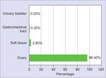
Glandular endometrial tissue and stromal tissue if and when ovaries (96.4%), followed by the soft tissue (2.8%), gastrointestinal
present outside the uterus are known as endometriosis. When tract (0.3%), and urinary tract (0.2%). 3
the lesion occurs as a well-circumscribed mass it is referred to as Fragments of endometrial tissue are inserted into the surgical
an endometrioma.2 It is commonly observed in women of active site during surgery leading to endometriotic implantation (Fig. 4).
The typical surgical procedures that give rise to AWE are abdominal
hysterectomies and cesarean sections. Several hypotheses have
been developed to analyze the causes and development of AWE.
The transport hypothesis states that categorical injection or transfer
of endometrial tissue into surgical incisions or nearby tissues in the
course of surgery is responsible for endometriosis of the abdominal
wall. The metaplastic hypothesis states that primitive pluripotent
mesenchymal cells have been isolated and metaplasia can give rise
to endometrioma of the abdominal wall. Prior obstetric surgery or
hysterectomy, grand multiparity, and menorrhagia are definitive
risk factors for AWEs. The Esquivel triad, which includes a palpable
tumor, cyclical pain, and a record of prior pelvic surgery all but
confirms a diagnosis of AWE.2 Minor differences in clinical features
may be noticeable and therefore a detailed history regarding
the timing of surgical events and the onset of symptoms is very
important. Typically, the gap between primary surgery and the
occurrence of endometriotic lesion-associated symptoms averages
around 3–6 years. Our case was presented for almost 6 years.
Fig. 2: Wide local excision of AWE Other criteria of the Esquivel triad were fulfilled in our case as
Fig. 3: AWE seen just below the rectus sheath
Journal of South Asian Federation of Obstetrics and Gynaecology, Volume 14 Issue 2 (March–April 2022) 203Abdominal Wall Endometrioma
of symptoms and so surgery remains a pillar of management in
most cases. The clinical features of AWE are usually severe pain,
pain, bleeding, dysmenorrhea, and dyspareunia. Most patients
can develop asymptomatic with an unpleasant and painless tumor
(96%). The appearance of lesion externally is based on which layer
in the abdominal wall the endometrioma is deposited: it could be
an invisible, painful mass if the lesion is deep-seated in the rectus
sheath, as was seen in our case, or, if the deposit is more superficial,
in the subcutaneous tissue or fat, it will be seen as a dark or black
nodule on the skin at the scar site. Pain, cyclical, or noncyclical are
present in most of the patients with the condition. Indications for
incomplete resuscitation are the seroma formation at the surgical
location and the onset of pain localized to the prior surgical site, not
responding to medical management. Clinical suspicion of AWE is
essential to develop prevention protocol during index surgery and
arrive at an accurate and early diagnosis when the condition occurs.
Fig. 4: Common sites of pelvic endometrioma
C o n c lu s i o n
well, which along with imaging reports ultimately lead us to Abdominal wall endometrioma is an entity usually occurring after
arrive at the correct diagnosis. Clinical suspicion is a prerequisite, a cesarean section or any surgery involving the opening of the
however, for further plan of management to move in the correct endometrial cavity. It is extremely rare after a hysterectomy. It is,
direction. A variety of thought patterns are available to validate however, essential that surgeons are more aware of the possibility
AWE diagnoses. As for imaging studies to confirm the diagnosis, of the development of endometriomas in the late postoperative
several tools are available. USG of the abdomen plus pelvis and period of obstetrical and gynecological surgeries and take the
a contrast-enhanced computed tomography (CECT) performed required measures to prevent it (high-pressure saline irrigation
during menstruation can be diagnostic as demonstrated in our case. of the wound edges). 2 In case prevention fails and an AWE is
Ultrasound and CECT, however, rank poorly when compared to MRI. suspected, further management would involve getting a USG or
Essentially, MRI is preferred as the final confirmatory study before CECT to confirm the need for surgery, and MRI to assist in precise
undertaking surgical resection as it provides a highly detailed surgical excision. Surgical excision is the gold standard of treatment
analysis of the resectable mass regarding the depth of invasion as it is the only management that ensures a zero recurrence of
into surrounding and underlying tissues. It also helps localize and symptoms can prevent the development of AWE at the time of the
delineate smaller endometriomas than which could be spotted on index surgery. Sclerotherapy uses ultrasound-guided intralesional
a USG and will indicate any bleeders that may be present around ethanol injection for AWE. Compared with the complications of
said endometrioma. Another, more invasive method of confirming surgical excision, the complications of sclerotherapy by ethanol
the diagnosis would be fine needle aspiration cytology (FNAC) to are at a more acceptable level. More recently, sclerotherapy by
demonstrate epithelial cells such as endometrial, stromal cells, and ethanol injection has been developed as an alternative treatment
macrophages filled with hemosiderin that would establish that it is to surgery for AWE.3 More studies on preventive strategies would
indeed an endometrioma and not one of the differential diagnosis further help in reducing the occurrence of AWE.
commonly presenting with a similar pattern of symptoms such as a
small abdominal wall abscess, lipoma, hematoma, sebaceous cyst, References
suture granuloma, hernia, lymphoma, and carcinoma.4 However, 1. Bozkurt M, Çil AS, Bozkurt DK. Intramuscular abdominal wall
there is the possibility that it could lead to the injection of the endometriosis treated by ultrasound-guided ethanol injection. Clin
endometriotic material along the tract created by the aspiration Med Res 2014;12(3–4):160–165. DOI: 10.3121/cmr.2013.1183.
cytology needle and thereby defeat the purpose of the study and 2. Vagholkar K, Vagholkar S. Abdominal wall endometrioma: a
in fact exacerbate symptoms. Fine needle aspiration cytology will diagnostic enigma–a case report and review of the literature. Case
show. Histopathological examination of resuscitated mass helps Rep Obstet Gynecol 2019;2019:6831545. DOI: 10.1155/2019/6831545.
to confirm a temporary diagnosis. Therefore, a detailed history 3. Leite GKC, Carvalho LFP de, Korkes H, et al. Scar endometrioma
following obstetric surgical incisions: retrospective study on 33 cases
with a positive clinical association of USG or MRI findings can
and review of the literature. Sao Paulo Med J 2009;127(5):270–277.
help to make an accurate preoperative diagnosis. The therapeutic DOI: 10.1590/s1516-31802009000500005.
regimens used by AWE include progesterone containing oral 4. Esquivel-Estrada V, Briones-Garduño JC, Mondragón-Ballesteros R.
contraceptives, danazol, and gonadotropic agonists such as Implante de endometriosis en cicatriz de operación cesárea
leuprolide acetate. However, it has been seen that medical [Endometriosis implant in cesarean section surgical scar. Cir Ciruj
therapy cannot ensure a complete resolution and nonrecurrence 2004;72(2):113–115]. PMID: 15175127.
204 Journal of South Asian Federation of Obstetrics and Gynaecology, Volume 14 Issue 2 (March–April 2022)You can also read


