A Reference Database of Standardised Continuous Lumbar Intervertebral Motion Analysis for Conducting Patient-Specific Comparisons
←
→
Page content transcription
If your browser does not render page correctly, please read the page content below
ORIGINAL RESEARCH
                                                                                                                                         published: 27 September 2021
                                                                                                                                        doi: 10.3389/fbioe.2021.745837

                                           A Reference Database of
                                           Standardised Continuous Lumbar
                                           Intervertebral Motion Analysis for
                                           Conducting Patient-Specific
                                           Comparisons
                                           Alexander Breen 1, Diana De Carvalho 2, Martha Funabashi 3,4, Greg Kawchuk 5,
                                           Isabelle Pagé 4, Arnold Y. L. Wong 6 and Alan Breen 1,7*
                                           1
                                            AECC University College, Bournemouth, United Kingdom, 2Division of Community Health and Humanities, Faculty of Medicine,
                                           Memorial University of Newfoundland, St. John’s, NL, Canada, 3Division of Research and Innovation, Canadian Memorial
                                           Chiropractic College, Toronto, ON, Canada, 4Département de chiropratique, Université du Québec à Trois-Rivières, Trois-
                                           Rivières, QC, Canada, 5Department of Physical Therapy, Faculty of Rehabilitation Medicine, University of Alberta, Edmonton, AB,
                                           Canada, 6Department of Rehabilitation Sciences, The Hong Kong Polytechnic University, Hong Kong, SAR China, 7Faculty of
                                           Science and Technology, Bournemouth University, Poole, United Kingdom

                          Edited by:
                     Babak Bazrgari,       Lumbar instability has long been thought of as the failure of lumbar vertebrae to maintain
University of Kentucky, United States      their normal patterns of displacement. However, it is unknown what these patterns consist
                       Reviewed by:        of. Research using quantitative fluoroscopy (QF) has shown that continuous lumbar
                       Navid Arjmand,
 Sharif University of Technology, Iran
                                           intervertebral patterns of rotational displacement can be reliably measured during
           Ameet Krishnan Aiyangar,        standing flexion and return motion using standardised protocols and can be used to
       Swiss Federal Laboratories for
                                           assess patients with suspected lumbar spine motion disorders. However, normative
  Materials Science and Technology,
                          Switzerland      values are needed to make individualised comparisons. One hundred and thirty-one
                                           healthy asymptomatic participants were recruited and performed guided flexion and return
                  *Correspondence:
                          Alan Breen       motion by following the rotating arm of an upright motion frame. Fluoroscopic image
                breenalan@gmail.com        acquisition at 15fps was performed and individual intervertebral levels from L2-3 to L5-S1
                                           were tracked and analysed during separate outward flexion and return phases. Results
                    Specialty section:
         This article was submitted to
                                           were presented as proportional intervertebral motion representing these phases using
                        Biomechanics,      continuous means and 95%CIs, followed by verification of the differences between levels
                a section of the journal
                                           using Statistical Parametric Mapping (SPM). A secondary analysis of 8 control participants
       Frontiers in Bioengineering and
                         Biotechnology     matched to 8 patients with chronic, non-specific low back pain (CNSLBP) was performed
            Received: 22 July 2021         for comparison. One hundred and twenty-seven asymptomatic participants’ data were
     Accepted: 08 September 2021           analysed. Their ages ranged from 18 to 70 years (mean 38.6) with mean body mass index
     Published: 27 September 2021
                                           23.8 kg/m2 48.8% were female. Both the flexion and return phases for each level
                            Citation:
Breen A, De Carvalho D, Funabashi M,
                                           evidenced continuous change in mean proportional motion share, with narrow
 Kawchuk G, Pagé I, Wong AYL and           confidence intervals, highly significant differences and discrete motion paths between
Breen A (2021) A Reference Database
                                           levels as confirmed by SPM. Patients in the secondary analysis evidenced significantly less
 of Standardised Continuous Lumbar
     Intervertebral Motion Analysis for    L5-S1 motion than controls (p < 0.05). A reference database of spinal displacement
                   Conducting Patient-     patterns during lumbar (L2-S1) intersegmental flexion and return motion using a
                Specific Comparisons.
Front. Bioeng. Biotechnol. 9:745837.
                                           standardised motion protocol using fluoroscopy is presented. Spinal displacement
     doi: 10.3389/fbioe.2021.745837        patterns in asymptomatic individuals were found to be distinctive and consistent for

Frontiers in Bioengineering and Biotechnology | www.frontiersin.org                1                                        September 2021 | Volume 9 | Article 745837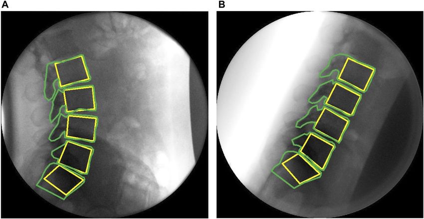
Breen et al.                                                                                                       Intervertebral Motion Sharing Database

                                           each intervertebral level, and to continuously change during bending and return. This
                                           database may be used to allow continuous intervertebral kinematics to drive dynamic
                                           models of joint and muscular forces as well as reference values against which to make
                                           patient-specific comparisons in suspected cases of lumbar spine motion disorders.
                                           Keywords: back pain, videofluoroscopy, lumbar spine, intervertebral motion, kinematics, reference database,
                                           instability

INTRODUCTION                                                                    been questioned as to whether this accurately represents
                                                                                vertebral orientation, for example, during dynamic bending
Pathological spinal motion, or lumbar instability, has long been                tasks (Nagel et al., 2014; Aiyangar et al., 2015).
thought of as the failure of the lumbar spine to maintain its                      In addition, “Despite RoM being a simple metric that could be
normal pattern of displacement (Panjabi, 1992). However, it is                  easily estimated within a clinical setting, it does not convey the
currently unclear what this normal pattern actually consists of, as             contribution over time of the related segments/joints to the
the motion segments of the spine are sited deep within the body,                movement performed, compensatory actions nor the
making them practically impervious to objective biomechanical                   movement variability, thus limiting our understanding of
measurement in living people. This tends to deny clinicians the                 movement strategies” (Papi et al., 2018). However, lumbar
tools to investigate relationships between symptoms and intrinsic               segmental contributions to motion, sometimes referred to as
biomechanics and constrains the identification of biomechanical                  “spinal rhythms”, have been demonstrated to change during
markers for spinal pain. Given that the spine has a complex                     simple tasks such as controlled flexion and return motion,
dynamic role in the normal activities of daily living, recent                   (Aiyangar et al., 2015; Breen and Breen, 2020), and even
proposals for future directions in spine biomechanics research                  during passive movement, where there is no measurable
have included the recommendation that “The dynamic properties                   muscle activation (Breen and Breen, 2018). As such, physical
of the (functional spinal unit) FSU . . . should be a focus of future           and computational models that are validated using only end
research efforts as they are likely very relevant to the in vivo                range of motion data may not accurately reproduce dynamic in
situation.” (Oxland, 2016). As non-invasive, in vivo measurement                vivo motion. Indeed, this may be one of the major causes of the
of the dynamic properties of the FSU generally requires imaging,                large differences found in inter-joint and muscle forces when
precision imaging measurement of in vivo segmental spine                        comparing models driven by generic patterns of rotational
dynamics is critical for gaining an understanding of spine                      displacement in the lumbar spine and those based on
biomechanics that could be applied in patient-specific                           kinematics acquired from dynamic imaging techniques
assessments.                                                                    (Eskandari et al., 2017; Byrne et al., 2020).
    Spine biomechanics also increasingly involves biomechanical                    With advancements in imaging and object tracking
modelling, where “the importance of verification, validation and                 technologies, continuous assessment of intersegmental spine
sensitivity testing in computational studies within the field of                 motion during bending using quantitative fluoroscopy (QF)
biomechanical engineering” has been highlighted (Jones and                      has been demonstrated to be relatively accurate and repeatable
Wilcox, 2008). These models are sometimes utilized to                           (Breen et al., 2019b). Thus, using QF for inter-image vertebral
estimate muscle and inter-joint forces within the lumbar spine,                 body tracking to quantify spine motion has allowed continuous
as they provide a relatively inexpensive and efficient method to                 intervertebral lumbar motion measurement in-vivo. However, the
estimate specific characteristics that are not otherwise possible or             precision (and therefore the application) of dynamic models that
practical to measure in-vivo. However, while there are studies that             integrate anthropometric and kinematic data will be limited if
provide in vivo information about intradiscal pressures, forces,                there is uncontrolled variation in subjects’ motion behaviour
and moments transmitted via instrumented vertebral implants,                    (Magee, 2015).
there is a lack of reference information with respect to multilevel                In previous work using QF, where both the motion task and
continuous intervertebral motion for use in dynamically                         the analysis were highly standardised for range and velocity, some
modelling loads (Dreischarf et al., 2016).                                      intervertebral motion sharing characteristics in the lumbar spine
    Although thorax and pelvis kinematics, used to drive such                   were found to be significantly different in chronic, nonspecific
models, have often been measured using skin-based motion                        back pain (CNSLBP) patients compared with asymptomatic
capture, the inherent errors associated with the proper                         controls, indicating their eligibility to be considered as pain
identification of underlying bony landmarks mean that skin-                      biomarkers (Breen and Breen, 2018; Breen and Breen, 2020).
based tracking is rarely used for measuring the motion of                       As some of these measurements were found to be relatively stable
individual vertebrae (Eskandari et al., 2017). Instead, the                     over 6 weeks in an asymptomatic population, this made these
kinematics of the lumbar vertebrae are often approximated                       measures potentially suitable for use in outcome and prognostic
from their segmental contributions to flexion motion based on                    studies. This, however, highlights the need for a reference
static end-of-range radiographs. These contributions are then                   database of normal values against which individuals could be
applied to the measured kinematics of the thorax-pelvis to                      compared (Breen et al., 2019a; Breen et al., 2019b). As the
estimate joint motion in the lumbar spine. However, it has                      differences between patients and controls found in these

Frontiers in Bioengineering and Biotechnology | www.frontiersin.org         2                                 September 2021 | Volume 9 | Article 745837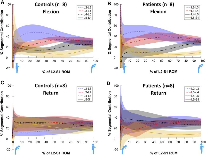
Breen et al.                                                                                                  Intervertebral Motion Sharing Database

studies were in terms of continuous proportional motion sharing               For the Patient-control subgroup study, 8 patients without any
parameters, it was decided to formulate a normative Reference              obvious mechanical disruption (for example surgery or
Database of these as information against which patient-specific             spondylolisthesis), who had been referred for QF imaging to
comparisons could be made.                                                 investigate CNSLBP using the same imaging protocol, were
   The present study therefore aimed to create a normative set of          recruited. Their imaging results were compared to those of 8 of
values for flexion and return dynamic lumbar segmental rotational           the asymptomatic controls, following written informed consent to
contributions from a sizeable population base that could be used to        inclusion on the study. The controls were chosen from the
drive future models. To support future patient-specific comparative         Reference Database as being of similar age, sex and BMI to the
studies and inform such musculoskeletal models, the project aimed to       patients. Their demographic information is shown in Table 1.
employ a standardised protocol, rather than a free-bending one, and
to identify the intersegmental contributions to motion from L2-S1          Reference Database Study Sample Size
during weight bearing flexion and return in asymptomatic                    The design criterion for determining the sample size needed to
individuals.                                                               establish a credible 95% reference interval (RI) is the ratio of the
   Given that more recent studies have focused on the return               confidence interval (CI) width on the RI cut-point to the RI
paths of lumbar flexion separately, to support dynamic loading              width. Practical values for this ratio suggested by Linnet range
models during lifting (Aiyangar et al., 2015; Pavlova et al., 2018),       from 0.1 to 0.3 (Linnet, 1987). Using a conservative ratio of 0.15,
the motion was separated into the flexion phase, and the return to          with a 90% cut-off CI and a single 95% upper RI cut-point, we
neutral phase for analysis. In addition, as proportional motion            required 134 participants (SSS software v.1, Wiley-Blackwell,
has been found to discriminate patients and controls in the past           Chichester United Kingdom). To allow for tracking failure in
(Breen and Breen, 2018; Breen et al., 2018; Breen and Breen,               approximately 10% of sequences, we rounded the sample size up
2020) but has not yet been analysed across the time series, this           to 148. However, assuming a non-Gaussian distribution for at
analysis protocol was also applied in a further secondary analysis         least some of the reference data, we employed the non-parametric
of a matched Patient-control subgroup.                                     RI methodology recommended in the Clinical Laboratory
                                                                           Standards Institute guidelines, for which the minimum
                                                                           recommended sample size is 120 (CLSI, 2008). This was
MATERIALS AND METHODS                                                      therefore selected as the minimum population for the
                                                                           Reference Database study.
The methods used for image acquisition and analysis in this project
were agreed by an international forum of QF users in 2009 (Breen           Data Collection
et al., 2012), and applied in the present Reference Database study.        The QF protocol for image acquisition and analysis procedures as
The participants in the Forum were the only four groups of QF              been detailed in previous studies (Breen et al., 2012; Breen and
users known to the authors in 2009, who all employed automated             Breen., 2018; du Rose et al., 2018; Breen and Breen, 2020). In brief
image registration and/or tracking for extracting vertebral                however, participants were guided through a standard active
kinematics data and used well documented data collection                   weight-bearing flexion and return motion task. This was
protocols. The focus of the Forum was to agree a standard                  designed to reduce behavioural variations in participant
protocol for data collection and analysis that could be employed           bending, while controlling the speed and range of motion in a
efficiently for investigating and comparing symptomatic and                 reproducible way. During this controlled motion, low dose
asymptomatic participants for clinical investigations and research.        fluoroscopic recordings of L2-S1 levels during continuous
                                                                           motion were acquired using a Siemens Arcadis Avantic digital
Participants                                                               C-arm fluoroscope (Siemens GMBH) at 15 frames per second. To
A convenience sample of 131 asymptomatic volunteer                         achieve this, participants stood with their right-hand side next to
participants was recruited to the Reference Database study                 a motion testing platform (Atlas Clinical Ltd. Lichfield,
from staff, students and visitors at the AECC University                   United Kingdom), which guided them through a 60° bending
College (Bournemouth, United Kingdom) between July 2011                    arc at 6°/s during both flexion and return phases (Figure 1).
and July 2020. Participants were included based on the                     Participants were positioned in a comfortable upright stance with
following inclusion criteria: between 21 and 80 years old, self-           the centre of rotation of the motion platform in line with the disc
reported body mass index less than 30 kg/m2 (to ensure image               space between the third and fourth lumbar vertebrae (This
quality), free of pain on the day of testing, free of any back pain        position was confirmed by single short pulse fluoroscopic
that limited normal activity for more than 1 day in the previous           images and the use of radiopaque markers temporarily aligned
year, no history of abdominal surgery or spondylolisthesis, no             with the platform’s centre of rotation.) A sacral brace and a belt
medical radiation exposure of >8 mSv in the previous 2 years               around the hips of participants were used to minimise pelvic
(self-identified by pre-study questionnaire detailing recent                motion and keep the spine in the field of view throughout the
medical imaging), and not currently pregnant. Ethical                      bending sequence. This was to ensure the best field of view for all
approval was obtained from the United Kingdom National                     the segments to be conveniently imaged throughout the whole
Research Ethics Service (SouthWest 3, 10/H0106/65) and                     range of motion.
written Informed consent was obtained from all participants                   Before the acquisition of the QF images, participants
prior to inclusion in the study.                                           undertook 3 practice bends. These standing movements,

Frontiers in Bioengineering and Biotechnology | www.frontiersin.org    3                                 September 2021 | Volume 9 | Article 745837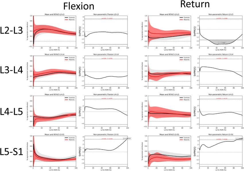
Breen et al. Intervertebral Motion Sharing Database
TABLE 1 | Participant characteristics (mean and SD).
Reference database Subgroup study
Controls Patients 2-tailed p
N 127 8 8
Females (%) 62 (48.8) 3 (38.8) 3 (38.8) 0.99
Age (years) 38.6 (13.8) 48.1 (13.4) 48.8 (14.4) 0.93
Height (m) 1.73 (0.09) 1.70 (0.1) 1.70 (0.1) 0.71
Weight (kg) 71.6 (12.7) 74.5 (12.7 75.4 (10.5) 0.41
BMI 23.8 (2.9) 25.8 (6.5) 25.3 (5.3) 0.42
SD: standard deviation; m: meters; kg: kilograms; BMI: body mass index.
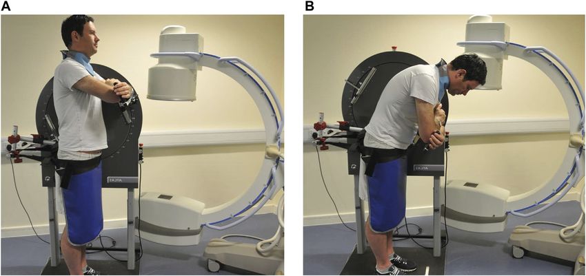
FIGURE 1 | Motion protocol used for fluoroscopic image acquisition (Courtesy Atlas Clinical Ltd., Lichfield, United Kingdom) (A) upright (B) flexed.
bending to 20° flexion and return, were followed by 40-degree and the L2-S1 angle was normalized to a percentage of its range of
60-degree bends. This ensured that participants could perform motion (RoM). Thus, during the flexion phase, standing was
their recorded motion confidently and smoothly. defined as 0% RoM and maximum flexion as 100% RoM, while in
the return phase, 0% RoM was defined as maximum flexion and
Intervertebral Motion Analysis 100% RoM as being returned to the original reference position.
A previously validated semi-automated tracking process was used Changes in intervertebral angles were then interpolated to
to determine the position of each vertebra (L2, L3, L4, L5 and S1) obtain each intervertebral motion segment’s rotation for every 1%
within each image recorded during the flexion and return trials increment of the L2–S1 RoM. The segmental contribution of each
(Breen et al., 2012). This process has been shown to have an intervertebral level as a percentage of the change in L2–S1 angle
accuracy for measuring intervertebral RoM of 0.52°, (du Rose and was then computed at every increment.
Breen, 2016), inter-and intra-observer repeatability ranging from
ICC 0.94–0.96 and SEM 0.23°–0.61° and acceptable intra-subject Statistical Analysis
repeatability (ICC 0.96, MDC over 6 weeks, 60%) (Breen et al., For the Reference Database study, the share of intervertebral
2006; du Rose and Breen, 2016; Breen et al., 2019b). segmental motion was calculated for all participants for each level
Rotations were extracted from the positions for each of the throughout the bending task. Statistical Parametric Mapping
tracked vertebrae (L2, L3, L4, L5 and S1, Figure 2) in each of the (SPM) was then used to compare the whole kinematic time-
QF images throughout the flexion and return movement. series between levels’ contributions to motion for both the flexion
Changes in the intervertebral angle from the starting position and return sequences (Friston et al., 2007). SPM analysis is an
at each level (L2-L3, L3-L4, L4-L5, and L5-S1) over time were open-source spm1d package (available from www.spm1d.org)
then computed. The motion outputs were separated into two based on Random Field Theory, and has been validated for 1D
phases, the flexion phase, and the return to neutral phase. To data (Adler et al., 2007; Pataky et al., 2016; Pataky, 2016).
standardise the representation of motion across all participants, Following normality testing, custom Python programs (Python
Frontiers in Bioengineering and Biotechnology | www.frontiersin.org 4 September 2021 | Volume 9 | Article 745837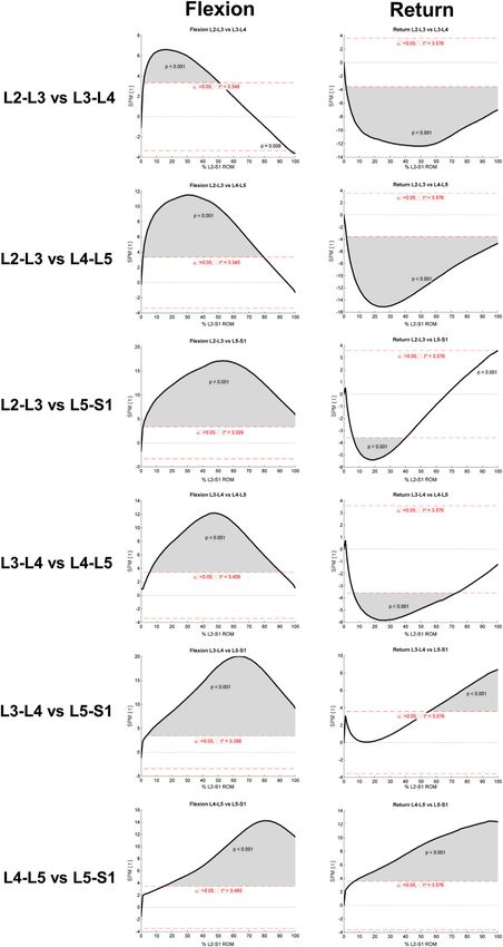
Breen et al.                                                                                                             Intervertebral Motion Sharing Database

  FIGURE 2 | Sagittal plane fluoroscopic images of the lumbar spine with computer templates (A) upright (B) flexed.

version 3.8) were used to conduct parametric two-tailed, two-                          For the Patient-control sub-study, 8 chronic back pain patients
sample t-tests across the time series. Statistical significance occurs               and 8 controls were imaged (43.8% female, mean age 48.1
when the SPM curves cross the critical threshold node at any                        (controls) and 48.8 years (patients). Thus, the sub-study
time, taking into account that each time point is related to those                  participants were approximately 10 years older than those in
on either side (Friston et al., 2007; Papi et al., 2020). Where                     the Reference Database study who had a mean age of 38.6.
multiple adjacent points of the SPM curves exceeded the critical                    This was the only substantial difference between the studies.
threshold, the associated p-values were calculated using Random
Field Theory.                                                                       Kinematics
   For the Patient-control secondary analysis, SPM analysis was                     The maximum intervertebral ranges throughout flexion and
conducted using non-parametric two tailed t-tests. This                             return motion (means) for the Reference Database study
compared segmental contributions to bending between                                 group and the Patient-control sub-study group are shown in
patients and controls throughout the motion. Previous                               Table 2. Maximum change in L5-S1 RoM was significantly less
measures of segmental contribution have been shown to have                          than the controls in the Patient-control sub-study.
high observer reliability and acceptable intrasubject repeatability                    Figure 3 shows statistically significant differences in
over 6 weeks (Breen et al., 2019b; To et al., 2020).                                contributions to bending during the motion, both between and
                                                                                    within levels. Each intervertebral level had its own characteristic
                                                                                    motion signature across the Reference Database study
RESULTS                                                                             population, with significant differences (p < 0.05, noted from
                                                                                    the lack of overlap of the 95%CI bands) between each level’s
For the Reference study, 131 participants were imaged. Four                         contribution throughout most of the motion. It is also notable
participants were excluded due to tracking errors of at least 1                     that these paths are in a state of constant change as the motion
vertebra. Full data sets were therefore obtained from 127                           progresses, although all levels exhibit more uniform motion
participants. Tables containing the Reference Database,                             sharing in the return phase than in the flexion phase. In
detailing the mean and 95%CI for the continuous proportional                        addition, there is a negative contribution to motion of L5-S1
segmental motion for flexion and return motion plus the Patient-                     at the beginning of flexion (Figure 3A). This is expected as
Control secondary analysis data can be found in Supplementary                       participants attempt to move their hips back to keep the centre of
Material I.                                                                         mass over the feet.
   Reference Database study participants received a mean (upper                        The SPM analysis reported in Figure 4 reveals these
third quartile) effective dose of 0.27 mSv (0.31) while secondary                   differences to be highly significant (p < 0.001) between levels
analysis patients received 0.26 mSv (0.30) for this investigation.                  for almost all data points across the motion for both flexion and
These values are approximately one quarter of the dose of a                         return in the Reference Database study cohort, confirming the
conventional plain radiographic examination of the lumbar spine                     presence of discrete motion paths for each motion segment.
(Mellor et al., 2014).                                                              During the outward flexion phase of movement, the superior

Frontiers in Bioengineering and Biotechnology | www.frontiersin.org             5                                   September 2021 | Volume 9 | Article 745837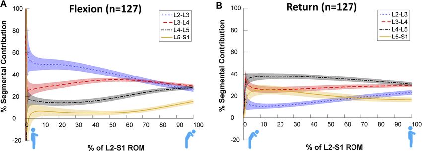
Breen et al.                                                                                                                      Intervertebral Motion Sharing Database

TABLE 2 | Mean maximum intervertebral rotational ranges (mean and SD).

Reference database                                                                                               Subgroup study
                                                                               Controls                              Patients                                2-tailed p

RoM L2-3                                 9.5 (3.87)                            10.2 (1.4)                             8.9 (5.2)                                 0.46
RoM L3-4                                10.6 (2.96)                            11.5 (2.8)                            10.1 (2.8)                                 0.21
RoM L4-5                                10.4 (3.93)                            11.9 (3.5)                             8.7 (1.1)                                 0.21
RoM L5-S1                                5.7 (5.60)                             7.2 (3.9)                             3.2 (2.9)                                 0.05
RoM: range of motion (degrees)

  FIGURE 3 | Average segmental contribution to lumbar flexion [(A): Flexion and (B): return to standing] with 95% confidence intervals (shaded areas) in the
  Reference Database cohort.

lumbar motion segment of each pair (Figure 4) consistently                            little difference between patients and controls in terms of motion
contributed more to the range of motion, exceeding the critical                       sharing at most intervertebral levels, although these have been
value for 50–99% of the task. In addition, the L2-3 vs. L3-4                          found to differentiate patients from controls in passive recumbent
motion segment combination also had a supra-threshold cluster                         studies (Breen and Breen, 2018). However, SPM analyses reveals
at the end of flexion where the inferior motion segment                                that there are statistically significant differences between the
contributed more (p  0.008).                                                         groups’ motion share at L2-L3 during the return to neutral
   During the return to upright position phase of the task, in 3                      phase of the task (p < 0.001) and at the end range of L5-S1
of the 6 inter joint combinations, the inferior motion segment                        motion (Flexion p  0.012 and Return p  0.004) (Figure 6).
of the pair constantly contributed significantly more to the
return phase of bending (p < 0.001). The exceptions were “L3-
4 vs. L5-S1” and “L4-5 vs. L5-S1”, where the superior motion                          DISCUSSION
segment contributed a greater amount (p < 0001), and at “L2-
3 vs. L5-S1”, where initially (between 5–40% of the RoM) L5-                          Reference Study
S1 contributed more (p < 0.001). In the late stages of bending                        To the best of the authors’ knowledge this study reports the largest
(at approximately 100% of RoM), L2-3 contributed more (p <                            database of continuous intersegmental lumbar spine kinematics
0.001).                                                                               during weight bearing in-vivo flexion and return, providing
                                                                                      normative reference values for making patient-specific kinematic
Patient-Control Secondary Analysis                                                    comparisons, for informing dynamic FE loading models, and to
The motion contributions in the secondary analysis are shown in                       help identify biomarkers for CNSLBP (Zanjani-Pour et al., 2018;
Figure 5. These subjectively demonstrate differences in the                           Breen and Breen, 2020). The Reference Database study used an
motion sharing patterns between patients and controls,                                established standardised protocol to measure the intersegmental
especially at L5-S1. Verification of these differences can be                          contributions to motion from L2-S1 during weight bearing flexion
seen in the non-parametric SPM analysis provided in                                   and return bending–unlike most conventional recording of lumbar
Supplementary Material III.                                                           flexion, which depends on participant co-ordination for its
   Figure 6 compares the motion sharing patterns for all 8                            consistency. Using this protocol, continuous change in mean
patients and 8 controls in the secondary analysis. There was                          proportional motion share was observed during both the flexion

Frontiers in Bioengineering and Biotechnology | www.frontiersin.org               6                                      September 2021 | Volume 9 | Article 745837
Breen et al. Intervertebral Motion Sharing Database
FIGURE 4 | Results of SPM parametric paired t-test (SPM{t}). Each row refers to a different intervertebral joint combination. Supra-threshold clusters indicate
significance differences between joint contributions to motion and are shown in grey. The critical threshold is shown as a red dashed line. Versions of these figures
alongside the mean and 95%CI bands can be found in Supplementary Material II.
Frontiers in Bioengineering and Biotechnology | www.frontiersin.org 7 September 2021 | Volume 9 | Article 745837Breen et al. Intervertebral Motion Sharing Database
FIGURE 5 | Average segmental contribution to return from flexion with 95% confidence intervals (shaded areas) in the Patient-control sub-study cohorts. (A):
Control Flexion, (B): Patient Flexion, (C): Control Return, (D): Patient Return.
and return phases and revealed significant differences in the motion or maximal flexion studies with continuous intervertebral motion
paths between levels. In addition, while the similarity between this studies, the distribution of sharing was found to be similar (Breen
study’s measures of lumbar segmental contributions and previous and Breen, 2020). Thus, the quasi-static spine kinematics
measures of lumbo-pelvic rhythms are interesting, these are not the literature, as reviewed by Widmer et al. (2019) exhibits a
same and should not be confused. degree of consistency with more recent continuous motion
It would be appropriate to compare this database with studies in terms of lumbar intervertebral motion sharing.
previous fluoroscopy studies, however no one study has The above considerations, plus the large number of participants
applied all the criteria required. We could find only four that in the Reference Database study, may account for the remarkably
attempted to employ completely continuous motion analysis consistent motion sharing patterns during both outward and
(Okawa et al., 1998; Harada et al., 2000; Nagel et al., 2014; return continuous motion, despite some heterogeneity in
Aiyangar et al., 2015). This may, in part, account for the participant characteristics. Although the age range in our
failure of studies that reported only quasi-static intersegmental sample was wide (21–70 years), body weight had an upper
motion to detect variations in the contributions of individual reference range of only 96 Kg, while weights of up to 119 Kg
segments during bending (Wong et al., 2006). Only three used have been shown to be associated with substantially increased L5-
proportional motion (Teyhen et al., 2007; Nagel et al., 2014; S1 compression in flexed postures (Hajihosseinali et al., 2015). This
Aiyangar et al., 2015), and none applied the degree of may affect the segmental contribution at that level and was also
standardisation of participant motion during imaging used in noted in relation to RoM in the Widmer et al. (2019) review and in
the present study (Breen et al., 2012). Return phase motion modelling studies by Zander et al. (2002). However, segmental
(which is not represented as flexion in reverse) was reported contributions, once thought to be RoM-dependent, did not exhibit
in only 4 (Okawa et al., 1998; Harada et al., 2000; Teyhen et al., this in our Reference Database study, nor in other studies that
2007; Aiyangar et al., 2015), while only 5 measured all levels from included all segments from L2 to S1 (Miyasaka et al., 2000; Ahmadi
L2-S1 (Takayanagi et al., 2001; Lee et al., 2002; Wong et al., 2006; et al., 2009; Aiyangar et al., 2015). Contribution patterns were also
Ahmadi et al., 2009; Aiyangar et al., 2015). However, when distinctly different in flexion and return, as one would expect with
comparing the segmental contributions derived from moderate different phasing of trunk muscle activation (Ouaaid et al., 2013).
Frontiers in Bioengineering and Biotechnology | www.frontiersin.org 8 September 2021 | Volume 9 | Article 745837Breen et al. Intervertebral Motion Sharing Database FIGURE 6 | Comparison of intervertebral motion sharing patterns between patients and controls at individual levels for flexion and return with 95% confidence intervals and SPM. Comparisons for all inter-joint combinations are shown in Supplementary Material III. Patient-Control Sub-Study patients is also reflected clinically in the Kinesiopathological Model The differences between patient and control subgroups in the of low back pain, and is considered to be an important factor in return phase also seem to complement those previously found in rehabilitation (Van Dillen et al., 2013). weight bearing studies that combined outward and return motion (Breen and Breen, 2020). Moreover, the standardised image Strengths and Limitations acquisition protocol would seem to make it unlikely that This is the largest dataset available to date to present normative abnormal patterns are attributable to artefact rather than values for continuous segmental contributions to motion in the motion pathology. However, it also raises the possibility that lumbar spine using variables that have been shown to distinguish other individual factors, such as lumbar geometry, may have an patients with CNSLBP from asymptomatic controls (Ahmadi influence, making clinical assessments based on motion sharing et al., 2009; Breen and Breen, 2020). Moreover, the Patient- patterns alone inadvisable. control sub-study provides further evidence of a kinematic The differences between patients and controls in the secondary biomarker for nonspecific back pain. However, standardising analysis may reflect differences in the respective roles of the deep the motion protocol involves a trade-off between natural multifidus and erector spinae muscle groups in people with motion and the repeatability necessary to make patient-specific CNSLBP (Wallwork et al., 2009) and/or passive tissue restraint. comparisons. In terms of the latter, the methodology used has Two trends are particularly apparent in this study of weight undergone extensive validation in terms of precision and validity bearing motion. Firstly, L5-S1 shares less motion in patients, (Breen et al., 2006; Breen and Breen, 2016; Breen et al., 2019b) albeit non-significantly until the end of range. This is also and has previously been used in preliminary dynamic loading reflected in the reduced RoM of L5-S1. Secondly, L2-3 studies using FE modelling (Zanjani-Pour et al., 2018). Thus, shares significantly more motion in patients during the return further subject-specific estimates of joint loading using dynamic phase, although this is only apparent in the mid-ranges and would imaging may be expected to improve the sensitivity of subject- not be measurable when merely investigating segmental range. specific model-based lumbar spinal loading estimates (Byrne Alterations in the readiness of lumbar joints to move in CNSLBP et al., 2020). However, like many other biomechanical studies Frontiers in Bioengineering and Biotechnology | www.frontiersin.org 9 September 2021 | Volume 9 | Article 745837
Breen et al.                                                                                                               Intervertebral Motion Sharing Database

that compare patients to controls, the sample size of our sub-                 CONCLUSION
study was small, which is a limitation that may be mitigated by
further replication. In addition, while evidence suggests that                 In asymptomatic people, provided a standardised QF imaging
magnitude of loading (beyond body weight), in vivo, does not                   protocol for measuring continuous proportional lumbar
have any significant effect on individual segmental contribution                intervertebral motion is used, consistent intervertebral motion
to motion (Aiyangar et al., 2015), biomechanical modelling                     patterns are revealed where each level follows its own discrete,
should exercise caution if using this database to model                        level-specific path that changes significantly during the motion.
unloaded or excessive loading states.                                          This is proposed to represent the human normative phenotype
   It would also have been useful for future biomechanical                     when using the present imaging protocol. These paths constantly
modelling studies if it had been possible to include the whole                 and consistently change as the bending motion progresses,
lumbar spine, but this could not be done owing to the limited                  although levels exhibit more uniform motion sharing in the
image intensifier diameter. This is a problem with most current                 return phase than in the flexion phase. Patients with CNSLBP
intensifiers which will be overcome as flat panel machines become                showed a significantly greater contribution at L2-3 and a
more plentiful.                                                                significantly smaller contribution at L5-S1 during the
                                                                               return phase.
Future Studies
Further studies are needed, not only to replicate the present
study’s findings, but also to explore the effects of other variables,           DATA AVAILABILITY STATEMENT
as well as coronal plane motion and passive recumbent motion,
where body mass and muscular contractions are mitigated.                       The original contributions presented in the study are included in
However, there is still considerable scope for elaboration of                  the article/Supplementary Material. Further inquiries can be
motion sharing studies of weight bearing flexion and return.                    directed to the corresponding author.
For example, variations in pelvic tilt may be an important source
of heterogeneity in light of the variations in the motion segment
flexibility that the QF procedure aims to measure (Retailleau and               ETHICS STATEMENT
Colloud, 2020).
    The specialised motion frame apparatus used in the current                 The studies involving human participants were reviewed and
work, in addition to standardising the velocity and range of                   approved by the United Kingdom National Research Ethics
bending, also partially stabilises the sacrum. This increases to               Service (South West 3). The patients/participants provided
varying degrees, the contribution of the lumbar spine to the                   their written informed consent to participate in this study.
flexion motion, regardless of the degree of lordosis or sacral
inclination. Although the degree of this restraint is not
standardised and depends on the individual’s natural lumbo-                    AUTHOR CONTRIBUTIONS
pelvic contribution to bending, this does not seem to disrupt the
consistency of the resulting motion sharing patterns.                          AB and AxB contributed to the conception and design of the
Nevertheless, there is likely to be some relationship between                  study and data collection. AxB organized the database, performed
lumbar sagittal shape and the motion contribution, albeit                      the statistical analysis, and wrote the first draft of the manuscript.
within the boundaries of the normative ranges of variation.                    AB provided interpretation of the outcomes. DDC, MF, GK, IP,
This should be explored. Given that spine shape has been                       and AW, contributed to the validation, writing the original draft
shown to influence a person’s preference for squatting or                       and critical revisions of the article. All authors read, contributed
stooping during lifting tasks, it would be useful to determine                 to manuscript revision, and approved the submitted version.
the relationships between spine shape and dynamic loading
stresses at individual levels based on their contributions to
flexion and return motion (Pavlova et al., 2018).
    It would also be useful to explore other kinematic indices in
                                                                               SUPPLEMENTARY MATERIAL
terms of motion contributions, as the present database provides                The Supplementary Material for this article can be found online at:
only rotational data, and there is evidence that the translational             https://www.frontiersin.org/articles/10.3389/fbioe.2021.745837/
component, although small, also affects inter-segment rotational               full#supplementary-material
stiffness (Affolter et al., 2020). However, in a previous study, we
did not find it to differentiate nonspecific back pain patients from             Supplementary Material I | Proportional intervertebral motion of individual level
                                                                               L2-S1 motion during forward bending and return in the Reference Database and
controls (Breen et al., 2018).                                                 Patient-control studies.
    Finally, it is now timely to explore possible relationships between
                                                                               Supplementary Material II | SPM of inter-joint contributions to motion from the
the motion variants that seem to be associated with CNSLBP and                 Reference Database, together with mean and 95% confidence intervals for each
possible sources of nociception. As these may not necessarily involve          combination.
disco-ligamentous micro-strain, it may be useful to explore muscular           Supplementary Material III | Differences in the motion sharing patterns for all joint
metabolic pain as a mechanism by including blood flow studies with              combinations between patients and controls analysed by 95% confidence intervals
those of motion contributions during bending.                                  and SPM.

Frontiers in Bioengineering and Biotechnology | www.frontiersin.org       10                                        September 2021 | Volume 9 | Article 745837
Breen et al. Intervertebral Motion Sharing Database
REFERENCES Eskandari, A. H., Arjmand, N., Shirazi-AdlFarahmand, A. F., and Farahmand, F.
(2017). Subject-specific 2D/3D Image Registration and Kinematics-Driven
Musculoskeletal Model of the Spine. J. Biomech. 57, 18–26. doi:10.1016/
Adler, R. J., Taylor, J. E., and Worsley, K. J. (2007). Applications of Random fields j.jbiomech.2017.03.011
and Geometry: Foundations and Case Studies. Netherlands: Springer. Friston, K. J., Ashburner, J. T., Kiebel, S. J., Nichols, T. E., and Penny, W. D. (2007).
Affolter, C., Kedzierska, J., Vielma, T., Weisse, B., and Aiyangar, A. (2020). Statistical Parametric Mapping: The Analysis of Functional Brain Images.
Estimating Lumbar Passive Stiffness Behaviour from Subject-specific Finite London: Elsevier.
Element Models and In Vivo 6DOF Kinematics. J. Biomech. 102, 109681. Hajihosseinali, M., Arjmand, N., and Shirazi-Adl, A. (2015). Effect of Body Weight
doi:10.1016/j.jbiomech.2020.109681 on Spinal Loads in Various Activities: A Personalized Biomechanical Modeling
Ahmadi, A., Maroufi, N., Behtash, H., Zekavat, H., and Parnianpour, M. (2009). Approach. J. Biomech. 48, 276–282. doi:10.1016/j.jbiomech.2014.11.033
Kinematic Analysis of Dynamic Lumbar Motion in Patients with Lumbar Harada, M., Abumi, K., Ito, M., and Kaneda, K. (2000). Cineradiographic Motion
Segmental Instability Using Digital Videofluoroscopy. Eur. Spine J. 18, Analysis of normal Lumbar Spine during Forward and Backward Flexion. Spine
1677–1685. doi:10.1007/s00586-009-1147-x 25, 1932–1937. doi:10.1097/00007632-200008010-00011
Aiyangar, A., Zheng, L., Anderst, W., and Zhang, X. (2015). Apportionment of Jones, A. C., and Wilcox, R. K. (2008). Finite Element Analysis of the Spine:
Lumbar L2-S1 Rotation across Individual Motion Segments during a Dynamic Towards a Framework of Verification, Validation and Sensitivity Analysis.
Lifting Task. J. Biomech. 48 (13), 3709–3715. doi:10.1016/j.jbiomech.2015.08.022 Med. Eng. Phys. 30 (10), 1287–1304. doi:10.1016/j.medengphy.2008.09.006
Breen, A., and Breen, A. (2016). Accuracy and Repeatability of Quantitative Lee, S.-w., Wong, K. W. N., Chan, M.-k., Yeung, H.-m., Chiu, J. L. F., and Leong,
Fluoroscopy for the Measurement of Sagittal Plane Translation and Finite J. C. Y. (2002). Development and Validation of a New Technique for Assessing
centre of Rotation in the Lumbar Spine. Med. Eng. Phys. 38, 607–614. Lumbar Spine Motion. Spine 27 (8), E215–E220. doi:10.1097/00007632-
doi:10.1016/j.medengphy.2016.03.009 200204150-00022
Breen, A., and Breen, A. (2020). Dynamic Interactions between Lumbar Linnet, K. (1987). Two-stage Transformation Systems for Normalization of
Intervertebral Motion Segments during Forward Bending and Return. Reference Distributions Evaluated. Clin. Chem. 33 (3), 381–386. doi:10.1093/
J. Biomech. 102, 109603. doi:10.1016/j.jbiomech.2020.109603 clinchem/33.3.381
Breen, A., and Breen, A. (2018). Uneven Intervertebral Motion Sharing Is Related Magee, J. (2015). Three Dimensional Digital Modelling of Human Spine
to Disc Degeneration and Is Greater in Patients with Chronic, Non-specific Low Anthropometrics and Kinematics from Meta-Analysis. How Relevant Is
Back Pain: an In Vivo, Cross-Sectional Cohort Comparison of Intervertebral Existing Anatomical Research? J. Spine 4 (1), 251–257. doi:10.4172/2165-
Dynamics Using Quantitative Fluoroscopy. Eur. Spine J. 27 (1), 145–153. 7939.1000205
doi:10.1007/s00586-017-5155-y Mellor, F. E., Thomas, P., and Breen, A. (2014). Moving Back: The Radiation Dose
Breen, A., Claerbout, E., Hemming, R., Ayer, R., and Breen, A. (2019a). Received from Lumbar Spine Quantitative Fluoroscopy Compared to Lumbar
Comparison of Intra Subject Repeatability of Quantitative Fluoroscopy and Spine Radiographs with Suggestions for Dose Reduction. Radiography 20,
Static Radiography in the Measurement of Lumbar Intervertebral Flexion 251–257. doi:10.1016/j.radi.2014.03.010
Translation. Sci. Rep. 9, 19253. doi:10.1038/s41598-019-55905-1 Miyasaka, K., Ohmori, K., Suzuki, K., and Inoue, H. (2000). Radiographic Analysis
Breen, A. C., Muggleton, J. M., and Mellor, F. E. (2006). An Objective Spinal of Lumbar Motion in Relation to Lumbosacral Stability. Spine 25 (6), 732–737.
Motion Imaging Assessment (OSMIA): Reliability, Accuracy and Exposure doi:10.1097/00007632-200003150-00014
Data. BMC Musculoskelet. Disord. 7 (1), 1–10. doi:10.1186/1471-2474-7-1 Nagel, T. M., Zitnay, J. L., Barocas, V. H., and Nuckley, D. J. (2014). Quantification
Breen, A. C., Teyhen, D. S., Mellor, F. E., Breen, A. C., Wong, K. W. N., and Deitz, of Continuous In Vivo Flexion-Extension Kinematics and Intervertebral
A. (2012). Measurement of Intervertebral Motion Using Quantitative Strains. Eur. Spine J. 23, 754–761. doi:10.1007/s00586-014-3195-0
Fluoroscopy: Report of an International Forum and Proposal for Use in the Okawa, A., Shinomiya, K., Komori, H., Muneta, T., Arai, Y., and Nakai, O. (1998).
Assessment of Degenerative Disc Disease in the Lumbar Spine. Adv. Orthop. Dynamic Motion Study of the Whole Lumbar Spine by Videofluoroscopy. Spine
2012, 1–10. doi:10.1155/2012/802350 23 (16), 1743–1749. doi:10.1097/00007632-199808150-00007
Breen, A., Hemming, R., Mellor, F., and Breen, A. (2019b). Intrasubject Repeatability Oxland, T. R. (2016). Fundamental Biomechanics of the Spine-What We Have
of In Vivo Intervertebral Motion Parameters Using Quantitative Fluoroscopy. Learned in the Past 25 Years and Future Directions. J. Biomech. 49 (6), 817–832.
Eur. Spine J. 28 (2), 450–460. doi:10.1007/s00586-018-5849-9 doi:10.1016/j.jbiomech.2015.10.035
Breen, A., Mellor, F. E., and Breen, A. C. (2018). Aberrant Intervertebral Motion in Panjabi, M. M. (1992). The Stabilizing System of the Spine. Part I. Function,
Patients with Treatment-Resistant Nonspecific Low Back Pain: a Retrospective Dysfunction, Adaptation, and Enhancement. J. Spinal Disord. 5 (4), 383–389.
Cohort Study and Control Comparison. Eur. Spine J. 27, 2831. doi:10.1007/ doi:10.1097/00002517-199212000-00001
s00586-018-5666-1 Papi, E., Bull, A. M. J., and McGregor, A. H. (2020). Alteration of Movement
Byrne, R. M., Aiyangar, A. K., and Zhang, X. (2020). Sensitivity of Musculoskeletal Patterns in Low Back Pain Assessed by Statistical Parametric Mapping.
Model-Based Lumbar Spinal Loading Estimates to Type of Kinematic Input and J. Biomech. 100, 109597. doi:10.1016/j.jbiomech.2019.109597
Passive Stiffness Properties. J. Biomech. 102, 109659. doi:10.1016/ Papi, E., Bull, A. M. J., and McGregor, A. H. (2018). Is There Evidence to Use
j.jbiomech.2020.109659 Kinematic/kinetic Measures Clinically in Low Back Pain Patients? A Systematic
CLSI (2008). Defining, Establishing and Verifying Reference Intervals in the Clinical Review. Clin. Biomech. 55, 53–64. doi:10.1016/j.clinbiomech.2018.04.006
Laboratory; Approved Guideline. 3rd Edn. Wayne, PA: Clinical Laboratory Pataky, T. C. (2016). rft1d: Smooth One-Dimensional Random Field Upcrossing
Standards Institute. Probabilities inPython. J. Stat. Soft. 71, i07. doi:10.18637/jss.v071.i07
Dreischarf, M., Shirazi-Adl, A., Arjmand, N., Rohlmann, A., and Schmidt, H. Pataky, T., Vanrenterghem, J., and Robinson, M. A. (2016). The Probability of False
(2016). Estimation of Loads on Human Lumbar Spine: A Review of In Vivo and Positives in Zero-Dimensional Analyses of One-Dimensional Kinematic, Force
Computational Model Studies. J. Biomech. 49, 833–845. doi:10.1016/ and EMG Trajectories. J. Biomech. 49, 1468. doi:10.1016/j.jbiomech.2016.03.032
j.jbiomech.2015.12.038 Pavlova, A. V., Meakin, J. R., Cooper, K., Barr, R. J., and Aspden, R. M. (2018).
du Rose, A., and Breen, A. (2016). Relationships between Lumbar Inter-vertebral Variation in Lifting Kinematics Related to Individual Intrinsic Lumbar
Motion and Lordosis in Healthy Adult Males: a Cross Sectional Cohort Study. Curvature: an Investigation in Healthy Adults. BMJ Open Sport Exerc. Med.
BMC Musculoskelet. Disord. 17 (121), 121. doi:10.1186/s12891-016-0975-1 4, e000374. doi:10.1136/bmjsem-2018-000374
du Rose, A., Breen, A., and Breen, A. (2018). Relationships between Muscle Electrical Retailleau, M., and Colloud, F. (2020). New Insights into Lumbar Flexion Tests
Activity and the Control of Inter-vertebral Motion during a Forward Bending Based on Inverse and Direct Kinematic Musculoskeletal Modeling. J. Biomech.
Task. J. Electromyogr. Kinesiol. 43, 48–54. doi:10.1016/j.jelekin.2018.08.004 105, 109782. doi:10.1016/j.jbiomech.2020.109782
El Ouaaid, Z., Shirazi-Adl, A., Plamondon, A., and Larivière, C. (2013). Trunk Takayanagi, K., Takahashi, K., Yamagata, M., Moriya, H., Kitahara, H., and
Strength, Muscle Activity and Spinal Loads in Maximum Isometric Flexion and Tamaki, T. (2001). Using Cineradiography for Continuous Dynamic-Motion
Extension Exertions: A Combined In Vivo-computational Study. J. Biomech. Analysis of the Lumbar Spine. Spine 26 (17), 1858–1865. doi:10.1097/
46, 2228–2235. doi:10.1016/j.jbiomech.2013.06.018 00007632-200109010-00008
Frontiers in Bioengineering and Biotechnology | www.frontiersin.org 11 September 2021 | Volume 9 | Article 745837Breen et al. Intervertebral Motion Sharing Database Teyhen, D. S., Flynn, T. W., Childs, J. D., Kuklo, T. R., Rosner, M. K., Polly, D. W., Bisegmental Stabilization. Clin. Biomech. 17, 439–445. doi:10.1016/s0268- et al. (2007). Fluoroscopic Video to Identify Aberrant Lumbar Motion. Spine 32 0033(02)00040-2 (7), E220–E229. doi:10.1097/01.brs.0000259206.38946.cb Zanjani-Pour, S., Meakin, J. R., Breen, A., and Breen, A. (2018). Estimation of In To, D., Breen, A., Breen, A., Mior, S., and Howarth, S. J. (2020). Investigator Vivo Inter-vertebral Loading during Motion Using Fluoroscopic and Magnetic Analytic Repeatability of Two New Intervertebral Motion Biomarkers for Resonance Image Informed Finite Element Models. J. Biomech. 70, 134–139. Chronic, Nonspecific Low Back Pain in a Cohort of Healthy Controls. doi:10.1016/j.jbiomech.2017.09.025 Chiropr Man. Therap 28 (62), 1–9. doi:10.1186/s12998-020-00350-5 Van Dillen, L. R., Sahrmann, S. A., and Norton, B. J. (2013). “The Conflict of Interest: The authors declare that the research was conducted in the Kinesiopathological Model and Mechanical Low Back Pain,” in Spinal absence of any commercial or financial relationships that could be construed as a Control: The Rehabilitation of Low Back Pain. Editors P.W. Hodges, potential conflict of interest. J. Cholewicki, and J. Van Dieen (Edinburgh: Churchill Livingstone Elsevier), 89–98. doi:10.1016/b978-0-7020-4356-7.00008-2 Publisher’s Note: All claims expressed in this article are solely those of the authors Wallwork, T. L., Stanton, W. R., Freke, M., and Hides, J. A. (2009). The Effect of and do not necessarily represent those of their affiliated organizations, or those of Chronic Low Back Pain on Size and Contraction of the Lumbar Multifidus the publisher, the editors and the reviewers. Any product that may be evaluated in Muscle. Man. Ther. 14 (5), 496–500. doi:10.1016/j.math.2008.09.006 this article, or claim that may be made by its manufacturer, is not guaranteed or Widmer, J., Fornaciari, P., Senteler, M., Roth, T., Snedeker, J. G., and Farshad, M. endorsed by the publisher. (2019). Kinematics of the Spine under Healthy and Degenerative Conditions: a Systematic Review. Ann. Biomed. Eng. 47, 1491–1522. doi:10.1007/s10439-019- Copyright © 2021 Breen, De Carvalho, Funabashi, Kawchuk, Pagé, Wong and 02252-x Breen. This is an open-access article distributed under the terms of the Creative Wong, K. W. N., Luk, K. D. K., Leong, J. C. Y., Wong, S. F., and Wong, K. K. Y. Commons Attribution License (CC BY). The use, distribution or reproduction in (2006). Continuous Dynamic Spinal Motion Analysis. Spine 31 (4), 414–419. other forums is permitted, provided the original author(s) and the copyright owner(s) doi:10.1097/01.brs.0000199955.87517.82 are credited and that the original publication in this journal is cited, in accordance Zander, T., Rohlmann, A., Klöckner, C., and Bergmann, G. (2002). Comparison of with accepted academic practice. No use, distribution or reproduction is permitted the Mechanical Behavior of the Lumbar Spine Following Mono- and which does not comply with these terms. Frontiers in Bioengineering and Biotechnology | www.frontiersin.org 12 September 2021 | Volume 9 | Article 745837
You can also read