A novel CT scoring method predicts the prognosis of interstitial lung disease associated with anti MDA5 positive dermatomyositis
←
→
Page content transcription
If your browser does not render page correctly, please read the page content below
www.nature.com/scientificreports
OPEN A novel CT scoring method predicts
the prognosis of interstitial lung
disease associated with anti‑MDA5
positive dermatomyositis
Wenwen Xu1,4, Wanlong Wu1,4, Danting Zhang1, Zhiwei Chen1, Xinwei Tao2, Jiangfeng Zhao1,
Kaiwen Wang1, Xiaodong Wang1, Yu Zheng3* & Shuang Ye1*
Anti-melanoma differentiation-associated gene 5-positive dermatomyositis-associated interstitial
lung disease (MDA5+ DM-ILD) is a life-threatening disease. This study aimed to develop a novel
pulmonary CT visual scoring method for assessing the prognosis of the disease, and an artificial
intelligence (AI) algorithm-based analysis and an idiopathic pulmonary fibrosis (IPF)-based scoring
were conducted as comparators. A retrospective cohort of hospitalized patients with MDA5+ DM-ILD
was analyzed. Since most fatalities occur within the first half year of the disease course, the primary
outcome was the six-month all-cause mortality since the time of admission. A ground glass opacity
(GGO) and consolidation-weighted CT visual scoring model for MDA5+ DM-ILD, namely ‘MDA5 score’,
was then developed with C-index values of 0.80 (95%CI 0.75–0.86) in the derivation dataset (n = 116)
and 0.84 (95%CI 0.71–0.97) in the validation dataset (n = 57), respectively. While, the AI algorithm-
based analysis, namely ‘AI score’, yielded C-index 0.78 (95%CI 0.72–0.84) for the derivation dataset
and 0.77 (95%CI 0.64–0.90) for the validation dataset. These findings suggest that the newly derived
‘MDA5 score’ may serve as an applicable prognostic predictor for MDA5+ DM-ILD and facilitate further
clinical trial design. The AI based CT quantitative analysis provided a promising solution for ILD
evaluation.
Anti-melanoma differentiation-associated gene 5-positive dermatomyositis ( MDA5+ DM) is a special subtype
of DM, associated with rapidly progressive interstitial lung disease (ILD). The overall prognosis is grave with
33–67% six-month mortality despite of aggressive immunosuppressive t herapy1–4.
Pulmonary high-resolution computed tomography (HRCT) is a main-stream imaging tool for identifying ILD
and measuring its severity. A semi-quantitative HRCT scoring system has been applied as a prognostic prediction
measurement in MDA5+ DM-ILD5, 6. However, the applied scoring system was initially designed for evaluation
of idiopathic pulmonary fibrosis (IPF)7, 8. Therefore, when referring to a more rapid progressive disease, such as
MDA5+ DM-ILD, the applicability has not been extensively validated. As examples, fibrosis components such
as traction bronchiectasis (TBE) and honeycombing changes were higher weighted in this ‘IPF score’; whereas
inflammation components, i.e., ground-glass opacity (GGO) and consolidation were less weighted. Only until
recently, another simplified scoring method for M DA5+ DM-ILD was proposed with equally weighted two
components of GGO and fibrosis9, 10. Unfortunately, the sample size was small with characteristic consolidation
feature being overlooked; further independent evaluation in a data-driven approach is warranted. It is noteworthy
that the time-consuming observer-dependent manner of these visual scorings is always an issue.
Under the pressure of the coronavirus disease 2019 (COVID-19) pandemic, advanced machine learning-based
technologies on pulmonary CT quantitative analysis have rapidly emerged, providing a promising solution for
diffuse lung disease HRCT evaluation in a more comprehensive and objective perspective11–13.
Thus, the aims of the current study were to establish a novel pulmonary HRCT visual scoring method for
predicting the six-month mortality in a large single-centered cohort of patients with M DA5+ DM; and in paral-
lel, to explore quantitative imaging assessment of this disease by applying artificial intelligence (AI) algorithm.
1
Department of Rheumatology, Shanghai Jiao Tong University School of Medicine Affiliated Renji Hospital,
No. 2000 Jiangyue Road, Shanghai 201112, China. 2CT Scientific Collaboration, Siemens Healthineers, Shanghai,
China. 3Department of Pulmonology, Shanghai Jiao Tong University School of Medicine Affiliated Renji Hospital,
No. 2000 Jiangyue Road, Shanghai 201112, China. 4These authors contributed equally: Wenwen Xu and Wanlong
Wu. *email: drzhengyu@yahoo.com; ye_shuang2000@163.com
Scientific Reports | (2021) 11:17070 | https://doi.org/10.1038/s41598-021-96292-w 1
Vol.:(0123456789)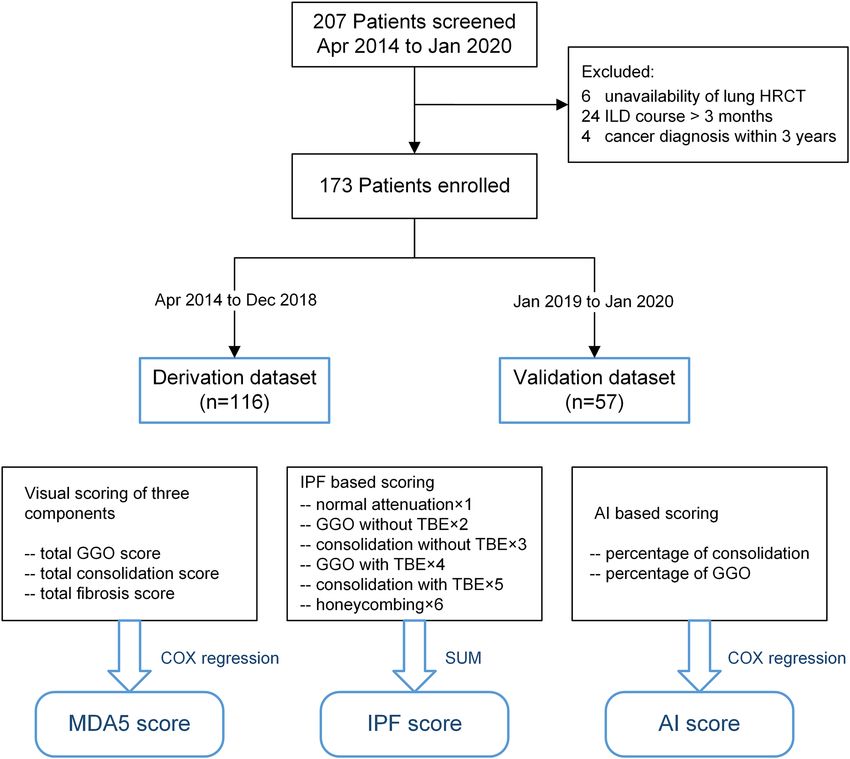
www.nature.com/scientificreports/

                                  Derivation dataset                                                  Validation dataset
                                                                       Non-survivors                                                       Non-survivors
                                  All (n = 116)   Survivors (n = 69)   (n = 47)          p-value*     All (n = 57)    Survivors (n = 36)   (n = 21)                  p-value*   p-value†
           Distribution pattern
           Bilateral lung
                                  113 (97.4)      66 (95.7)            47 (100.0)           0.39      54 (94.7)       33 (91.7)            21 (100.0)                  0.46     0.64
           involved
           RU lobe involved       95 (81.9)       50 (72.5)            45 (95.7)            0.003     48 (84.2)       27 (75.0)            21 (100.0)                  0.03     0.87
           RM lobe involved       76 (65.5)       36 (52.2)            40 (85.1)            0.001     42 (73.7)       22 (61.1)            20 (95.2)                   0.01     0.36
           RL lobe involved       113 (97.4)      66 (95.7)            47 (100.0)           0.39      57 (100.0)      36 (100.0)           21 (100.0)                  1        0.55
           LU lobe involved       99 (85.3)       54 (78.3)            45 (95.7)            0.02      51 (89.5)       30 (83.3)            21 (100.0)                  0.13     0.61
           LL lobe involved       113 (97.4)      67 (97.1)            46 (97.9)            1         54 (94.7)       33 (91.7)            21 (100.0)                  0.46     0.64
           No. involved lobe                                                                0.001                                                                      0.02     0.17
           1                      2 (1.7)         2 (2.9)              0 (0.0)                        3 (5.3)         3 (8.3)              0 (0.0)
           2                      12 (10.3)       11 (15.9)            1 (2.1)                        1 (1.8)         1 (2.8)              0 (0.0)
           3                      7 (6.0)         5 (7.2)              2(4.3)                         4 (7.0)         4 (11.1)             0 (0.0)
           4                      26 (22.4)       21 (30.4)            5 (10.6)                       10 (17.5)       9 (25.0)             1 (4.8)
           5                      69 (59.5)       30 (43.5)            39 (83.0)                      39 (68.4)       19 (52.8)            20 (95.2)
           No. involved
                                  95 (81.9)       51 (73.9)            44 (93.6)            0.01      49 (86.0)       28 (77.8)            21 (100.0)                  0.05     0.65
           lobes > 3
           No. involved
                                  69 (59.5)       30 (43.5)            39 (83.0)           < 0.001    39 (68.4)       19 (52.8)            20 (95.2)                   0.002    0.33
           lobes > 4
           PNM                    5 (4.3)         2 (2.9)              3 (6.4)              0.39      3               1 (2.8)              2 (9.5)                     0.55     0.72
           Scoring of three components
           Total GGO score        4 [2–8]         3 2–6]               7 [3–10]            < 0.001    6 [2–11]        5 [1–8]              10 [7–12]                   0.002    0.13
           Total CON score        4 [2–8]         3 [15]               8 [5–11]            < 0.001    6 4–9]          5 [2–7]              9 [7–13]                   < 0.001   0.06
           Total fibrosis score   3 [2–5]         2 [2–4]              3 [2–5]              0.51      4 [2–5]         4 [2–5]              5 [2–5]                     0.51     0.01
           IPF score              126 [116–151]   118 [112–127]        147 [125–171]      < 0.001     135 [121–155]   127 [112–141]        153 [134–188]              < 0.001   0.12

                                                  Table 1.  Comparison of visual CT features between two datasets with different outcome. Data are presented as
                                                  median [IQR] for continuous variables and number (frequency) (%) for categorical variables. * A comparison
                                                  between the survivors and non-survivors groups. † A comparison between the derivation and validation
                                                  datasets. CT, computed tomography; RU, right upper; RM, right middle; RL, right lower; LU, left upper; LL, left
                                                  lower; PNM, pneumomediastinum or pneumothorax; GGO, ground-glass opacity; CON, consolidation; IPF
                                                  score, idiopathic pulmonary fibrosis based visual scoring method.

                                                                                           HR [95%CI]                      P-value                   β coefficient
                                                  ‘MDA5 score’ model
                                                  Total GGO score                          1.14 [1.06–1.22]                     < 0.001              0.13
                                                   Total consolidation score               1.25 [1.16–1.34]                     < 0.001              0.22
                                                  ‘AI score’ model
                                                  Percentage of GGO                        1.02 [1.00, 1.04]                     0.07                0.02
                                                  Percentage of consolidation              1.15 [1.08, 1.22]                    < 0.001              0.14

                                                  Table 2.  Multivariable COX regression analysis for ‘MDA5 score’ and ‘AI score’ models. HR, hazard ratio;
                                                  95%CI, 95% confidence interval; MDA5, anti-melanoma differentiation-associated gene 5; AI, artificial
                                                  intelligence; GGO, ground-glass opacity.

                                                  Results
                                                  Comparable baseline clinical features, treatment and outcomes of the derivation dataset and validation dataset
                                                  were listed in Supplementary table S1. Of which, 47 (40.5%) and 21 (36.8%) patients died within six-month fol-
                                                  low up since the time of admission, respectively (p = 0.764).

                                                  ‘MDA5 score’: a novel CT visual semi‑quantitative analysis. The pulmonary HRCT findings from
                                                  visual semi-quantitative analysis of patients between survivors and non-survivors in both datasets were pre-
                                                  sented in Table 1. As expected, the ILD pattern distributed bilaterally. It was noteworthy that only GGO and con-
                                                  solidation patterns were significantly associated with outcome according to univariable analysis; as opposed to
                                                  neither fibrosis nor the presence of pneumomediastinum or pneumothorax (PNM) at baseline. Then, the GGO
                                                  and consolidation score were included in further multivariable COX regression analysis. Both total GGO score
                                                  (β coefficient = 0.13, p < 0.001) and total consolidation score (β coefficient = 0.22, p < 0.001) were determined
                                                  to be significantly associated with all-cause mortality (Table 2). To simplify, a linear equation, namely ‘MDA5

          Scientific Reports |       (2021) 11:17070 |                     https://doi.org/10.1038/s41598-021-96292-w                                                                      2

Vol:.(1234567890)
www.nature.com/scientificreports/
Figure 1. Flow chart of patients and three CT scoring models. MDA5, melanoma differentiation-associated
gene 5; HRCT, high-resolution computed tomography; ILD, interstitial lung disease; GGO, ground-glass
opacity; TBE, traction bronchiectasis; IPF, idiopathic pulmonary fibrosis; AI, artificial intelligence; DM,
dermatomyositis.
score’, by combining defined prognostic factors weighted by their β coefficients was finally generated: total GGO
score + 2*total consolidation score.
ROC curve analysis indicated that the optimal cutoff value for ‘MDA5 score’ was 18, which could efficiently
predict the six-month all-cause mortality in the derivation dataset (sensitivity 70.2%; specificity 82.6%) and the
validation dataset (sensitivity 85.7%; specificity 63.9%). The prediction accuracy of ‘MDA5 score’ calculated by
AUC was 0.85 (95%CI 0.78–0.91) for the derivation dataset and 0.87 (95%CI 0.78–0.96) for the validation data-
set, far ahead of the ‘IPF score’, which was 0.81 (95%CI 0.73–0.89) for the derivation dataset and 0.79 (95%CI
0.68–0.91) for the validation dataset. Additionally, The Kaplan–Meier survival plots of patients in both datasets
presented significant difference between the high-risk (‘MDA5 score’ > 18) and low-risk (‘MDA5 score’ ≤ 18)
groups (Figs. 1, 2). The mortality of high-risk patients was 73.3% in the derivation dataset and 58.1% in the
validation dataset; while the mortality of low-risk patients was 19.7% in the derivation dataset and 11.5% in the
validation dataset.
‘AI score’: an AI algorithm‑based quantitative analysis. The redundancy of the baseline fibrosis
component in terms of outcome prediction made pneumonia-trained AI algorithm plausible for our M DA5+
DM-ILD patients’ CT quantitative analysis (Fig. 3A). Percentage of consolidation was determined as the only
significant predictor for the overall survival in the final multivariable COX model (p < 0.001) (Table 2). Thus, the
percentage of consolidation was defined to represent ‘AI score’. Interestingly, the radar charts in Fig. 3B showed
Scientific Reports | (2021) 11:17070 | https://doi.org/10.1038/s41598-021-96292-w 3
Vol.:(0123456789)www.nature.com/scientificreports/
a 1.00 Low risk (MDA5 score ≤18)
High risk (MDA5 score >18)
+
0.75
Survival probability
0.50
0.25 +
p < 0.001, HR = 6.17 [95%CI 3.28-11.59]
0.00
0 5 10 15 20 25
Follow up time (week)
No. at risk
Low Risk 71 66 58 57 57 57
High Risk 45 22 15 12 12 12
Low risk (MDA5 score ≤18)
b 1.00
High risk (MDA5 score >18)
+
0.75
Survival probability
0.50
+
0.25
p < 0.001, HR = 7.36 [95%CI 2.16-25.12]
0.00
0 5 10 15 20 25
Follow up time (week)
No. at risk
Low Risk 26 26 23 23 23 23
High Risk 31 18 13 13 13 13
Figure 2. Survival curves of ‘MDA5 score’ in the derivation (a) and validation (b) datasets. MDA5, melanoma
differentiation-associated gene 5; HR, hazard ratio; 95%CI, 95% confidence interval.
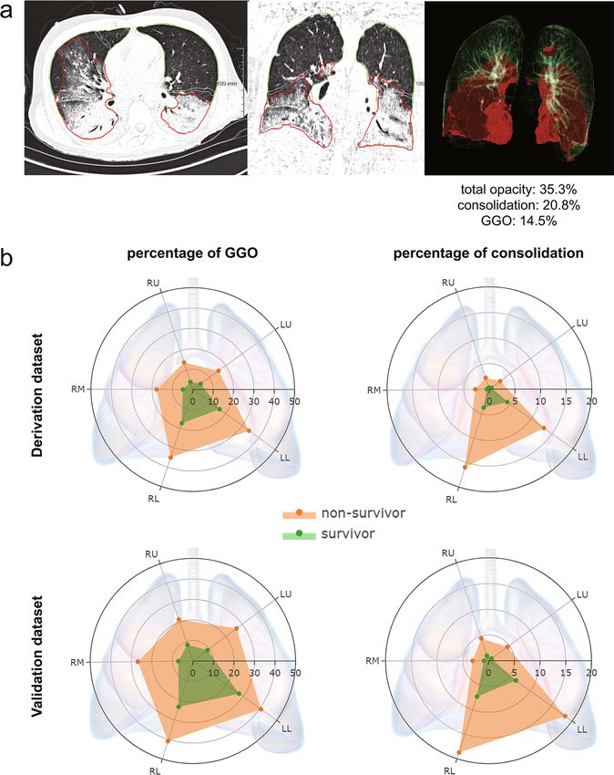
that the GGO and consolidation patterns were symmetrically distributed, with an evident ’gravity gradient’ pro-
pensity to the lower area of the lungs, especially for the consolidation distribution.
Comparisons of clinical performance between ‘IPF score’, ‘MDA5 score’ and ‘AI score’
model. The inter-observer consistency of the two visual scoring models was assessed with an ICC of 0.69
(95% CI 0.57–0.78) for ‘IPF score’ and an ICC of 0.93 (95% CI 0.89–0.96) for ‘MDA5 score’. Therefore, ‘MDA5
score’ attained a better inter-observer reproducibility. As a comparator, the detailed data of six domains for
calculating ‘IPF score’ was presented in Supplementary table S2. Hereafter, the comparisons of model discrimi-
nation between ‘IPF score’, ‘MDA5 score’ and ‘AI score’ were shown in Table 3. Notably, ‘MDA5 score’ had the
best performance with C-index values of 0.80 (95%CI 0.75–0.86) in the derivation dataset and 0.84 (95%CI
0.71–0.97) in the validation dataset, respectively. While, ‘AI score’ yielded C-index 0.78 (95%CI 0.72–0.84) for
Scientific Reports | (2021) 11:17070 | https://doi.org/10.1038/s41598-021-96292-w 4
Vol:.(1234567890)www.nature.com/scientificreports/
Figure 3. Artificial intelligence algorithm-based CT quantitative analysis. (a) The segmentation results of the
lung and its total opacity in representative CT images were shown in green and red borders respectively. The
percentage of total opacity, consolidation, and ground-glass opacity (GGO) of the whole lung were automatically
calculated to be 35.3%, 20.8%, and 14.5% respectively. (b) The average distributions of GGO and consolidation
in each lobe of the lung were displayed by the radar charts in both datasets. The axial line of the radar chart
referred to the mean percentage (%) of either GGO or consolidation of each lobe. RU, right upper lobe; RM,
right middle lobe; RL, right lower lobe; LU, left upper lobe; LL, left lower lobe.
the derivation dataset and 0.77 (95%CI 0.64–0.90) for the validation dataset. Finally, the DCA further demon-
strated that the ‘MDA5 score’ also presented with a higher overall net benefit than the other two models in terms
of clinical applicability (Fig. 4).
Discussion
As a highly progressive disease, M DA5+ DM-ILD remains to be a big challenge despite of recent treatment
advances4, 14. Several prognostic indicators of the disease had been reported involving respiratory physiology
parameters, laboratory biomarkers, and radiology f eatures1, 6, 15, 16. The current study focused on patients’ base-
line pulmonary HRCT and attempted to quantitatively assess the disease in the regard of predicting six-month
mortality.
Our study takes a step-forward from the previous visual scoring methods, and extensively evaluates the
distribution and extent of three basic imaging components of MDA5+ DM-ILD, i.e., GGO, consolidation and
Scientific Reports | (2021) 11:17070 | https://doi.org/10.1038/s41598-021-96292-w 5
Vol.:(0123456789)www.nature.com/scientificreports/
C-index [95%CI]
Scoring model Derivation dataset (n = 116) Validation dataset (n = 57)
‘IPF score’ 0.78 [0.71–0.84] 0.78 [0.65–0.91]
‘MDA5 score’ * 0.80 [0.75–0.86] 0.84 [0.71–0.97]
Total GGO score + 2*total consolidation score
‘AI score’ 0.78 [0.72–0.84] 0.77 [0.64–0.90]
Percentage Of consolidation
Table 3. Comparison of the prediction performance of each model. * ‘MDA5 score’ model performed
significantly better than ‘IPF score’ model (p = 0.02). C-index, concordance index; 95%CI, 95% confidence
interval; IPF, idiopathic pulmonary fibrosis; MDA5, anti-melanoma differentiation-associated gene 5; GGO,
ground-glass opacity; AI, artificial intelligence.
Figure 4. Decision curve analysis for ‘IPF score’, ‘MDA5 score’ and ‘AI score’ model. The concept of population
net benefit (NB) is fundamental to decision curves (measured in the y-axis) and referred to classification
accuracy of a model. Suppose high risk is defined as risk above some risk threshold R (x-axis); such high-risk
patients are recommended an intervention. The NB of using the risk model was calculated by the true-positive
rate, the proportion of cases with risk above risk threshold R; and the false-positive rate, the proportion of
controls with risk above risk threshold R. The horizontal dotted line at NB = 0 mean a simple policy of no
intervention to all patients (treat none); the gray curve in the plot depicted the NB of another simple policy:
recommend the intervention to everyone regardless of risk. In our result, the ‘MDA5 score’ model (red line) had
the highest net benefit compared to the others, almost across the full range of threshold probabilities.
fibrosis. In line with prior reports, our data confirmed that the presence of fibrosis or TBE in the context of GGO
or consolidation, is not of predictive value on prognosis in MDA5+ DM-ILD5, 10. The probable explanation is that
those fibrotic features are less common in this rapid progressive disease and likely to be presented, if it happens,
in later stage instead of baseline. The same notion apparently holds true for the presence of PNM, which is a
known severity indicator rather than a baseline p redictor17.
The combination of the extent of GGO and consolidation was found to have the best yield in terms of outcome
prediction, with the area of consolidation contributing more than GGO. The image ‘snapshot’ might reflect the
dynamic transformation from GGO to consolidation as disease progresses, just like the imaging changes observed
in severe COVID-19 p atients11, 12, 18. A possible shared underlying mechanism of acute lung injury in the two
diseases is a very intriguing question deserves further investigation. After all, the highly activated type I inter-
feron pathway in M DA5+ DM-ILD which suggested a possible virus-triggered response has been p ostulated19, 20.
+
To apply AI algorithm-based quantitative imaging analysis in M DA5 DM-ILD is a preliminarily yet novel
attempt. The initial primary applicable population of this algorithm was pneumonia, or more specifically,
COVID-19 disease. Of interest, our data suggested that this AI algorithm performs fairly well among MDA5+
DM-ILD patients. The performance might be further enhanced given more M DA5+ DM-ILD imaging data could
be fed into its machine-learning processes.
Scientific Reports | (2021) 11:17070 | https://doi.org/10.1038/s41598-021-96292-w 6
Vol:.(1234567890)www.nature.com/scientificreports/
The major limitation of our study was the single-center design. Although we presented a relatively large cohort
for this rare disease and performed internal validation, large-scale multi-center external validation is mandatory
before the CT scoring models being utilized in a clinical setting. Based upon this, the biases of different machine
conditions, patient selection and treatment protocols could be taken into consideration and subjected to better
control and adjustment. In addition, longitudinal analysis on the changes of ILD patterns over time remains
untouched in the current study, which deserves further exploration.
In conclusion, we have shown that a GGO and consolidation-weighted CT scoring model, along with an AI
algorithm, might serve as prognostic predictors for six-month mortality in MDA5+ DM-ILD. This might facilitate
future clinical trial design and precision management for this tricky disease.
Methods
Patients. A retrospective cohort of hospitalized patients with M
DA5+ DM-ILD was setup since April 2014
in our center. All patients initially fulfilled Bohan and Peter’s criteria for DM or Sontheimer’s criteria for clini-
cally amyopathic dermatomyositis on admission21, 22, were re-evaluated and considered eligible as long as they
also met the recent 239th ENMC classification criteria for DM23. All patients were with imaging-confirmed ILD
and positive anti-MDA5 antibody. ILD course was defined as time from the first abnormal pulmonary CT which
revealed ILD changes to admission. Patients with ILD course > 3 months or with coexisting malignancy (within
3 years) or with pre-existing chronic obstructive pulmonary disease were excluded. The primary outcome was
the six-month all-cause mortality since the time of admission.
A total of 173 eligible patients were enrolled and were further divided into two datasets. Patients admitted
between April 2014 and December 2018 (n = 116) versus those admitted between January 2019 and January 2020
(n = 57), were defined as the derivation dataset and the validation dataset, respectively (Fig. 1).
Clinical data including age, gender, physical findings, respiratory function, treatment history and outcomes
were obtained from medical records. The study was approved by the Shanghai Jiao tong University School of
Medicine, Renji Hospital Ethnics Committee. The need to obtain informed consent was waived by the same com-
mittee. All methods performed in the study involving human participants were in accordance with the ethical
standards of the Helsinki Declaration and its later amendments or comparable ethical standards.
Measurement of autoimmune antibodies. The semi-quantitative detection of anti-MDA5 and other
myositis specific antibodies (MSAs) was performed with EUROLINE Autoimmune Inflammatory Myopathies
16 Ag (IgG) (Euroimmun, Germany).
Quantification of anti-MDA5 antibody as confirmatory was conducted by the enzyme linked immunosorbent
assay (ELISA). Firstly, purified recombinant MDA5 antigen (rMDA5) (Freezone Biotechnology co., LTD, Shang-
hai, China) diluted to 5 μg/mL in phosphate-buffered saline (PBS), was coated onto 96-well Microtiter plates
(Maxisorp; Nunc, Rochester, NY, USA) overnight at 4 °C. The plates were washed twice with PBS and blocked
with PBS containing 1% bovine serum albumin (BSA) and 5% sucrose overnight at 4 °C. Secondly, the serum
samples were diluted at 1:101 in PBS containing 0.5% sodium chloride, 0.15% Tween 20, 0.2% BSA. Incubated for
30 min at room temperature. The plates were then washed four times with PBS containing 0.05% Tween 20 and
incubated with Goat-conjugated anti-human IgG (PROMEGA, USA) diluted 1:60,000 in Conjugate Stabilizer
(Thermo, USA). Finally, after incubation for 30 min at room temperature, the plates were washed 4 times and the
bound antibodies were detected with the peroxidase substrate, 3, 3’, 5, 5’-tetramethylbenzidine. After incubation
for 10 min at room temperature, the reaction was stopped by the addition of 0.5 N sulfuric acid. Absorbance at
450 nm (A) was measured, and unit values (IU/mL) were calculated from the following formula: 100 × (sample
OD—blank OD) / (anti-MDA5-positive reference OD—blank OD). The cut-off level was set at 35 IU/ml.
HRCT images acquisition and visual scoring. Patients underwent non-contrast pulmonary HRCT at
the day around admission (median, 2 days; range, 1–6 days), using multidetector CT scanner (United Imaging,
Shanghai, China; Siemens Healthineers, Forchheim, Germany). CT slice thickness was 1.0–1.5 mm at 10 mm
intervals in the whole lungs.
All CT images were reviewed by two observers (YZ with 10-years’ experience and CZ with 5-years’ experi-
ence in chest HRCT imaging evaluation) who were blinded to patients’ outcome. Inter-observer variability was
evaluated by Intraclass correlation coefficient (ICC). The results were agreed upon by consensus between the
two observers.
For the previously reported IPF-based visual scoring method (‘IPF score’), HRCT findings were graded on
a scale of 1–6 based on the classification system: 1, normal attenuation; 2, GGO without TBE; 3, consolidation
without TBE; 4, GGO associated with TBE; 5, consolidation associated with TBE; and 6, honeycombing (Fig. 1)8.
The overall ‘IPF score’ was calculated by summing the average score of six zones (upper, middle, and lower on
both sides) as described; and was used as a comparator for the following analysis.
Three components, i.e. GGO, consolidation and fibrosis, were separately rated and recorded according to
pulmonary involvement area of the five lobes (right upper, right middle, right lower, left upper and left lower
lobes of the lung). The 0–5 scoring for GGO or consolidation at each lobe was adopted (0, no involvement; 1, ≤ 5%
involvement; 2, 5 to < 25% involvement; 3, 25–49% involvement; 4, 50–75% involvement; 5, > 75% involvement).
Similarly, the fibrotic change in each lobe was classified into 5 grades (0, no fibrosis; 1, interlobular septal thick-
ening without honeycombing; 2, honeycombing < 25%; 3, 25–49%; 4, 50–75%; 5, > 75% of the lobe) as fibrosis
score9, 10. The respective total score of each component (GGO, consolidation and fibrosis) was the sum of each
lobe’s score and ranged from 0 (no involvement) to 25 (maximum involvement).
Scientific Reports | (2021) 11:17070 | https://doi.org/10.1038/s41598-021-96292-w 7
Vol.:(0123456789)www.nature.com/scientificreports/
AI algorithm‑based CT quantitative analysis. The Digital Imaging and Communications in Medi-
cine files of CT images were inputted and run on a software package named “CT Pneumonia Analysis” (syngo.
via Frontier 1.0, Siemens Healthineers, Forchheim, Germany). The algorithm had been first trained on a large
cohort of patients with various diseases, then fine-tuned with a cohort with abnormal patterns including GGO,
consolidation, effusions, and masses, to improve the robustness of the lung segmentation over the involved areas.
Based on 3D segmentations of lesions, lungs, and lobes, the AI algorithm automatically detected and quantified
abnormal tomographic patterns commonly present in pneumonia, such as GGO and consolidation both globally
and lobe-wise.
The percentage of total opacity (total lesions) as well as the percentage of consolidation (with a cutoff of CT
value ≥ -200 Hounsfield unit) was directly calculated for the whole lung. Then by subtracting consolidation from
total lesion, the percentage of GGO was obtained for further analysis.
Statistical analysis. Clinical data were described and compared between the derivation and validation
datasets by univariable analysis. The Mann–Whitney U test, Chi-square test and Fisher’s exact test were con-
ducted, as appropriate. Clinical features with > 5% missing data were excluded for analysis. Due to the retrospec-
tive observational design, no sample size calculation was performed in the current analysis.
Among the three visual scoring components, i.e. GGO, consolidation and fibrosis, variables significantly
associated with outcome in the univariable analysis were subsequently included in the multivariable COX pro-
portional hazards model. The derived β regression coefficients were used to construct a linear weighted scoring
model, defined as ‘MDA5 score’. Likewise, the percentage of GGO and consolidation from AI algorithm based
quantitative analysis were used to construct another weighted scoring model, defined as ‘AI score’.
The optimal cutoff value of CT score was identified by receiver operating characteristic curve analysis. The
association between CT score and six-month survival were assessed by Kaplan–Meier survival plot and log-rank
test.
Model discrimination of the ‘IPF score’, ‘MDA5 score’ and ‘AI score’ models were quantified and compared
by the Harrell concordance index (C-index) with 95% confidence interval (CI). A decision curve analysis (DCA)
was built to determine and compare the clinical usefulness of each model24. Significance was defined as p < 0.05.
Statistical analyses were performed by SPSS software version 25 (IBM Corp., Armonk, NY, USA), and R
software version 3.6.1 (http://www.Rproject.org). All the R codes were available at Github (https://github.com/
tomato08217/MDA5).
Data availability
The datasets used and/or analysed during the current study are available from the corresponding author upon
reasonable request.
Received: 14 April 2021; Accepted: 2 August 2021
References
1. Gono, T. et al. Anti-MDA5 antibody, ferritin and IL-18 are useful for the evaluation of response to treatment in interstitial lung
disease with anti-MDA5 antibody-positive dermatomyositis. Rheumatology (Oxford) 51, 1563–1570 (2012).
2. Ye, S. et al. Adult clinically amyopathic dermatomyositis with rapid progressive interstitial lung disease: a retrospective cohort
study. Clin. Rheumatol. 26, 1647–1654 (2007).
3. Wu, W. et al. Interstitial lung disease in anti-MDA5 positive dermatomyositis. Clin. Rev. Allergy Immunol. 60, 293–304 (2021).
4. Tsuji, H. et al. Multicenter prospective study of the efficacy and safety of combined immunosuppressive therapy with high-dose
glucocorticoid, tacrolimus, and cyclophosphamide in interstitial lung diseases accompanied by anti-melanoma differentiation-
associated gene 5-positive dermatomyositis. Arthritis Rheumatol. 72, 488–498 (2020).
5. Zou, J., Guo, Q., Chi, J., Wu, H. & Bao, C. HRCT score and serum ferritin level are factors associated to the 1-year mortality of
acute interstitial lung disease in clinically amyopathic dermatomyositis patients. Clin. Rheumatol. 34, 707–714 (2015).
6. Lian, X. et al. Mortality risk prediction in amyopathic dermatomyositis associated with interstitial lung disease: the FLAIR model.
Chest 158, 1535–1545 (2020).
7. Kazerooni, E. A. et al. Thin-section CT obtained at 10-mm increments versus limited three-level thin-section CT for idiopathic
pulmonary fibrosis: correlation with pathologic scoring. AJR Am. J. Roentgenol. 169, 977–983 (1997).
8. Ichikado, K. et al. Prediction of prognosis for acute respiratory distress syndrome with thin-section CT: validation in 44 cases.
Radiology 238, 321–329 (2006).
9. Fujiki, Y. et al. Evaluation of clinical prognostic factors for interstitial pneumonia in anti-MDA5 antibody-positive dermatomyositis
patients. Mod.. Rheumatol. 28, 133–140 (2018).
10. Yamaguchi, K. et al. Clinical features of patients with anti-melanoma differentiation-associated gene-5 antibody-positive dermato-
myositis complicated by spontaneous pneumomediastinum. Clin. Rheumatol. 38, 3443–3450 (2019).
11. Sun, D. et al. CT Quantitative analysis and its relationship with clinical features for assessing the severity of patients with COVID-
19. Korean J. Radiol. 21, 859–868 (2020).
12. Wang, Y. et al. Quantitative analysis of chest CT imaging findings with the risk of ARDS in COVID-19 patients: a preliminary
study. Ann. Transl. Med. 8, 594 (2020).
13. Lanza, E. et al. Quantitative chest CT analysis in COVID-19 to predict the need for oxygenation support and intubation. Eur.
Radiol. 30, 6770–6778 (2020).
14. Chen, Z., Wang, X. & Ye, S. Tofacitinib in amyopathic dermatomyositis-associated interstitial lung disease. N. Engl. J. Med. 381,
291–293 (2019).
15. Wang, K. et al. CD4+CXCR4+ T cells as a novel prognostic biomarker in patients with idiopathic inflammatory myopathy-
associated interstitial lung disease. Rheumatology (Oxford) 58, 511–521 (2019).
16. Wu, W. et al. Forced vital capacity predicts the survival of interstitial lung disease in anti-MDA5 positive dermatomyositis: a
multi-centre cohort study. Rheumatology (Oxford) 25, keab305; https://doi.org/10.1093/rheumatology/keab305 (2021).
17. Zhou, M. et al. Noninvasive positive pressure ventilator deteriorates the outcome of pneumomediastinum in anti-MDA5 antibody-
positive clinically amyopathic dermatomyositis. Clin. Rheumatol. 39, 1919–1927 (2020).
Scientific Reports | (2021) 11:17070 | https://doi.org/10.1038/s41598-021-96292-w 8
Vol:.(1234567890)www.nature.com/scientificreports/
18. Ruch, Y. et al. CT lung lesions as predictors of early death or ICU admission in COVID-19 patients. Clin. Microbiol. Infect. 26,
1417 (2020).
19. Sato, S. et al. RNA helicase encoded by melanoma differentiation-associated gene 5 is a major autoantigen in patients with clini-
cally amyopathic dermatomyositis: Association with rapidly progressive interstitial lung disease. Arthritis Rheum 60, 2193–2200
(2009).
20. Nakashima, R. et al. The RIG-I-like receptor IFIH1/MDA5 is a dermatomyositis-specific autoantigen identified by the anti-
CADM-140 antibody. Rheumatology (Oxford) 49, 433–440 (2010).
21. Bohan, A. & Peter, J. B. Polymyositis and dermatomyositis (first of two parts). N. Engl. J. Med. 292, 344–347 (1975).
22. Sontheimer, R. D. Would a new name hasten the acceptance of amyopathic dermatomyositis (dermatomyositis sine myositis) as
a distinctive subset within the idiopathic inflammatory dermatomyopathies spectrum of clinical illness?. J. Am. Acad. Dermatol.
46, 626–636 (2002).
23. Mammen, A.L., Allenbach, Y., Stenzel, W., Benveniste, O. & Group, E.t.W.S. 239th ENMC International Workshop: Classification
of dermatomyositis, Amsterdam, the Netherlands, 14–16 December 2018. Neuromuscul Disord 30, 70–92 (2020).
24. Guidance for Correct Interpretation and Appropriate Use. Kf, K., Md, B., K, Z. & Oncology, J.H.J.J.o.c.o.o.j.o.t.A.S.o.C. assessing
the clinical impact of risk prediction models with decision curves. J. Clin. Oncol. 34, 2534–2540 (2016).
Author contributions
W.X., W.W., Y.Z. and S.Y. are the guarantors of and take responsibility for the content of the manuscript. W.X.,
W.W., X.W. and D.Z. recruited patients and collected the clinical data. J.Z. and K.W. were responsible for labora-
tory data. Y.Z. contributed to the assessment of pulmonary function tests. Z.C. and Y.Z. carried out visual HRCT
evaluation. W.X. and X.T. operated the CT pneumonia analysis software. W.X., W.W., D.Z. and XT analyzed the
data. All authors read and approved the final manuscript.
Competing interests
The authors declare no competing interests.
Additional information
Supplementary Information The online version contains supplementary material available at https://doi.org/
10.1038/s41598-021-96292-w.
Correspondence and requests for materials should be addressed to Y.Z. or S.Y.
Reprints and permissions information is available at www.nature.com/reprints.
Publisher’s note Springer Nature remains neutral with regard to jurisdictional claims in published maps and
institutional affiliations.
Open Access This article is licensed under a Creative Commons Attribution 4.0 International
License, which permits use, sharing, adaptation, distribution and reproduction in any medium or
format, as long as you give appropriate credit to the original author(s) and the source, provide a link to the
Creative Commons licence, and indicate if changes were made. The images or other third party material in this
article are included in the article’s Creative Commons licence, unless indicated otherwise in a credit line to the
material. If material is not included in the article’s Creative Commons licence and your intended use is not
permitted by statutory regulation or exceeds the permitted use, you will need to obtain permission directly from
the copyright holder. To view a copy of this licence, visit http://creativecommons.org/licenses/by/4.0/.
© The Author(s) 2021
Scientific Reports | (2021) 11:17070 | https://doi.org/10.1038/s41598-021-96292-w 9
Vol.:(0123456789)You can also read