A New Hendra Virus Genotype Found in Australian Flying Foxes
←
→
Page content transcription
If your browser does not render page correctly, please read the page content below
A New Hendra Virus Genotype Found in Australian Flying
Foxes
Jianning Wang ( Jianning.Wang@csiro.au )
CSIRO AAHL: CSIRO Australian Animal Health Laboratory https://orcid.org/0000-0002-2178-8242
Danielle E Anderson
Duke-NUS Medical School
Kim Halpin
CSIRO Australian Animal Health Laboratory
Xiao Hong
CSIRO AAHL: CSIRO Australian Animal Health Laboratory
Honglei Chen
CSIRO AAHL: CSIRO Australian Animal Health Laboratory
Som Walker
CSIRO AAHL: CSIRO Australian Animal Health Laboratory
Stacey Valdeter
CSIRO AAHL: CSIRO Australian Animal Health Laboratory
Brenda van der Heide
CSIRO AAHL: CSIRO Australian Animal Health Laboratory
Matthew J Neave
CSIRO AAHL: CSIRO Australian Animal Health Laboratory
John Bingham
CSIRO AAHL: CSIRO Australian Animal Health Laboratory
Dwane O’Brien
CSIRO AAHL: CSIRO Australian Animal Health Laboratory
Debbie Eagles
CSIRO AAHL: CSIRO Australian Animal Health Laboratory
Lin-Fa Wang
Duke-NUS Medical School
David T Williams
CSIRO Australian Animal Health Laboratory
Research Article
Keywords: Hendra virus, HeV genotype 2, henipavirus, flying fox, fruit bat, Next-generation sequencing, zoonosis
Posted Date: June 18th, 2021
DOI: https://doi.org/10.21203/rs.3.rs-616496/v1
Page 1/22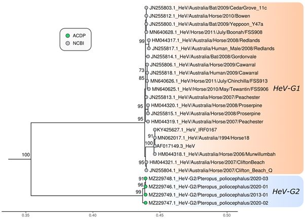
License: This work is licensed under a Creative Commons Attribution 4.0 International License. Read Full
License
Page 2/22
Abstract
Background
Hendra virus (HeV) has caused lethal disease outbreaks in humans and horses in Australia. Pteropid bats (flying
foxes) are the wildlife reservoir from which the virus was first isolated in 1996. Following a heat stress mortality
event in Australian flying foxes in 2013, a novel HeV variant was discovered. This study describes the subsequent
surveillance of Australian flying foxes for this novel virus over a nine year period using qRT-PCR testing of bat
tissues submitted primarily for Australian bat lyssavirus (ABLV) diagnosis. Genome sequencing and
characterisation of the novel HeV variant was also undertaken.

Methods
Spleen and kidney samples harvested from flying fox carcasses were initially screened with two real-time qRT-PCR
assays specific for the prototype HeV. Two additional qRT-PCR assays were developed specific for the HeV variant
first detected in samples from a flying fox in 2013. Next-generation sequencing and virus isolation was attempted
from selected samples to further characterise the new virus.

Results
Since 2013, 98 flying foxes were tested and 11 were positive for the new HeV variant. No samples were positive for
the original HeV. Ten of the positive samples were from grey-headed flying foxes (GHFF, Pteropus poliocephalus),
however this species was over-represented in the opportunistic sampling (83% of bats tested were GHFF). The
positive GHFF samples were collected from Victoria and South Australia and one positive Little red flying fox
(LRFF, Pteropus scapulatus) was collected from Western Australia. Immunohistochemistry (IHC) confirmed the
presence of henipavirus antigen, associated with an inflammatory lesion in cardiac blood vessels of one GHFF.
Positive samples were sequenced and the complete genome was obtained from three samples. When compared to
published HeV genomes, there was 84% sequence identity at the nucleotide level. Based on phylogenetic analyses,
the newly detected HeV belongs to the HeV species but occupies a distinct lineage. We have therefore designated
this virus HeV genotype 2 (HeV-G2). Attempts to isolate virus from PCR positive samples have not been successful.

Conclusions
A novel HeV genotype (HeV-G2) has been identified in two flying fox species submitted from three states in
Australia, indicating that the level of genetic diversity for HeV is broader than first recognised. Given its high
genetic relatedness to HeV, HeV-G2 should be considered a zoonotic pathogen.

Background
Hendra virus (HeV) belongs to the genus Henipavirus (family Paramyxoviridae, order Mononegavirales), which
currently includes five species: Cedar virus (CedV), Ghana virus (GhV), Hendra virus (HeV), Mojiang virus (MojV)
and Nipah virus (NiV) (1). Two members of the Henipavirus genus, HeV and CedV, have been detected in pteropid

                                                        Page 3/22
bats in Australia (2). HeV is highly pathogenic to horses and humans, while the pathogenicity of CedV remains
unknown (2).
HeV first emerged in Hendra, Australia in 1994 where the outbreak was responsible for the death of a horse trainer,
illness in a horse strapper, and the death of 20 horses (3). Five more human HeV cases have since occurred, of
which three have died, and all became infected after close contact with an infected horse (4; 5). Horses become
infected after exposure to excretions from flying foxes, also known as fruit bats, or pteropid bats (6).
Bats comprise the order Chiroptera, which is divided into two suborders, the Microchiroptera (microbats) and the
Megachiroptera (megabats). Megachiroptera comprise > 200 species and 43 genera confined to a single family, the
Pteropodidae (7). Pteropid bats belonging to the genus Pteropus within the Pteropodidae, have been identified as
reservoir hosts for several zoonotic viruses from the order Mononegavirales, namely HeV, NiV, Menangle virus and
Australian bat lyssavirus (ABLV).
HeV was first isolated from the uterine fluid of a pregnant grey headed flying fox (GHFF, Pteropus poliocephalus)
which aborted twin foetuses after being caught on a barbed-wire fence (8). Virus was also recovered from pooled
foetal liver and lung from the same flying fox, and from neonatal lung of a black flying fox (BFF, Pteropus alecto),
all collected in Brisbane in September 1996. Since 1996 there have been numerous surveillance studies, most
utilising a method of collecting urine underneath flying fox camps and testing by real-time PCR. Most positive
results have been at the limit of detection of real-time PCR and attempts to isolate the virus have usually not been
successful. Sero-surveys of flying foxes have been employed to determine infection prevalence but require the
capture and restraint of bats for serum collection, which is a labour-intensive activity with welfare implications for
the captured animals. To address these limitations and impacts, we have undertaken passive surveillance for the
molecular detection of HeV in flying fox specimens submitted to our laboratory for the primary purpose of ABLV
testing.
Here, we report the detection and identification of a novel HeV genotype (HeV-G2) from pteropid bats in Australia.
The virus was first detected from a GHFF in 2013 in Adelaide, South Australia, and has since been detected in two
other states. The findings from this study provides critical information about the diversity of henipaviruses in
pteropid bats in Australia and highlights the importance of enhanced surveillance of henipaviruses and other bat-
borne pathogens.
Methods
Sample collection and processing
Clinical specimens were collected from flying foxes, submitted to the Australian Centre for Disease Preparedness
(ACDP) for the diagnosis of ABLV during 2013 to 2021 from South Australia, Victoria, New South Wales,
Queensland, and Western Australia. Tissues were prepared as 10% (w/v) homogenates in Dulbecco's PBS (ph 7.6;
Oxoid) containing antibiotics (Sigma-Aldrich) using 1-mm silicon carbide beads (BioSpec Products) in a
FastPrep24 tissue homogenizer (MP Biomedical). Samples were clarified by low speed centrifugation (1000 xg, 5
min, 4oC) and the supernatant used for nucleic acid extraction and virus isolation.
Cell culture and virus isolation
Page 4/22Vero cells (ATCC CCL-81) and primary kidney cells derived from P. alecto (PaKi) (9) were used in this study. Vero
cells were cultured at 37°C in 25 cm2 flasks in EMEM (Gibco®, ThermoFisher Scientific) containing 10% foetal calf
serum (FCS; Gibco®, ThermoFisher Scientific), supplemented with 1% v/v L-glutamine (Sigma-Aldrich), 10mM
HEPES, 0.25% v/v penicillin-streptomycin (Sigma-Aldrich) and 0.5% v/v amphotericin B (Sigma-Aldrich) and). PaKi
cells were cultured in DMEM/F-12 media (Gibco®, ThermoFisher Scientific) with 5% FCS and the same
supplements.
For virus isolation, growth media was removed from tissue culture flasks, and cell monolayers were washed once
with PBS. For each specimen tested, cells were inoculated with 200 µl of tissue homogenate and incubated for 40
min to allow virus adsorption. Inoculum was then removed, and cell monolayers were washed with PBS, then
overlaid with culture media containing supplements and 1% (v/v) FCS. Cells were incubated at 37°C for 7 days and
observed regularly for signs of cytopathic effect by light microscopy. Cells were then frozen, thawed and the cell
suspension was centrifuged at 1000 xg at 4°C to remove cellular debris. Clarified supernatant (500 µl) was then
passaged on a fresh cell monolayer. A total of three passes per sample were performed on each cell line, except
one sample, for which Vero cells only were used. Passage 3 samples were tested by real time qRT-PCR to detect the
presence of replicating HeV genome.
Histopathology
Formalin fixed paraffin embedded (FFPE) tissues were available from one HeV-G2 positive bat. These were
processed by routine histological methods and sections were stained by haematoxylin and eosin and by HeV IHC
according to methods previously described (10). For the latter, the primary antibody was an antiserum from a
rabbit immunised against recombinant expressed Nipah virus nucleoprotein that cross-reacts with HeV.
Polymerase chain reaction (PCR)
RNA extraction
Fifty microlitre of supernatant from processed clinical specimens or following cell culture passage was used for
nucleotide extraction using the MagMax 96 Viral RNA Kit (ThermoFisher Scientific) in a MagMAX Express
Magnetic Particle Processor (ThermoFisher Scientific) following manufacturer’s instructions. Extracted RNA was
used for PCR testing immediately or stored at -80°C for further use.
Quantitative RT-PCR (qRT-PCR)
Two qRT-PCR assays for detection of HeV matrix (M) gene and nucleocapsid (N) gene (11; 12) were used during
initial screening of bat samples. Two additional qRT-PCR assays specific for the M and N genes of HeV-G2 were
developed. Primers and probes were designed by using Primer Express 3 (Applied BioSystems). The M gene assay
was used for initial testing of samples and the N gene assay was then used as a confirmatory test. Sequences of
primers and probes are shown in Table 1. The PCR was performed in 96-well plates in a 25 µL reaction volume
containing 5 µL of RNA, 12.5 µL of AgPath One-step RT-PCR buffer (Ambion), 1 µL of 25 x reverse transcriptase, 18
µM each primer, 1.25 µL of 5 µM TaqMan probe, and 2.75 µL of nuclease free water. The qRT-PCR assays were
performed under the following conditions: 10 min at 45°C for reverse transcription of RNA, 10 min at 95°C for
inactivation of reverse transcriptase, followed by 45 cycles of 95°C for 15sec, 60°C for 45sec using a 7500 Real-
time PCR system (Applied Biosystems)
Page 5/22Table 1
Primers and probes for qRT-PCR and conventional PCR
qRT-PCR
HeV-G2 M gene
Forward primer HeV-G2-M-F: 5’-CTGATCTACGTTACGGCAAACCTT-3’
Reverse primer HeV-G2-M-R: 5’-GGCCCGCTTCACCATCTCTTAC-3’
Probe HeV-G2-M-P: 5’-FAM-CAGCATTGAATATTGACCCGCCAGTCA-BHQ1-3’
HeV-G2 N gene
Forward primer HeV-G2-N-F: 5’-TGCGACAGATCCCAGTAGTATTAAAT-3’
Reverse primer HeV-G2-N-R: 5’-GGCAGCTTATTCGGCAAAAG-3’
Probe HeV-G2-N-P: 5’-FAM-CTCTGGTGACGGAACACAAATGCAAATTTC-BHQ1-3’
Conventional PCR
Primary RT-PCR Primer sequences PCR product
Forward primer HeV-M-5481F 5’- GCCCGCTTCATCATCTCTT-3’ 300 bp
Reverse primer HeV-M-5871R1: 5’CCACTTTGGTTCCGTCTTTG-3’
Semi-nested PCR
Forward primer HeV-M-5481F 5’- GCCCGCTTCATCATCTCTT-3’ 210 bp
Reverse primer HeV-M-5691R2: 5’-GCAATAGCGTTGTTCCTTCTG-3’
Conventional PCR and Sanger sequencing
A nested PCR for detection of HeV M gene was conducted on extracts of tissue samples positive by qRT-PCR.
Primer sequences are listed in Table 2. The RT-PCR was performed using SuperScript III One step RT-PCR System
with Platinum DNA Polymerase (Invitrogen) according to manufacturer’s instructions. Briefly, a 25 µl reaction
volume consisted of 12.5 µl 2 x reaction buffer, 1 µl of SuperScript III RT/Taq Mix, 0.5 µl of each forward and
reverse primer (10 µM), and 5 µl RNA. The PCR reaction was performed in a thermocycler (Eppendorf) under the
following conditions: 48°C for 30 mins, 94°C for 2 min, 40 cycles of 94°C 15 sec, 50°C for 30 sec, and 68°C for 45
sec. Nested PCR was performed using HotStarTaq Plus Master Mix Kit (Qiagen) according to the manufacturer’s
instructions. Each PCR reaction consisted of 12.5 µl of 2 x Taq polymerase buffer, 0.5 µl (10 µM) of each forward
and reverse primer 10 µM), 2.5 µl of CoralLoad concentrate (10x), 5 µl of the first round RT-PCR product.
Thermocycling conditions were: 95°C for 5 min, and 30 cycles of 95°C for 15 sec, 53°C for 30 sec, and 72°C for 45
sec. PCR products were visualised by gel electrophoresis and products of expected size were purified using
QIAQuick gel purification system (Qiagen). Purified PCR products were sequenced using the BigDye terminator v3.1
kit on an Applied Biosystems 3130xl Genetic Analyser (Applied biosystem). Sequences were analysed using
Geneious R11 (Geneious).
Page 6/22Table 2
Details of flying foxes tested for HeV-G2
Species No. tested No. positive State of Origin (No.)
Grey-headed flying fox 81 10 Victoria (73), South Australia (7), NSW (1)
Little red flying fox 3 1 Western Australia (3)
Black flying fox 3 0 Western Australia (3)
Unspecified flying fox 11 0 Western Australia (2), Queensland (3), Victoria (6)
Total 98 11
Next generation sequencing (NGS) and sequence analysis
Preparation of Illumina DNA libraries from total RNA
The NGS of initial samples from GHFF from South Australia in 2013 was conducted at Duke-NUS Medical School,
Singapore in 2016, and subsequent work with three GHFF from Victoria was carried out at ACDP, Australia. Two
different sequencing library preparation methods were used. At Duke-NUS, Illumina libraries were constructed from
total RNA using the NEBNext Ultra Directional RNA Library Prep Kit for Illumina (New England Biolabs) in
conjunction with NEBNext Multiplex Oligos for Illumina (New England Biolabs), according to the method described
previously (13). At ACDP, TruSeq RNA library Prep Kit V2 (Illumina) was utilised for DNA library preparation,
according to manufacturer’s instructions. The purified libraries were quantified using a Qubit Fluorometer and an
Agilent 2100 before enrichment.
Enrichment of viral library
An approach for viral sequence library enrichment was used as previously described (13). Targeted HeV-G2
genome enrichment was achieved using custom designed, biotinylated 120-mer xGen Lockdown baits (Integrated
DNA Technologies). Biotinylated DNA baits complementary to the consensus sequences of HeV and NiV (Table 2),
plus additional baits specific to HeV-G2 were designed (Table 3). The DNA baits were designed to tile the
henipavirus genome at intervals of approximately 500 nt. The xGen hybridisation and washing kit (Integrated DNA
Technologies) were used, and capture workflow was followed according to manufacturer’s instructions. The
enriched library was purified using the MinElute PCR Purification Kit (Qiagen) and quantified using a Qubit
Fluorometer and an Agilent 2100. The concentration of the final libraries was normalised and pooled in equimolar
ratios. The library pool was then loaded into a MiSeq Reagent Kit V2 (2 x 150 cycles; Illumina) and sequenced in a
MiSeq platform (Illumina) according to the manufacturer’s instructions.
Page 7/22Table 3
Details of HeV-G2 positive flying foxes; GHFF = grey-headed flying fox; LRFF = little red flying fox. *This bat
presented with clinical signs consistent with ABLV infection and tested positive to ABLV by qRT-PCR and FAT
(details available on request).
Year- Species Location Result Result Confirmed by Cause of death
Sample ID spleen (CT) kidney (CT) sequencing?
2013-01 GHFF South Positive Positive Yes suspect heat
Australia stress event
2015-01 LRFF Western Positive Negative No dog attack
Australia
2019-01 GHFF Victoria Positive Negative Yes caught in fruit
netting
2019-02 GHFF Victoria Positive Negative Yes caught in fruit
netting
2020-01 GHFF Victoria Positive Positive Yes no history
supplied
2020-02 GHFF Victoria Positive Positive Yes unspecified
trauma
2020-03 GHFF Victoria Negative Positive Yes suspect dog
attack
2021-01 GHFF Victoria Positive Negative No fractured wing
2021-02 GHFF South Positive Negative Yes dog attack
Australia
2021-03 GHFF South Positive Negative No dog attack
Australia
2021-04 GHFF Victoria Negative Positive Yes ABLV*
NGS data analysis
The NGS sequence data was analysed using CLC Genomic Workbench 20 (Qiagen) using standard parameters.
The raw reads were quality-trimmed. De novo assembly was then conducted with the unaligned sequence reads to
generate longer sequence contigs. The resultant sequences were analysed using the NCBI nonredundant
nucleotide database (BlastN) and protein database (BlastX)( https://blast.ncbi.nlm.nih.gov/Blast).
Gaps in the genome sequence obtained from NGS were resolved with multiple PCRs and Sanger sequencing, using
primers designed to flank missing regions (sequences available upon request).
Phylogenetic analysis
The assembled genomes were phylogenetically analysed with selected reference sequences from GenBank.
Multiple sequence alignments for the complete genome, N gene, G gene and L gene were undertaken using MAFFT
v.7.301 (14) with the auto option to select the most appropriate alignment parameters. Maximum likelihood trees
were then created using IQ-TREE v.2.0.6 (15) with model test enabled to choose the best fitting evolutionary models
for each dataset. The trees were visualised using the R package ggtree v.1.14.6 (16).
Page 8/22Results
Initial detection and identification of a novel HeV genotype
Spleen and kidney samples from a GHFF from South Australia, submitted in 2013 for ABLV exclusion, were tested
by qRT-PCR assays targeting the HeV M and N genes. While the M PCR was negative, HeV N was detected in spleen
and kidney samples, with a cycle threshold (Ct) of 38 and 34, respectively. This led us to further investigations
using Sanger sequencing to confirm the qRT-PCR results. NGS using two platforms, Roche 454 and Illumina MiSeq,
using unbiased methods was attempted but was unsuccessful and no viral sequences were detected (methods
available on request).
Nested PCR for HeV M produced a specific amplicon, approximately 300 bp in length, from the kidney sample only.
The PCR amplicon was sequenced and BLASTn analysis revealed 89% nucleotide sequence identity to HeV, 77–
79% to NiV, 66% to CedV, and 61% GhV.
The PCR was subsequently used for analysis of positive samples detected by qRT-PCR. Multiple single nucleotide
polymorphisms were observed between the different positive samples from different bats. Alignment of the
sequences is shown in Supplementary Fig. 1.
In the original submission, a range of tissues were collected into formalin and examined microscopically.
Histopathology showed infiltration and cuffing with mononuclear inflammatory cells within the full circumference
of one large blood vessel of the heart (Fig. 1A), and IHC showed granular henipavirus antigen present diffusely in
the wall of this vessel (Fig. 1B). Antigen was also detected in the wall of one small blood vessel in the same area
of myocardium.
Taken together, these results indicated the detection of a novel henipavirus, most closely related to HeV.
Screening of bat samples using qRT-PCR assay
Following the initial detection of HeV-G2 in 2013, a more sensitive qRT-PCR targeting the HeV-G2-M gene was
developed and applied to screening bat samples from a broader range of locations in Australia. From January
2013 to April 2021, spleen and kidney samples from 98 flying foxes were tested. The details of species, collection
location, and PCR results are summarised in Table 2. Of the 98 bats tested, the majority (n = 81) were GHFF, most
of which (n = 79) were from Victoria. Eleven of 98 bats tested positive by qRT-PCR, 10 of which were GHFF, and 1
was a little red flying fox (LRFF) (Table 3). No samples were positive for prototype HeV. The overall prevalence of
HeV-G2 in this study was 11.2%. Of the 11 positive bats, seven were from Victoria, three from South Australia, and
one LRFF from Western Australia (Table 3); ten spleen specimens tested positive, whilst only six kidney specimens
were positive (Table 4).
Page 9/22Table 4
Comparison of three qRT-PCR assays for the detection of HeV-G2, HeV N assay,
HeV-G2 M assay and HeV-G2 N assay using bat samples from 2013 to 2021;
values shown are CT values
Sample ID- sample type HeV N assay HeV-G2 M assay HeV-G2 N assay
2013-01-kidney* 31.3 28.8 NT**
2013-01-spleen 40.9 33.4 NT
2015-01-spleen Negative 38.4 Negative
2019-01-kidney Negative Negative Negative
2019-01-spleen Negative 34.2 NT
2019-02-kidney Negative Negative Negative
2019-02-spleen Negative 29.6 31.8
2020-01-kidney 43.2 30.6 28.3
2020-01-spleen 34.8 30.6 32.3
2020-02-kidney Neg 34.9 35.5
2020-02-spleen 34.4 23.1 23
2020-03-kidney 37.1 27.4 26.6
2020-03-spleen Negative Negative Negative
2021-01-kidney Negative Neg 41.8
2021-01-spleen Negative 33.2 40.3
2021-02-kidney Negative Negative Negative
2021-02-spleen Negative 37 41.3
2021-03-kidney Negative Negative Negative
2021-03-spleen Negative 36.9 40.2
2021-04-kidney 39 31.1 31.4
2021-04-spleen Negative Negative 36.8
Total No. of Positive 7/21 (14%) 14/21 (67%) 12/18(67%)
*Virus isolation was attempted for samples in bold
**NT, not tested
Establishing improved qRT-PCR assays for HeV-G2
In this study, HeV-G2 was detected by three qRT-PCR assays, HeV-N, HeV-G2-M and HeV-G2-N from the flying fox
samples. Samples that tested positive using the HeV-G2-M screening assay were then tested by HeV-G2-N gene
assay for confirmation. The sensitivity of the three qRT-PCR assays were compared using tissue samples (kidney
and spleen) from bats that tested positive. The two HeV-G2 assays developed in this study are more sensitive than
Page 10/22HeV-N gene assay for detection of HeV-G2, while both HeV-G2-M and N gene assay have equivalent sensitivity on
samples tested. (Table 4). To evaluate the specificity of the newly developed qRT-PCR assays, nine viral RNA
samples derived from HeV isolates from equine disease outbreaks in different years were tested. The HeV-G2-M
assay detected HeV in all nine samples, while all samples were negative in the HeV-G2-N assay, indicating that the
N assay is specific for HeV-G2 (Table 5). Two NiV RNA samples extracted from NiV-Malaysia and NiV-Bangladesh
isolates tested negative by both HeV-G2-M and -N assays, further supporting the specificity of the HeV assays.
Table 5
Comparison of qRT-PCR assays for the detection of HeV isolates belonging to
the prototype (G1) lineage
Year and Sample ID HeV-N assay HeV-G2-M assay HeV-G2-N assay
2010-01 30.8 29.9 Negative
2015-01 22.7 23.4 Negative
2015-02 21.9 24.8 Negative
2011-01 19.2 22.3 Negative
2011-02 20.3 22.4 Negative
2011-03 20.6 23.3 Negative
2011-04 19.4 18.9 Negative
2011-05 20.7 23.1 Negative
2011-06 30.8 32.4 Negative
Genomic characterisation by NGS
Spleen or kidney samples that had higher viral loads indicated by qRT-PCR from four GHFF were selected for
hybridization probe enrichment-based NGS analysis, which was not available when NGS was first attempted with
the 2013 HeV-G2. The samples generated 1.14–3.59 million paired-end reads per sample with 88–92% of bases ≥
Q30. Following quality trimming of the raw reads, de novo assembly was used to generate long sequence contigs.
Blastn analysis demonstrated highest nucleotide identity to HeV genomes, in agreement with the sequences of
conventional PCR amplicons. The viral genomes were then assembled based on the HeV genome (GenBank
Accession no. AF017149). The gaps in the genome were then filled using multiple PCRs followed by Sanger
sequencing.
Using this approach, we have successfully sequenced three full HeV-G2 genomes from three flying foxes from
Victoria, as well as a partial genome sequence (approximately 11,530 nt, equivalent to 63% genome) from the
flying fox from South Australia originally tested in 2013. The complete genomes are 18,234 nt in length, obeying
the ‘Rule-of-Six', which is observed for all known members of the subfamily Orthoparamyxovirinae (17) and the
genome organisation is identical to HeV. The predicted viral genes and their ORFs are as follows: nucleocapsid
protein (N) gene of 1599 nt encoding 532 aa; phosphoprotein (P) gene of 2124 nt encoding 707 aa; matrix protein
(M) gene of 1059 nt encoding 352 aa; fusion protein gene of 1641 nt encoding 546 aa; glycoprotein (G) gene of
1812 nt encoding 603 aa; large polymerase (L) protein gene of 6735 nt encoding 2244 aa. The sequences from the
four viruses have been submitted to GenBank under Accession Nos: MZ229746-MZ229749.
Page 11/22Comparative analysis of the novel bat HeV full genome with those of other henipaviruses available from GenBank
indicated highest nt identity (83.6%) to HeV (AF017149), followed by 71% nt identity to NiV Malaysia and
Bangladesh strains (AJ627196 and AY988601), respectively, 57% to CedV (NC025351), 55% to GhV (HQ660219),
and 55% to MojV (NC025352). The amino acid sequences of six individual proteins are highly homologous to
those of HeV (AF017149), with sequence identities of 96% for N protein, 82% for P protein, 96% M protein, 95% F
protein, 92% for G protein, and 95% for L protein. Comparison of nucleotide and protein sequences of each of the
six individual genes is demonstrated in Table 6. In addition, there are minor sequence variations among the novel
bat HeV genomes from this study, though they share over 99% nucleotide identity at genome level. HeV-G2/2020-
01 and − 03 are nearly identical with only 5nt differences and one aa change. HeV-G2/2020-02 is slightly diverse
from the other two, with 75 nt changes across the genome, and 13 aa changes in different coding regions (Table
7). Analysis of partial genome sequences from HeV-G2/2013-01 from South Australia revealed minor variations in
comparison with the 3 viruses from Victoria (data not shown).
Table 6
Comparison of sequences of HeV-G2 with other henipaviruses (figures are % similarity)
Virus Nucleocapsid Phosphatase Matrix Fusion Glycoprotein Polymerase
(N) (P) (M) (F) (G) (L)
nt2 aa3 nt aa nt aa nt aa nt aa nt aa
HeV 87 96 83 82 88 96 87 95 85 92 87 95
(G1)
NiV-M 77 91 70 64 77 90 75 87 71 79 75 87
NiV-B 77 91 71 64 77 90 74 86 71 79 75 87
CedV 62 60 53 27 62 61 54 42 54 30 58 52
GhV 60 56 54 28 62 62 57 52 54 29 59 53
MojV 56 48 53 21 61 60 53 40 51 21 58 52
1
Abbreviations: HeV: Hendra virus (AF017149); NiV-M: Nipah virus Malaysian strain (AJ627196); NiV-B, Nipah virus
Bangladesh strain (AY988601); ), CedV: Cedar virus (NC_025351), GhV: Ghana virus (HQ660129); MojV: Mojiang
virus (NC_025352)
2nucleotide sequence identities (%) against cognate nucleotides of HeV-G2 virus
3
amino acid sequence identities (%) against cognate proteins of HeV-G2 virus
Page 12/22Table 7
Comparison of nucleotide and amino acid sequence variations of HeV-G2 viruses when compared to the
consensus sequence for HeV-G2
Virus open reading frame
length (nt)
Full N P M F G L
genome
1,599 2,124 1,059 1,641 1,812 6,735
(%)
nt nt nt nt nt nt
aa aa aa aa aa aa
HeV/Australia/Pteropus poliocephalus/2020- 5* 1 0 1 0 0 0 1 0 1 1 0
01 (0.02%)# 0
HeV/Australia/Pteropus poliocephalus/2020- 75 7 0 9 3 4 0 11 1 6 24 7
02 (0.4%) 2
HeV/Australia/Pteropus poliocephalus/2020- 5 1 0 1 0 0 0 0 0 0 0 3
03 (0.03%) 0
*total number of nt and aa changes across the full genome, including NCRs
# percentage variation across the full genome, including NCRs, compared to?
Phylogenetic analysis
Maximum likelihood phylogenetic analysis based on the full genome, nucleocapsid (N), glycoprotein (G) and
polymerase (L) of the 3 novel genomes sequenced in this study and reference genomes of other members of the
genus Henipavirus, and subfamily Orthoparamyxovirinae was undertaken. The 3 sequences clustered together with
HeV, but as a separate sub-lineage in the phylogenetic trees of full genome, N and L protein, with 100% bootstrap
support for the two HeV sub-lineages (Figs. 2–4). Analysis of a partial region of the G gene, including the HeV-
G2/2013-01 virus revealed a very similar tree topology to the full genome and N gene phylogenetic trees
(Supplementary Fig. 2). Based on the ICTV species demarcation criteria, a new species in the genus Henipavirus is
defined as having a distance greater than 0.03 between the tip of the branch and the nearest node to the reference
sequence (GenBank Accession No. AAC83194.3) in a phylogenetic tree of the complete L protein (1). Phylogenetic
analysis of the complete L protein of the novel HeV detected in this study indicate that these viruses are HeV
species (branch length to the nearest node is < 0.03). As these viruses form a distinct sub-lineage, we propose to
designate this group as HeV genotype 2 (HeV-G2), and designate the previously identified sub-lineage, containing
the reference HeV sequence, as genotype 1 (HeV-G1). Therefore, the strain names of the 3 viruses with full genome
from 2020, and one virus with partial genome from 2013 are: HeV-G2/Australia/Pteropus poliocephalus/2020-01,
HeV-G2/Australia/Pteropus poliocephalus /2020-02, HeV-G2/Australia/Pteropus poliocephalus /2020-03, and HeV-
G2/Australia/Pteropus poliocephalus /2013-01.
Discussion
A new genotype of HeV has been identified in flying foxes in Australia. This follows the first detection of HeV-G1 in
Australian flying foxes in 1996 (8), and Cedar virus in 2012 (2). Histopathology, including IHC, indicated that this
Page 13/22virus replicates within blood vessels and is able to cause significant vascular disease in flying foxes. Molecular
characterisation of the HeV-G2 demonstrated highest genetic similarity to HeV-G1, when compared to other
members of the genus. Sequence variation was observed within HeV-G2 despite high sequence similarity (> 99% at
genome level) between each other. This is similar to the findings from HeV (G1) in flying foxes and horses (18; 19).
Due to the high genetic similarity between HeV-G1 and HeV-G2 it is highly possible that HeV-G2 is a zoonotic
pathogen posing potential risk to other species, including horses and humans. To date, there has been no report of
spill-over events of HeV-G2 in the areas where the virus has been detected in flying foxes. However, the recent
retrospective identification of a virus belonging to this sub-lineage from a 2015 case of equine neurological
disease in Queensland shows the HeV-G2 is able to infect horses and potentially cause disease (Annand et al.
submitted 2021). Further studies on epidemiology and pathogenicity of HeV-G2 are warranted.
In this study, 98 bats were tested by PCR and HeV-G2 was detected in 11, with a detection rate of 11.2%. In a
previous study (20), 2840 individual black flying-foxes were tested by qRT-PCR, and HeV was detected in 43
animals with 1.48% prevalence of detection. This is significantly lower than that for HeV-G2 in the current study,
however in Edson’s study (20) the sample type was not tissues; they sampled urine and swabs suggesting that
those samples have a lower sensitivity for the detection of virus. Additionally, the sensitivity and specificity of the
assay used in Edson’s study may not have been optimal for the detection of different henipaviruses, and they were
sampling healthy bats in the wild.
Ten of the eleven bats that tested positive for HeV-G2 were GHFF (Table 3). In another study by Edson et al. (21),
HeV was not detected in 1168 GHFF. Once again, the sample types were different; they were not tissues, but urine,
packed blood cells, serum and swabs. This, along with other studies where HeV was either not found or difficult to
find in excreted samples from GHFF (22–26) strongly suggests that excretion from GHFF is significantly lower
than in BFF. However, it is still unclear whether infection of HeV-G2 occurs in BFF, and what level of prevalence
exists, as only very limited numbers (n = 3) of BFF were tested in this study. It is interesting that one of three LRFF
from Western Australia tested positive for HeV-G2. However, this was at the limit of detection and could not be
confirmed by sequencing. Additional surveillance in these populations is needed.
Hendra and Nipah virus are serologically cross-protective (10). Although serologic evidence of HeV infection has
been found in all four flying foxes species in Australia, including the spectacled flying-fox, BFF, LRFF and GHFF
(27), the discovery of HeV-G2 raises the question of which variant the flying foxes have been exposed to – HeV-G1,
HeV-G2 and/or other variants. Current serological tests would not be able to differentiate between the different
variants.
Recent epidemiological studies have suggested that only the BFF and spectacled flying-fox, are the primary
reservoir hosts for HeV (20; 21; 28–30). Neither GHFF nor LRFF have been identified as the principal source of HeV
in spill-over events to horses, despite reports of high levels of seroprevalence to HeV in these species, and the
isolation of virus from GHFF (8; 23; 31). It is notable that in this study HeV-G2 appears to have a broad geographic
distribution, in areas of Australia that were previously deemed as low risk for HeV spill-over events.
Co-infection with HeV-G2 and ABLV was detected in one GHFF. This is the first time that this has been reported.
Virus co-infection in flying foxes has not been studied extensively but is known to occur. While mean viral
prevalence was low, multi-viral shedding from flying fox populations was common in one report, with up to eight
paramyxoviruses, which included HeV-G1, detected in one mixed colony (containing BFF and GHFF) at a single
point in time, and referred to as a ‘synchronous shedding pulse’ (32).
Page 14/22A qRT-PCR assay targeting the HeV N gene was used during initial detection of the HeV-G2 from GHFF from South
Australia in 2013, in addition to the HeV-G1-specific M gene-specific assay that failed to detect HeV-G2. This
demonstrates the benefit of using diagnostic qRT-CR tests that target multiple gene for disease investigations to
address circulating genetic variants. Subsequently, a new qRT-PCR assay based on the HeV-G2 M gene was
developed and applied to the screening of samples from flying foxes. This assay is more sensitive than the HeV N
gene assay for detection of HeV-G2, and it is also able to detect HeV belonging to the prototype lineage. The broad
reactivity of this assay could potentially be beneficial to the detection of mutant/variant forms of HeV in the future.
Through obtaining more sequences of HeV-G2 from GHFFs during this study, we developed an additional HeV-G2
specific qRT-PCR assay targeting the N gene. This assay is specific for HeV-G2 and is used as the confirmatory test
in our laboratory. Preliminary validation of both assays using bat samples submitted for disease exclusion, equine
samples from previous outbreaks and NiV isolates indicated that these tests perform with high levels of sensitivity
and specificity. Further assessment of these tests will be required for full validation according to World
Organisation for Animal Health principles for veterinary diagnostic tests (33).
As it is likely that HeV-G2 can spill over from flying foxes to other species, this novel virus is a zoonotic pathogen
that threatens animal and human health. These new assays enhance diagnostic capability through rapid and
specific detection of HeV-G2. The addition of capability for detection of HeV-G2 to current laboratory diagnostic
testing algorithms for the diagnosis of HeV infections in animals of different species, particularly horses, is of
critical importance. An updated testing protocol to incorporate these tests has been applied at ACDP.
In the present study, virus isolation was attempted using the continuous Vero cell line and primary Pteropus alecto
(kidney) cell culture. HeV-G2 was unable to be isolated from selected PCR positive GHFF samples. Previously, HeV
has been isolated from flying fox urine using these cell cultures, with equal success in either Vero cells or primary
bat cell lines (19). The unsuccessful attempts for isolation of HeV-G2 may be due to non-viability of the virus in the
samples. These samples were collected either from carcasses exposed to ambient temperature for unknown
periods, or euthanised and stored under suboptimal conditions for the preservation of live virus. The apparent low
virus load in some of the samples, as indicated by qRT-PCR results, may have also been a contributing factor.
The finding of viral antigen within an artery of the heart in association with a prominent mononuclear cell
inflammation indicates that HeV is able to cause vasculitis. This is the first time, to our knowledge, that a
henipavirus has been demonstrated to cause significant pathology in a flying fox. The lesion was localised and
limited in extent and therefore is unlikely to have caused serious illness in that individual. Nevertheless, this finding
challenges the notion that henipavirus infections in flying boxes do not cause disease. Further investigation on the
ability of these viruses to cause disease and illness in flying foxes is warranted.
Conclusions
The identification and characterisation of a novel HeV genotype, designated HeV-G2, in two species of flying fox
previously deemed to be low risk for HeV cross-species transmission, in regions where HeV spill-over has not
occurred, is a significant contribution to HeV epidemiology in Australia. The new variant causes vasculitis in the
flying fox host and it is probable it may cause illness in these and other species. Due to high levels of genome
similarity with the HeV prototype lineage, HeV-G2 is likely to be a zoonotic pathogen, posing a potentially
significant risk to different species of animals, particularly horses and humans. Indeed, the recent retrospective
detection of HeV-G2 infection in a 2015 equine neurological disease case strongly supports this hypothesis.
Further in-depth studies on the epidemiology, pathogenicity, and risk of spill-over of this new virus are warranted.
Page 15/22Abbreviations
ACDP: Australian Centre for Disease Preparedness; HeV: Hendra virus; HeV-G2: Hendra virus genotype 2; ABLV:
Australian bat lyssavirus; GHFF: grey-headed flying fox; Quantitative reverse transcription polymerase PCR (qRT-
PCR); NGS: Next-Generation Sequencing
Declarations
Ethics approval and consent to participate
Clinical specimens and deceased animals were submitted for diagnostic testing and were not subject to animal
ethics approval.
Consent for publication
Not available
Availability of data and materials
All data generated or analysed during this study are included in this published article and supplementary
information files. The genome sequences of the three HeV-G2 are available at the
GenBank(https://www.ncbi.nlm.nih.gov/genbank/).
Competing interests
The authors declare that they have no competing interests.
Funding
This work was supported by the Australian Government Department of Agriculture, Water and the Environment.
Authors’ contributions
JW, DW, and LFW conceived and designed the study. DEA and JW performed hybridization enrichment of NGS
library, XH performed bat post mortem and collected and processed samples; HC performed NGS; SW and SV
performed qRT-PCR testing, Bv performed virus isolation, MN performed phylogenetic analysis, JB performed
pathological analysis, JW and KH drafted and reviewed the manuscript, DW, DO and DE supervised the study and
reviewed manuscript. All authors have read and approved the final manuscript.
Acknowledgements
The authors acknowledge the capabilities of the Australian Centre for Disease Preparedness (grid.413322.5) in
undertaking this research, including infrastructure funded by the National Collaborative Research Infrastructure
Strategy. We are grateful to staff in the Molecular Diagnostics, Diagnostic Virology, Agent Characterization and
Sequencing, Histopathology and Veterinary Investigation teams, at the Australian Centre for Disease Preparedness,
for their excellent technical assistance. We also thank Department of Primary Industries and Regions (DPIR), South
Australia; AgriBio, Centre For AgriBioscience, Victoria; Department of Primary Industries and Regional Development,
Western Australia; Department of Primary Industries, New South Wales; and the Department of Agriculture and
Page 16/22Fisheries, Queensland, for providing samples. We acknowledge Dr Ina Smith (CSIRO) for contributions to the 2013
laboratory investigation at ACDP. We thank Dr Tom Zsiazek (US Centers for Disease Control) for providing NiV-
Bangladesh and Prof. Kenneth Lam (University of Malaya) for providing NiV-Malaysia.
Author details
1Australian Centre for Disease Preparedness (ACDP), Commonwealth Scientific and Industrial Research
Organisation (CSIRO), Geelong, Australia
2 Programme in Emerging, Infectious Diseases, Duke-NUS Medical School, Singapore
3SingHealth Duke-NUS Global Health Institute, Singapore
References
1. Rima B, Balkema-Buschmann A, Dundon WG, Duprex P, Easton A, Fouchier R, et al. ICTV Report Consortium
(December 2019). ICTV Virus Taxonomy Profile: Paramyxoviridae. J Gen Virol. 2019;100(12):1593–4.
2. Marsh GA, Jong Cde, Barr JA, Tachedjian M, Smith C, Middleton D, et al. Cedar virus: a novel henipavirus
isolated from Australian bats. PloS Pathog. 2012;8(8):e1002836. doi:10.1371/journal.ppat.1002836.
3. Murray K, Selleck P, Hooper P, Hyatt A, Gould A, Gleeson L, et al. (1995) A morbillivirus that caused fatal
disease in horses and humans. Science 1995;268:94–97.
4. Playford EG, McCall B, Smith G, Slinko V, Allen G, Smith I, Moore F, Taylor C, Kung Y-H, Field H. Human Hendra
virus encephalitis associated with equine outbreak, Australia, 2008. Emerg Infect Dis. 2010;16(2):219–23.
5. Taylor C, Playford EG, McBride WJH, McMahon J, Warrilow D. No evidence of prolonged Hendra virus
shedding by 2 patients. Emerg Infect Dis. 2012;18(12):2025–27.
6. Halpin K, Hyatt AD, Fogarty R, Middleton D, Bingham J, Epstein JH, et al. Pteropid bats are confirmed as the
reservoir hosts of henipaviruses: a comprehensive experimental study of virus transmission. Am J Trop Med
Hyg. 2011;85(5):946–51.
7. Nesi N, Tsagkogeorga G, Tsang SM, Nicolas V, Lalis A, Scanlon AT, et al. Interrogating Phylogenetic
Discordance Resolves Deep Splits in the Rapid Radiation of Old World Fruit Bats (Chiroptera: Pteropodidae).
Syst Biol. 2021 Mar 4:syab013. doi: 10.1093/sysbio/syab013.
8. Halpin K, Young PL, Field HE, Mackenzie JS. Isolation of Hendra virus from pteropid bats: a natural reservoir of
Hendra virus. J Gen Virol. 2000;81:1927–32.
9. Crameri G, Shawn T, Grimley S, McEachern JA, Marsh GA, Smith C, et al. Establishment, Immortalisation and
Characterisation of Pteropid Bat Cell Lines. PLoS One. 2009;11(12):e8266. doi:10.1371/journal.pone.0008266.
4).
10. World Organisation for Animal Health (OIE). (2018). – Chap. 3.1.14. Nipah and Hendra Virus Diseases. Manual
of Diagnostic Tests and Vaccines for Terrestrial Animals, 8th ed. OIE, Paris.
https://www.oie.int/fileadmin/Home/eng/Health_standards/tahm/3.01.14_NIPAH_HENDRA.pdf.
11. Smith IL, Halpin K, Warrilow D, Smith GA. Development of a fluorogenic RT-PCR assay (TaqMan) for the
detection of Hendra virus. Virol Methods. 2001;98(1):33–40.
12. Feldman KS, Foord A, Heine HG, Smith IL, Boyd V, Marsh GA, et al. Design and evaluation of consensus PCR
assays for henipaviruses. J Virol Methods. 2009;161(1):52–7.
Page 17/2213. Singanallur NB, Anderson DE, Sessions OM, Kamaraj US, Bowden TR, Horsington J, et al. Probe capture
enrichment next-generation sequencing of complete foot-and-mouth disease virus genomes in clinical
samples. J Virol Methods. 2019;272:113703. doi:10.1016/j.jviromet.2019.113703.
14. Katoh K, Standley DM. MAFFT multiple sequence alignment software version 7: Improvements in performance
and usability. Mol Biol Evol. 2013;30:772–80.
15. Nguyen LT, Schmidt HA, Von Haeseler A, Minh BQIQ-TREE. A fast and effective stochastic algorithm for
estimating maximum-likelihood phylogenies. Mol Biol Evol. 2015;32:268–74.
16. Yu G, Smith DK, Zhu H, Guan Y, Lam TTY. Ggtree: an R Package for Visualization and Annotation of
Phylogenetic Trees With Their Covariates and Other Associated Data. Methods Ecol Evol. 2017;8:28–36.
17. Halpin K, Bankamp B, Harcourt BH, Bellini WJ, Rota PA. Nipah virus conforms to the rule of six in a
minigenome replication assay. J Gen Virol. 2004;85(3):701–7.
18. Marsh GA, Todd S, Foord A, Hansson E, Davies K, Wright L, et al. Genome sequence conservation of Hendra
virus isolates during spillover to horses, Australia. Emerg Infect Dis. 2010 Nov;16(11):1767–69.
doi:10.3201/eid1611.100501.
19. Smith I, Broos A, Jong C de3, Zeddeman A, Smith C, Greg Smith G, et al. Identifying Hendra virus diversity in
Pteropid bats. PLoS One. 2011;6(9):e25275. doi:10.1371/journal.pone.0025275.
20. Edson DW, Field HE, McMichael LA, Vidgen M, Goldspink L, Broos A, et al. Routes of Hendra virus excretion in
naturally-infectedflying-foxes: implications for viral transmission and spillover risk. PLoS One.
2015a;10(10):e0140670. doi:10.1371/journal.pone.0140670.
21. Edson DW, Field HE, McMichael LA, Jordan D, Kung N, Mayer D, et al. Flying-fox roost disturbance and Hendra
virus spillover risk. PLOS One. 2015b;10(5):e0125881. doi:10.1371/journal.pone.0125881.
22. Breed AC, Breed MF, Meers J, Field HE. Evidence of Endemic Hendra Virus Infection in Flying-Foxes (Pteropus
conspicillatus)-Implications for Disease Risk Management. PLoS One. 2011; 6(12):: e28816. doi:
10.1371/journal.pone.0028816.
23. Burroughs AL, Durr PA, Boyd V, Graham K, White JR, Todd S, et al. Hendra Virus Infection Dynamics in the Grey-
Headed Flying Fox (Pteropus poliocephalus) at the Southern-Most Extent of Its Range: Further Evidence This
Species Does Not Readily Transmit the Virus to Horses. PLoS One. 2016;15(6):e0155252.
doi:10.1371/journal.pone.0155252. 11 ) .
24. Field H, Crameri G, Kung NY, Wang LF. Ecological aspects of hendra virus. Curr Top Microbiol Immunol.
2012;359:11–23. doi:10.1007/82_2012_214.
25. Field H, Jordan D, Edson D, Morris S, Melville D, Parry-Jones K, et al. Spatiotemporal aspects of Hendra virus
infection in pteropid bats(flying-foxes) in eastern Australia. PloS ONE. 2015;10:e0144055.
doi:10.1371/journal.pone.0144055.
26. Plowright RK, Eby P, Hudson PJ, Smith IL, Westcott D, Bryden WL, et al. Ecological dynamics of emerging bat
virus spillover. Proceedings of the Royal Society B-Biological Sciences. 2015; 282(1798) :20142124. doi:
10.1098/rspb.2014.2124.
27. Field HE. (2005) The ecology of Hendra virus and Australian bat lyssavirus. PhD thesis, University of
Queensland.
28. Field HE, de Jong C, Melville D, Smith C, Smith I, Broos A, et al. Hendra Virus Infection Dynamics in Australian
Fruit Bats. PLoS One. 2011;6(12):e28678. doi:10.1371/journal.pone.0028678.
Page 18/2229. Goldspink LK, Edson DW, Vidgen ME, Bingham J, Field HE, Smith CS. Natural Hendra virus infection in flying-
foxes - tissue tropism and risk factors. PloS One. 2015;10(6):e0128835.
30. Smith C, Skelly C, Kung N, Roberts B, Field H. (2014) Flying-fox species density–a spatial risk factor for Hendra
virus infection in horses in eastern Australia. 2014; 9(6): e99965. doi: 10.1371/journal.pone.0099965.
31. Plowright RK, Field HE, Smith C, Divljan A, Palmer C, Tabor G, et al Reproduction and nutritional stress are risk
factors for Hendra virus infection in little red. flying foxes (Pteropus scapulatus). Proceedings of the Royal
Society B-Biological Sciences. 2008;275(1636):861–9.
32. Peel AJ, Wells K, Giles J, Boyd V, Burroughs A, Edson D, Crameri G, Baker ML, Field H, Wang LF, McCallum H,
Plowright RK, Clark N. Synchronous shedding of multiple bat paramyxoviruses coincides with peak periods of
Hendra virus spillover. Emerg Microbes Infect. 2019;8(1):1314–23.
33. World Organisation for Animal Health (OIE). (2019). – Chap. 1.1.6. Principles and methods of validation of
diagnostic assays for infectious diseases. In Manual of Diagnostic Tests and Vaccines for Terrestrial Animals,
8th Ed. OIE, Paris, France. Available at: .
Figures
Page 19/22Figure 1
Heart tissue from the grey headed flying fox from South Australia in 2013, showing mononuclear cell inflammation
of an artery (A, haematoxylin and eosin stain). The walls of this artery (B) and a nearby small blood vessel (C)
contained viral antigen (IHC for henipavirus nucleoprotein).
Page 20/22Figure 2
Maximum likelihood phylogenetic tree of complete Hendra virus genomes available in GenBank. The TN model
with gamma rate heterogeneity was used as the best fit by IQ-TREE v.2.0.6. The results from 1000 bootstrap
replicates are given on the nodes (if greater than 70) and the scale represents the number of nucleotide
substitutions per site. The tree was drawn with Nipah virus outgroups which were removed for visualisation.
Figure 3
Page 21/22Maximum likelihood phylogenetic tree of N gene nucleotide sequences. The TIM2 model allowing for a proportion
of invariant sites was used as the best fit by IQ-TREE v.2.0.6. The results from 1000 bootstrap replicates are given
on the nodes (if greater than 70) and the scale represents the number of nucleotide substitutions per site. The tree
was drawn with Nipah virus outgroups which were removed for visualisation.
Figure 4
Maximum likelihood phylogenetic tree of L gene amino acid sequences. The tree was created using the JTT
substitution model in IQ-Tree v.2.0.6 as per ICTV guidelines for the family Paramyxoviridae (Rima et al., 2019). The
results from 100 bootstrap replicates are given on the nodes (if greater than 70) and the scale represents the
number of substitutions per site. The zoomed inset has lengths written on the branches specifically showing the
distance between HeV-G1 and HeV-G2.
Supplementary Files
This is a list of supplementary files associated with this preprint. Click to download.
Additionalfile1TableS1.docx
Additionalfile2TableS2.docx
Additionalfile3FigS1.docx
Additionalfile4FigS2.docx
Page 22/22You can also read