A near miss: subclinical saddle pulmonary embolism diagnosed by handheld ultrasound - Oxford Academic Journals
←
→
Page content transcription
If your browser does not render page correctly, please read the page content below
Oxford Medical Case Reports, 2021;4,127–130

                                                                              doi: 10.1093/omcr/omab011
                                                                              Case Report

CASE REPORT

A near miss: subclinical saddle pulmonary embolism

                                                                                                                                                         Downloaded from https://academic.oup.com/omcr/article/2021/4/omab011/6255677 by guest on 25 December 2021
diagnosed by handheld ultrasound
Travis M. Skipina† , S. Allan Petty and Christopher T. Kelly
Department of Internal Medicine, Wake Forest Baptist Medical Center, Winston-Salem, NC, USA
*Correspondence address. Department of Internal Medicine, Wake Forest Baptist Medical Center, 1 Medical Center Boulevard, Winston Salem, NC 27157,
USA. Tel: +1 336 716 5039; Fax: +1 336 716 4995; E-mail: tskipina@wakehealth.edu

Abstract
Introduction: Pulmonary embolism (PE) is a life-threatening condition characterized by occlusive disease of the pulmonary
vasculature. Point-of-care ultrasound (POCUS) of right ventricular strain patterns have high specificity and low sensitivity
for diagnosis. Here, we describe a patient with a saddle PE and low pre-test probability who was diagnosed primarily by
handheld POCUS. Case Report: An 80-year old female was admitted to the intensive care unit with hypotension and lactic
acidosis. She also had mild leukocytosis and troponinemia. No other clinical or metabolic abnormalities were present. After
transfer to the f loor, handheld POCUS demonstrated D-sign and McConnell’s sign. Computed tomography angiography
showed a saddle PE involving both main pulmonary arteries. The patient was immediately initiated on anticoagulation
without further complications. Conclusion: Handheld POCUS is inexpensive, carries a low risk of harm and is an invaluable
extension of the physical exam when interpreted in the appropriate context.

INTRODUCTION                                                                  admitted to the intensive care unit (ICU) with hypotension and
                                                                              lactic acidosis following a 2-week history of progressive altered
Pulmonary embolism (PE) is a life-threatening condition charac-
                                                                              mental status and physical decline. Initial blood pressure was
terized by partial or complete occlusion of the pulmonary vascu-
                                                                              89/56 with a normal heart rate and oxygen saturation. No fever
lature potentially leading to cardiovascular collapse [1]. Despite a
                                                                              or tachypnea was present. Lactic acid was 3.6 mmol/L. There was
variety of validated clinical scoring systems, there are still cases
                                                                              a leukocytosis of 14.7 x 10³ cells/μL with neutrophilic predom-
that go unrecognized thus conferring increased morbidity and
                                                                              inance. Troponin peaked at 1104 pg/mL. No other laboratory or
mortality risk. Bedside point-of-care ultrasound (POCUS) of right
                                                                              clinical abnormalities were present. Wells score was 0 points; age
ventricular (RV) strain patterns have high specificity and low
                                                                              was the only of the PE rule-out criteria met [4]. D-dimer was not
sensitivity for diagnosing PE [2]. Despite these diagnostic advan-
                                                                              obtained on admission. She was admitted for presumptive septic
tages, POCUS is not routinely used by internists in non-critical
                                                                              shock and treated with fluids and broad-spectrum antibiotics.
care settings [3]. Here, we describe a patient with a saddle PE and
                                                                              Following these interventions, her blood pressure improved and
low clinical pre-test probability who was diagnosed primarily by
                                                                              her lactic acidosis resolved. The troponin elevation was felt to be
POCUS using a handheld ultrasound device (HUD).
                                                                              secondary to demand ischemia from sepsis. Blood cultures were
                                                                              negative. Per family, she had reached her pre-hospital baseline.
CASE REPORT                                                                   After two nights of hemodynamic stability and improvement in
An 80-year old female with a history of Alzheimer’s dementia,                 the ICU, she was transferred to the gerontology unit for further
diabetes mellitus, hypertension, and chronic kidney disease was               care and discharge planning.

† Travis
       M. Skipina, http://orcid.org/0000-0002-5577-0307
Received: December 6, 2020; Revised: January 19, 2021; Accepted: January 22, 2021
© The Author(s) 2021. Published by Oxford University Press. All rights reserved. For Permissions, please email: journals.permissions@oup.com
This is an Open Access article distributed under the terms of the Creative Commons Attribution Non-Commercial License (http://creativecommons.org/
licenses/by-nc/4.0/), which permits non-commercial re-use, distribution, and reproduction in any medium, provided the original work is properly cited.
For commercial re-use, please contact journals.permissions@oup.com

                                                                                                                                                127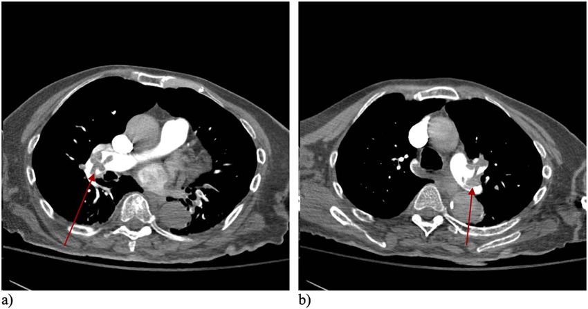
128 T.M. Skipina et al.
Downloaded from https://academic.oup.com/omcr/article/2021/4/omab011/6255677 by guest on 25 December 2021
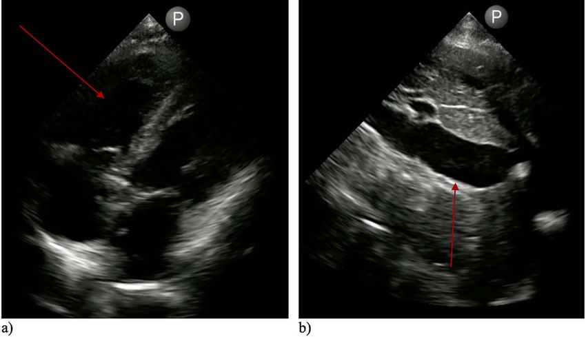
Figure 1: Bedside POCUS demonstrating McConnell’s sign (a) and a dilated IVC with minimal inspiratory variation (b).
Figure 2: ECG showing new S1Q3T3 pattern of right heart strain.
As part of initial assessment on arrival to the gerontology Table 1: Sensitivity and specificity of various diagnostic modalities
unit, the patient was afebrile, heart rate 80, blood pressure for the diagnosis of PE
117/62, SpO2 99% on room air. Jugular venous distension was
Diagnostic modality Sensitivity Specificity
present with a normal cardiopulmonary auscultatory exam. No
peripheral edema or asymmetric lower extremity swelling was CT angiography 83–100% 89–96%
present. Cardiac POCUS using a HUD revealed a dilated RV with POCUS 45–61% 74–90%
akinesis of the mid-RV free wall with apical sparing, consistent ECG (S1Q3T3 pattern) 54% 62%
with McConnell’s sign (Fig. 1a). Diastolic septal flattening was D-dimer 99.5% 41%
also present in the parasternal short-axis view, consistent with
D-sign. The inferior vena cava (IVC) was dilated to 3.0 cm with
minimal inspiratory variation (Fig. 1b). These findings were
confirmed with formal transthoracic echocardiography. An
electrocardiogram (ECG) showed evidence of an S1Q3T3 pattern apixaban without any hemodynamic compromise or further
not present in prior exams (Fig. 2). D-dimer was 51 190 ng/mL. complications for the remainder of her hospitalization. On
Computed tomography (CT) angiography of the chest showed a review of her home medications, the patient was taking
saddle PE involving both main pulmonary arteries with evidence megestrol as an outpatient. This was thought to be the etiology
of RV strain (Fig. 3). The patient was immediately initiated of her thromboembolism, so no further hypercoagulability
on a heparin infusion and subsequently transitioned to oral workup was pursued.Subclinical saddle pulmonary embolism 129
Downloaded from https://academic.oup.com/omcr/article/2021/4/omab011/6255677 by guest on 25 December 2021
Figure 3: Axial CT angiography showing saddle pulmonary emboli with evidence of filling defects in the right (a) and left (b) main pulmonary arteries.
DISCUSSION in any organization or entity with any financial interest or
non-financial interest in the subject matter or materials dis-
This case illustrates the diagnostic potential of POCUS using
cussed in this manuscript.
an HUD in patients with low pretest probabilities of disease,
especially in the context of undifferentiated shock or critical
illness. More specifically, since echocardiographic assessment of
RV strain patterns have high specificity and low sensitivity for PE FUNDING
[2], POCUS using HUDs can be successfully used as a ‘rule-in’ test
None.
for patients with more ambiguous presentations. Table 1 details
the sensitivity and specificity of various diagnostic modalities
for the diagnosis of PE.
POCUS is already standard of care in most emergency depart- ETHICAL APPROVAL
ments and critical care settings. However, it is only sparingly
This case report was conducted in accordance with the Decla-
used among non-critically ill patients in hospital wards tradi-
ration of Helsinki. The collection and evaluation of all protected
tionally managed by internists [3]. With the advent of HUDs,
patient health information was performed in a HIPAA-compliant
this technology is becoming increasingly more affordable and
manner. This case report does not generate generalizable knowl-
accessible for providers [5]. Further, providers can achieve exam-
edge about a disease or condition and therefore does not meet
ination and interpretation proficiency in a relatively short period
the national policy for the protection of human subjects’ defini-
of time (∼25–50 exams), especially if training is received in a
tion of research, so approval from the institutional review board
structured fashion [5, 6]. Standardization of POCUS competency
was not sought.
is still in its infancy, but current data shows that simulation-
based training is the most efficacious [7, 8]. HUDs have less
spatial and temporal resolution and lack many of the more
advanced features of standard echocardiography; however, mul- CONSENT
tiple studies have shown that HUDs have more diagnostic accu-
racy than physical exam alone and competent practitioners pro- The authors have obtained informed consent from the patient’s
duce results that correlate well with standard echocardiography healthcare power of attorney. Measures have been taken to pro-
[5, 9, 10]. tect the patient’s anonymity including omission of the patient’s
Overall, POCUS is inexpensive, carries a low risk of harm name, initials, medical record numbers and other identifiable
and continues to serve as an invaluable extension of the phys- characteristics from the manuscript and figures. An informed
ical exam when interpreted in the appropriate context by a consent document is on file with the authors which conforms
competent practitioner. With the advent of HUDs offering rapid with HIPPA compliance standards.
assessment, this technology adds to the diagnostic repertoire of
the general internist and may be routinely used to improve the
pathway of PE diagnosis.
GUARANTOR
Travis M. Skipina.
ACKNOWLEDGEMENTS
There were no further contributions to this manuscript beyond
those of the listed authors. REFERENCES
Conflicts of Interest Statement. The authors whose names are 1. Goldhaber SZ, Bounameaux H. Pulmonary embolism and
listed certify that they have no affiliations with or involvement deep vein thrombosis. Lancet 2012;379:1835–46.130 T.M. Skipina et al.
2. Fields JM, Davis J, Girson L, Au A, Potts J, Morgan CJ et al. 7. Jensen JK, Dyre L, Jorgensen ME, Andreasen LA, Tolsgaard
Transthoracic echocardiography for diagnosing pulmonary MG. Simulation-based point-of-care ultrasound training: a
embolism: a systematic review and meta-analysis. J Am Soc matter of competency rather than volume. Acta Anaesthesiol
Echocardiogr 2017;30:714–723.e4. Scand 2018;62:811–9.
3. LoPresti CM, Schnobrich DJ, Dversdal RK, Schembri F. A 8. Hu KC, Salcedo D, Kang YN, Lin CW, Hsu CW, Cheng CY
road map for point-of-care ultrasound training in internal et al. Impact of virtual reality anatomy training on ultrasound
medicine residency. Ultrasound J 2019;11:10. competency development: a randomized controlled trial.
4. Kline JA, Courtney DM, Kabrhel C, Moore CL, Smithline HA, PLoS One 2020;15:e0242731.
Plewa MC et al. Prospective multicenter evaluation of the 9. Cardim N, Dalen H, Voigt JU, Ionescu A, Price S, Neskovic
pulmonary embolism rule-out criteria. J Thromb Haemost AN et al. The use of handheld ultrasound devices: a posi-
2008;6:772–80. tion statement of the European Association of Cardiovas-
5. Savino K, Ambrosio G. Handheld ultrasound and focused cular Imaging (2018 update). Eur Heart J Cardiovasc Imaging
Downloaded from https://academic.oup.com/omcr/article/2021/4/omab011/6255677 by guest on 25 December 2021
cardiovascular echography: use and information. Medicina 2019;20:245–52.
(Kaunas) 2019;55:423. doi:10.3390/medicina55080423 10. Giusca S, Jurcut R, Ticulescu R, Dumitru D, Vladaia A, Savu
6. Schnobrich DJ, Mathews BK, Trappey BE, Muthyala BK, Olson O et al. Accuracy of handheld echocardiography for bedside
APJ. Entrusting internal medicine residents to use point of diagnostic evaluation in a tertiary cardiology center: com-
care ultrasound: towards improved assessment and super- parison with standard echocardiography. Echocardiography
vision. Med Teach 2018;40:1130–5. 2011;28:136–41.You can also read