A CROSS-SECTIONAL OVERVIEW OF SARS-COV-2 GENOME VARIATIONS IN TURKEY TÜRKIYE KAYNAKL I SARS-COV-2 GENOM VARYASYONLAR ININ KESITSEL DE ĞERLENDIRMESI
←
→
Page content transcription
If your browser does not render page correctly, please read the page content below
Turk J Biochem 2021; aop

Research Article

Koray Ergünay, Mücahit Kaya, Muhittin Serdar, Yakut Akyön and Engin Yılmaz*

A cross-sectional overview of SARS-CoV-2
genome variations in Turkey
[Türkiye kaynaklı SARS-CoV-2 genom
varyasyonlarının kesitsel değerlendirmesi]
https://doi.org/10.1515/tjb-2021-0119                                  genomes (98.3%) belonged in the SARS-CoV-2 haplogroup
Received May 22, 2021; accepted August 11, 2021;                       A. No evidence for recombination was identified in genomes
published online August 20, 2021                                       representing sub-haplogroup branches. The variants B.1.1.7,
                                                                       B.1.351 and P.1 were detected, with a statistically-significant
Abstract
                                                                       time-associated increase in B.1.1.7 prevalence.
                                                                       Conclusions: We described prominent SARS-CoV-2 varia-
Objectives: We assessed SARS-CoV-2 genome diversity
                                                                       tions as well as comparisons with global virus diversity.
and probable impact on epidemiology, immune response
                                                                       Continuing a molecular surveillance in agreement with
and clinical disease in Turkey.
                                                                       local disease epidemiology appears to be crucial, as
Materials and methods: Complete genomes and partial
                                                                       vaccination and mitigation efforts are ongoing.
Spike (S) sequences were accessed from the Global Initia-
tive on Sharing Avian Influenza Data (GISAID) database.                Keywords: COVID-19; genome; mutation; SARS-CoV-2;
The genomes were analysed for variations and recombi-                  Turkey; variant.
nations using appropriate softwares.
Results: Four hundred ten complete genomes and 206 S                   Öz
region sequences were included. Overall, 1,200 distinct
nucleotide variations were noted. Mean variation count was             Amaç: Türkiye kaynaklı SARS-CoV-2 genomik dizi çeşitli-
14.2 per genome and increased significantly during the                  liğinin ve epidemiyoloji, bağışık yanıt ve hastalık seyri
course of the pandemic. The most frequent variations were              üzerine muhtemel etkili varyasyonların incelenmesi.
identified as A23403G (D614G; 92.9,%), C14408T (P323L,                  Gereç ve Yöntem: “Global Initiative on Sharing Avian
92.2%), C3037T (89.8%), C241T (83.4%) and GGG28881AAC                  Influenza Data” (GISAID) veri tabanında yer alan tüm
(RG203KR, 62.6%). The A23403G mutation was the most                    genom ve “Spike” (S) bölgesine ait verilere ulaşılarak
frequent variation in the S region sequences (99%). Most               uygun yazılımlar yardımıyla varyasyon ve muhtemel
                                                                       rekombinasyonlar araştırıldı.
                                                                       Bulgular: Toplam 410 tam genom ve 206 S bölgesi dizisi
The manuscript is available as a preprint as https://doi.org/10.       incelendi, 1200 farklı nükleotid düzeyinde varyasyon
21203/rs.3.rs-472330/v1.                                               saptandı. Genom başına toplam varyasyon ortalaması 14.2
*Corresponding author: Engin Yılmaz, PhD, Department of Medical
                                                                       olarak hesaplandı ve salgının ilerleyişi süresince ista-
Biology, Faculty of Medicine, Hacettepe University, Sihhiye, 06100,    tistiksel olarak anlamlı artış izlendi. En sık saptanan var-
Ankara, Turkey, E-mail: eyilmaz@hacettepe.edu.tr. https://orcid.org/   yasyonlar A23403G (D614G; 92.9,%), C14408T (P323L,
0000-0001-8873-7645
                                                                       92.2%), C3037T (89.8%), C241T (83.4%) ve GGG28881AAC
Koray Ergünay and Yakut Akyön, Department of Medical
Microbiology, Faculty of Medicine, Hacettepe Üniversity, Ankara,       (RG203KR, 62.6%) olarak sıralandı. S bölgesi dizilerinde
Turkey. https://orcid.org/0000-0001-5422-1982 (K. Ergünay)             de A23403G, en yaygın varyasyon (%99) şeklinde izlendi.
Mücahit Kaya, Department of Molecular Biology and Genetics, Faculty    İncelenen genomların sıklıkla (%98.3) SARS-CoV-2 A
of Arts and Sciences, Ondokuz Mayıs University, Samsun, Turkey
                                                                       haplogrubuna ait olduğu görüldü. Alt haplogrupları temsil
Muhittin Serdar, Department of Medical Biochemistry, Faculty of
Medicine, Acıbadem Mehmet Ali Aydınlar University, Istanbul, Turkey.   eden genomlarda yapılan incelemelerde rekombinasyon
https://orcid.org/0000-0002-3014-748X                                  bulgusu saptanmadı. Çalışma grubunda B.1.1.7, B.1.351 ve
  Open Access. © 2021 Koray Ergünay et al., published by De Gruyter.     This work is licensed under the Creative Commons Attribution 4.0
International License.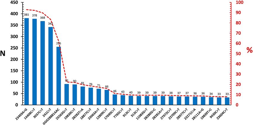
2        Ergünay et al.: SARS-CoV-2 genome variations

P.1 varyantları tespit edildi. B.1.1.7 prevalansında izlenen      available. As the vaccination efforts are spreading in many
zamanla ilişkili artış, istatistiksel olarak anlamlı bulundu.     countries, the monitorization of the virus genetic diversity
Sonuçlar: Çalışmada ülkemiz kaynaklı SARS-CoV-2                   will continue to be crucial to identify variants with enhanced
genomlarında belli başlı varyasyonlar belirlenmiş ve küre-        transmissibility or clinical disease as well as potential
sel virüs çeşitliliği ışığında değerlendirilmiştir. Kontrol ve    escape mutants [9]. In this study, we aimed to evaluate
aşılama çalışmaları ile eşzamanlı olarak önemli varyantların      SARS-CoV-2 genomic diversity from Turkey since the emer-
tanımlanabilmesi için, bölgesel epidemiyoloji ile uyumlu          gence of initial cases, to gain insights into origin, patterns of
moleküler sürveyans çalışmaları sürdürülmelidir.                  variation and their probable impact on epidemiology.

Anahtar kelimeler: COVID-19; genom; mutasyon; SARS-CoV-
2; Türkiye; varyant.
                                                                  Materials and methods
                                                                  The “Global Initiative on Sharing Avian Influenza Data” (GISAID) [10]
Introduction                                                      database (https://www.gisaid.org) was screened for SARS-CoV-2
                                                                  sequences originating from Turkey and all complete virus genomes
The severe acute respiratory syndrome coronavirus 2               dated between 16.03.2020 and 30.01.2021 were accessed (Supplement
                                                                  1). High quality virus genomes larger than 29.000 nucleotides with less
(SARS-CoV-2) is the causative agent of the pandemic of the
                                                                  than 1% N content and 0.05% specific amino acid variation were
21st century, also called as Coronavirus Disease-2019             selected. Recurrent submissions and genomes with unverified inser-
(COVID-19) [1, 2]. Initially emerging as cases of atypical        tion/deletions were omitted. The SARS-CoV-2 Wuhan-Hu-1 isolate
viral pneumonia in the Wuhan city, Hubei province of              genome sequence (GISAID access: EPI_ISL_402125, GenBank access:
China, SARS-CoV-2 have readily spread around the globe,           MN908947.3, NC045512) was used as the reference. Sequences were
                                                                  aligned using MUSCLE within the MSA package (v1.22.0), based on the R
currently affecting millions of people in more than 220
                                                                  platform (v4.0.3, 2020-10-10) [11, 12]. Nucleotide variations were iden-
countries. More than a year after its official announcement,
                                                                  tified by the Sequence Variation (SNP) package in the Virus Pathogen
the control of the pandemic and mitigating its health,            Resource (ViPR) database and further confirmed via MEGAX [13–15].
economic and social impact have become the foremost               Probable recombinations were screened using the R8DP, Geneconv,
priority in all countries and international organizations.        Bootscan, MaxChi, Chimaera, SiScan, 3Seq tools of the RDP4 software in
     SARS-CoV-2, classified in the Betacoronavirus genus of       the default settings [16]. A total of 410 genomes were analysed.
                                                                        To incorporate recent data in the analysis, we further screened
the family Coronaviridae, is an enveloped particle with a
                                                                  spike region sequences uploaded at a later date in the database and
positive-stranded RNA as its genome [3]. The viral genome         accessed a total of 206 non-redundant sequences, collected between
is approximately 29.9 kb in size and flanked by untrans-           30.04.2020–03.02.2021 but mostly during January 2021 (170/206)
lated regions (UTRs) in the 3′ and 5′ ends. Virus proteins        (Supplement 1). Descriptive statistics, data distribution and correla-
comprising the structural components nucleocapsid (N),            tions were analysed using Analyse-it v4.20.1 (Analyse-it Software Ltd.
                                                                  Leeds, United Kingdom).
envelope (E), membrane (M) and spike (S), as well as
                                                                        The study and its findings are based on virus genome sequences
several accessory and non-structural proteins (NSPs) are
                                                                  obtained from online sources and is not related to either human or
encoded by several open reading frames (ORFs) on the              animal use. Therefore, no institutional ethics board approval was
genome [4, 5]. Variations occur spontaneously during              required nor sought. The study was approved by the COVID-19
SARS-CoV-2 replication but mutation frequency is relati-          Scientific Research Evaluation Committee, the Ministry of Health of
vely low compared to other RNA viruses, owing to the              Turkey (2021-03-22T11_13_48).

activity of nsp14 exoribonuclease [6, 7].
     The exploration of SARS-CoV-2 genetic diversity in has
been pivotal for control efforts since the beginning of the       Results
pandemic. Sequencing of the virus genome has provided
critical information for the investigation of origin and spread   Among the 410 SARS-CoV-2 genomes included in the
of the infection as well as development of appropriate            study, 261 (63%) originated from specimens collected
diagnostics. It further enables surveillance of virus mole-       during March–June 2020, 27 (6.58%) in July–October 2020
cular epidemiology and identification of variants that can        and 122 (29.75%) in December 2020–January 2021. Age,
undermine mitigation and vaccination efforts [8, 9]. Aided        gender, residence and other demographic or clinical fea-
by the widespread availability of next generation sequen-         tures of infected cases were not evaluated due to the lack of
cing technologies and online sharing of genome informa-           sufficient information in the database.
tion, a significant number of virus genomes originating from            Complete genome sequencing revealed a total of 1,200
several countries affected by the pandemic has now been           different nucleotide variations (Table 1). Majority of the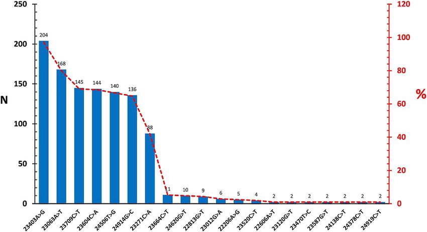
Ergünay et al.: SARS-CoV-2 genome variations 3
variations were missense (661/1,200, 55.1%) and silent as well as B cell epitopes in S1 spike region [18, 19] (Table 2).
mutations (441/1,200, 36.7%). The variations were fre- Other include the S region T cell epitope variations C24378T
quently located in ORF1a/1b region (684/1,200, 57%), that (n=2) ve C24381T (n=2), and N region B cell epitope varia-
encodes for the proteins and cofactors participating in viral tions G29383A (n=1) and G29405C (n=2).
replication; as well as in S region (153/1,200, 12.5%), We further evaluated the diversity of the SARS-CoV-2
involved in virus attachment on the respiratory epithelium genomes as previously-described mutation-annotated
and the main target for neutralizing antibodies (Table 1). virus lineages and haplotypes [8]. Majority of the genomes
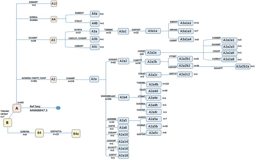
Median variation count per genome was calculated as (403/410, 98.3%) were observed to belong in haplogroup
12 (mean + standard deviation: 14.2 + 6.5, range: 4–36). A, from which various subhaplogroups were further iden-
Temporal distribution of the total variation count per tified (A2: n=381, 92.9%; A3: n=39, 9.5%; A9: n=29, 7.1%;
genome according to sampling date revealed a positive A12: n=2, 0.5%) (Figure 3). The most frequent subhaplog-
correlation and statistically-significant increase during the roup was noted as A2a, that diversifies from A2 by the
course of the pandemic (Figure 1). C14408T variation. Haplogroup B genomes were relatively
Variations frequently-identified in the SARS-CoV-2 few (7/410, 1.7%), with subhaplogroups B4 and B4a rep-
genomes were overviewed in Figure 2. Here, the most resented with 8 and 23 genomes, respectively (Figure 3). No
commonly-observed variations representing 62.2–92.9% evidence of recombination was observed among viruses of
of the diversity repertoire resulted in amino acid substi- the A2a2b2a, A3a1a, A12 and B4a subhaplogroups, repre-
tution; whereas silent changes and non-ORF mutations senting highly-diversified genomes within the group.
were also noted (Table 2). Among these, the A23403G, In the spike region dataset, a total of 44 nucleotide
C14408T and GGG28881AAC variations stood out, as variations comprising missense (32/206, 72.7%) and silent
they are reported to be associated with severe infections (12/206, 29.3%) mutations (Figure 4). The most frequent
requiring hospitalization [8]. Further screening for variation was observed as A23403G (204/206, 99%), similar
previously-reported globally-prevalent variations revea- to the complete genome dataset. Of the virus epitopes
led G25563T (ORF3, Q57H) (n=92), C1059T (nsp2, L37F) located in the spike protein, the C24378T (S939F) variation,
(n=9), C8782T (n=7) and C17747T (nsp13, P504L) (n=1) in affecting predicted T cell epitope was detected in 2 (0.9%)
the study groups [8, 17]. sequences.
We also checked particular mutations, previously- Finally, particular SARS-CoV-2 variants with potential
reported to potentially affect predicted T and B cell epito- impact on transmission, disease course and immune pro-
pes in S, N ve M proteins [18]. The most frequent of such tection were screened in the study groups [20–22]. These
mutations is the A23403G, resulting in the D614G substi- were detected in 30.2% (186/616) of the individual viruses
tution, which is also reported to enhance virus infectivity where the B.1.1.7 variant (variant 1 or 501Y.V 1) was noted
Table : Spatial distribution of the variations observed in SARS-CoV- genomes.
Region Location Variation type Total
Missense mutation Silent mutation Nonsense mutation Deletion Non-ORF change
′UTR – – – – –
ORFa –, – –
ORF b ,–, – – –
S ,–, – – –
ORFa ,–, – – –
E ,–, – – –
M ,–, – – –
ORF ,–, – – –
ORFa ,–, – –
ORFb ,–, – – –
ORF ,–, – –
N ,–,
ORF ,–, – – –
′UTR ,–, – – – –
Total ,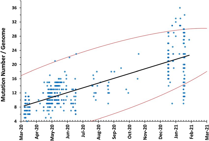
4         Ergünay et al.: SARS-CoV-2 genome variations

                                                                                             Figure 1: Temporal distribution of the total
                                                                                             variation counts in the study. Spearman
                                                                                             test was used to calculate correlation
                                                                                             coefficient. Red lines indicate 95%
                                                                                             confidence intervals. A time-related, sta-
                                                                                             tistically-significant increase total varia-
                                                                                             tion count per genome was observed
                                                                                             (r=0.776, p
Ergünay et al.: SARS-CoV-2 genome variations 5
Table : Common variations and predicted outcomes in the course of the pandemic [8]. However, SARS-CoV-2
SARS-CoV- genomes. isolates from Turkey was proposed to exhibit an elevated
variation rate in a study focusing on 166 virus genomes
Variation Frequency, Target region Outcome
accessed during July 2020, where frequently-detected
n, %
variations C14408T and C18877T, affecting viral polyme-
AG , . S – spike protein Amino acid
rase (nsp12) and exoribonuclease (nsp14), respectively;
change:
were suggested as a possible precipitating factors [26–29].
DG
CT , . Non-structural protein Amino acid These variations were also noted in our study with varying
– RNA-dependent change: rates (Table 2, Figure 2). In addition, the co-detection of
RNA polymerase PL C14408T and A23403G variations were suggested to be
CT , . Non-structural protein Silent associated with increased diversity [27, 28] In parallel
– phosphoesterase
with global isolates, the SARS-CoV-2 genome variations in
CT , . ′UTR Non-ORF
GGGAAC , . N – nucleocapsid Amino acid Turkey are mostly missense or silent mutations, fre-
protein change: quently involving the enzymes and co-factors, participa-
RGKR ting in replication in ORF1a/1b or the S regions of the virus
genome [8, 17, 27].
In the study, the most frequently-detected variations,
namely the A23403G, C14408T and GGG28881AAC muta-
statistically-significant accumulation of variations during tions resulting in amino acid substitutions in the corres-
the 10-month period examined (Figure 1). Comparable ponding virus proteins, were reported in previous analyses
findings were reported in globally-distributed virus isola- from Turkey [23–26]. However, they seem to be positively-
tes, with more than 3,000 specific point mutations being selected in the local virus population pool, as their abun-
detected and an increased frequency of variation during dance seem to be elevated. For example, the A23403G
Figure 3: Distribution of the SARS-CoV-2 haplogroups in the study group. Branch-delineating variations and genome frequencies are
indicated.6 Ergünay et al.: SARS-CoV-2 genome variations
Figure 4: Distribution of the frequently-observed variations in spike region dataset.
variation was reported as low as 56.2% in previous reports, epidemiology [8, 17]. Previous reports on SARS-CoV-2
while it is detected in 92.9% of the complete genomes and genetic diversity have described particular virus lineages
99% in S regions in this study (Figure 2, Figure 4). This and clades, mostly in overall agreement but lacking a uni-
observation is also evident in global genome data, where form nomenclature [8, 30]. The size of accumulating
viruses with the A23403G and C14408T variations were sequence data further warrants more practical approaches
steadily increased in frequency during the course of the to indicate phylogeographic relationships than standard
pandemic and have become the majority in late 2020 [8]. phylogenetic reconstruction. Here, we adopted a previously-
The amino acid substitutions occurring as a result of these reported mutation-annotated reference strategy to describe
variations, namely P323L, D614G ve RG203KR, were also intraspecific phylogeny of SARS-CoV-2. We observed the
associated with a more severe COVID-19 clinical pre- majority of the SARS-CoV-2 genomes from Turkey to belong
sentation [8]. Moreover, the D614G mutation, a defining in the haplogroup A (98.3%), with main subhaplotype
component of the variant of concern B.1.1.7, is also likely to diversification into A2 (Figure 3). SARS-CoV-2 haplogroup A
affect immune responses to the S protein. In addition to the isolates constitute the ancestral node and predominant
high frequency of this substitution in the study, we further clade across the world. They are frequently-represented in
identified other variations that might affect T and B cell isolates from Europe (97%) Africa (93%) and Asia (77%), but
epitopes, albeit with lower rates. relatively scarce in South America (68%) and North America
Throughout the pandemic, the availability of virus (53%) [8]. Among global haplogroup A subclades, A2 and
genome sequences and powerful online tools have enabled A2a appears as the majority, with the phylogeographic
a nearly real-time monitorization of SARS-CoV-2 molecular inferences indicating a European origin. We observed hap-
logroup B is with a much lower frequency, and B4a sub-
Table : Distribution of the current variants of concern in the study haplotype representing the majority within this group
groups. (Figure 3). Haplogroup B viruses have been identified in all
continents, with higher prevalence in North America (47%),
Variant Genome S region Total
South America (32%), Asia and Oceania (23%), with all
(n=) (n=)
major and minor subclades present in Asia [8]. The hap-
n % n % lotype B viruses were introduced in Turkey likely by travel to
B... . . endemic regions and further local spread was presumably
B.. . . prevented by isolation. Our analyses employing highly-
P. – – . diversified subclades of each main haplogroup failed to
Total –
identify any evidence for recombination among localErgünay et al.: SARS-CoV-2 genome variations 7
SARS-CoV-2 genomes. Overall, the findings on virus genome Competing interests: The authors declare no actual or
diversity in Turkey suggest several introductions originating potential conflict of interest in relation with the article.
from multiple sources and subsequent local adaptation, also Çıkar çatışması beyanı: Yazarların makale içeriği ile ilgili
noted in previous reports using smaller datasets [23]. herhangi bir gerçek ya da muhtemel çıkar çatışması
The emergence and rapid spread of SARS-CoV-2 bulunmamaktadır.
variants has raised significant concern, due to their potential
for enhanced transmissibility, altered clinical progression
and escape from protective immune response induced by
previous infection or widely-available vaccines [20, 31]. Also References
dubbed as the variant of concern (VOC), these viruses
1. Zhu N, Zhang D, Wang W, Li X, Yang B, Song J, et al. A novel
exhibit a wide array of amino acid changes accumulated in
coronavirus from patients with pneumonia in China, 2019. N Engl
several regions of the virus genome including the spike J Med 2020;382:727–33.
protein [20–22]. The rapid spread of particular VOCs in 2. Cucinotta D, Maurizio Vanelli M. WHO declares COVID-19 a
several countries during fall 2020 called for more stringent pandemic. Acta Biomed 2020;91:157–60.
public health measures as well as targeted monitorization, 3. Gorbalenya AE, Baker SC, Baric RS, de Groot RJ, Drosten C,
which is also initiated and currently ongoing in Turkey. We Anastasia A, et al. The species severe acute respiratory
syndrome-related coronavirus: classifying 2019-nCoV and
detected three major VOCs in the study group, with increa-
naming it SARS-CoV-2. Nat Microbiol 2020;5:536–44.
sed prevalence of B.1.1.7 and B.1.351 in the recently-dated 4. Lu R, Zhao X, Li J, Niu P, Yang B, Wu H, et al. Genomic
dataset (Table 3). Moreover, the detection of P.1 in this group characterisation and epidemiology of 2019 novel coronavirus:
suggests not only an elevated prevalence but also a broader implications for virus origins and receptor binding. Lancet 2020;
repertoire of variants in the population. These findings 395:565–74.
justify the efforts to identify and monitor known and 5. Wu A, Peng Y, Huang B, Ding X, Wang X, Niu P, et al. Genome
composition and divergence of the novel coronavirus
potentially-emerging virus variants.
(2019-nCoV) originating in China. Cell Host Microbe 2020;27:
Particular limitations of this study need to be addres- 325–8.
sed. An important issue is the heterogeneity in temporal 6. Grubaugh ND, Petrone ME, Holmes EC. We shouldn’t worry when a
and spatial distribution of the samples employed for virus mutates during disease outbreaks. Nat Microbiol 2020;5:
genome sequencing, which suggests a lack of organized 529–30.
7. Ogando NS, Zevenhoven-Dobbe JC, van der Meer Y, Bredenbeek
sampling strategy for screening. In addition, missing
PJ, Posthuma CC, Snijder EJ. The enzymatic activity of the nsp14
demographic and location data in many instances also exoribonuclease Is critical for replication of MERS-CoV and
prevented further evaluations. Therefore, it is not possible SARS-CoV-2. J Virol 2020;94:e01246–20.
to assess whether the current dataset fully represents the 8. Flores-Alanis A, Cruz-Rangel A, Rodríguez-Gómez F, González J,
epidemiology and diversity in circulating viruses in Tur- Torres-Guerrero CA, Delgado G, et al. Molecular epidemiology
key. A continuous and organized surveillance strategy in surveillance of SARS-CoV-2: mutations and genetic diversity one
year after emerging. Pathogens 2021;10:184.
conjunction with local transmission dynamics and infec-
9. Gómez-Carballa A, Bello X, Pardo-Seco J, Martinón-Torres F, Salas
tion epidemiology, will provide a better understanding of A. Mapping genome variation of SARS-CoV-2 worldwide
the SARS-CoV-2 molecular epidemiology in Turkey. highlights the impact of COVID-19 super-spreaders. Genome Res
In conclusion, in this analysis of complete and partial 2020;30:1434–48.
SARS-CoV-2 genome sequences almost covering the first 10. Shu Y, McCauley J. GISAID: global initiative on sharing all
influenza data – from vision to reality. Euro Surveill 2017;22:
year since emergence, we described main variations asso-
30494.
ciated with epidemiology and immune response, with the 11. Bodenhofer U, Bonatesta E, Horejš-Kainrath C, Hochreiter S.
observation of increased incidence of VOCs in Turkey. With MSA: an R package for multiple sequence alignment.
the ongoing pandemic and accelerated vaccination cam- Bioinformatics 2015;31:3997–9.
paigns, such investigations should be performed periodi- 12. Edgar RC. MUSCLE: multiple sequence alignment with high
cally for precise screening and coordination of control accuracy and high throughput. Nucleic Acids Res 2004;32:
measures. 1792–7.
13. Pickett BE, Greer DS, Zhang Y, Stewart L, Zhou L, Sun G, et al.
Virus pathogen database and analysis resource (ViPR): a
Acknowledgment: No funding was received for the study.
comprehensive bioinformatics database and analysis resource
The authors are grateful to Diagen Biyoteknolojik Sistemler
for the coronavirus research community. Viruses 2012;4:
Inc., for technical support during sequence handling. 3209–26.
Information on GISAID sequence records is provided as a 14. Crooks GE, Hon G, Chandonia JM, Brenner SE. WebLogo: a
separate file (Acknowledgement Table). sequence logo generator. Genome Res 2004;14:1188–90.8 Ergünay et al.: SARS-CoV-2 genome variations
15. Kumar S, Stecher G, Li M, Knyaz C, Tamura K. MEGA X: molecular 24. Demir AB, Benvenuto D, AbacioĞlu H, Angeletti S, Ciccozzi M.
evolutionary genetics analysis across computing platforms. Mol Turk J Biol 2020;44:178–84.
Biol Evol 2018;35:1547–9. 25. Hanifehnezhad A, Kehribar EŞ, Öztop S, Sheraz A, Kasırga S,
16. Martin DP, Murrell B, Khoosal A, Muhire B. Detecting and Ergünay K, et al. Characterization of local SARS-CoV-2 isolates
analyzing genetic recombination using RDP4. In: Keith J, editor. and pathogenicity in IFNAR-/- mice. Heliyon 2020;6:e05116.
Bioinformatics. methods in molecular biology. New York: 26. Eskier D, Akalp E, Dalan Ö, Karakülah G, Oktay Y. Current
Humana Press; 2017, vol 1525:433–60 pp. mutatome of SARS-CoV-2 in Turkey reveals mutations of interest.
17. Mercatelli D, Giorgi FM. Geographic and genomic Turk J Biol 2021;45:104–13.
distribution of SARS-CoV-2 mutations. Front Microbiol 2020;11: 27. Koçhan N, Eskier D, Suner A, Karakülah G, Oktay Y. Different
1800. selection dynamics of S and RdRp between SARS-CoV-2 genomes
18. Koyama T, Weeraratne D, Snowdon JL, Parida L. Emergence of drift with and without the dominant mutations. Infect Genet Evol 2021;
variants that may affect COVID-19 vaccine development and 91:104796.
antibody treatment. Pathogens 2020;9:324. 28. Eskier D, Karakülah G, Suner A, Oktay Y. RdRp mutations are
19. Korber B, Fischer WM, Gnanakaran S, Yoon H, Theiler J, Abfalterer associated with SARS-CoV-2 genome evolution. PeerJ 2020;8:
W, et al. Tracking changes in SARS-CoV-2 spike: evidence that e9587.
D614G increases infectivity of the COVID-19 virus. Cell 2020;182: 29. Eskier D, Suner A, Oktay Y, Karakülah G. Mutations of
812–27. SARS-CoV-2 nsp14 exhibit strong association with increased
20. Tang JW, Tambyah PA, Hui DS. Emergence of a new SARS-CoV-2 genome-wide mutation load. PeerJ 2020;8:e10181.
variant in the UK. J Infect 2020;S0163-4453:30786–6. 30. Alm E, Broberg EK, Connor T, Hodcroft EB, Komissarov AB,
21. Tegally H, Wilkinson E, Giovanetti M, Iranzadeh A, Maurer-Stroh S, et al. Geographical and temporal distribution of
Fonseca V, Giandhari J, et al. Emergence and rapid spread of a SARS-CoV-2 clades in the WHO European region, January to June
new severe acute respiratory syndrome-related coronavirus 2020. Euro Surveill 2020;25:2001410.
2 (SARS-CoV-2) lineage with multiple spike mutations in South 31. Davies NG, Jarvis CI, CMMID COVID-19 Working Group, Edmunds
Africa. Nature 2021;6:e0035321. WJ, Jewell NP, Diaz-Ordaz K, et al. Increased mortality in
22. Toovey OTR, Harvey KN, Bird PW, Tang JWW. Introduction of community-tested cases of SARS-CoV-2 lineage B.1.1.7. Nature
Brazilian SARS-CoV-2 484K.V2 related variants into the UK. J 2021;593:270–4.
Infect 2021;82:e23–4.
23. Adebali O, Bircan A, Çirci D, İşlek B, Kilinç Z, Selçuk B, et al.
Phylogenetic analysis of SARS-CoV-2 genomes in Turkey. Turk J Supplementary Material: The online version of this article offers
Biol 2020;44:146–56. supplementary material (https://doi.org/10.1515/tjb-2021-0119).You can also read