A complete joint treatment - therapy
←
→
Page content transcription
If your browser does not render page correctly, please read the page content below
therapy

A complete
joint treatment

    I-ONE® THERAPY DELIVERS A SIGNAL PERMEATING THE ENTIRE EXTENSION
      AND DEPTH OF THE ARTICULAR CARTILAGE AS WELL AS THE ARTICULAR
                 STRUCTURES AND THE SUBCHONDRAL BONE.

                                                                 Sinovia
     Cartilage                                                   I-ONE® therapy exerts an anti-
                                                                 inflammatory effect, decreasing
                                                                 the release of catabolic factors
     I-ONE® therapy stimulates
                                                                 (TNF-α, IL-6, IL-8, IL-1β, PGE2)
     the synthesis of the cartilage
                                                                 and increasing the production
     matrix and exercises a
                                                                 of anabolic factors (IL-10,
     chondroprotective effect.
                                                                 TGF-β1).

                                                                  Subchondral
                                                                  bone
                                                                  I-ONE® therapy prevents the
                                                                  sclerosis of the subchondral
                                                                  bone and facilitates the bone
                                                                  edema reabsorption.

I-ONE® therapy performs a triple action:
                                                    1      ANTI-INFLAMMATORY ACTION
                                                           ON THE WHOLE ARTICULATION

                                                2       ANABOLIC ACTION ON
                                                        THE CARTILAGE

                                           3        TROPHIC ACTION
                                                    ON THE SUBCONDRAL BONE

2
» ACTUAL SIZE IMAGE «
A modern, innovative
and reliable
technology
95,5 mm
69 mm
»PORTABLE DEVICE«
Sites treatable with
I-ONE® therapy
Uniqueness
A biophysical signal covered by international patents
makes I-ONE® therapy unique
and not reproducible. Shoulder
Safety
The therapy parameters are preset by IGEA Elbow
and can not be modified by the patient, in
compliance with the current legislation. To guarantee
safety, effectiveness and simplicity of use I-ONE®
therapy is entirely manageable with a single button.
Wrist
Compliance
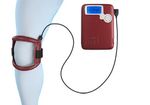
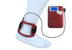
A light, flexible and ergonomic coil guarantees the best Knee
possible freedom of movement.
Efficacy
Similarly to what happens with drug, the effectiveness
of the therapy is guaranteed by a homogeneous and
correct distribution of the biophysical signal in the area
Ankle
to be treated.
3Early Osteoarthritis
I-ONE® therapy is indicated in patients with grade 0-2 osteoarthritis, according to the Kellgren-
Lawrence classification, presenting pain and functional limitation.
therapy
• Exerts a chondroprotective effect
• Controls pain
• Improves joint functionality
KOOS SCALE
100
90
pBone Edema / SONK
I-ONE® therapy is indicated in symptomatic patients with acute or chronic bone edema of
idiopathic, post-traumatic or degenerative origin.
therapy
• Enhances the process of edema reabsorption
• Resolves pain and improves activity level
• Delays the arthroplasty surgery
VAS
10
6
pAlgodistrophy
I-ONE® therapy is indicated in patients with Type I algodistrophy or CRPS (Complex Regional Pain
Syndrome).
therapy
• Controls the joint inflammatory process
• Resolves pain
• Inhibits osteoclastogenesis
QUOTED by SICM GUIDELINES and by
TUSCANY REGION GUIDELINES.
TRIGGER EVENT
Uncontrolled response
of the sympathetic Inflammation,
nervous system edema, pain
I-ONE® therapy
ACTING ON
INFLAMMATION,
Localized osteoporosis Lack of joint edema and pain,
Osteonecrosis mobility ALLOWS TO STOP THE
VICIOUS CIRCLE
CLINICAL CASE
BEFORE I-ONE® therapy AFTER
Borelli PP. Chir Mano, Vol. 54(3) 2017
6Patellofemoral Pain Syndrome
I-ONE® therapy is indicated in patients with Patellofemoral Pain Syndrome (PFPS) complaining
about pain located in the anterior part of the knee when walking or doing sporting activity.
therapy
• Resolves pain
• Reduces NSAIDs consumption
• Allows a fast return to sporting activity
VISA score variation
50
40
I-ONE® therapy
30
20 p = 0.001
RESUMPTION p = 0.010
10
OF SPORTING CONTROL
ACTIVITY
0
-10
0 2 4 6 8 10 12 14
Months
VAS scale variation
1
0
-1
p = 0.012 CONTROL
-2
-3 p = 0.015
-4
p = 0.003
PAIN -5
I-ONE® therapy
RESOLUTION -6
-7
-8
0 2 4 6 8 10 12 14
Months
Iammarrone Servodio C et al. Bioelectromagnetics, 2015
7Clinical indications
• EARLY OSTEOARTHRITIS • BONE EDEMA / SONK
• JOINT INFLAMMATION • PATELLOFEMORAL PAIN SYNDROME
• INTRA ARTICULAR EFFUSION • ALGODISTROPHY (CRPS)
Daily treatment time: 4 hours. Treatment duration: 30-60 days.
The therapy can be repeated.
References
• Benazzo F et al. Cartilage repair with osteochondral autografts in sheep: • Veronesi F et al. In vivo effect of two different pulsed electromagnetic
effect of biophysical stimulation with pulsed electromagnetic fields. J field frequencies on osteoarthritis. J Orthop Res. 2014 May;32(5):677-85
Orthop Res. 2008 May;26(5):631-42 • Fini M et al. Razionale d’uso della stimolazione biofisica nell’algodistrofia.
• Fini M et al. Effect of pulsed electromagnetic field stimulation on knee Chirurgia della Mano - Vol. 52 (3) 2015
cartilage, subchondral and epyphiseal trabecular bone of aged Dunkin • Veronesi F et al. Experimentally induced cartilage degeneration treated
Hartley guinea pigs. Biomed Pharmacother. 2008 Dec;62(10):709-15 by pulsed electromagnetic field stimulation; an in vitro study on bovine
• Gobbi A et al. L’uso dei campi elettromagnetici pulsati in pazienti cartilage. BMC Musculoskelet Disord. 2015 Oct 20;16(1):308
sintomatici con lesioni degenerative della cartilagine del ginocchio: un • Veronesi F et al. Pulsed electromagnetic fields combined with
rapporto preliminare. Journal of Sports Traumatology. 2011;Vol. 28, No. a collagenous scaffold and bone marrow concentrate enhance
4. Dicembre 2011 osteochondral regeneration: an in vivo study. BMC Musculoskelet
• Ongaro A et al. Chondroprotective effects of pulsed electromagnetic fields Disord. 2015 Sep 2;16:233
on human cartilage explants. Bioelectromagnetics. 2011 Oct;32(7):543-51 • Massari L. La stimolazione biofisica articolare. Giornale Italiano di
• Bruscoli R. Necrosi del CFM del ginocchio in un podista master. Ortopedia e Traumatologia. 2016;42(Suppl.1):S73-S78
Trattamento con CEMP. ReaLiMe, Maggio 2012;14:15 • Servodio Iammarrone C et al. Is there a role of pulsed electromagnetic
• Ongaro A et al. Electromagnetic fields (EMFs) and adenosine receptors fields in management of patellofemoral pain syndrome? Randomized
modulate prostaglandin E(2) and cytokine release in human osteoarthritic controlled study at one year follow-up. Bioelectromagnetics. 2016
synovial fibroblasts. J Cell Physiol. 2012 Jun;227(6):2461-9 Feb;37(2):81-8
• Marcheggiani Muccioli GM et al. Conservative treatment of spontaneous • Massari L et al. Impiego clinico della stimolazione elettrica in ortopedia
osteonecrosis of the knee in the early stage: Pulsed electromagnetic e traumatologia. Giornale Italiano di Ortopedia e Traumatologia.
fields therapy. Eur J Radiol. 2013 Mar;82(3):530-7 2017;43:105-106
• Gobbi A et al. Symptomatic Early Osteoarthritis of the Knee Treated With • Pagani S et al. Complex Regional Pain Syndrome Type I, a Debilitating and
Pulsed Electromagnetic Fields: Two-Year Follow-up. Cartilage. 2014 Apr ; Poorly Understood Syndrome. Possible Role for Pulsed Electromagnetic
5(2) :76-83 Fields: A Narrative Review. Pain Physician. 2017 Sep;20(6):E807-E822
• Ongaro A et al. Pulsed electromagnetic fields stimulate osteogenic • Varani K et al. Adenosine Receptors as a Biological Pathway for the Anti-
differentiation in human bone marrow and adipose tissue derived Inflammatory and Beneficial Effects of Low Frequency Low Energy Pulsed
mesenchymal stem cells. Bioelectromagnetics. 2014 Sep;35(6):426-36 Electromagnetic Fields,” Mediators of Inflammation, vol. 2017, Article ID
2740963, 2017. doi:10.1155/2017/2740963
This folder refers to the medical device Model CBA-03, Series I-ONE.
The device complies with the Medical Device Directive 93/42/EEC and its revised version. The device is marked 0051.
The device complies with the standard IEC 60601-1 - for the basic safety and essential performance of Medical electrical equipment.
The device complies with the standard IEC 60601-1-11 for the Medical electrical equipment used in the home healthcare environment.
IGEA/XXX
Via Parmenide, 10/A | 41012 Carpi (MO) Italy | phone +39 059 699600 | www.igeamedical.com | info@igeamedical.comYou can also read


