Where do we Stand after Decades of Studying Human Cytomegalovirus? - MDPI
←
→
Page content transcription
If your browser does not render page correctly, please read the page content below
microorganisms
Review
Where do we Stand after Decades of Studying
Human Cytomegalovirus?
Francesca Gugliesi 1,† , Alessandra Coscia 2,† , Gloria Griffante 1 , Ganna Galitska 1 ,
Selina Pasquero 1 , Camilla Albano 1 and Matteo Biolatti 1, *
 1   Laboratory of Pathogenesis of Viral Infections, Department of Public Health and Pediatric Sciences,
     University of Turin, 10126 Turin, Italy; francesca.gugliesi@unito.it (F.G.); gloria.griffante@unito.it (G.G.);
     ganna.galitska@unito.it (G.G.); selina.pasquero@unito.it (S.P.); camilla.albano@unito.it (C.A.)
 2   Complex Structure Neonatology Unit, Department of Public Health and Pediatric Sciences, University of
     Turin, 10126 Turin, Italy; alessandra.coscia@unito.it
 *   Correspondence: matteo.biolatti@unito.it
 †   These authors contributed equally to this work.

 Received: 19 March 2020; Accepted: 5 May 2020; Published: 8 May 2020

 Abstract: Human cytomegalovirus (HCMV), a linear double-stranded DNA betaherpesvirus
 belonging to the family of Herpesviridae, is characterized by widespread seroprevalence,
 ranging between 56% and 94%, strictly dependent on the socioeconomic background of the country
 being considered. Typically, HCMV causes asymptomatic infection in the immunocompetent
 population, while in immunocompromised individuals or when transmitted vertically from the
 mother to the fetus it leads to systemic disease with severe complications and high mortality
 rate. Following primary infection, HCMV establishes a state of latency primarily in myeloid cells,
 from which it can be reactivated by various inflammatory stimuli. Several studies have shown that
 HCMV, despite being a DNA virus, is highly prone to genetic variability that strongly influences its
 replication and dissemination rates as well as cellular tropism. In this scenario, the few currently
 available drugs for the treatment of HCMV infections are characterized by high toxicity, poor oral
 bioavailability, and emerging resistance. Here, we review past and current literature that has greatly
 advanced our understanding of the biology and genetics of HCMV, stressing the urgent need for
 innovative and safe anti-HCMV therapies and effective vaccines to treat and prevent HCMV infections,
 particularly in vulnerable populations.

 Keywords: human cytomegalovirus; genetic variability; viral dissemination; pathogenesis;
 antiviral therapy

1. Introduction
     Human cytomegalovirus (HCMV), also called human herpesvirus 5 (HHV-5), is one of the
nine herpesviruses capable of successfully infecting humans. HCMV belongs to the Group I of the
Baltimore classification, and specifically to the subfamily Betaherpesvirinae within the Herpesviridae
family (Table 1) [1].

Microorganisms 2020, 8, 685; doi:10.3390/microorganisms8050685                www.mdpi.com/journal/microorganisms
Microorganisms 2020, 8, x FOR PEER REVIEW 2 of 30
Microorganisms 2020, 8, 685 2 of 30
Table 1. Classification of human herpesviruses.
Table 1. Classification of human herpesviruses. Global
Subfamily Genus Species Tropism prevalence
Subfamily Genus Species Tropism Global Prevalence (%)(%)
Human
Human herpesvirus
herpesvirus 1 1(HHV-1) / Mucoepithelial cells
Mucoepithelial cells
(HHV-1)/Herpes simplex
Herpes simplex virus type 1
(mainly (mainlytract),
oro-facial oro-facial tract),
Simplexvirus
Simplexvirus virus type 1 (HSV-1) 40–90 40–90
(HSV-1) 2 neurons neurons
Human herpesvirus
Mucoepithelial cells
Alphaherpesvirina
Alphaherpesvirinae Human herpesvirus
(HHV-2)/Herpes 2 (HHV-2)
simplex / genital
(mainly Mucoepithelial
tract), cells10–60
virus type 2 (HSV-2) 10–60
e Herpes simplex virus type 2 (mainly genital tract),
neurons
Varicellovirus Human herpesvirus 3 50–95
(HSV-2)zoster Mucoepithelial cells,neurons
Varicellovirus (HHV-3)/Varicella 50–95
T cells, neurons
Human herpesvirus
virus (VZV) 3 (HHV-3) / Mucoepithelial cells, T
Varicella
Human zoster virus5 (VZV)
herpesvirus cells, neurons
Human(HHV-5)/Human
herpesvirus 5 (HHV-5) /
cytomegalovirus Epithelial cells,
Human(HCMV) cytomegalovirus Epithelial cells, 56–94
monocytes, lymphocytes, 56–94
(HCMV) monocytes,
fibroblasts, and more lymphocytes,
Cytomegalovirus
Cytomegalovirus Human herpesvirus 6A Epithelial cells, T cells, and more
fibroblasts, 60–100
(HHV-6A)
Betaherpesvirinae
Human herpesvirus 6A (HHV- fibroblasts
Epithelial cells, T cells,
Roseolovirus
Epithelial cells, T cells,
40–100 60–100
Betaherpesvirinae 6A)
Human herpesvirus 6B fibroblasts fibroblasts
Roseolovirus (HHV-6B) Epithelial
Epithelial cells, T cells,cells, T cells,
44–98 40–100
Human herpesvirus 6B (HHV- fibroblasts fibroblasts
Human herpesvirus 7
6B)
(HHV-7) Epithelial cells, T cells,
44–98
Human herpesvirus 4
fibroblasts
Human herpesvirus 7 (HHV-7)
(HHV-4)/Epstein-Barr
Mucoepithelial cells,
Lymphocryptovirus virus (EBV) 4 (HHV-4) /
Human herpesvirus 80–100
Lymphocryptovir B cells
Mucoepithelial cells, B
Gammaherpesvirinae Human herpesvirus 8
Epstein-Barr virus (EBV) 80–100
Gammaherpesvirin Rhadinovirus
us (HHV-8)/Kaposi’s 6–50
Human herpesvirus 8 (HHV-8) /Lymphocytes cells
sarcoma associated
ae
Kaposi’s sarcoma
herpesvirus associated
(KSHV) 6–50
Rhadinovirus Lymphocytes
herpesvirus (KSHV)
The expression
The expression of HCMV genes,
of HCMV genes, similar
similar to that of
to that of all
all other
other herpesviruses,
herpesviruses, occurs
occurs in
in aa temporal
temporal
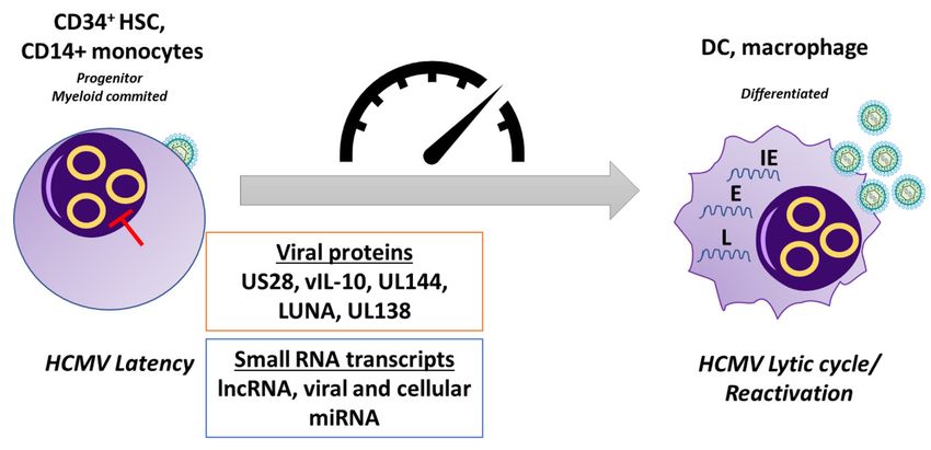
cascade consisting of immediate-early (IE), early (E), and late (L) genes. The viral particles are formed
cascade consisting of immediate-early (IE), early (E), and late (L) genes. The viral particles are formed
by a double-stranded DNA (dsDNA) genome (~230 kb), an icosahedral capsid, followed by
by a double-stranded DNA (dsDNA) genome (~230 kb), an icosahedral capsid, followed the
by the
tegument (a proteinaceous layer), and a coating known as pericapsid or envelope, which
tegument (a proteinaceous layer), and a coating known as pericapsid or envelope, which confers the confers the
virion a quasi-spherical shape (Figure 1), a feature shared with all other herpesviruses.
virion a quasi-spherical shape (Figure 1), a feature shared with all other herpesviruses.
Figure 1. Structure of HCMV virion. Mature virions are coated by an envelope, from which viral
Figure 1. Structure of HCMV virion. Mature virions are coated by an envelope, from which viral
glycoproteins protrude, and contain a double-stranded DNA genome enclosed within an icosahedral
glycoproteins protrude, and contain a double-stranded DNA genome enclosed within an icosahedral
symmetry capsid, that is surrounded by tegument.
symmetry capsid, that is surrounded by tegument.
HCMV can infect a broad cell range that includes epithelial cells of glandular and mucous
tissues, smooth muscle cells, fibroblasts, macrophages, hepatocytes, dendritic cells, and vascular
endothelial cells (ECs) [2]. After primary infection, similar to other members of the herpesvirus
Microorganisms 2020, 8, 685                                                                          3 of 30

      HCMV can infect a broad cell range that includes epithelial cells of glandular and mucous tissues,
smooth muscle cells, fibroblasts, macrophages, hepatocytes, dendritic cells, and vascular endothelial
cells (ECs) [2]. After primary infection, similar to other members of the herpesvirus family, HCMV can
establish latency in the host that can be reversed even after many years by any number of stimuli [3,4].
      A typical characteristic of HCMV, which originally granted the virus its name, is that of forming
in the infected cell a voluminous intranuclear inclusion body and one or more intra-cytoplasmic
inclusion bodies, the so-called “owl’s eye” inclusions, made up of clusters of newly formed viruses and
lysosomes. The formation of such bodies generally results in increased cellular volume, a phenomenon
defined as cytomegaly. Studies on HCMV began in the early 1900s when particular attention was paid
to the owl’s eyes found in biopsies from stillborn fetuses and later in the kidneys and parathyroid
gland cells of organ transplant patients [4].
      HCMV efficiently spreads through infected body fluids, and it can also be transmitted vertically
from the mother to the fetus through the placenta, causing congenital pathologies. Furthermore,
even when the primary infection is resolved by an effective cellular immune response, a population of
latently infected myeloid cells can persist in bone marrow monocyte precursors, thereby contributing
to the risk of transferring HCMV along with organs and tissues following transplantation.
      HCMV is a common pathogen of global clinical relevance, with worldwide seroprevalence
ranging from 56% to 94% [5]. The viral spread in the global population is enormous, mainly due to the
asymptomatic mode of infection, followed by a constant shedding of the virus through body fluids
(e.g., milk, saliva, cervical secretions, and tears), which can last for months or even years.
      HCMV is particularly dangerous for the following target categories of individuals [6]:
(i) immunocompetent hosts, where it causes asymptomatic infections or a slight form of
mononucleotic-like pathology; (ii) immunocompromised individuals such as patients suffering from
human immunodeficiency virus (HIV) or undergoing bone or organ transplants; and (iii) congenitally
infected newborns, who can be infected in utero, postnatally, or via breastfeeding. Of note, the prevalence
of congenital HCMV (cHCMV) disease is much higher than that of Down syndrome, spina bifida,
or fetal alcohol syndrome [7].
      The host immune status ultimately determines the outcome of the infection as
immunocompromised conditions predispose the patient to a primary infection or determine the
reactivation of a latent one. In this regard, HCMV is notoriously famous for its ability to cause
congenital anomalies and long-term neurological sequelae in newborns. Furthermore, it can also
trigger the development of serious pathologies in solid organ or stem cell transplant recipients that are
not always resolved by currently available antivirals, thereby leading in some cases to death [8].
      This review provides a summary of the general characteristics of HCMV as well as its strain
variability, dissemination, latency, reactivation, pathogenesis, prevention, and treatment.

2. Pathogenesis
   HCMV pathogenesis and clinical features of infection in various patient populations are
summarized below (Figure 2).
Microorganisms 2020, 8, x FOR PEER REVIEW                                                                           4 of 30

   2. Pathogenesis
Microorganisms 2020, 8, 685
      HCMV pathogenesis and clinical features of infection in various patient populations 4are
                                                                                            of 30

   summarized below (Figure 2).

      Figure   2. HCMV
          Figure  2. HCMV clinical manifestations
                               clinical             in immunocompetent
                                        manifestations    in immunocompetentindividuals with severe
                                                                                  individuals  with HCMV    infection,
                                                                                                     severe HCMV
      in immunocompromised          people, especially
          infection, in immunocompromised               in acquired
                                                  people,   especiallyimmune   deficiency
                                                                        in acquired       syndrome
                                                                                     immune           (AIDS)
                                                                                              deficiency     patients,
                                                                                                         syndrome
          (AIDS) patients,
      transplant            transplant
                   recipients, and upon recipients, andinfection.
                                           congenital    upon congenital infection.

2.1.2.1.
     Infection   of of
         Infection  Immunocompetent
                       ImmunocompetentAdults
                                       Adults
      HCMV
         HCMV    infection
                    infection commonly
                                 commonlyoccurs
                                              occursin inhealthy
                                                          healthy adults andand children,
                                                                                 children,with
                                                                                             withaaprevalence
                                                                                                       prevalence    gradually
                                                                                                                   gradually
increasing
   increasing with   age
                  with  age [9].
                              [9].When
                                   Whensymptomatic,
                                           symptomatic, itit results
                                                               results in a mononucleosis-like
                                                                             mononucleosis-likesyndromesyndromewith with   a less
                                                                                                                        a less
prominent
   prominent   cervical
                  cervicallymphadenopathy
                             lymphadenopathythan     thanthat
                                                           that caused
                                                                 caused by the Epstein-Barr
                                                                                 Epstein-Barrvirus virus(EBV)
                                                                                                           (EBV)[10].
                                                                                                                  [10].One
                                                                                                                        One of of
   the  symptoms      is rash,   which   only  manifests   in 30%  of HCMV     mononucleosis
the symptoms is rash, which only manifests in 30% HCMV mononucleosis cases [11]. Noteworthy,       cases  [11]. Noteworthy,
   a minority
a minority         of primary
              of primary      HCMV HCMV      infections
                                        infections        result
                                                     result       in relapsing
                                                             in relapsing        symptoms
                                                                            symptoms            (i.e., fever,
                                                                                         (i.e., fever,  night night
                                                                                                               sweats,sweats,
                                                                                                                        fatigue,
   fatigue,arthralgia,
myalgia,       myalgia, and arthralgia,    and transaminitis),
                                  transaminitis),    which can lastwhich    can lastweeks
                                                                        for several    for several
                                                                                               [10] or,weeks    [10] or, less
                                                                                                         less frequently,   lead
to multi-organ failure [12,13], even though the severe tissue-invasive disease is usually limitedis to
   frequently,     lead   to  multi-organ     failure   [12,13], even   though   the  severe    tissue-invasive    disease
   usuallyilllimited
critically               to critically ill patients
                or immunodeficient           or immunodeficient
                                                       [14]. A studypatients    [14].the
                                                                        describing     A various
                                                                                          study describing       the various
                                                                                                      clinical manifestations
   clinical   manifestations      of 290  immunocompetent         patients  with  severe  HCMV
of 290 immunocompetent patients with severe HCMV infection showed that the gastrointestinal          infection  showed    that
   the gastrointestinal tract is the preferential organ affected, primarily in the form of colitis, followed
tract is the preferential organ affected, primarily in the form of colitis, followed by morbidities of the
   by morbidities of the central nervous system (CNS) (i.e., meningitis, encephalitis, and transverse
central nervous system (CNS) (i.e., meningitis, encephalitis, and transverse myelitis), hematological
   myelitis), hematological abnormalities (i.e., hemolytic anemia, and thrombocytopenia), the
abnormalities (i.e., hemolytic anemia, and thrombocytopenia), the involvement of the eye (uveitis and
   involvement of the eye (uveitis and retinitis), liver (hepatitis), and lung (pneumonitis), and
retinitis), liver (hepatitis), and lung (pneumonitis), and thrombosis of the arterial and venous system [15].
   thrombosis of the arterial and venous system [15]. Although several studies have reported a rapid
Although several studies have reported a rapid clinical improvement in immunocompetent patients
   clinical improvement in immunocompetent patients with severe HCMV infection after anti-HCMV
with  severe criteria
   therapy,     HCMV for   infection   after
                                specific      anti-HCMV
                                           antiviral          therapy, criteria
                                                      pharmacological             for specific
                                                                           treatments    are not antiviral   pharmacological
                                                                                                     well established    [12].
treatments     are  not  well    established   [12].  Randomized      controlled   trials should
   Randomized controlled trials should therefore be conducted to determine in which cases anti-HCMV  therefore   be conducted
to determine      in which cases anti-HCMV
   therapy in immunocompetent              patients withtherapy   in immunocompetent
                                                            symptomatic     HCMV infections    patients    with symptomatic
                                                                                                  is needed.
HCMV infections is needed.

2.2. Infection of Immunocompromised Patients
     In the immunocompetent host, HCMV and immunity coexist in a delicate balance. When the
host immune system is compromised—i.e., in individuals with acquired immune deficiency syndrome
Microorganisms 2020, 8, 685                                                                            5 of 30

(AIDS) and other immune diseases, post-transplant and intensive care unit (ICU) patients, and, to some
extent, elderly people—the virus can exert its full pathogenic potential. Its reactivation in
immunocompetent hosts, which occurs intermittently throughout life, triggers a lifelong IgG-mediated
immunologic response that keeps in check viral replication. In contrast, uncontrolled viral replication
occurs when populations of HCMV-specific CD4+ and CD8+ T cells are not well preserved, as observed
in immunocompromised hosts, leading to severe clinical disease [16].

2.3. Cytomegalovirus and HIV
      HIV-infected individuals are generally co-infected with HCMV [17]. Prior to the introduction
of highly active antiretroviral therapy (HAART) in developed countries, about 40% of HIV-infected
patients would suffer from severe HCMV disease [18]. Currently, durable suppression of HIV viremia
has increased the overall patient life quality and expectancy and reduced to a minimum the pathologies
associated with HCMV viral reactivation. Nevertheless, comorbidities remain problematic for HIV
patients. In this regard, a close relationship between HCMV infection and HIV persistence has been
reported. This is probably due to the fact that HIV-driven CD4+ T-cell loss and dysfunction may lead
to HCMV replication and subsequent expansion of CD8+ T cells. Indeed, an elevated number of
CD8+ T cells and a low CD4+ /CD8+ T-cell ratio have been observed in individuals co-infected with
both viruses but not in patients infected with HIV or HCMV alone [19]. Fittingly, Hunt et al. [20]
demonstrated that CD8 T-cell activation could be reduced in HAART-treated HIV patients with
incomplete CD4 T-cell recovery by administering anti-HCMV drugs, attesting that HCMV plays
a significant role in immune activation in HIV patients. Moreover, persistent HCMV replication
modulated longevity and proliferation of HIV-infected cells, improved the recruitment of new HIV
target cells, and stimulated HIV transcription, thereby creating an HIV reservoir favoring AIDS
progression. Clinically, retinitis has been shown to be the predominant pathology in AIDS patients
(20–30%), and it usually appears at the late stages of the syndrome in patients with low CD4 count [21].
HCMV retinitis in HIV patients is commonly observed in two different forms: fulminant or indolent,
both characterized by minimal or completely absent vitreous and anterior chamber inflammation [22].
If left untreated, HCMV infection of retinal cells may cause subacute retinal destruction, which can
result in irreversible blindness. Paradoxically, HAART therapy while restoring the patient immune
system can lead to a new pathology, known as immune recovery uveitis (IRU), which is equally
destructive to the host tissue and deleterious to the quality of the patient’s life [23]. Following retinitis,
the most prevalent clinical manifestations are the following: colitis(in the US, 5–10% of HIV patients
with low CD4 lymphocyte counts were affected by enterocolitis prior to the availability of HAART
therapy [24]), esophagitis (most commonly due to co-infection with either herpes simplex virus or
Candida albicans), pneumonitis, encephalitis, hepatitis, and adrenalitis [25].

2.4. Cytomegalovirus and Transplant Patients
      HCMV is one of the most frequently encountered opportunistic viral pathogens in transplant
patients: a primary infection can occur in seronegative individuals after organ transplantation while
a latent infection can be reactivated in seropositive individuals due to immunosuppressive treatment.
The risks of HCMV-related complications in transplant recipients (R) vary according to the serostatus
of the donor (D): HCMV D− /R− transplantation is classified as low-risk, HCMV D+ /R+ or D− /R+ as
medium-risk, and HCMV D+ /R− as high-risk [26,27]. The most prevalent clinical manifestations in
HCMV-transplanted patients are gastrointestinal symptoms, mainly affecting the upper digestive
tract, whereas diarrhea is a rare occurrence indicative of colon involvement. Meningoencephalitis,
clinical hepatitis, myocarditis, and pancreatitis are more common than respiratory symptoms, which in
fact indicate more severe disease and may require admission to an ICU. Transplant patients without
HCMV prophylaxis may display a spectrum of clinical manifestations that vary in severity from patient
to patient, depending on additional personal illness risk factors, type of transplant procedure,
the immunological match between donor and recipient, and immunosuppressive drugs being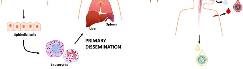
Microorganisms 2020, 8, 685                                                                         6 of 30

administered. For instance, patients treated with mammalian target of rapamycin (mTOR) inhibitors
display a very low incidence of HCMV. The incidence of HCMV also correlates with the type of
transplant: about 50% among pancreas or kidney–pancreas recipients, 50–75% in lung or heart–lung
recipients, 9–23% in heart recipients, 22–29% in liver recipients, and 8–32% in kidney recipients [28,29].
Moreover, in patients undergoing allogeneic hematopoietic stem cell transplantation (HSCT), HCMV can
lead to fatal infectious complications related to host immune recovery—about 30–70% of non-autologous
and 5% of autologous HSCT-patients develop HCMV disease. Pneumonia is the disease most highly
associated with HCMV infection of HSCT patients, frequently leading to death despite aggressive
treatment with antiviral agents and adjunctive therapies [30]. The reasons for the development of
different clinical sequelae among all the aforementioned types of transplanted patients is probably
due to a combination of the following factors: (i) the nature of the proinflammatory cytokine cocktail
arising after organ transplantation; (ii) the duration of HCMV replication—most transplant recipients
display acute HCMV infection, which subsequently results in disease in a relatively short time frame,
whereas, for example, congenitally infected infants and HIV patients can display high levels of HCMV
replication for several months; and (iii) the status of the immune system response. For instance,
HCMV pneumonitis only occurs when patients can activate their immune system [31].
      Interestingly, conflicting results have been reported regarding the association between early
cytomegalovirus reactivation and relapse after HSCT. Some studies suggest that HCMV replication
after transplantation is associated with a decreased relapse risk [32–37], while others highlight
that HCMV’s protective effect is restricted to patients with acute myeloid leukemia (AML) [38,39]
and cannot be extended to patients with acute lymphoblastic leukemia (ALL) [40], lymphoma [41],
myelodysplastic syndrome (MDS) [42], or observed in pediatric leukemias [43]. Furthermore, a more
recent study by Peric et al. [44] reports that the protective HCMV effect has been pronounced in
patients with myeloproliferative malignancies, while, at the same time, confirming the fact that such
effect has not been observed in patients with lymphoproliferative disorders, in concordance with other
studies [40,41]. Moreover, the same study highlights a significant reduction of relapse in patients
with myeloproliferative neoplasms (MPN) associated with early CMV reactivation [44]. Finally, some
evidence suggests that the beneficial effect of HCMV is mostly related to the conditioning regimen
and restricted only to patients who receive myeloablative chemotherapy (MAC) before undergoing
HSCT [45]. Despite the mounting evidence, the impact of HCMV reactivation on the patients’ overall
survival (OS) has been largely regarded as controversial due to its known negative effect on non-relapse
mortality (NRM). Thus, it remains largely unclear whether these conflicting reports can provide
a more detailed insight into the distinct protective mechanism or they simply reflect other variables,
including the sample size of the studied transplant groups. Therefore, larger prospective studies
with a significant follow-up on all patients with different malignancies, monitored and treated in
a homogeneous manner are needed to fully elucidate the underlying mechanism responsible for the
exact effect of HCMV reactivation. These findings may ultimately lead to a significant improvement in
patient management, donor selection strategies, or more personalized preemptive treatment of HCMV
infection in posttransplant patients with particular malignancies.

2.5. Congenital and Neonatal Infection
      HCMV is the major infectious cause of congenital abnormalities. The incidence of cHCMV
infection due to primary and non-primary maternal HCMV infection is ~0.4–0.8% in developed
countries. In general, the risk of transmission correlated with the stage of pregnancy is higher in later
stages and lower in earlier ones, but in either case HCMV infection is generally associated with severe
clinical sequelae in the fetus. In developed countries, ~40% of women in reproductive age are HCMV
seronegative, 1–3% of whom may contract primary HCMV infection during pregnancy. The most
vulnerable groups include adolescents, mothers, and caregivers in close contact with young children
(e.g., teachers, nurses, etc.). Primary maternal HCMV infection has a 30–40% risk of transmission to the
Microorganisms 2020, 8, 685 7 of 30
fetus. Importantly, HCMV reactivation or reinfection can also occur in women who are seropositive
prior to pregnancy, but, in such cases, the rate of HCMV transmission is only ~1% [46,47].
The hypothesis that pre-existing maternal immunity may favor low HCMV transmission rates
has long been debated. In this regard, Coppola et al. [48] performed a systemic review of the literature
using Preferred Reporting Items for Systematic Review and Meta-Analyses (PRISMA) [49] guidelines
and identified 19 studies assessing congenital HCMV birth prevalence in HCMV-seropositive mothers.
All these studies reported low levels of congenital HCMV birth prevalence (0.4–0.6%) in seropositive
mothers, in good agreement with previous findings by Lanzieri et al. [50], who systematically reviewed
postnatal HCMV prevalence in developing countries. Moreover, 11 studies reported HCMV maternal
seroprevalence and HCMV birth prevalence rates of 84–100% and 0.6–6.1%, respectively.
Even though most infants with cHCMV are asymptomatic, they may develop health problems
at birth or later. In the most severe cases, cHCMV can cause the death of the unborn baby. In all
other cases, the most clinically relevant signs at birth may include microcephaly, hepatosplenomegaly,
retinitis, intrauterine growth restriction, seizures, rash, and jaundice. Some children with cHCMV
infection may also suffer from long-term health problems, such as hearing loss, which can be present at
birth or may develop later even in asymptomatic infants, developmental and motor delay, vision loss,
and seizures. Cognitive impairment and retinitis have also been observed in asymptomatic children
but at much lower rates compared to symptomatic children [51]. Prevention of HCMV remains elusive
given the lack of drugs capable of treating HCMV infection in pregnant women. This aspect, together
with the fact that maternal immunity has a protective role against cHCMV infection, suggests that
vaccine development remains the most viable option to avoid HCMV vertical transmission.
3. Dissemination
HCMV exploits both vertical and horizontal transmission. Vertical transmission occurs through
the placenta [52–54], during birth (with genital secretions), or postnatally through breast milk [55–57].
Horizontal transmission takes place via organ transplant [58,59], blood transfusion, or direct contact
with contaminated body fluids, such as urine, breast milk, and genital secretions [60–62].
In most of these cases, because they cover all body surfaces, epithelial cells of the skin and internal
mucosa are the first site of HCMV infection. For instance, infection through breastfeeding starts
from the oral mucosa, moves to the gastrointestinal tract, which can support a productive infection,
and eventually disseminates throughout the body. In contrast, studies using murine CMV (MCMV)
have shown that, after oral/intranasal inoculation, the infection can only evolve in the upper respiratory
tract but not in the gut [63]. It remains a matter of debate how HCMV disseminates from the upper
respiratory tract throughout the body.
It is widely acknowledged that HCMV can spread systemically via leucocytes, a process associated
with short-duration viremia, during which infection of the lungs, liver, and spleen occurs primarily
through viral dissemination [64]. Subsequent secondary dissemination leads to the infection of salivary
glands, breast, and kidneys, all secretion-producing organs that release the virus into the environment
for months, even years, favoring intra-host transmission [64]. According to a model whereby primary
dissemination produces many viral particles that then infect other organs generating even more
virus progeny, it would be expected a gradual increase in viral burden during primary infection [64]
(Figure 3).Microorganisms 2020, 8, 685 8 of 30
Microorganisms 2020, 8, x FOR PEER REVIEW 8 of 30
Figure
Figure HCMV
3. 3. HCMVcan canbebetransmitted
transmitteddirectly
directly from
from person
person toto person
personthrough
throughbodily
bodilyfluids
fluids including
including
saliva,
saliva,urine, cervical,
urine, and
cervical, andvaginal secretions,
vaginal secretions,breast
breastmilk, semen,
milk, semen,blood, and
blood, andtears. It infects
tears. a new
It infects a newhost
usually by getting
host usually in through
by getting the upper
in through gastrointestinal
the upper gastrointestinal
gastrointestinal tracttract
gastrointestinal or the respiratory
or the tract.
respiratory
tract.
Here, theHere, the epithelial
epithelial cells arecells
oftenare
theoften the first
first site site of infection
of infection and fromand from
there HCMVthereinfects
HCMVleucocytes
infects
leucocytes
that that traffic
traffic around the body.around
Thisthe body. This
is correlated is correlated
with with aprimary
a process called processviral
called primary viral
dissemination that
dissemination
leads that of
to the infection leads to the
multiple infection
tissues, suchofasmultiple tissues,
lungs, liver, such as Afterwards,
and spleen. lungs, liver,secondary
and spleen.viral
Afterwards, secondary
dissemination spreads the viral dissemination
infection spreads the infection
to secretion-producing to secretion-producing
organs, such as salivary and organs, such
mammary
as salivary and mammary glands
glands and kidneys, which shed the virus. and kidneys, which shed the virus.
However,
However, animal
animal studies
studies using
using the the
MCMV MCMV model model
showedshowed a biphasic
a biphasic ratherrather
than athan a gradual
gradual increase
increase in viremia, suggesting a much
in viremia, suggesting a much more complex scenario [65].more complex scenario [65].
Recently,
Recently, Jacksonand
Jackson andSparer
Sparer[66][66]demonstrated
demonstrated that that cells
cells ofofthe
theupper
upperrespiratory
respiratory tract,
tract,once
once
infected, release not only viral progeny but also chemotactic factors. According
infected, release not only viral progeny but also chemotactic factors. According to the proposed model, to the proposed
model,
these these chemokines
chemokines trigger the trigger the recruitment
recruitment of innate ofimmune
innate immune cells, which
cells, which after being
after being infected
infected further
further spread the virus to secondary organs and body fluids [66]. Fittingly, HCMV DNA has never
spread the virus to secondary organs and body fluids [66]. Fittingly, HCMV DNA has never been found
been found in the form of free circulating viral particles, except for highly fragmented DNA [67].
in the form of free circulating viral particles, except for highly fragmented DNA [67]. Consistent with
Consistent with the lack of free circulating HCMV, leukocyte-depleted blood from seropositive
the lack of free circulating HCMV, leukocyte-depleted blood from seropositive donors prior to blood
donors prior to blood transfusion prevents HCMV transfer [68,69], indicating that HCMV viremia is
transfusion prevents HCMV transfer [68,69], indicating that HCMV viremia is mostly cell-associated.
mostly cell-associated. More recently, Farrell et al. [63] showed that the first cells to be infected after
More recently, Farrell et al. [63] showed that the first cells to be infected after nasal inoculation with
nasal inoculation with MCMV are alveolar macrophages and type 2 alveolar epithelial cells. Entry
MCMV are alveolar
into epithelial cells macrophages
and macrophages andoccurs
type 2through
alveolar epithelial and
endocytosis cells.is Entry intobyepithelial
followed subsequent cells
pH- and
macrophages
dependent fusion occurswith
through endocytosis
the endosomal and is followed
membrane, mediated bybysubsequent pH-dependent
the viral envelope fusion gB
glycoproteins with
theand
endosomal
gH/gL/gO and the pentameric complex formed by gH/gL/UL128, UL130 and UL131A [70,71].the
membrane, mediated by the viral envelope glycoproteins gB and gH/gL/gO and
pentameric
Then, the complex
local spread formed by gH/gL/UL128,
is thought to occur through UL130 and
direct UL131Atransmission,
cell-to-cell [70,71]. Then, the local
mediated in spread
part
is thought
by the HCMV to occurgenethrough
US28 [72]. direct cell-to-cell transmission, mediated in part by the HCMV gene
US28 [72].
The main cell types contributing to hematogenous dissemination, albeit to different extents,
The main
include cell types contributing
polymorphonuclear cells (PMNs), to hematogenous
monocytes, ECs,dissemination,
and dendritic cells.albeit to different
After recruitment extents,
to
the first
include site of infection, these
polymorphonuclear cells aremonocytes,
cells (PMNs), highly prone ECs,toand
infection
dendriticthemselves, thereby
cells. After becoming
recruitment to the
potential
first vehicles for
site of infection, theseHCMV transmission,
cells are highly prone even though most
to infection of themthereby
themselves, are unable to support
becoming a
potential
complete viral replication cycle [73–76]. Consistently, HCMV is frequently
vehicles for HCMV transmission, even though most of them are unable to support a complete viral found in PMNs from
immunocompromised
replication patients [74],HCMV
cycle [73–76]. Consistently, in which viral replication
is frequently found in is PMNsgenerally abortive and non-
from immunocompromised
patients [74], in which viral replication is generally abortive and non-productive between
productive [73]. The infection of PMNs most likely occurs by transient microfusion [73]. TheECs and
infection
PMNs after an initial direct contact mediated by the pentameric complex.
of PMNs most likely occurs by transient microfusion between ECs and PMNs after an initial direct Successively, infected
PMNsmediated
contact transfer theby thevirus particles to
pentameric other cell
complex. types [77]. infected
Successively, On the other
PMNshand, other
transfer thestudies using
virus particlesMicroorganisms 2020, 8, 685 9 of 30
to other cell types [77]. On the other hand, other studies using MCMV do not seem to support the
hypothesis that neutrophils play a role in HCMV dissemination, since their depletion did not alter
primary or secondary viral diffusion [78], whereas depletion of monocytes, macrophages, and NK
led to reduced viral dissemination [63,79,80]. However, it is important to point out that there are
substantial differences between human and murine CMV, exemplified by the lack of the MCMV CXC
chemokine homolog involved in neutrophil migration [78].
HCMV carries two genes, UL146 and UL147, which encode for the two chemokine homologs
vCXCL-1 and vCXCL-2, respectively, involved in the recruitment of innate immune cells [81–84].
pUL128, a key component of the pentameric complex, is an important chemokine that, once released in
the extracellular milieu, regulates monocyte migration [85]. Likewise, MCK2 in MCMV acts as a strong
attractant of monocytes, which appear to be conserved dissemination vehicles across species [86,87].
Monocyte-driven hematogenous spread is probably the result of the close proximity of these cells to
the vascular epithelium, which renders them particularly susceptible to infection with viral particles
originating from productively infected ECs. Once fully differentiated into tissue macrophages [75],
they can, in turn, spread the infection to the organs where they transmigrated [88–90].
Infected ECs also play a fundamental and active role in HCMV dissemination. In fact, HCMV
infection of ECs supports viral replication and promotes the enhanced expression of the adhesion
molecules ICAM-1 and vCAM-1 [91,92], as well as increased vascular permeability, which promotes
recruitment of leucocytes, direct contact [92] and migration through the endothelial layer.
Dendritic cells (DCs) are antigen-presenting cells that keep in check foreign pathogens by
influencing T-cell activation and differentiation in the draining lymph node through different
mechanisms [93]. Immature DCs localize in all mucosal and epidermal surfaces of the body where they
uptake HCMV infectious particles, thereby initiating the maturation process during their migration to
the draining lymph node. Upon localization in this new site, the newly mature and permissive DCs
are capable of transferring the virus to other cells [94].
In summary, there is still certainly a long way to go before we can fully understand the pathogenesis
of HCMV infection, but the aforementioned mechanism of HCMV dissemination proposed by Jackson
and Sparer [66] appears to be putting together many pieces of the puzzle.
4. Latency
Viral latency is defined as the maintenance of the viral genome without any production of
infectious progeny until this dormant genome can reactivate in response to specific stimuli and initiate
a productive infection. It is, therefore, becoming increasingly clear that a better understanding of latency
and subsequent reactivation may be crucial to elucidate HCMV pathogenesis and develop therapeutics
targeting latent virus reservoirs. This is a particularly important aspect given that all commercially
available drugs for the treatment of HCMV diseases only target lytic but not latent infections.
For decades, latency was considered as a silent state of the infection, characterized by overall
suppression of viral gene expression aimed at preventing the detection and activation of the immune
system. However, several recent studies have shown latency to be a dynamic phase of the infection,
where viral gene expression triggers a transcriptional cascade responsible for subverting host cell
functions, such as cell survival, genome carriage, and immune evasion [95–97].
The main site where HCMV is known to establish latency is in cells of the myeloid lineage.
The idea that infectious viral particles could be carried by white blood cells came from the observation
that blood transfer from healthy seropositive donors to immunosuppressed seronegative recipients
often resulted in HCMV disease [98–100], and that transfusion of leukocyte-depleted blood reduced
the incidence of HCMV disease [101].
However, it was only thanks to the increased sensitivity of the PCR technique that HCMV DNA
could be found in naturally latently infected peripheral blood mononuclear cells (PBMCs), in particular
monocytes and CD34+ progenitor cells isolated from the bone marrow [102,103]. Consistent with theMicroorganisms 2020, 8, 685 10 of 30
notion of myeloid cells being a bona fide site of latency, HCMV IE RNA expression has also recently
been detected in DCs isolated from peripheral blood of healthy individuals [104].
Investigations on viral gene expression during natural infection are limited by the fact that
only 0.004–0.01% of mononuclear cells from seropositive granulocyte colony-stimulating factor
(G-CSF)-stimulated donors carry viral genomes, with a low copy number of 2–13 genomes per infected
cell, as judged by PCR-driven in situ hybridization [105]. For this and other reasons, leukemic
cell line models such as THP-1 and CD34+ Kasumi 3, as well as several embryonic stem cell lines,
have been preferentially used as bona fide and low-cost models to study latency and reactivation
in vitro [106–109].
As CD34+ cells are also lymphoid cell progenitors, several studies have tried to explain why
myeloid cells are then the only cell lineage able to carry latent viral genome. By performing
experimentally latent HCMV infection of CD34+ progenitor cells, Poole et al. [110] observed an increase
in the cellular transcription factor GATA-2, a key regulator of myeloid differentiation, suggesting
that the virus may only engage myeloid-committed cells, promoting their survival. GATA-2 is also
involved in the differentiation of hematopoietic progenitors along the endothelial lineage. However,
HCMV DNA could not be detected by PCR in ECs from the saphenous vein of healthy individuals,
contrary to the hypothesis that microvasculature is a site of HCMV latency [111].
To establish latency, HCMV must stop the production of infectious viral particles through
suppression of viral gene expression and, at the same time, induce the expression of latency-associated
viral genes [112–116]. One of the key events required to initiate a state of latency involves the repression
of the viral major immediate-early promoter (MIEP), which is sufficient to prevent the expression of E
and late L genes as well. Transcriptional inactivation of this region is achieved through induction of
repressive chromatin marks—e.g., histones methylation and recruitment of heterochromatin protein
1 (HP-1)—and repressive transcription factors [117]. Concurrently, differentiation of latent CD34+
cells or monocytes to macrophages or DCs induces re-activation of the promoter through histone
acetylation and loss of HP-1, with subsequent expression of IE genes and re-entry into the lytic
cycle [76,104,118,119], indicating that dynamic regulation of the MIEP is a first and crucial step to
control latency/reactivation.
One of the most widely accepted hypotheses is that the virus gene expression upon latency is
mainly characterized by a robust suppression and shut down of almost all viral genes, an expression
profile similar to that of the late lytic cycle. In this regard, it has been proposed that, in latently infected
cells, the timely transcriptional cascade of productive infection may be prematurely interrupted by
cellular mechanisms. Alternatively, there could be, right after viral entry, early induction of viral gene
expression followed by massive repression of viral transcription [120].
As mentioned above, rather than being quiescent, latent HCMV infection induces the expression
of a certain amount of viral genes. The most sophisticated mechanism for modulating the host cell
environment without attracting an immune response is mediated by non-immunogenic molecules,
such as small RNA transcripts. Assessing both experimentally and naturally latent infected cells by
next-generation sequencing, Rossetto et al. [121] identified two long non-coding (nc) RNAs (lncRNAs),
RNA4.9 and RNA2.7, and mRNAs encoding replication factors UL84 and UL44. Of note, RNA lnc4.9 in
concert with latently expressed UL84 was shown to interact with members of the polycomb repressor
complex 2 (PRC2), which potentially represents an additional step of silencing of the MIEP through
their histone methyltransferase activity [122].
Across its genome, HCMV also encodes at least 20 viral microRNAs (miRNAs) identified first
in lytically-infected cells [123], but also in latently-infected cells THP-1 by Meshesha et al. [124],
using deep-sequencing analysis. More recently, two similar studies were performed using instead
primary latently-infected cells that more resemble the in vivo situation, even though they showed
conflicting results to some extent [125,126]. The advantage of using miRNAs, besides their
non-immunogenic state, stems from their ability to modulate the expression of multiple targets
involved in immune evasion, survival, and proliferation of HCMV-infected cells, as well as virusMicroorganisms 2020, 8, 685 11 of 30
reactivation [127]. One example is the miR-UL148D that during the lytic cycle promotes T-cell
chemotaxis by targeting CCL5 (RANTES), while during latency it may trigger activin signaling,
Microorganisms 2020, 8, x FOR PEER REVIEW 11 of 30
thereby inhibiting pro-inflammatory cytokine secretion [126,128]. In addition, even cellular miRNAome
was shown tocytokine
inflammatory be widely affected
secretion by HCMV
[126,128]. latent infection
In addition, [129].
even cellular miRNAome was shown to be
A restricted amount of viral proteins
widely affected by HCMV latent infection [129]. is detected in naturally latent infected cells, even though their
exactArole in latency
restricted amountis only partially
of viral proteins clear. These include:
is detected US28,
in naturally a constitutively
latent activated
infected cells, even though chemokine
their exact
receptor role in
acting aslatency is only partially
a chemokine sink [130]; clear. These
viral include:
IL-10, which US28,cana downregulate
constitutively activated
MHC II surface
chemokine and
expression receptor acting CD4
modulate + T-cell recognition
as a chemokine sink [130];[131],
viral IL-10,
UL144,which
a decoycan downregulate
tumor necrosis MHCfactorII receptor
surface expression and modulate CD4 + T-cell recognition [131], UL144, a decoy tumor necrosis factor
(TNFR), which inhibits T cell proliferation in vitro [132] and subverts the TH1 immune response in
receptor (TNFR), which inhibits T cell proliferation in vitro [132] and subverts the TH1 immune
a TNF ligand-independent fashion [133]; LUNA (latency unique natural antigen), a protein required
response in a TNF ligand-independent fashion [133]; LUNA (latency unique natural antigen), a
for reactivation [134]; and UL138, which maintains latent infection and suppresses reactivation [115].
protein required for reactivation [134]; and UL138, which maintains latent infection and suppresses
Interestingly, this latter
reactivation [115]. is a potentially
Interestingly, this latterdruggable targetdruggable
is a potentially in latently infected
target cells as
in latently it inhibits
infected cells a cellular
drug transporter [134].
as it inhibits a cellular drug transporter [134].
As latency
latencyappears
appears toto
bebe a very
a very complex
complex phenomenon,
phenomenon, reactivation
reactivation is oftenis theoften
resultthe
of result
a closely of a closely
intertwined crosstalk between
intertwined crosstalk betweencellular
cellularand andviralviralsignals
signals triggering
triggering multiple
multiple pathways.
pathways. Indeed,
Indeed, IE IE gene
gene expression
expression alone alone
does does
notnot
seemseem totobebesufficient
sufficient to to induce
induce thetheproduction
production of infectious particles
of infectious particles [76].
[76]. In this regard, the observation that differentiation of experimentally latently
In this regard, the observation that differentiation of experimentally latently infected monocytes into infected monocytes
into monocyte-derived macrophages can lead to a fully permissive phenotype [88,89] implies that the
monocyte-derived macrophages can lead to a fully permissive phenotype [88,89] implies that the
differentiation status is a critical determinant of reactivation. In addition, mounting evidence
differentiation status is a critical determinant of reactivation. In addition, mounting evidence indicates
indicates that inflammation may also play a role in HCMV reactivation. For instance, virus
that inflammation may also play a role in HCMV reactivation. For instance, virus reactivation has
reactivation has been observed in several progenitor cell types under a variety of inflammatory
been observed
conditions in several
[135–137], progenitor
and HCMV diseasecell types under
prevalence a variety
is directly of inflammatory
associated conditions [135–137],
with highly inflammatory
and HCMV disease
environments prevalence is directly associated with highly inflammatory environments [137–139].
[137–139].
In light
light of
ofthetheabove,
above, it it
is is becoming
becoming increasingly
increasingly evident
evident how ahow a multiplicity
multiplicity of latency ofandlatency and
reactivation pathways can determine the course of HCMV infection.
reactivation pathways can determine the course of HCMV infection. These pathways appear These pathways appear to be to be
independent ofofthe
independent theclinical strains
clinical strainsbutbut seemingly
seemingly dependent
dependenton a combination
on a combination of viral and cellular
of viral and cellular
factors working
factors working cooperatively
cooperatively totocross
cross thethe
threshold
threshold for reactivation of latently
for reactivation infected
of latently cells (Figure
infected cells (Figure 4).
4).
Figure 4. Latency. Following primary infection, HCMV can establish latency in CD34+ myeloid+
Figure 4. Latency. Following primary infection, HCMV can establish latency in CD34 myeloid
progenitor cells and is carried down the myeloid lineage. In latently-infected CD34+ cells and
progenitor cells and is carried down the myeloid lineage. In latently-infected CD34+ cells and
monocytes, there is a targeted suppression of lytic viral gene expression. HCMV utilizes several viral
monocytes, there is a targeted suppression of lytic viral gene expression. HCMV utilizes several viral
proteins and small RNA transcripts, including viral and cellular miRNAs, during latent infection to
proteins and small RNA transcripts, including viral and cellular miRNAs, during latent infection to
alter the signaling environment within the cell to maintain the status of latency. Differentiation of
alter
these cellssignaling
the environment
to macrophages and DCs within thederepression
causes the cell to maintain the status
of the MIEP of latency.
and allows Differentiation
initiation of the of
these cells to macrophages and DCs causes the derepression of the MIEP and allows
lytic transcription program, which involves a temporal cascade of viral gene transcription, allowing initiation of the
lytic transcription
reactivation program,
of de novo which involves
virus production. HCMV,a human
temporal cascade of viral
cytomegalovirus; DC,gene transcription,
dendritic cell; HSC, allowing
reactivation
hematopoieticof de novo
stem virus production.
cell; lncRNA, HCMV,
long non-coding RNA;human cytomegalovirus;
IE, immediate-early; DC,
E, early; dendritic cell; HSC,
L, late.
hematopoietic stem cell; lncRNA, long non-coding RNA; IE, immediate-early; E, early; L, late.Microorganisms 2020, 8, 685 12 of 30
5. HCMV Strain Variation
The massive spread of HCMV infection and the wide spectrum of disease manifestations in
infected patients sparked a major interest in determining the origin and mechanisms of HCMV
pathogenicity. It has been 66 years since Margaret Gladys Smith isolated for the first time MCMV from
salivary glands and propagated the virus in mouse cell culture—such strain is still in use and commonly
called “Smith strain” [140]. One year later, she succeeded in growing submaxillary salivary-derived
HCMV in human cell culture. As early as 1960, Thomas Weller isolated the so-called David strain
from a liver biopsy and managed to identify serological differences between cytomegalic inclusion
disease (CID) isolates [141].. Subsequently, five different HCMV clinical isolates were sequenced
(FIX GenBank AC146907; TR GenBank KF021605; PH GenBank AC146904; Toledo GenBank AC146905;
Merlin GenBank AY446894). The pioneering work on sequencing of the complete genome of an HCMV
laboratory strain (AD169 GenBank AC146999) [142], along with numerous in vitro findings and data
from several vaccine studies, revealed the existence of a substantial genetic variation among HCMV
strains. In particular, the highly passaged laboratory strains AD169 and Towne (GenBank FJ616285)
appeared attenuated when administered as vaccine candidates [143,144]. By contrast, the Toledo
strain, which had only been passaged several times in culture, caused disease when administered to
seropositive individuals [145]. Taken together, these findings suggest that the pathogenic potential of
HCMV was correlated with the genetic composition of each distinct strain.
The differences among the widely used laboratory strains AD169, Towne, and Toledo were localized
to multiple ORFs in the UL/b0 region of the genome, encoding viral proteins with immunomodulatory
or evasive functions [145,146]. Those genes that were lost upon extensive passaging in vitro played
a crucial role in promoting viral replication and immune manipulation in vivo [147]. Consistently,
extensive culture passaging led to the selection of HCMV mutants lacking these genes within weeks of
propagation, and it also gave rise to variations between commonly used laboratory strains [146,148–150].
In the following years, with the development of more sensitive sequencing techniques, such as
Sanger and high-throughput sequencing [151,152], a higher number of HCMV genomes were sequenced
from bacterial artificial chromosomes [153–155], virion DNA [156], or overlapping PCR amplicons [148].
The widespread implementation of these new techniques allowed assessing different aspects of
HCMV genome variation in clinical HCMV isolates from different cohorts of infected patients,
thus providing novel insights into the genetic variation upon natural infection. Up to date, the complete
genomes of 351 full-length HCMV strains have been published and analyzed (National Institute of
Allergy and Infectious Diseases (NIAID)-sponsored Virus Pathogen Database and Analysis Resource
(ViPR) [157]) [158]. Interestingly, these sequencing data show that HCMV can be highly polymorphic
among and within hosts [159–161], with a high level of intra-host variability comparable to that of RNA
viruses [159]. Given the fact that HCMV is a large double-stranded DNA virus, a high degree of genetic
variation contradicted the logical expectation that the virus would retain high genome stability [162].
This unexpected intra-host HCMV diversity was initially attributed to the rapid occurrence of de novo
mutations [159,160]—i.e. new mutations occur every time the virus infects a new host, thereby giving
rise to a unique viral strain for each infected individual. Eventually, HCMV infection triggers
a selection event where a new genotype becomes dominant due to the selective pressure of the immune
response [159]. An alternative explanation was based on the evidence that viral and host factors
can contribute to the onset of HCMV genome mutations, thus fostering virus genetic drift upon
infection [163,164]. However, more recent data indicate that in non-mixed infections the mutation
rate of HCMV is no different from that of other DNA viruses, while HCMV acquires a high degree of
variability upon mixed infections [165–167], extensive recombination [152,166–168], or reactivation of
the latent virus within a single individual. Many of these genetic alterations may in turn influence
HCMV cell tropism, immune evasion, and disease outcomes. Indeed, the contribution of superinfection
and recombination to viral genetic variability, an intensively debated topic, could have important
ramifications in viral evolution, immune adaptation, and pathogenesis, especially in congenital or
transplant patients [169,170].Microorganisms 2020, 8, 685 13 of 30
Substantial efforts have been undertaken by various groups to correlate infection outcomes with
variation in HCMV-specific genes [145,171,172]. Even though the selection of these genes was based
on data supporting their potential role in viral pathogenicity and dissemination, these studies were
only limited to Sanger sequencing of polymerase chain reaction (PCR) amplicons and often focused on
a small number of polymorphic (hypervariable) genes. Furthermore, in such cases, low-abundance
viral populations might have been missed, and the overall viral diversity underestimated. Thus, future
studies should take advantage of high-throughput sequencing for fast detection and characterization
of multiple-strain infections. Ideally, as recently put forward by Davison and co-workers, the definition
of HCMV natural populations should be carried out by whole genome sequencing of HCMV strains
directly from clinical samples [166].
6. Prevention
Although the development of an HCMV vaccine had already started in the 1970s, research in
HCMV vaccine discovery received a major push when in 2000 the US Institute of Medicine placed
HCMV among the top priorities for vaccine development [173]. Despite these increasing efforts,
an effective vaccine against HCMV is currently missing, de facto leaving high-risk populations, chiefly
immunocompromised patients and immunocompetent seronegative pregnant mothers, exposed to
primary infection [174]. Given that one of the main obstacles to the development of an efficient
vaccine against HCMV is the lack of protection against HCMV re-infection and/or reactivation, the first
objective of a newly designed HCMV vaccine should be that of shielding vulnerable populations
from primary infection. The long-range goal would then be to grant permanent protection against
new infections with other HCMV strains and reactivated infections, which can occur repeatedly
throughout life. To reach these goals, the ideal HCMV vaccine should be able to trigger a strong
humoral response, in the form of binding and neutralizing antibodies, and an HCMV-specific CD8+
and CD4+ T-cell response. With this in mind, the experimental and clinical results achieved so far
predict that the ideal HCMV vaccine should include: (i) gB, promoting both humoral—primarily
antibody-binding—and T-cell-mediated response [164]; (ii) pp65, triggering a potent T-cell response;
and (iii) the pentameric complex (PC), which prompts a quite strong neutralizing antibody (NAb)
response [175]. Indeed, PC-induced NAbs are powerful cell-to-cell spread viral inhibitors in numerous
cell types, not just fibroblasts. In the following sections, we summarize the most relevant approaches
for designing effective HCMV vaccines (Table 2).
Table 2. HCMV Vaccines. NAb, Neutralizing Antibody; HELF, human embryonic lung fibroblasts; PC,
pentameric complex; VLPs, virus like particles.
Live HCMV Vaccine Description Clinical Trials
Phase I/II clinical studies evidences:
(a) no virus excretion;
(b) no virus latency;
(c) NAb induction;
Towne vaccine HCMV attenuated strain.
(d) generation of both
HCM-specific CD4 and CD8 T-cell;
(e) partial protection against
a secondary infection.
In Phase I clinical trials they were
Genetic recombinant Towne well tolerated and with no virus
Towne-Toledo chimera vaccines
and Toledo. excretion. One chimera was more
immunogenic than Towne.
Patients did not to display
AD169 vaccine HCMV attenuated strain. cell-mediated immunity depression
or any systemic reactions.You can also read