Water structure around a left-handed Z-DNA fragment analyzed by cryo neutron crystallography
←
→
Page content transcription
If your browser does not render page correctly, please read the page content below
4782–4792 Nucleic Acids Research, 2021, Vol. 49, No. 8                                                                  Published online 19 April 2021
doi: 10.1093/nar/gkab264

Water structure around a left-handed Z-DNA fragment
analyzed by cryo neutron crystallography
Joel M. Harp1 , Leighton Coates2 , Brendan Sullivan2 and Martin Egli                                                    1,*

1
 Department of Biochemistry and Center for Structural Biology, Vanderbilt University, School of Medicine, Nashville,
TN 37232, USA and 2 Neutron Scattering Division, Oak Ridge National Laboratory, 1 Bethel Valley Road, Oak Ridge,
TN 37831, USA

Received August 29, 2020; Revised March 28, 2021; Editorial Decision March 29, 2021; Accepted March 31, 2021

                                                                                                                                                                  Downloaded from https://academic.oup.com/nar/article/49/8/4782/6238407 by guest on 03 December 2021
ABSTRACT                                                                          and B-form duplexes as a result of different sugar puckers
                                                                                  and consequently phosphate distances (4). The water con-
Even in high-quality X-ray crystal structures of                                  tent of even tightly packed crystals of duplexes and other
oligonucleotides determined at a resolution of 1 Å or                            DNA and RNA folding motifs that diffract to high resolu-
higher, the orientations of first-shell water molecules                           tion is typically 25% or higher. X-ray crystallography can
remain unclear. We used cryo neutron crystallogra-                                then pinpoint the locations of water molecules in the first
phy to gain insight into the H-bonding patterns of wa-                            and often also the second hydration shell. Examples of the
ter molecules around the left-handed Z-DNA duplex                                 quality of the electron density computed at ca. 1 Å around
[d(CGCGCG)]2 . The neutron density visualized at 1.5                              first-shell waters forming H-bonds to base and backbone
Å resolution for the first time allows us to pinpoint                            atoms of a left-handed Z-DNA duplex are depicted in Fig-
the orientations of most of the water molecules di-                               ure 1. However, even in this high-quality X-ray structure
rectly contacting the DNA and of many second-shell                                and a few others determined to resolutions beyond 1 Å,
                                                                                  donor-acceptor patterns of water H-bonds remain hidden.
waters. In particular, H-bond acceptor and donor pat-
                                                                                  One might speculate that water molecules bridging oxygen
terns for water participating in prominent hydration                              atoms, as seen in Figure 1B and C, act as donors in both
motifs inside the minor groove, on the convex sur-                                H-bonds, but there is no experimental evidence in support
face or bridging nucleobase and phosphate oxygen                                  of this. Further, it is readily apparent that trying to reliably
atoms are finally revealed. Several water molecules                               assign the roles of donors and acceptors for H-bonds such
display entirely unexpected orientations. For exam-                               as those shown in Figure 1D and E is futile, especially if one
ple, a water molecule located at H-bonding distance                               considers bifurcated cases in addition to more common uni-
from O6 keto oxygen atoms of two adjacent gua-                                    directional H-bonding.
nines directs both its deuterium atoms away from                                      The first single crystal structure reported for an oligo-
the keto groups. Exocyclic amino groups of gua-                                   2 -deoxynucleotide, the hexamer d(CGCGCG), revealed a
nine (N2) and cytosine (N4) unexpectedly stabilize                                left-handed duplex (5). Z-DNA is stabilized by negative su-
                                                                                  percoiling during transcription when polymerases and heli-
waters H-bonded to O2 keto oxygens from adjacent
                                                                                  cases in their wake generate underwound DNA (6–11). The
cytosines and O6 keto oxygens from adjacent gua-                                  transition from B- to Z-DNA (B−Z junction) occurs within
nines, respectively. Our structure offers the most de-                            a single A−T pair, whereby both bases are extruded from
tailed view to date of DNA solvation in the solid-state                           the duplex when the phosphate backbone reverses direction
undistorted by metal ions or polyamines.                                          (12). The discovery of the Z␣ protein domain that specifi-
                                                                                  cally binds to Z-DNA left little doubt as to a biological sig-
                                                                                  nificance of left-handed DNA (13). The domain recognizes
INTRODUCTION                                                                      left-handed DNA without specific contacts to bases by fol-
X-ray crystal structures of double helical nucleic acid frag-                     lowing the zig-zag conformation of the backbone (14). The
ments have revealed intricate hydration patterns inside                           Z␣ domain binds both Z-DNA and Z-RNA and is notably
the grooves and around the sugar-phosphate backbones.                             associated with double-stranded RNA-specific adenosine
Among them are the water spine or ribbon in the minor                             deaminase (ADAR (15)), zipcode binding protein 1 (ZBP1
groove of the DNA duplex [d(CGCGAATTCGCG)]2 (1,2),                                (16)) and viral orthologs that regulate innate immunity. In
water tandems that link 2 -OH groups across the minor                            recent years, the case for an involvement of Z-DNA and Z-
groove of double-stranded RNA (3), and distinct water                             RNA in human disease has been strengthened considerably
bridges between intra-strand phosphate groups in A-form                           (17,18).

* To   whom correspondence should be addressed. Tel: +1 615 343 8070; Fax: +1 615 343 0704; Email: martin.egli@vanderbilt.edu

C The Author(s) 2021. Published by Oxford University Press on behalf of Nucleic Acids Research.
This is an Open Access article distributed under the terms of the Creative Commons Attribution-NonCommercial License
(http://creativecommons.org/licenses/by-nc/4.0/), which permits non-commercial re-use, distribution, and reproduction in any medium, provided the original work
is properly cited. For commercial re-use, please contact journals.permissions@oup.com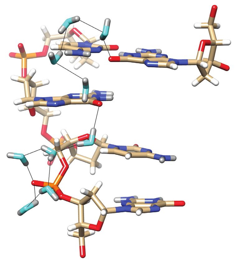
Nucleic Acids Research, 2021, Vol. 49, No. 8 4783
form). The density of mixed spermine/Mg2+ form crystals
A was experimentally determined to be 1.49 g/cm3 . The calcu-
lated density, including 74 refined water positions per asym-
metric unit, amounts to 1.41 g/cm3 . The difference between
the experimental and calculated densities of this Z-DNA
crystal form thus accounts for another 16 water molecules,
consistent with 90 water molecules surrounding an individ-
ual duplex in the asymmetric unit. The exceptional quality
of the diffraction data and the level of hydration, with up to
B C 85 water molecules visualized in the Mg2+ form (19), ren-
der Z-DNA a perfect target for an in-depth analysis of the
water structure around DNA using neutron diffraction.
Neutron crystallography provides insight into the posi-
Downloaded from https://academic.oup.com/nar/article/49/8/4782/6238407 by guest on 03 December 2021
tions of hydrogen (deuterium) atoms in the structures of
nucleic acids and proteins (20–24). A key difference be-
tween oligonucleotide and protein crystals in regard to neu-
tron diffraction experiments is that proteins can be re-
D combinantly expressed in their perdeuterated form. The
synthesis of perdeuterated DNA oligos using solid phase
phosphoramidite approaches would be prohibitively expen-
sive by comparison. Synthetic strategies for the prepara-
tion of nucleosides and nucleotides deuterated at single or
multiple sites have been reported (25). However, facile ac-
cess to phosphoramidite building blocks for solid-phase
oligonucleotide synthesis remains currently out of reach.
The preparation of perdeuterated DNA polynucleotides
E for neutron fiber diffraction experiments has also been de-
scribed (26), but the approach cannot be applied to produce
milligram amounts of an oligonucleotide for single crystal
neutron diffraction studies. As with all other previously re-
ported neutron structures of DNA oligonucleotides, a limi-
tation of the present analysis of the Z-DNA hexamer is that
the majority of backbone and base hydrogen atoms is not
exchanged by deuterium in D2 O-based crystallization solu-
tions. Thus, the levels of background scattering are expected
to be higher in neutron diffraction experiments with DNA
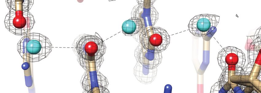
Figure 1. Prominent first-shell hydration patterns around [d(CGCGCG)]2
(PDB ID 1ICK (47)). (A) Standard C:G base pair. Longitudinal patterns compared to protein crystals.
bridging base atoms from opposite strands include (B) a water spine inside Previously reported neutron structures of DNA frag-
the minor groove that links cytosine O2 atoms, (C) waters between guanine ments revealed the orientations of a number of water
O6 atoms on the convex surface and (D) water tandems that link cytosine molecules, but did not provide a complete picture of the first
N4 atoms on the convex surface. (E) Water molecules at the periphery of
the minor groove linking guanine N2 atoms to 5 - and 3 -adjacent phos-
hydration shell around DNA duplexes (20,21,27,28). Com-
phate groups in a transverse fashion. Fourier 2Fo -Fc sum electron den- pared to establishing protonation states of nucleobases and
sity drawn at the 1 level surrounds water molecules and individual nu- protein sidechains or the orientation of water molecules in
cleotides. Water molecules are cyan spheres, atoms engaged in H-bonds channels or cavities by neutron crystallography, reliable de-
are highlighted as spheres and H-bonds are drawn with thin solid lines. termination of H-bond donor-acceptor patterns for water
molecules on the surface of DNA duplexes is more challeng-
ing. This is because the mobility of such water molecules is
In Z-DNA, bases form standard Watson-Crick pairs, but typically increased relative to those situated in sheltered re-
cytosine and guanosine adopt different sugar puckers (C2 - gions such as protein active sites or inside the deep (major)
endo and C3 -endo, respectively). Phosphates exhibit a zig- grooves of A- and B-form DNA duplexes.
zag like arrangement along the two backbones such that ad- Neutron diffraction experiments are mostly conducted at
jacent base pairs display alternating, low (−10◦ , CpG steps) room temperature (RT) with crystals sealed in capillaries
and high twists (−50◦ , GpC steps). Infinite stacks of du- as neutrons, unlike X-rays, do not induce radiation dam-
plexes exhibit close lateral spacings in Z-DNA crystals, with age. However, given the expected increased mobility of first-
a record low volume per base pair of ca. 1000 Å3 . The or- shell and certainly higher shell water compared to DNA
thorhombic crystals belonging to space group P21 21 21 grow atoms, RT neutron data are unlikely to provide detailed in-
under various conditions and include the Mg2+ , spermine, sight into the water structure around a DNA duplex. In-
and mixed spermine/Mg2+ forms (19). The ordered water deed, the neutron density for the Z-DNA duplex computed
contents (w/w) for these three range from 21% (spermine using RT data at ca. 1.6 Å showed deuterium atoms for
form) to 29% (Mg2+ form), with calculated crystal densi- bases, but did not unambiguously resolve the orientation
ties of between 1.37 (Mg2+ form) and 1.43 g/cm3 (spermine of many water molecules even in the first hydration shell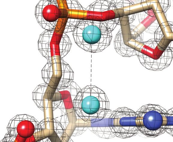
4784 Nucleic Acids Research, 2021, Vol. 49, No. 8
(our unpublished data). Also supporting the notion that RT tion drop volumes were 30 l. The reservoir was filled with
data may be insufficient for a detailed analysis of the sol- 500 l of D2 O crystallization buffer diluted to 50% with
vent structure in DNA crystals are the RT and low temper- D2 O and allowed to equilibrate for 3–4 days. The strength of
ature (LT) X-ray crystal structures of the Z-DNA spermine the crystallization buffer in the reservoir was then gradually
form. Thus, in the RT structure, separate electron density increased over a period of weeks by adjusting the strength of
peaks between adjacent cytosines in the minor groove were the buffer by 5% and allowing for equilibration for 3–4 days
assigned to a string of water molecules (29). However, the before again increasing the buffer strength. This procedure
LT structure clearly showed a second spermine molecule in- was continued until a crystal appeared.
side the minor groove (30). A comparison of the mobilities
of water and spermine molecules in the RT and LT struc-
tures demonstrates the benefits of data collected at cryo- Crystal mounting and cryo-protection
genic temperature in that the B-factors of water molecules Cryoprotection was achieved by transferring the crystal into
are distinctly lower in the latter (31). More evidence that a large drop of Paratone-N oil. Mother-liquor adhering to
Downloaded from https://academic.oup.com/nar/article/49/8/4782/6238407 by guest on 03 December 2021
cryo-cooling of crystals can reduce the mobility of water the surface of the crystal was removed using a nylon loop to
molecules is provided by the LT neutron structure (15K) swiftly shift the crystal in the drop allowing the viscosity of
of concanavalin A that led to the identification of twice as the oil to pull the mother liquor away. The crystal was then
many water molecules (32) compared to the RT structure transferred to a fresh drop of Paratone-N oil from which
(33). In addition, the B-factors of water molecules in the LT the crystal was mounted on a MiTeGen dual-thickness mi-
structure were around fourfold lower than those of waters in cromount of appropriate size. Flash-cooling was done by
the RT structure. The benefits of cryo-neutron crystallogra- plunging into liquid nitrogen. Crystals were stored in liquid
phy were recently also demonstrated in studies investigating nitrogen before transporting them by hand to the SNS. No
transient protein ligand complexes in -lactamases (34) and modifications to the cryoprotection protocol were required
aimed at clarifying the protonation states of cytochrome c even given the large crystal volume.
peroxidase (35).
In order to analyze the water structure that surrounds
Z-DNA, we crystallized the duplex without divalent metal Neutron data collection and processing
ions and polyamines from an (ND4 )2 SO4 /D2 O solution
(36). This prevents the disruption of the first and the second Neutron diffraction data were collected using the macro-
hydration shell around the DNA by metal ions and organic molecular neutron diffractometer (MaNDi) instrument
polycations. The crystal used had an approximate volume of (37,38) at the SNS. A total of 15 diffraction images were
1 mm3 (31), and cryo neutron diffraction data were collected collected with an exposure time of 12 h each. The crystal
at 100K on the Macromolecular Neutron Diffractometer was held static during each diffraction image and was ro-
(MaNDi (37)) at the Spallation Neutron Source (SNS), Oak tated 10◦ between images. Data collection was completed in
Ridge National Laboratory (ORNL, Oak Ridge, TN) to a 7.5 days, yielding a dataset of good completeness to a res-
resolution of 1.5 Å. Here, we report the most detailed anal- olution of 1.50 Å (Table 1). These data were reduced with
ysis yet of the orientation of water molecules that surround the Mantid program (39) using newly developed 3D profile
a DNA double helix. fitting for time-of-flight diffraction data (40,41). Integrated
intensities were then scaled using Lauenorm from the Laue-
gen suite (42,43). The reduced and corrected data statistics
MATERIALS AND METHODS are shown in Table 1.
Synthesis and purification of d(CGCGCG)
The Z-DNA hexamer d(CGCGCG) was synthesized X-ray data collection and processing
and HPLC purified by Integrated DNA Technologies
(Coralville, IA, USA; www.idtdna.com). Following The crystal used for neutron diffraction was returned to
lyophilization, a D2 O stock solution of the DNA hexamer the laboratory but did not survive the journey. A very sim-
with a concentration of 60 mg/ml was prepared. ilar crystal from the same batch and with nearly identi-
cal unit cell parameters was used. X-ray diffraction data
were collected using the D8 Venture (Bruker AXS, Madi-
Crystallization
son, WI, USA) system in the Biomolecular Crystallogra-
We used previously reported conditions without spermine phy Facility in the Vanderbilt University Center for Struc-
to grow crystals of the Z-DNA hexamer (31,36). Briefly, tural Biology. The system includes an Excillum D2+ Met-
crystals were obtained by mixing equal volumes of DNA alJet X-ray source with Helios MX optics providing Ga
with a D2 O crystallization buffer containing 2.5 M am- K␣ radiation at 1.3418 Å wavelength. The crystal was
monium sulfate (ND4 )2 SO4 , 10 mM magnesium acetate, mounted on a kappa axis goniometer and maintained at
and 50 mM perdeuterated 2-(N-morpholino)ethanesulfonic 100 K using an Oxford Cryosystems Cryostream 800 cryo-
acid (MES), pH 6.1, and equilibrating droplets by vapor stat. The detector was a PHOTON III charge-integrating
diffusion against a reservoir of the crystallization buffer. pixel array detector. Data collection was performed in
Crystallization experiments were assembled using Nextal shutterless mode. Data were reduced using Proteum3 soft-
EasyXtal Tools with sitting-drops bridges inserted. The ware (Bruker AXS, Madison, WI, USA). Selected crys-
greaseless closures of the EasyXtal Tool allow for effi- tal data and data collection statistics are summarized in
cient modification of the reservoir conditions. Crystalliza- Table 1.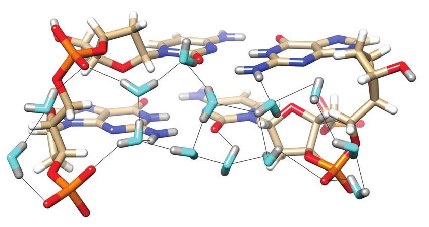
Nucleic Acids Research, 2021, Vol. 49, No. 8 4785
Table 1. Selected crystal data, data collection and processing, and refine- similar size was used for X-ray diffraction data collection
ment statistics on an in-house Bruker MetalJet instrument to a resolution
Diffraction method Cryo X-ray data Cryo neutron data of 1.0 Å. Coordinates based on the X-ray structure of Z-
DNA crystals grown under identical conditions (21) served
PDB ID code 7JY2 as the starting model for a joint cryo X-ray/neutron refine-
Space group P21 21 21
a, b, c [Å] 17.99, 31.16, 44.12 ment of the left-handed duplex. Crystal data, neutron, and
α, β, γ [◦ ] 90, 90, 90 X-ray data collection statistics and refinement parameters
Resolution range [Å] 16.66–1.00 14.69–1.50 are summarized in Table 1. The asymmetric unit of the final
(1.08–1.00) (1.55–1.50) joint X-ray/neutron Z-DNA structure comprises a single
Total no. of reflections 140 000 (18 713) 21 134 (1 182)
No. of unique reflections 13 361 (2 407) 3587 (307)
DNA duplex, 64 D2 O molecules, and an additional 14 wa-
Completeness [%] 95.9 (88.6) 84.2 (78.52) ter oxygen atoms, the latter surrounded by electron density
Multiplicity 10.5 (7.6) 5.89 (3.85) but no or insufficient neutron density to establish water ori-
I/(I) 36.9 (12.6) 13.8 (4.2) entation. Examples of the quality of the superimposed elec-
Downloaded from https://academic.oup.com/nar/article/49/8/4782/6238407 by guest on 03 December 2021
R-merge [%] 4.1 (14.8) 27.7 (38.1) tron and neutron densities around water molecules are de-
R-meas [%] 4.3 (15.9) 30.3 (43.2)
R-pim [%] 1.3 (5.5) 11.60 (19.7) picted in Figure 2. For the following description of the water
CC1/2 [%] 99.9 (98.5) 91.80 (48.2) structure, nucleotides in the first strand are numbered C1 to
Wilson B factor [Å2 ] 3.05 n/a G6, and nucleotides in the second strand are numbered C7
R-work 0.182 (0.424) 0.277 (0.390) to G12. Refinement using the neutron data indicates near
R-free 0.204 (0.456) 0.303 (0.389)
R-free test set size (%) 10.26 4.49
complete H to D exchange for nucleobase N2(G) and N4(C)
R-free test set count 2 507 161 amino groups (Table 2) and 5 - and 3 -terminal hydroxyls.
No. of non-hydrogen atoms 321 321 The N2 amino group of G4 constitutes an outlier in that
No. of DNA atoms 244 244 D21 and D22 exhibit occupancies of 0.23 and 0.53, respec-
No. of solvent molecules 77 77 tively. Similarly, most D1(G) occupancies are quite high (be-
R.m.s.d. bonds (Å) 0.016 0.012
R.m.s.d. angles (◦ ) 1.65 1.44 tween 0.5 and 0.94), but G4 again constitutes an exception
Avg. B-factor, DNA (Å2 ) 25.4 25.4 in that the occupancy of its D1 is only 0.25. Guanine H8
Avg. B-factor, water (Å2 ) 33.4 33.4 hydrogen atoms are also partially replaced by deuterium,
whereby the exchange is highest for 3 -terminal guanosines
(G6 0.33, G12 0.76; Table 2). Superimposed electron and
Joint XN structure refinement neutron density maps provide no evidence of ammonium
or sulfate ions binding to the Z-DNA duplex.
Initial phases were obtained by the molecular replacement
technique, using the high-resolution model of the Z-DNA
duplex based on the X-ray structure of crystals grown un-
der identical conditions (PDB ID 3QBA). All crystallo- Water structure in the minor groove
graphic refinements were carried out with the program In the minor groove, the most prominent H-bond donor
PHENIX (44,45), and model building was performed using and acceptor atoms presented by nucleobases are N2(G)
the COOT molecular graphics program (46). Nonexchange- and O2(C), respectively, and they form H-bonds to water
able H atoms were treated with a group occupancy in the molecules in all cases. By comparison, N3(G) is not con-
joint refinement (X-ray term), typically resulting in values tacted by water as it lies in close proximity of the sugar as a
that are below 1. In the case of partially exchanged hydro- result of the syn conformation of guanosine in Z-DNA. The
gens, occupancies were handled differently: they add up to C2 , C3 and C5 atoms are positioned between 3.2 and 3.5 Å
1 for H and D. from N3 and hydrogens of these sugar atoms are directed to-
ward the base nitrogen. Thus, the N3(G) acceptor is tucked
Separate X-ray and neutron refinements away at the border of the minor groove and inaccessible to
water. Instead, its lone electron pair is surrounded by three
To investigate potential variations in the water positions sugar H atoms with average N3. . . H2 /H3 /H5 distances
and orientations as a result of using different crystals and of 2.9 Å (G4) and 2.8 Å (G10) for central guanosines. The
slightly different cryoprotection protocols, we carried out syn orientation of guanosine brings N2 in relatively close
separate X-ray and neutron refinements of the Z-DNA proximity of the 3 and 5 phosphates of the nucleotide, such
structure. For details see the Supplementary Data (Table that two water molecules can link the exocyclic amino group
S1). to both. A single water accepts a hydrogen from N2 and in
turn donates one of its hydrogens to the 3 -phosphate (OP2).
Its other hydrogen is used for a H-bond to a second water
RESULTS
that itself donates a hydrogen to the 5 phosphate (Figure
Crystals of the Z-DNA duplex [d(CGCGCG)]2 were grown 3). This water structure is observed quite systematically in
by sitting drop vapor diffusion in deuterium oxide at pH the Z-DNA duplex, but is disrupted for G6 and G12 at the
6.1 (50 mM MES buffer) using as precipitant (ND4 )2 SO4 ends that do not carry a 3 phosphate. A ZII conformation
at a concentration of 2.5 M. Neutron diffraction data were occasionally adopted by a phosphate and observed (partial
collected at 100 K on the MaNDi/SNS instrument for one occupancy) between G10 and C11 also disrupts the pattern
week and yielded a data set with a resolution of 1.5 Å and as the distance between OP2 and N2 becomes too long to
good completeness. A crystal from the same drop and of a be bridged by a single water (Figure 3B).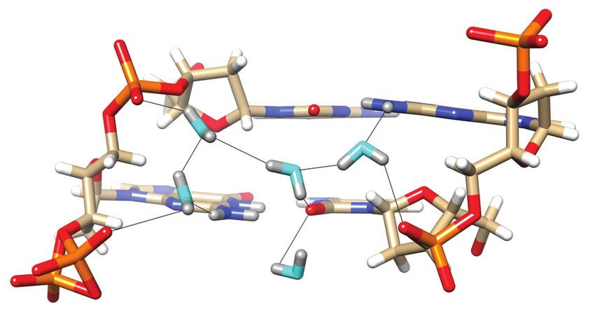
4786 Nucleic Acids Research, 2021, Vol. 49, No. 8
A B
C D
Downloaded from https://academic.oup.com/nar/article/49/8/4782/6238407 by guest on 03 December 2021
Figure 2. Quality of the final joint cryo X-ray/neutron crystal structure of Z-DNA. Examples of the superimposed 2Fo -Fc electron (1.0 Å, blue) and
neutron (1.5 Å, green) densities drawn at the 1.5 threshold. (A) Water pentagons spanning the minor groove at one end of the duplex. (B) Three water
molecules linking guanine to the phosphate of the adjacent residue on the convex surface. (C) Water pentagon nestled against the phosphate backbone
in the minor groove. (D) Water tandem bridging exocyclic amino groups of adjacent cytosines on the convex surface. Selected residues and/or atoms are
labeled and H-bonds are drawn with thin solid lines.
Table 2. Deuterium occupancies for exchangeable hydrogen atoms of G and C nucleobases
Base pair D1(G) D21(G) D22(G) D8(G) D41(C) D42(C)
C1 : G12 0.50 0.46 1.00 0.76 1.00 0.39
G2 : C11 0.93 1.00 0.95 0.04 1.00 1.00
C3 : G10 0.94 0.87 0.56 0.27 0.70 0.62
G4 : C9 0.25 0.23 0.53 0.00 0.99 1.00
C5 : G8 0.58 0.89 0.84 0.05 0.95 0.71
G6 : C7 0.61 1.00 1.00 0.33 1.00 1.00
Owing to the syn conformation of guanosine in Z-DNA, There is only a single example of a water molecule link-
N2 atoms lie closer to 5 - and 3 -phosphate groups on av- ing O2 atoms of cytosines from adjacent base pairs in the
erage than O2 atoms of cytidine. Thus, the average dis- minor groove. The water sits halfway between O2 atoms of
tance of N2 to 5 - and 3 -phosphates (OP2 atoms) is 6.1 and C3 and C11, with OW . . . O2 distances of 2.80 and 2.79 Å,
5.1 Å, respectively. The average distance of O2 to 5 - and respectively (Figure 3C). Despite the fact that the two cyto-
3 -phosphates (OP2 atoms) is 6.8 and 6.6 Å, respectively. sine keto oxygens are the water’s closest neighbors, its ori-
The shorter distances between N2 and OP2 atoms of 3 - entation suggests a more complicated involvement with the
phosphates enable a single water bridge between them as G2:C11 and C3:G10 base pairs than simply as a bridge be-
discussed above. However, distances closer to 6 Å and be- tween O2(C3) and O2(C11). The deuterium atoms of the
yond preclude the formation of a one-water bridge be- water do not lie in the plane defined by OW , O2(C3) and
tween guanine base and phosphate. Hence the two-water O2(C11), and are thus not pointing straight at the keto oxy-
bridge linking N2(G) to OP2 from the 5 -phosphate and gens. Instead, the water molecule is oriented more sideways
O2(C) to OP2 atoms from both the 5 -phosphate and the 3 - vis-à-vis C:G base pair edges, with OW ––DW ––O(C) an-
phosphate (Figure 3). With water bridges involving O2 and gles of ca. 100◦ (Figure 3C). The N2(D)2 amino nitrogen of
OP2, both waters have to act as H-bond donors to DNA G10 is positioned 3.60 Å from the water oxygen. The DW
atoms. These hydration patterns on the borders of the mi- atom close to O2(C11) (distance of 2.39 Å) is positioned
nor groove are then extended across the entire groove. At 2.91 Å from N2(G10), leading us to believe that the par-
one end of the duplex, an intricate ribbon involving two ticular orientation of the water molecule enables an elec-
hexagons and two pentagons links backbones and nucle- trostatically favorable interaction between water deuterium
obase edges (Figure 3A). The hexagon on one side of the mi- and nitrogen lone electron pair. The N2(D)2 amino nitro-
nor groove is composed of two phosphate groups (G8 and gen of G2 is positioned 3.70 Å from the water oxygen with
C9) and four waters. On the other side, the hexagon involves a lone pair of the latter directed toward the amino group.
five waters and the phosphate of C5. The two hexagons are The distance between OW and the amino deuterium partic-
linked by central water pentagons, with the entire hydra- ipating in G2:C11 pairing is 3.18 Å and thus somewhat too
tion network being composed of first and second shell water long to contribute favorably to the water-base pair interac-
molecules. tion. However, given the orientation of the water and the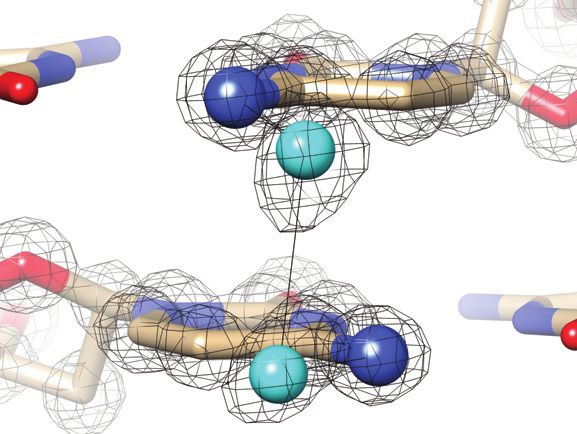
Nucleic Acids Research, 2021, Vol. 49, No. 8 4787
A A
Downloaded from https://academic.oup.com/nar/article/49/8/4782/6238407 by guest on 03 December 2021
B B
C
C
Figure 4. Water molecules bridging either pairs of cytosine N4 amino
groups or pairs of guanine O6 keto groups on the convex surface. The Z-
DNA neutron structure reveals H-bond donor-acceptor patterns for water
bridges between cytosines (A) C1 and C11, and (B) C5 and C7. Water oxy-
gen and deuterium atoms are colored in cyan and gray, respectively, and
Figure 3. Water structure in the minor groove. Transverse hydration pat- DNA deuterium and hydrogen atoms are colored in gray and white, respec-
terns that link cytosine O2 keto oxygens and guanine N2 amino groups tively. H-bonds (D. . . O) are drawn as thin solid lines. (C) Water molecule
to backbone phosphates. (A) Base pairs C5:G8 and G6:C7 and (B) base bridging O6 keto oxygens of adjacent guanines on the convex surface (cen-
pairs C3:G10 and C4:C9. Water oxygen and deuterium atoms are colored ter), flanked by water molecules forming H-bonds to N7 of G6 and G12#
in cyan and gray, respectively, and DNA deuterium and hydrogen atoms (# marks a symmetry-related residue). Electrostatically favorable interac-
are colored in gray and white, respectively. H-bonds (D. . . O) are drawn as tions by the central water are not limited to O6 (guanine) but also involve
thin solid lines, and strand polarities are indicated. ZI and ZII refer to two N4(C1# ), i.e. a lone electron pair of water and the deuterium atoms of the
different phosphate orientations that are characterized by distinct ε and amino group. D. . . N and D. . . O distances are indicated by thin solid lines.
backbone torsion angles. (C) The sole example of a water molecule bridg-
ing O2 keto oxygens of adjacent cytosines in the minor groove. Electrostat-
ically favorable interactions by this water are not limited to O2 (cytosine)
but also involve N2 (guanine), i.e. the lone electron pair of N2(G10) and and phosphate groups much less common than in the nar-
one of the deuterium atoms of N2(G2). D. . . N and D. . . O distances are row minor groove. Exceptions can occur when a phosphate
indicated by thin solid lines.
flips from the ZI to the ZII conformation, thereby rotating
non-bridging OP1 and OP2 oxygen atoms away from the
observed distances to acceptor and donor atoms of the two minor groove (Figures 3B and 4A). The alternating posi-
C:G pairs, the notion that the water molecule interacts with tive and negative twists between adjacent base pairs in Z-
O2(C3), O2(C11) and N2(G10) is reasonable. Thus, water DNA shift pairs of N4 amino groups roughly above one
in the central minor groove appears to optimize H-bonds another on the convex surface. The distances between N4
with the edges of base pairs rather than solely interacting atoms in the three pairs range from 3.6 to 3.9 Å. Amino
with cytosine O2 keto groups (Figure 3C). pairs are linked by water tandems such that water oxygen
atoms are positioned more or less in the planes defined by
cytosine bases (Figure 4A, B). Both waters accept in H-
Water structure on the convex surface
bonds with amino groups, and one donates, and the other
Z-DNA lacks a major groove, and instead, the O6 and N7 accepts in the inter-water H-bond. However, water orien-
(guanine) acceptors and the N4 amino group (cytosine) tations vis-à-vis cytosines differ somewhat between pairs
donor are presented on the so-called convex surface. The of N4 amino groups as the examples in Figure 4A, and B
particular orientation of backbones relative to base pairs in demonstrate. These differences can likely be traced to the
Z-DNA renders water-mediated bridges between base edges hydration networks that individual waters participate in, as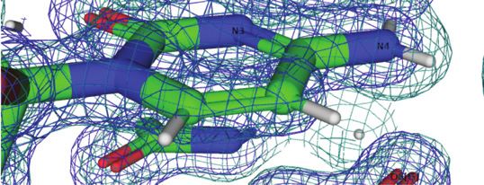
4788 Nucleic Acids Research, 2021, Vol. 49, No. 8

orientations of waters are not simply dictated by their inter-
actions with DNA atoms, but are also influenced by other
water molecules that are part of the first and second hydra-
tion shells.
   The particular orientation of a water molecule linking
O6 keto oxygens of adjacent guanines on the convex sur-
face is somewhat reminiscent of the situation in the minor
groove. As in the case of the aforementioned water link-
ing O2 atoms of cytosines from adjacent base pairs there,
the water situated between guanine keto groups does not
project its deuterium atoms in the direction of O6 atoms.
Rather, it turns away slightly so as to share one of its deuteri-
ums with both guanine keto oxygens. The distances between

                                                                                                                                                   Downloaded from https://academic.oup.com/nar/article/49/8/4782/6238407 by guest on 03 December 2021
the water oxygen and O6(G6) and O6(C12# ) are 3.02 and
3.00 Å, respectively, and the corresponding DW . . . O6 dis-
tances are 2.58 and 2.68 Å, respectively (Figure 4C). This
orientation of the water results in one of its lone electron
pairs being directed toward the deuterium atoms of the N4
amino group of cytosine C1# . The OW . . . N4 distance is 3.25
Å, and the OW . . . D(N4) distances are 2.89 Å (D41) and 3.02
Å. These distances are not significantly different from those
between water OW /DW and O6 keto oxygens (Figure 4C).
Just like with the water bridging O2 keto oxygens of cytosine       Figure 5. Longitudinal hydration pattern linking O6 keto oxygen of G and
in the minor groove, a water on the convex surface can inter-       backbone phosphates on one side of the convex surface. Water oxygen and
act with two O6 keto oxygens of guanosines and an amino             deuterium atoms are colored in cyan and gray, respectively, and DNA deu-
group of one of the cytidines. Therefore, water in the cen-         terium and hydrogen atoms are colored in gray and white, respectively. H-
                                                                    bonds (D. . . O) are drawn as thin solid lines. Note the formation of pen-
tral portion of the convex surface does not just engage with        tagons involving four water molecules and either a phosphate oxygen or
O6 keto groups of adjacent guanines but interacts with the          an O6 keto oxygen. A water molecule positioned between G4 and G8 lies
DNA by H-bonding to acceptors and donors of G:C pairs.              within H-bonding distance from O6 keto oxygens (dashed lines). However,
   The water molecule interacting with O6 keto oxygens of           its particular orientation, i.e. both deuterium atoms are directed away from
G6 and G12# , as well as the N4 amino group of C1# , is             O6 atoms, renders H-bond formation unlikely.
flanked by water molecules that form H-bonds to N7 of ad-
jacent guanines (Figure 4C). The waters contacting N7(G6)
and N7(G12# ) are too far removed from the central wa-              and the pair of O6 atoms (3.07 and 2.95 Å, Figure 5) are
ter for effective interactions (3.5 Å and 4.0 Å, respectively),   not incompatible with H-bond formation, but the relative
but are embedded in water networks spanning the convex              orientations of lone pairs among the three oxygen atoms
surface. Except for one guanine, all N7 acceptors form H-           are. Perhaps this is an indication that waters participating
bonds to a water molecule, but in some cases, the neutron           in networks around DNA occasionally prefer interactions
density did not permit us to settle the orientations of these       with neighboring solvent molecules instead of an H-bond
water molecules. In cases where the orientations were es-           to a DNA atom. In any case, the water molecule located
tablished, for example, the water molecule associated with          below the base plane of G4, as depicted in Figure 5, does
N7 of G10, it is H-bonded to a water from a pair of solvent         form an H-bond to the O6 keto oxygen of that guanine and
molecules that bridges N4 amino groups (C1/C11). The wa-            then bridges the oxygen to another water pentagon. Here,
ter molecule H-bonded to N7 of G12 is linked to the water           the pentagon is composed of four water molecules and OP1
tandem associated with the N4 amino groups of C7# and               of the phosphate group linking G2 and C3. An example
C5# via an intermediate water.                                      of a transverse hydration network on the convex surface
   Water networks on the convex surface include longitudi-          is shown in Figure 6. Nine water molecules span the en-
nal and transverse patterns. An example of the former is de-        tire width of the duplex, starting with a water that forms an
picted in Figure 5, with a string of water molecules spanning       H-bond to the 5 -hydroxyl group of C7 and ending with wa-
four layers of bases along the edge of the convex surface. As       ters that either H-bond to N7 of G6 or the phosphate group
in the minor groove, pentagons constitute the predominant           from a neighboring duplex. The neutron density did not al-
ring-shaped arrangement of waters that are part of the first        low a definitive assignment of the orientations of all waters
and second hydration shells. The participation of either a          in the network (cyan spheres in Figure 6). However, it is
DNA base or backbone atom in the pentagons is common.               encouraging that neutron crystallography allows visualiza-
An example that includes O6(G8) is shown at the top of              tion of donor-acceptor patterns in water networks around
Figure 5, although one side of that pentagon is not con-            a DNA duplex, even on the more exposed convex surface
stituted by an actual H-bond. Instead, the water molecule,          compared to the narrow minor groove. Higher resolution
unlike in the case of that between O6 atoms of G6 and G12#          data might reveal the orientations of the remaining waters
(Figure 4C), turns its back––i.e. lone pairs––toward the O6         that lack neutron density for deuterium atoms in the present
keto groups of G4 and G8. The distances between water               structure.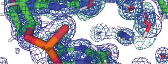
Nucleic Acids Research, 2021, Vol. 49, No. 8 4789

                                                                                        A

Figure 6. Transverse hydration pattern linking base edges and backbones
across the convex surface. Water oxygen and deuterium atoms are colored
in cyan and gray, respectively, and H-bonds (D. . . O/N) are drawn as thin
solid lines. Dashed lines link a water molecule to an adjacent one as well

                                                                                                                                                          Downloaded from https://academic.oup.com/nar/article/49/8/4782/6238407 by guest on 03 December 2021
as to O6(G6), whereby the distances are compatible with H-bond forma-
tion. However, the relative orientations of the two water molecules and O6
render H-bonding unlikely. Symmetry-related residues are marked by #.                   B

Phosphate hydration
The shortest distances between non-bridging oxygen atoms
of adjacent intra-strand phosphate groups are typically
around 4.5 Å with phosphate pairs that both assume the
more common ZI orientation. Such distances become much
longer (6–7 Å) in cases where the local backbone confor-
mation flips to ZII, thereby rotating the phosphate group
outwards and away from the minor groove (Figure 3). De-
spite the relatively short spacing between adjacent phos-
phates with a regular ZI backbone conformation, they are
most often linked by two water molecules (Figures 2A, C
and 3A, B). Both waters act as donors in H-bonds to                          Figure 7. Phosphate hydration. Water structure around the backbones of
phosphate oxygens and either the water closer to the 5 -                    nucleotides (A) G4 to G6, and (B) G10 to G12. Water molecules without
                                                                             significant neutron density for deuterium atoms are drawn as cyan spheres,
end or that closer to the 3 -end accepts/donates in the                     and H-bonds are thin solid lines. ZI and ZII designate particular confor-
inter-water H-bond (Figures 3A,B and 7B). Such waters                        mations of phosphate groups in Z-DNA.
are then embedded in hydration motifs that extend across
the minor groove (Figure 3A) or along the convex surface
(Figure 5) and commonly entail pentagon motifs. Patterns
seen in the water structure around phosphates, among oth-                    molecules that are part of the first and second hydration
ers, include 5-membered (Figure 5), 7-membered (Figure                       shells. As the examples depicted in Figure 2 demonstrate,
3A) and even 10- (Figure 7B) and 12-membered rings (Fig-                     the overlaid neutron and electron densities around DNA
ure 7A). In regions with ZII backbone conformations and                      atoms and water molecules are of good quality and the
thus longer distances between adjacent phosphates, two wa-                   maps complement each other well. The neutron diffrac-
ter molecules are not sufficient to bridge the former. Thus,                 tion data collected at cryo temperature on MaNDi/SNS at
the phosphates of nucleotides C5 and C11 exhibit ZII con-                    ORNL are of high completeness to a maximum resolution
formations (mixed ZI/ZII in the latter case; Figures 3B, 4A,                 of 1.5 Å. The dataset was collected in a week-long experi-
7). As a consequence, the phosphates of G4 and G6 are close                  ment using a crystal that was quite large with a volume of
enough to be linked by two water molecules (Figure 7A).                      ca. 1 mm3 . We believe that further optimization (e.g. wave-
Conversely, the ZI phosphate of G4 and the ZII phosphate                     length) and a somewhat longer exposure can yield complete
of C5 are linked by four water molecules. A further four                     data with a resolution around 1.2 Å. It is likely that the ori-
waters then connect the C5 phosphate to the O4 atom of                      entations of many of the water molecules that currently lack
G6. The sugar oxygen occasionally accepts in H-bonds to                      neutron density for deuterium atoms can then also be es-
water but is poorly hydrated compared to base edges and                      tablished. The neutron data were initially processed to 1.4
phosphate groups. The shortest distance between adjacent                     Å resolution and the  in the 1.45–1.40 Å resolu-
intra-strand phosphate groups in the Z-DNA duplex stud-                      tion bin was 3.6 with a completeness of 62.6%. We decided
ied here is found between OP2(C11) and OP1(G12): 4.25 Å                     to cut the data at 1.5 Å ( of 4.2 and completeness
(Figure 7B). This pair is joined by a single water molecule,                 of 78.5% in the 1.55–1.50 Å resolution bin; Table 1) in or-
the only example of a one-water bridge between phosphates                    der to work with a more complete data set. Still, it should
observed in the DNA structure.                                               be possible to collect neutron data to higher resolution with
                                                                             a Z-DNA crystal of this size as the mean intensity did not
                                                                             drop to a level of, say,  = 2 at 1.4 Å. The MaNDi
DISCUSSION
                                                                             setup is such that the detector cannot be moved; instead it
The joint X-ray and neutron refinement of the Z-DNA                          was necessary to use a more optimal wavelength and collect
duplex has thus far revealed the orientations of 64 D2 O                     longer frames.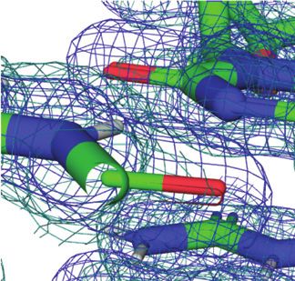
4790 Nucleic Acids Research, 2021, Vol. 49, No. 8
As expected, hydrogen atoms in the Z-DNA duplex ing adjacent pairs of O2 and O6 keto oxygens in the minor
that are exchanged with deuterium are limited to exocyclic groove and on the convex surface, respectively, as implied
amino groups in cytosine and guanine and 5 - and 3 - by electron density alone (Figure 1). More work using high-
terminal hydroxyls. We also found an indication for partial resolution cryo neutron crystallography is necessary to es-
H-D exchange by H8 hydrogen atoms in some guanines, as tablish whether such features are commonplace in the water
reported by Niimura et al. (28). It is possible that differ- structure that surrounds DNA duplexes.
ent crystallization conditions can affect exchange to some
degree. However, the level of background scattering in neu-
CONCLUSIONS
tron experiments with crystals of synthetic oligonucleotides
is bound to be higher than with crystals of perdeuterated We used cryo neutron crystallography to analyze the water
proteins because none of the H atoms in the DNA back- structure around the DNA duplex [d(CGCGCG)]2 at near-
bone are exchanged. The Z-DNA crystal form described atomic resolution. The resulting neutron density reveals the
here was grown using ammonium sulfate as the precipitant orientation of the majority of first-shell water molecules.
Downloaded from https://academic.oup.com/nar/article/49/8/4782/6238407 by guest on 03 December 2021
to avoid binding of divalent metal ions or polyamines to Cryo neutron data allow insight into acceptor and donor
the DNA, thereby displacing first and second shell water functions for water molecules that are part of intricate hy-
molecules and distorting the water structure around the du- dration patterns in the minor groove and on the Z-DNA
plex. It is noteworthy that the density maps did not reveal convex surface, thus complementing information previously
any coordinated sodium, ammonium, or sulfate ions. The obtained from room temperature neutron structures or X-
ammonium sulfate crystal form displays unit cell constants ray crystal structures determined to ultra-high resolution.
that are very similar to those grown from magnesium, sper- The discovery based on the determination of deuterium po-
mine, or mixed magnesium/spermine conditions (19) and sitions that water is shared among guanine and cytosine
all these Z-DNA crystals belong to space group P21 21 21 . keto and amino groups in the minor groove and on the con-
X-ray crystal structures of Z-DNA at atomic resolution vex surface rather than just clamping keto oxygens of ad-
revealed regular longitudinal and transverse hydration pat- jacent cytidines (minor groove) or guanosines (convex sur-
terns (19), and the former included a chain of waters in face) is particularly noteworthy.
the minor groove that linked cytosine O2 atoms, and wa- Several challenges remain to be overcome in order to vi-
ter molecules bridging pairs of guanine O6 keto oxygens sualize at truly atomic resolution––1 Å or higher––the wa-
and cytosine N4 amino groups (Figure 1). By comparison, ter structure around oligonucleotides based on joint neu-
transversal hydration patterns both in the minor groove and tron and X-ray diffraction data collected at cryogenic tem-
on the convex surface appear to be more common in the perature. Even with the higher flux of the neutron beam
cryo-neutron structure of Z-DNA analyzed here. In particu- at MaNDi/SNS, a crystal with a volume of, say, 0.1 mm3
lar, repetitive patterns involving water molecules linking the will not deliver atomic resolution neutron data. Obviously,
edges of guanine and cytosine bases to phosphate groups the resolution of the X-ray data that can be achieved even
are commonly seen in the minor groove (A, B). Longitudi- with small Z-DNA crystals or other well diffracting DNA
nal water motifs include water tandems bridging adjacent and RNA oligo crystals is not an issue. As well, crystals of
pairs of cytosine N4 amino groups (Figure 4A,B) and ad- Z-DNA with volumes of 1 mm3 and larger can be grown
jacent phosphate groups (Figures 3A, B and 7). Hydration within weeks as we have shown. Cryoprotection and freez-
motifs such as single waters linking O2 keto oxygens and O6 ing of very large crystals requires special caution and trans-
keto oxygens of neighboring cytidines in the minor groove port using a dryshipper via courier is not encouraged. The
(Figure 3C) and guanosines on the convex surface (Figure crystal has to remain cooled and stable for two weeks and
4C), respectively, are not observed repeatedly. more in the neutron beam and then recovered for X-ray
The cryo-neutron structure at high resolution has re- data collection. Hence, collecting the latter data on a home
vealed an interesting hydration pattern that involves amino source nearby like the MetalJet in our case––without the
groups of either cytosine or guanine and pairs of keto oxy- need to ship the crystal to an X-ray synchrotron––is bene-
gens in both the minor groove and on the convex surface. ficial. The opportunities to collect neutron diffraction data
In this motif, a water donates H-bonds to two keto oxygens at cryogenic temperature worldwide are very scarce. Once
and also establishes electrostatically favorable interactions data collection time is granted, conducting an optimal ex-
with adjacent amino groups. In the minor groove, the water periment in order to push the resolution limit closer to 1
interacts with the N2 amino group of guanine in addition to Å is expected to take at least two weeks and may require
forming H-bonds to O2 keto oxygens of stacked cytosines changing the wavelength to avoid limiting data resolution.
(Figure 3C). On the convex surface, the water interacts with Finally, joint refinement of cryogenic X-ray and neutron
the N4 amino group of cytosine in addition to forming H- data for oligonucleotides is not a routine affair and more ex-
bonds to O6 keto oxygens of stacked guanines (Figure 4C). perience needs to be gained to understand differences seen
The neutron density does not indicate a change in the sp2 in the temperature factors of ‘X-ray’ and ‘neutron’ water
hybridization of the nitrogen atom with a concomitant loss molecules.
of conjugation with the nucleobase system as a result of
this hydration motif. The orientation of water molecules vis-
DATA AVAILABILITY
à-vis C:G pairs observed here likely serves the optimization
of H-bonding interactions with the edges of DNA bases and Atomic coordinates and structure factors for the reported
perhaps other water molecules. Thus, this hydration motif crystal structure have been deposited with the Protein Data
may be energetically beneficial compared to simply bridg- bank under accession number 7JY2.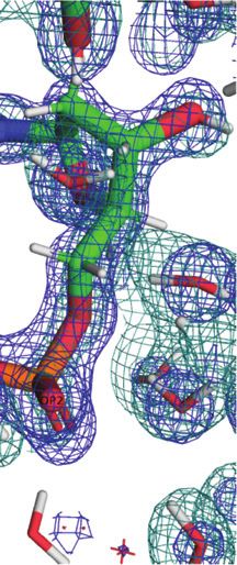
Nucleic Acids Research, 2021, Vol. 49, No. 8 4791

SUPPLEMENTARY DATA                                                         17. Ravichandran,S., Subramani,V.K. and Kim,K.K. (2019) Z-DNA in
                                                                               the genome: from structure to disease. Biophys. Rev., 11, 383–387.
Supplementary Data are available at NAR Online.                            18. Herbert,A. (2019) Z-DNA and Z-RNA in human disease. Commun.
                                                                               Biol., 2, 7.
                                                                           19. Gessner,R.V., Quigley,G.J. and Egli,M. (1994) Comparative studies of
ACKNOWLEDGEMENTS                                                               high-resolution Z-DNA crystal structures. Part 1: common hydration
                                                                               patterns of alternating dC-dG. J. Mol. Biol., 236, 1154–1168.
The research at O.R.N.L.’s Spallation Neutron Source was                   20. Leal,R.M.F., Callow,S., Callow,P., Blakeley,M.P., Cardin,C.J.,
sponsored by the Scientific User Facilities Division, Office                   Denny,W.A., Teixeira,S.C.M., Mitchell,E.P. and Forsyth,V.T. (2010)
of Basic Energy Sciences, US Department of Energy.                             Combined neutron and X-ray diffraction studies of DNA in crystals
                                                                               and solution. Acta Cryst. D, 66, 1244–1248.
                                                                           21. Fenn,T.D., Schnieders,M.J., Mustyakimov,M., Wu,C., Langan,P.,
FUNDING                                                                        Pande,V.S. and Brunger,A.T. (2011) Reintroducing electrostatics into
                                                                               macromolecular crystallographic refinement: application to neutron
L.C. and B.S. acknowledge support from the National Insti-                     crystallography and DNA hydration. Structure, 19, 523–533.
tutes of Health under grant number [R01 GM071939]. The                     22. Chen,J.C.-H., Hanson,L., Fisher,S.C., Langan,P. and

                                                                                                                                                         Downloaded from https://academic.oup.com/nar/article/49/8/4782/6238407 by guest on 03 December 2021
open access publication charge for this paper has been                         Kovalevsky,A.Y. (2012) Direct observation of hydrogen atom
waived by Oxford University Press – NAR Editorial Board                        dynamics and interactions by ultrahigh resolution neutron protein
members are entitled to one free paper per year in recogni-                    crystallography. Proc. Natl. Acad. Sci. U.S.A., 109, 15301–15306.
                                                                           23. Cuypers,M.G., Mason,S.A., Blakeley,M.P., Mitchell,E.P.,
tion of their work on behalf of the journal                                    Haertlein,M. and Forsyth,V.T. (2013) Near-atomic resolution
Conflict of interest statement. None declared.                                 neutron crystallography on perdeuterated Pyrococcus furiosus
                                                                               rubredoxin: implication of hydronium ions and protonation state
                                                                               equilibria in redox changes. Angew. Chem. Int. Ed., 52, 1022–1025.
REFERENCES                                                                 24. Chen,J.C.-H. and Unkefer,C.J. (2017) Fifteen years of the protein
 1. Drew,H. and Dickerson,R.E. (1981) Structure of a B-DNA                     crystallography station: the coming of age of macromolecular
    dodecamer. III. Geometry of hydration. J. Mol. Biol., 151, 535–556.        neutron crystallography. IUCrJ, 4, 72–86.
 2. Tereshko,V., Minasov,G. and Egli,M. (1999) The Dickerson-Drew          25. Földesi,A., Trifonova,A., Kundu,M.K. and Chattopadhyaya,J. (2000)
    B-DNA dodecamer revisited - at atomic resolution. J. Am. Chem.             The synthesis of deuterionucleosides. Nucleot. Nucleos. Nucleic Acids,
    Soc., 121, 470–471.                                                        19, 1615–1656.
 3. Egli,M., Portmann,S. and Usman,N. (1996) RNA hydration: a              26. Fuller,W., Forsyth,T. and Mahendrasingam,A. (2004) Water–DNA
    detailed look. Biochemistry, 35, 8489–8494.                                interactions as studied by X-ray and neutron fibre diffraction. Phil.
 4. Egli,M., Tereshko,V., Teplova,M., Minasov,G., Joachimiak,A.,               Trans. R. Soc. Lond. B, 359, 1237–1248.
    Sanishvili,R., Weeks,C.M., Miller,R., Maier,M.A., An,H. et al.         27. Arai,S., Chatake,T., Ohhara,T., Kurihara,K., Tanaka,I., Suzuki,N.,
    (2000) X-ray crystallographic analysis of the hydration of A- and          Fujimoto,Z., Mizuno,H. and Niimura,N. (2005) Complicated water
    B-form DNA at atomic resolution. Biopolymers (Nucleic Acid Sci.),          orientations in the minor groove of the B-DNA decamer
    48, 234–252.                                                               d(CCATTAATGG)2 observed by neutron diffraction measurements.
 5. Wang,A.H.-J., Quigley,G.J., Kolpak,F.J., Crawford,J.L., van                Nucleic Acids Res., 33, 3017–3024.
    Boom,J.H., van der Marel,G.A. and Rich,A. (1979) Molecular             28. Chatake,T., Tanaka,I., Umino,H., Arai,S. and Niimura,N. (2005) The
    structure of a left-handed double helical DNA fragment at atomic           hydration structure of a Z-DNA hexameric duplex determined by a
    resolution. Nature, 282, 680–686.                                          neutron diffraction technique. Acta Cryst. D, 61, 1088–1098.
 6. Peck,L.J. and Wang,J.C. (1983) Energetics of B-to-Z transition in      29. Egli,M., Williams,L. D., Gao,Q. and Rich,A. (1991) Structure of the
    DNA. Proc. Natl. Acad. Sci. U.S.A., 80, 6206–6210.                         pure-spermine form of Z-DNA (magnesium free) at 1Å resolution.
 7. Rich,A., Nordheim,A. and Wang,A.H.-J. (1984) The chemistry and             Biochemistry, 30, 11388–11402.
    biology of left-handed Z-DNA. Ann. Rev. Biochem., 53, 791–846.         30. Bancroft,D., Williams,L. D., Rich,A. and Egli,M. (1994) The low
 8. Lancillotti,F., Lopez,M.C., Arias,P. and Alonso,C. (1987) Z-DNA in         temperature crystal structure of the pure-spermine form of Z-DNA
    transcriptionally active chromosomes. Proc. Natl. Acad. Sci. USA,          reveals binding of a spermine molecule in the minor groove.
    84, 1560–1564.                                                             Biochemistry, 33, 1073–1086.
 9. Wittig,B., Dorbic,T. and Rich,A. (1991) Transcription is associated    31. Harp,J., Coates,L., Sullivan,B. and Egli,M. (2018) Cryo-neutron
    with Z-DNA formation in metabolically active permeabilized                 crystallographic data collection and preliminary refinement of
    mammalian cell nuclei. Proc. Natl. Acad. Sci. U.S.A., 88, 2259–2263.       left-handed Z-DNA d(CGCGCG). Acta Cryst. F, 74, 603–609.
10. Wittig,B., Wölfl,S., Dorbic,T., Vahrson,W. and Rich,A. (1992)         32. Blakeley,M.P., Kalb,A.J., Helliwell,J.R. and Myles,D.A.A. (2004)
    Transcription of human C-MYC in permeabilized nuclei is associated         The 15-K neutron structure of saccharide-free concanavalin A. Proc.
    with formation of Z-DNA in three discrete regions of the gene.             Natl. Acad. Sci. U.S.A., 101, 16405–16410.
    EMBO J., 11, 4653–4663.                                                33. Habash,J., Raftery,J., Nuttall,R., Price,H.J., Wilkinson,C., Kalb,A.J.
11. Rich,A. and Zhang,S. (2003) Z-DNA: the long road to biological             and Helliwell,J.R. (2000) Direct determination of the positions of the
    function. Nat. Rev. Genet., 4, 566–572.                                    deuterium atoms of the bound water in -concanavalin A by neutron
12. Ha,S.C., Lowenhaupt,K., Rich,A., Kim,Y.G. and Kim,K.K. (2005)              Laue crystallography. Acta Cryst. D, 56, 541–550.
    Crystal structure of a junction between B-DNA and Z-DNA reveals        34. Coates,L., Tomanicek,S., Schrader,T.E., Weiss,K.L., Ng,J.D.,
    two extruded bases. Nature, 437, 1183–1186.                                Juttner,P. and Ostermann,A. (2014) Cryogenic neutron protein
13. Herbert,A., Alfken,J., Kim,Y.G., Mian,I.S., Nishikura,K. and               crystallography: routine methods and potential benefits. J Appl.
    Rich,A. (1997) A Z-DNA binding domain present in the human                 Cryst., 47, 1431–1434.
    editing enzyme, double-stranded RNA adenosine deaminase. Proc.         35. Casadei,C.M., Gumiero,A., Metcalfe,C.L., Murphy,E.J., Basran,J.,
    Natl. Acad. Sci. U.S.A., 94, 8421–8426.                                    Concilio,M.G., Teixeira,S.C.M., Schrader,T.E., Fielding,A.J.,
14. Schwartz,T., Rould,M.A., Lowenhaupt,K., Herbert,A. and Rich,A.             Ostermann,A. et al. (2014) Heme enzymes. Neutron
    (1999) Crystal structure of the Zalpha domain of the human editing         cryo-crystallography captures the protonation state of ferryl heme in
    enzyme ADAR1 bound to left-handed Z-DNA. Science, 284,                     a peroxidase. Science, 345, 193–197.
    1841–1845.                                                             36. Langan,P., Li,X., Hanson,B.L., Coates,L. and Mustyakimov,M.
15. Bass,B.L. (2002) RNA editing by adenosine deaminases that act on           (2006) Synthesis, capillary crystallization and preliminary joint X-ray
    RNA. Ann. Rev. Biochem., 71, 817–846.                                      and neutron crystallographic study of Z-DNA without polyamine at
16. Kesavardhana,S., Kuriakose,T., Guy,C.S., Samir,P., Malireddi,R.K.,         low pH. Acta Cryst. F, 62, 453–456.
    Mishra,A. and Kanneganti,T.D. (2017) ZBP1/DAI ubiquitination           37. Coates,L., Cuneo,M.J., Frost,M.J., He,J.H., Weiss,K.L.,
    and sensing of influenza vRNPs activate programmed cell death. J.          Tomanicek,S.J., McFeeters,H., Vandavasi,V.G., Langan,P. and
    Exp. Med., 214, 2217–2229.                                                 Iverson,E.B. (2015) The macromolecular neutron diffractometer
4792 Nucleic Acids Research, 2021, Vol. 49, No. 8
MaNDi at the spallation neutron source. J. Appl. Cryst., 48, 42. Campbell,J.W., Hao,Q., Harding,M.M., Nguti,N.D. and
1302–1306. Wilkinson,C. (1998) LAUEGEN version 6.0 and INTLDM. J. Appl.
38. Coates,L., Cao,H.B., Chakoumakos,B.C., Frontzek,M.D., Cryst., 31, 496–502.
Hoffmann,C., Kovalevsky,A.Y., Liu,Y., Meilleur,F., dos 43. Helliwell,J.R., Habash,J., Cruickshank,D.W.J., Harding,M.M.,
Santos,A.M., Myles,D.A.A. et al. (2018) A suite-level review of the Greenhough,T.J., Campbell,J.W., Clifton,I.J., Elder,M., Machin,P.A.,
neutron single-crystal diffraction instruments at Oak Ridge National Papiz,M Z. et al. (1989) The recording and analysis of synchrotron
Laboratory. Rev. Sci. Instrum., 89, 092802. X-radiation Laue diffraction photographs. J. Appl. Cryst., 22,
39. Arnold,O., Bilheux,J.C., Borreguero,J.M., Buts,A., Campbell,S.I., 483–497.
Chapon,L., Doucet,M., Draper,N., Leal,R.F., Gigg,M.A. et al. 44. Adams,P.D., Afonine,P.V., Bunkoczi,G., Chen,V.B., Davis,I.W.,
(2014) Mantid - data analysis and visualization package for neutron Echols,N., Headd,J.J., Hung,L.W., Kapral,G.J.,
scattering and SR experiments. Nucl. Instrum. Methods A, 764, Grosse-Kunstleve,R.W. et al. (2010) PHENIX: a comprehensive
156–166. Python-based system for macromolecular structure solution. Acta
40. Sullivan,B., Archibald,R., Langan,P.S., Dobbek,H., Bommer,M., Cryst. D, 66, 213–221.
McFeeters,R.L., Coates,L., Wang,X.P., Gallmeier,F., Carpenter,J.M. 45. Afonine,P.V., Mustyakimov,M., Grosse-Kunstleve,R.W., Moriarty,N.
et al. (2018) Improving the accuracy and resolution of neutron W., Langan,P. and Adams,P.D. (2010) Joint X-ray and neutron
crystallographic data by three-dimensional profile fitting of Bragg refinement with phenix.refine. Acta Cryst. D, 66, 1153–1163.
Downloaded from https://academic.oup.com/nar/article/49/8/4782/6238407 by guest on 03 December 2021
peaks in reciprocal space. Acta. Cryst. D, 74, 1085–1095. 46. Emsley,P., Lohkamp,B., Scott,W.G. and Cowtan,K. (2010) Features
41. Sullivan,B., Archibald,R., Vandavasi,V., Langan,P., Langan,P.S., and development of Coot. Acta Cryst. D, 66, 486–501.
Coates,L., Azadmanesh,J. and Lynch,V.E. (2019) BraggNet: 47. Dauter,Z. and Adamiak,D.A. (2001) Anomalous signal of
integrating Bragg peaks using neural networks. J. Appl. Cryst., 52, phosphorus used for phasing DNA oligomer: importance of data
854–863. redundancy. Acta Cryst. D, 57, 990–995.You can also read